Abstract.
The most trusted hypothesis to explain
how 2-adrenergic agonists may preserve pulmonary
functions in critically ill patients is that they directly
act on macrophages by interfering with an
autocrine/paracrine adrenergic system that controls
cytokine
release
through
locally
synthetized
noradrenaline and 1- and 2-adrenoreceptors. We
tested this hypothesis in primary cultures of resident
macrophages from human lung (HLMs). HLMs were
isolated by centrifugation on percoll gradients from
macroscopically healthy human lung tissue obtained
from four different patients at the time of lung
resection for cancer. HLMs from these patients
showed a significant expression of α2A, α2B and
α2C adrenoreceptors both at the mRNA and at the
protein
level.
To
evaluate
whether
α2
adrenoreceptors controlled cytokine release from
HMLs, we measured IL-6, IL-8 and TNF-
concentrations in the culture medium in basal
conditions and after preincubation with several
2-adrenergic agonists or antagonists. Neither the
pretreatment with the 2-adrenergic agonists
clonidine, medetomidine or dexdemetomidine or
with the 2-adrenergic antagonist yohimbine caused
significant changes in the response of any of these
cytokines to LPS. These results show that, different
from what reported in rodents, clonidine and
dexdemetomidine do not directly suppress cytokine
release from human pulmonary macrophages. This
suggests that alternative mechanisms such as effects
on immune cells activation or the modulation of
autonomic neurotransmission could be responsible
for the beneficial effects of these drugs on lung
function in critical patients.
Keywords: clonidine; dexmedetomidine; medetomidine; yohimbine; cytokines; macrophages.
I. INTRODUCTION
2-adrenergic agonists including dexmedetomidine and clonidine exert sedative, analgesic, and anxiolytic effects by acting at different levels. More specifically, they decrease noradrenaline release in locus coeruleus hence activating descending inhibitory pathways that reach the posterior horns of the spinal cord to inhibit the transmission of painful stimuli to the brain (1). 2-adrenergic receptors are also expressed in projecting neurons of the dorsal horn and inhibit their activity. In addition, a possible direct blocking effect of 2-adrenergic agonists on A and C fibres has been described and could account for the ability of these drugs to enhance the peripheral nerve blocking activity of local anaesthetics (2). Finally, 2-adrenergic agonists exert central sedative effects acting at the level of thalamic nuclei. By this mechanism they also synergize with opioids hence lowering the need for opiate medication during post-operative pain. Since they can induce a good level of sedation with less respiratory and cardiovascular depression than conventional centrally-acting sedative drugs, 2-adrenergic agonists are used very often in the intensive care unit (ICU). Their sedative effects may be helpful, indeed, in seriously ill patients such as those with sepsis or respiratory distress syndrome. Clonidine is usually preferred in the pediatric patients whereas dexmedetomidine is the 2-adrenergic agonist more commonly used in adults (3). Although many side effects have been described for these drugs such as hypotension, rebound tachycardia and hypertension after withdrawal, a moderate, not clinically relevant bradycardia is the only one that is frequently observed in critical patients (4). An important argument supporting the use of 2-adrenergic drugs in the ICU is the clinical evidence that they may favourably impact on the clinical course in critical patients. For instance, it has been shown that the length of stay in the ICU and time to extubation are significantly shorter in critically ill patients that receive dexmedetomidine instead of propofol or benzodiazepines (5). In addition, a systematic review of literature (6)
Effect Of 2-Adrenergic Agonists And Antagonists On Cytokine Release From
Human Lung Macrophages Cultured In Vitro
O. Piazza
1, R.I. Staiano
2, E. De Robertis
3-4, G. Conti.
5, V. Di Crescenzo
1, S. Loffredo
2, G.
Marone
2, G. Zito Marinosci
1, M. Cataldi M.
3-41Università di Salerno, Department of Medicine and Surgery, Via Allende, 84081 Baronissi, (SA) Italy; 2Università degli Studi di Napoli Federico II, Department of Translational Medical Sciences and Center for Basic and Clinical
Immunology Research (CISI), Naples, Italy; 3Università degli Studi di Napoli Federico II, Department of Neurosciences, Naples, Italy; 4Federico II University Hospital, Naples, Italy 5Università Cattolica del Sacro Cuore,
Anaesthesiology and Intensive Care, Rome, Italy.
reported that dexmedetomidine improves short-term mortality in sepsis patients, compared with other sedatives, even though this conclusion needs further studies to be confirmed.
It is currently unknown by which mechanism dexmedetomidine could exert these positive effects on morbidity and mortality of critical patients. Among the proposed explanations it has been suggested that it could have significant antinflammatory effects that could be useful, for instance, in maintaining alveolar gas exchanges in the inflamed lung. Several preclinical studies seem to support this hypothesis. For instance, a decrease in cytokine release in response to LPS has been observed upon incubation with dexmedetomidine in the macrophagic cell line RAW264.7 (7) and in cultured astrocytes (8) and microglia (9). Moreover, Xu et al (10) showed that in a mouse model of acute lung injury, the pretreatment with dexmedetomidine significantly blunted the increase in plasma TNF-α the by a mechanism dependent on the inhibition of the MAPK signalling pathway. Few studies investigated the anti-inflammatory effects of dexmedetomidine in humans and most of them were based on indirect evidence. For instance, in 20 patients with sepsis, Memis et al showed that TNF-, IL-1β and IL-6 levels in plasma were significantly lower after 24 hours of sedation with dexedemetomidine in comparison with controls that did not receive this drug (11). Gao et al (12) compared two groups of patients undergoing one-lung ventilation for lung cancer surgery, the first receiving dexmedetomidine intraoperatively and the latter not. They showed that the peripheral concentrations of TNF- were significantly lower in patients treated with the 2-adrenergic agonist. Moreover, in this group of patients the levels of the heme oxygenase mRNA in the removed lung tissue was higher than in the other group. Considering that heme oxygenase has antinflammatory effects this finding stands for a direct antinflammatory effect of dexmedetomidine on the lung. Up to date no study investigated yet whether the possible antinflammatory effects of dexmedetomidine on the lung are directly exerted at the level of inflammatory cells such as lung monocytes although the evidence that this drug lowers proinflammatory mediator production in whole human blood in vitro stands for a direct effect on inflammatory cells (13). Therefore, we designed the present study to investigate whether primary macrophages isolated from human lung do express 2-adrenergic receptors and respond to 2-adrenergic agonists by decreasing cytokine release.
II. METHODOLOGY
This study was performed on macroscopically normal tissue obtained from four patients. No patient was allergic, asthmatic or affected by bronchitis. The study protocol was approved by the ethics authority and all patients gave their written informed consent to take part to this study.
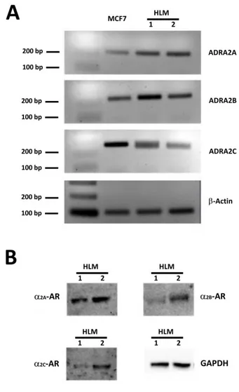
Figure 1. ADRA2A, ADRA2B, ADRA2C expression inHLMs.
Panel A: RT-PCR performed with primers specific for ADRA2A, ADRA2B and ADRA2C. Gels were loaded with retrotranscripts from lung tissues of two different patients (Lines 1 and 2) and from MCF-7 cells, a breast cancer cell known to express all the three 2
-adrenoreceptor isoforms that was used as an internal reference. In all the PCR reactions bands of about 200 bp were amplified, a size corresponding to what expected for ADRA2A (211 bp; first line), ADRA2B (230 bp; second line) and ADRA2C (242 bp; third line). β actin was used as housekeeping gene product or normalization. Panel B: Western blotting performed using anti-α2A-, anti α2B- and anti- α2C-adrenoreceptor antibodies and cellular lysates from HLMs obtained from two different patients preparations. GAPDH immunoreactivity was used for normalization. In both HLM preparations, immunoreactive bands of the expected size were obtained for all the three α2-adrenoreceptor isoforms (70, 62 and 60 kDa for α2A-, α2B- and α2C-adrenoreceptors, respectively).
2.1. HLM isolation, purification and culture
Human macrophages were purified as described elsewhere (14). Lung tissue was mechanically dispersed and the macrophage suspension was enriched (75–85%) by flotation over Percoll® density gradients. After being
suspended in RPMI-1640 containing 5% FCS, 2mM L-glutamine, and 1% antibiotic-antimycotic solution at the final density of 2x10⁶ cells/mL, cells were allowed to
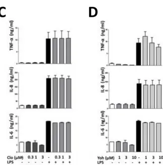
Figure 2. Effect of 2-adrenergic agonists and antagonists on in vitro
cytokine release from HMLs.
The bar graphs show the effect of vehicle and of progressively higher concentrations of the 2-adrenergic agonists dexmedetomidine (0.1-10
µM) (panel A), medetomidine (0.1-10 µM) (panel B) and clonidine (0,3-3 µM) (panel C) and of the 2-adrenergic antagonist yohimbine (1-10
µM) (panel D) on LPS-induced TNF-α, IL-8 and IL-6 release in the culture medium from HLMs. HLMs were preincubated with the aforementioned drugs for 30 min before adding LPS (100 ng/ml) to induce TNF-α, IL-8 and IL-6 release. The concentrations of these cytokines in culture medium were measured by ELISA 18 hours after LPS addition. Each bar represents as the mean± SEM of the concentrations of the respective cytokine obtained in four different experimental sessions.
adhere to plastic dishes at 37°C in a 5% CO2atmosphere. After 12 h, the medium was removed and the plates were gently washed with RPMI-1640. More than 98% of adherent cells were macrophages, as assessed by flow cytometry analysis and a-naphthylacetate esterase staining.
2.2. RT-PCR for ADRA2A, ADRA2B and ADRA2C
Total RNA from HLMs and from breast adenocarcinoma MCF7 cells, that were used as a positive control because constitutively expressing all the three ADR2A isoforms (15) was extracted using the SV 96 total RNA isolation system (Promega, Madison, WI) and treated with RNase-free DNase I. Reverse transcription was performed as previously described (16). ADRA2A, ADRA2B and ADRA2C mRNA expression was evaluated by semiquantitative PCR using specific primers that were
designed with Beacon Designer 3.0 software (Biorad Laboratories, Milan, Italy) according to the corresponding cDNA sequences published in GenBank accession No. NM_000681.3, Homo sapiens adrenoreceptor alpha 2 (ADRA2A); NM_000682.5 ADRA2B; NM_000683.3 ADRA2C) and that were synthesized by Invitrogen (Carlsbad, CA). Specifically, we used the following
primers: ADRA2A forward: 5’-
ACTGGACTACAAGGGCATGG -3’, reverse: 5’-
ACATCAAAACCAAGGCCAAG -3’; ADRA2B
forward: 5’- CCTGTTTTCGGATCTGTGGT -3’, reverse: 5’- CTGCAAAGCCTTTCATCTCC -3’; ADRA2C forward: 5’- CCGGTCATCTACACGGTCTT -3’, reverse: 5’- ATCTCTCTGCCAAGCTCCTG -3’. Similar amounts of cDNAs from HLMs and MCF-7 were amplified according the following PCR protocol: denaturation at 95°C for 10 minutes, amplification with 35 cycles of 20 second, denaturation (94°C) and 30 second annealing (55°C). The PCR products were separated on 2% agarose gel, stained with ethidium bromide and visualized under the image analysis system Chemidoc XRS (Biorad).
2.3. Western blot analysis of α2A-AR, α2B-AR e α2C-AR
Total proteins were extracted from HLMs and MCF-7 cells using the following lysis buffer: 20 mM Tris pH 7.5, 5 mM EDTA, 1 mM PMSF, 2 mM benzamidine, 10 μg/ml leupeptin, 10 mM NaF, 150 mM NaCl, 1% Nonidet P-40 and 5% glycerol (17).
Cell lysates were kept on ice for 20 minutes then microfuged (14,000 rpm, 4°C, 20 min). An aliquot of protein extracts from cell lysates was drawn and the protein content was measured with the BCA Protein Assay Kit (Novagen, Merck Biosciences, San Diego, CA). The remaining protein extracts were diluted in lithium dodecyl-sulfate sample buffer with 2.5% 2-mercaptoethanol, and boiled before storage at -80°C. Equal protein extracts (30-50 g per sample) were separated on 4-12% Bis-Tris gels (NuPAGE, Novex, Invitrogen) and transferred to a nitrocellulose membrane (Schleicher & Schuell, Dassel, Germany) together with a biotinylated protein ladder (Cell Signaling, Beverly, MA). After immersion overnight in TBST (50 mM Tris pH 7.5, 150 mM NaCl and 0.05% Tween 20) containing 5% non-fat dry milk (Biorad), membranes were washed three times (10 min each) with TBST then incubated (4°C, overnight) with goat anti-α2A-adrenoreceptor (1:500), goat anti- α2B- adrenoreceptor (1:500), or rabbit anti- α2C- adrenoreceptor (1:1000). The membranes were washed then incubated (22°C, 1 hour) with HRP-conjugated rabbit anti-goat IgG Ab or donkey anti-rabbit IgG Ab together with HRP-conjugated anti-biotin Ab. Membrane-bound antibodies were visualized with the ECL Plus Western blotting detection system (GE Healthcare) under the Chemidoc XRS.
2.4. Pharmacological treatments and Cytokine assay
TNF-α, IL-8 and IL-6 were measured in the culture medium of HLM cells stimulated with LPS (100 ng/ml for 6 hours) after a 30 min preincubation either with vehicle or with metedomidine or dexmedetomidine (0.1-10 M), clonidine (0,3-3 µM)or yohimbine (1-10 µM). At the end of the incubation with LPS, cell culture medium was collected and centrifuged at 1,000 g for 5 min. Then the supernatant was collected and stored at -80°C till the assay. To assess whether the pharmacological treatments affected cell viability, the trypan blue exclusion test was performed at the end of all the experiments and it always showed that more than 95% of the plated cells were still alive. Cytokine assays were all performed in duplicate using commercially available ELISA kits from R&D System, (Minnesota, USA). The results were expressed in nanograms per ml of supernatant fluids.
2.5. Statistical analysis
All data were expressed as mean ± SEM. Statistical analysis was performed with one-way analysis of variance (ANOVA) followed by Bonferroni post-hoc test using the Analyse-it 2.16 statistics package for Microsoft Excel (Analyse-it Software, Ltd., Leeds, United Kingdom). Differences were considered statistically significant when p< 0.05.
III. RESULTS
To establish whether human lung macrophages do express 2-adrenergic receptors we performed PCR and Western blot experiments on HMLs purified from the pulmonary tissue of four patients undergoing surgery for lung cancer. As detailed in the methods section, only macroscopically healthy portions of resected lung not infiltrated by cancer were used for HLM preparation. As shown in Fig. 1A, PCR amplified DNA fragments of the expected size for ADRA2A (211 bp), ADRA2B (230 bp) and ADRA2C (242 bp) indicating that all these three adrenergic receptor isoforms are expressed in HLMs. Western blot experiments showed a significant expression of all the three 2-adrenergic receptor isoforms also at the protein level (Fig. 1B).
To assess whether 2-adrenergic drugs could directly affect macrophage activity, we evaluated the effect of the 2-adrenergic agonist yohimbine, and of the antagonists, clonidine, medetomidine, and dexdemetomidine on LPS-induced cytokine release from cultured HLMs. When HLMs were incubated with LPS for 18 hours in the absence of these drugs, the concentrations of TNF-α, IL-6 and IL-8 in culture medium increased six-, three- and eight-fold, respectively (TNF-α, from 0.71±0.37 to 11.52±2.85 ng/ml, IL-6 from 6.93±0.09 to 21.84±0.54 ng/ml; IL-8 from 20.08±3.09 to 166.87±27.77 ng/ml; p ≤ 0.05 for all these cytokines). As shown in Fig. 2, no significant difference in the LPS-induced stimulation of the release of any of these cytokines was observed when HML cells were preincubated for 30 min with yohimbine, clonidine, medetomidine, or dexmedetomidine.
IV. DISCUSSION
In this study, we evaluated the effect on human lung macrophages of several drugs acting on -adrenergic receptors. The main result that we obtained was that although HLM do express multiple isoforms of 2-adrenergic receptors neither the pharmacological stimulation or the inhibition of these receptors does affect cytokine release by these cells.
The main reason that prompted us to investigate the issue of the adrenergic regulation of lung macrophages was the clinical evidence that the 2-adrenergic agonist dexmedetomidine reduces inflammation and injury in critically ill humans (5) and in several experimental models of lung damage including sepsis (18), liver transplantation (19), ischemia-reperfusion (20, 21) ventilator-induced lung injury (22) or hemorrhagic shock (23). Although the mechanism responsible for this clinically relevant effect remains obscure, it has been repeatedly suggested that it could relay on the ability of 2-adrenergic drugs to inhibit the activity of macrophagic cells hence preventing lung inflammation, which is a major contributor of respiratory impairment in critically ill patients. This hypothesis, however, is mainly supported by data obtained in experimental systems that are significantly different from a failing lung. Starting from
the seminal observations of Spengler et al (24), it has been repeatedly shown that 2 adrenoreceptors control cytokine release in primary macrophagic cell cultures (25) and in continuous macrophagic cell lines such as RAW264.7, cells in which 1M dexmedetomidine inhibited LPS-induced IL-1β, TNF-α, IL-6, and IL-10 production (7) and decreased the release of proinflammatory High Mobility Group Box 1 (HMGB1) proteins (26). Direct anti-inflammatory effects of dexmedetomidine have also been documented in whole blood (13), in primary cultures of rat astrocytes (8) or microglia (27). Conversely, very few data are available on the effect exerted by dexmedetomidine on lung macrophages. Jiang et al (28) showed that this 2-adrenergic agonist prevents H2O2-induced oxidative cell damage in NR8383 cells, a continuous cell line derived from lung macrophages. Moreover, dexmedetomidine significantly lowers the concentration of cytokines in bronchoalveolar lavage fluid of rats with lung ischemia-reperfusion injury (21) or sepsis (29). Our study is the first to directly investigate the effect of dexmedetomidine on human pulmonary macrophages. A wealth of data has been accumulated supporting the idea that resident macrophages differ from one tissue to the other (30-32). Therefore, the choice of the source of macrophages on which pharmacologically active substances should be tested could be not a trivial issue. In this perspective, our study adds new information on what is already known on the effect of dexmedetomidine and other drugs acting on 2-adrenergic receptors in the human lung.
Our finding that drugs acting on 2-adrenergic receptors do not affect cytokine release from HLMs is in contrast with current evidence of an anti-inflammatory role of 2-adrenergic agonists in a plethora of serious lung diseases, also in humans. This raises the question of how these anti-inflammatory effects could be exerted independently from a direct action on HLMs. A first point that should be considered is that, although it has been clearly demonstrated that both macrophages and polymorphonucleates produce and release their own cathecholamines that can autocrinally and paracrinally act on these cells (33), a clear demonstration that this is the main mechanism responsible for the adrenergic regulation of lung inflammation in humans has never been provided. Conversely, a much more relevant source of cathecolamines, especially in the setting of a critically ill patient, could be represented by the massive activation of the orthosympathetic system that has been shown to have an important role both in the formation of pulmonary edema and in lung inflammation (34). In this perspective, testing the effect of 2-adrenergic drugs on isolated macrophages in vitro could not reliably reproduce what happens in the lung in vivo when cathecolamines are released from adrenergic terminals and strongly stimulate not only 2- but also 1-adrenoreceptors on macrophages. It has been suggested, indeed, that cathecolamines stimulate cytokine release from inflammatory cells by a 1-adrenoreceptor dependent mechanism that is
negatively modulated by 2-adrenoreceptors (35). This implies that when the effect of 2-adrenergic drugs was tested on isolated macrophages these compounds could have been ineffective simply because the 1-system that they are supposed to modulate was not stimulated at all. It also has to be considered that part of the effects of adrenergic drugs on pulmonary inflammation could be indirect and be exerted at the level of adrenergic modulation of cholinergic system. It has been reported, indeed, that macrophages do express nicotinic receptors whose stimulation suppress cytokine synthesis by inhibiting the translocation of NF-κB from the cytoplasm to the nucleus (36) and Liu et al recently (37) showed that in a model of sepsis-induced lung damage in the rat, the antinflammatory effect of dexmedetomidine is attenuated by the nicotinic antagonist α-bungarotoxin. This suggests that 2-adrenergic agonists could reduce lung inflammation by enhancing the activity of the parasympathetic system and consequently the local release of acethylcholine. An additional hypothesis that could explain why 2-adrenergic drugs were ineffective in our experimental system is that that the main effect of these drugs could be exerted not on resident macrophages but on inflammatory cells that penetrate into the lung in the presence of a strong inflammatory stimulus such as monocytes or polymorphonucleates. For instance, 2-adrenergic drugs could reduce lung inflammation by impairing chemotaxis. Previous evidence has been reported, indeed, that 2-adrenergic receptors control phagocytosis and chemotaxis in primary cultures of rat peritoneal macrophages (38) and exert a modulatory role on pleural neutrophilia elicited by the evoked by the instillation of LPS in the pleural cavity in the rat (39). Our finding that dexedemetomidine and clonidine do not directly suppress cytokine release from resident lung macrophages could have interesting clinical implications. It suggests, indeed, that these drugs do not inhibit the basal “immunological surveillance” activity of these cells whereas they could impair by any of the aforementioned proposed mechanisms the supramaximal macrophagic activation that takes place in the presence of serious lung damage such as in sepsis or in ventilator-induced lung injury (40). If this conclusion would be confirmed this could represent an additional important argument in support of the use of 2-adrenergic agonists in critically ill patients. It would provide indeed a rationale to exclude that these drugs could depress local lung defenses, which could be extremely dangerous in the ICU.
V. CONCLUSION
In conclusion, we demonstrated that the documented ability of dexdemetomidine and clonidine to reduce lung inflammation in critically ill patients is not dependent on a direct suppression of the activity of resident lung macrophages. While further studies will be necessary to clarify the mechanism responsible for the
antinflammatory effect of these drugs, our data suggest that it does not directly impair the “immunological surveillance” activity of these cells.
REFERENCES
[1] International Practice of Anaesthesia. London, United Kingdom: Elsevier Health Sciences; 1997.
[2] Sardesai SP, Patil KN, Sarkar A. Comparison of clonidine and dexmedetomidine as adjuncts to intravenous regional anaesthesia. Indian J Anaesth. 2015;59(11):733-738.
[3] Jing Wang G, Belley-Cote E, Burry L, Duffett M, Karachi T, Perri D, Alhazzani W, D'Aragon F, Wunsch H, Rochwerg B. Clonidine for sedation in the critically ill: a systematic review and meta-analysis (protocol). Syst Rev. 2015;4:154.
[4] Chen K, Lu Z, Xin YC, Cai Y, Chen Y, Pan SM. Alpha-2 agonists for long-term sedation during mechanical ventilation in critically ill patients. Cochrane Database Syst Rev. 2015;1:CD010269.
[5] Conti G, Ranieri VM, Costa R, Garratt C, Wighton A, Spinazzola G, Urbino R, Mascia L, Ferrone G, Pohjanjousi P, Ferreyra G, Antonelli M. Effects of dexmedetomidine and propofol on patient-ventilator interaction in difficult-to-wean, mechanically ventilated patients: a prospective, open-label, randomised, multicentre study. Crit Care. 2016;20(1):206.
[6] Zamani MM, Keshavarz-Fathi M, Fakhri-Bafghi MS, Hirbod-Mobarakeh A, Rezaei N, Bahrami A, Nader ND. Survival benefits of dexmedetomidine used for sedating septic patients in intensive care setting: A systematic review. J Crit Care. 2016;32:93-100.
[7] Lai YC, Tsai PS, Huang CJ. Effects of dexmedetomidine on regulating endotoxin-induced up-regulation of inflammatory molecules in murine macrophages. J Surg Res. 2009;154(2):212-219.
[8] Zhang X, Wang J, Qian W, Zhao J, Sun L, Qian Y, Xiao H. Dexmedetomidine inhibits tumor necrosis factor-alpha and interleukin 6 in lipopolysaccharide-stimulated astrocytes by suppression of c-Jun N-terminal kinases. Inflammation. 2014;37(3):942-949.
[9] Peng M, Wang YL, Wang CY, Chen C. Dexmedetomidine attenuates lipopolysaccharide-induced proinflammatory response in primary microglia. J Surg Res. 2013;179(1):e219-225.
[10] Xu Y, Zhang R, Li C, Yin X, Lv C, Wang Y, Zhao W, Zhang X. Dexmedetomidine attenuates acute lung injury induced by lipopolysaccharide in mouse through inhibition of MAPK pathway. Fundam Clin Pharmacol. 2015;29(5):462-471.
[11] Memis D, Hekimoglu S, Vatan I, Yandim T, Yuksel M, Sut N. Effects of midazolam and dexmedetomidine on inflammatory responses and gastric intramucosal pH to sepsis, in critically ill patients. Br J Anaesth. 2007;98(4):550-552.
[12] Gao S, Wang Y, Zhao J, Su A. Effects of dexmedetomidine pretreatment on heme oxygenase-1
expression and oxidative stress during one-lung ventilation. Int J Clin Exp Pathol. 2015;8(3):3144-3149. [13] Kawasaki T, Kawasaki C, Ueki M, Hamada K, Habe K, Sata T. Dexmedetomidine suppresses proinflammatory mediator production in human whole blood in vitro. J Trauma Acute Care Surg. 2013;74(5):1370-1375.
[14] Triggiani M, Granata F, Petraroli A, Loffredo S, Frattini A, Staiano RI, Monaco G, Marone G. Inhibition of secretory phospholipase A2-induced cytokine production in human lung macrophages by budesonide. Int Arch Allergy Immunol. 2009;150(2):144-155.
[15] Vazquez SM, Mladovan AG, Perez C, Bruzzone A, Baldi A, Luthy IA. Human breast cell lines exhibit functional alpha2-adrenoceptors. Cancer Chemother Pharmacol. 2006;58(1):50-61.
[16] Triggiani M, Petraroli A, Loffredo S, Frattini A, Granata F, Morabito P, Staiano RI, Secondo A, Annunziato L, Marone G. Differentiation of monocytes into macrophages induces the upregulation of histamine H1 receptor. J Allergy Clin Immunol. 2007;119(2):472-481.
[17] Granata F, Frattini A, Loffredo S, Del Prete A, Sozzani S, Marone G, Triggiani M. Signaling events involved in cytokine and chemokine production induced by secretory phospholipase A2 in human lung macrophages. Eur J Immunol. 2006;36(7):1938-1950. [18] Koca U, Olguner CG, Ergur BU, Altekin E, Tasdogen A, Duru S, Girgin P, Gunduz K, Cilaker Micili S, Guzeldag S, Akkus M. The effects of dexmedetomidine on secondary acute lung and kidney injuries in the rat model of intra-abdominal sepsis. ScientificWorld Journal. 2013;2013:292687.
[19] Chi X, Wei X, Gao W, Guan J, Yu X, Wang Y, Li X, Cai J. Dexmedetomidine ameliorates acute lung injury following orthotopic autologous liver transplantation in rats probably by inhibiting Toll-like receptor 4-nuclear factor kappa B signaling. J Transl Med. 2015;13:190. [20] Gu J, Chen J, Xia P, Tao G, Zhao H, Ma D. Dexmedetomidine attenuates remote lung injury induced by renal ischemia-reperfusion in mice. Acta Anaesthesiol Scand. 2011;55(10):1272-1278.
[21] Shen J, Fu G, Jiang L, Xu J, Li L, Fu G. Effect of dexmedetomidine pretreatment on lung injury following intestinal ischemia-reperfusion. Exp Ther Med. 2013;6(6):1359-1364.
[22] Yang CL, Tsai PS, Huang CJ. Effects of dexmedetomidine on regulating pulmonary inflammation in a rat model of ventilator-induced lung injury. Acta Anaesthesiol Taiwan. 2008;46(4):151-159.
[23] Yang CH, Tsai PS, Wang TY, Huang CJ. Dexmedetomidine-ketamine combination mitigates acute lung injury in haemorrhagic shock rats. Resuscitation. 2009;80(10):1204-1210.
[24] Spengler RN, Allen RM, Remick DG, Strieter RM, Kunkel SL. Stimulation of alpha-adrenergic receptor augments the production of macrophage-derived tumor necrosis factor. J Immunol. 1990;145(5):1430-1434. [25] Ignatowski TA, Chou RC, Spengler RN. Changes in noradrenergic sensitivity to tumor necrosis factor-alpha in
brains of rats administered clonidine. J Neuroimmunol. 1996;70(1):55-63.
[26] Chang Y, Huang X, Liu Z, Han G, Huang L, Xiong YC, Wang Z. Dexmedetomidine inhibits the secretion of high mobility group box 1 from lipopolysaccharide-activated macrophages in vitro. J Surg Res. 2013;181(2):308-314.
[27] Mori K, Ozaki E, Zhang B, Yang L, Yokoyama A, Takeda I, Maeda N, Sakanaka M, Tanaka J. Effects of norepinephrine on rat cultured microglial cells that express alpha1, alpha2, beta1 and beta2 adrenergic receptors. Neuropharmacology. 2002;43(6):1026-1034. [28] Jiang L, Qi Z, Li L, Shen J. Effect of dexmedetomidine hydrochloride on H2O2-induced oxidative stress in alveolar macrophages. Journal of Central South University. Medical sciences. 2013;38(10):1014-1019.
[29] Wu Y, Liu Y, Huang H, Zhu Y, Zhang Y, Lu F, Zhou C, Huang L, Li X, Zhou C. Dexmedetomidine inhibits inflammatory reaction in lung tissues of septic rats by suppressing TLR4/NF-kappaB pathway. Mediators Inflamm. 2013;2013:562154.
[30] Davies LC, Jenkins SJ, Allen JE, Taylor PR. Tissue-resident macrophages. Nat Immunol. 2013;14(10):986-995.
[31] Davies LC, Taylor PR. Tissue-resident macrophages: then and now. Immunology. 2015;144(4):541-548. [32] Gautier EL, Yvan-Charvet L. Understanding macrophage diversity at the ontogenic and transcriptomic levels. Immunol Rev. 2014;262(1):85-95.
[33] Cosentino M, Marino F, Bombelli R, Ferrari M, Lecchini S, Frigo G. Endogenous catecholamine synthesis, metabolism, storage and uptake in human neutrophils. Life Sci. 1999;64(11):975-981.
[34] Piazza O, Venditto A, Tufano R. Neurogenic pulmonary edema in subarachnoid hemorrage. Panminerva Med. 2011 Sep;53(3):203-10. [35] Flierl MA, Rittirsch D, Nadeau BA, Chen AJ, Sarma JV, Zetoune FS, McGuire SR, List RP, Day DE, Hoesel LM, Gao H, Van Rooijen N, Huber-Lang MS, Neubig RR, Ward PA. Phagocyte-derived catecholamines enhance acute inflammatory injury. Nature. 2007;449(7163):721-725. [36] Wang H, Yu M, Ochani M, Amella CA, Tanovic M, Susarla S, Li JH, Wang H, Yang H, Ulloa L, Al-Abed Y, Czura CJ, Tracey KJ. Nicotinic acetylcholine receptor alpha7 subunit is an essential regulator of inflammation. Nature. 2003;421(6921):384-388.
[37] Liu Z, Wang Y, Wang Y, Ning Q, Zhang Y, Gong C, Zhao W, Jing G, Wang Q. Dexmedetomidine attenuates inflammatory reaction in the lung tissues of septic mice by activating cholinergic anti-inflammatory pathway. Int Immunopharmacol. 2016;35:210-216.
[38] Garcia JJ, del Carmen Saez M, De la Fuente M, Ortega E. Regulation of phagocytic process of macrophages by noradrenaline and its end metabolite 4-hydroxy-3-metoxyphenyl-glycol. Role of alpha- and beta-adrenoreceptors. Mol Cell Biochem. 2003;254(1-2):299-304.
[39] Altenburg SP, Paixao e Silva S, Ventura DG, Gomes RN, Bozza PT, Castro-Faria-Neto HC. A role for adrenoceptors in the regulation of pleural neutrophilia induced by LPS. J Neuroimmunol. 2000;111(1-2):15-22. [40] Piazza O, Leggiero E, De Benedictis G, Pastore L, Salvatore F, Tufano R, De Robertis E. S100B induces the release of pro-inflammatory cytokines in alveolar type I-like cells. Int J Immunopathol Pharmacol. 2013 Apr-Jun;26(2):383-91.