UNIVERSITY OF SALERNO
DEPARTMENT OF PHARMACEUTICAL SCIENCES
PhD Course in Pharmaceutical Sciences IX-Course N.S (XXIII)
2007-2010
DESIGN AND SYNTHESIS OF NEW POLYCYCLIC COMPOUNDS WITH POTENTIAL ANTICANCER ACTIVITY
Tutor PhD Student Prof. Pio Iannelli Simona Musella
Coordinator
INDEX
ABSTRACT I
CHAPTER I: INTRODUCTION
Proliferation, cell cycle and Apoptosis in cancer
1
1.1 Evolution of cancers 2
1.2 The commonality of cancers 5
1.2.1. Carcinogenesis 5
1.3 The cell cycle and its regulation 7
1.3.1 Cancer as a disease of deregulated cell proliferation 11
1.4. Apoptosis in health and disease 12
1.4.1 The apoptotic process 13
1.4.2 Cancer as a disease of deregulated survival 15 1.5 Therapeutic targeting of cell proliferation and apoptosis 19 CHAPTER II:
INTRODUCTION
Target for cancer therapy
22
2.1. p53 network 23
2.1.1 Role of p53: normal versus cancer cells 23
2.2 The role of p53 in human cancer 25
2.2.1 p53; the guardian of the genome 26 2.2.1.1. p53; The policeman of the oncogenes 29
2.2. p53 co-activators 30
2.3. Downstream events of the p53 pathway 32
2.3.1 Growth arrest 32
2.3.2 DNA repair 33
2.3.3 Apoptosis 34
2.3.3.1 Cell cycle arrest or apoptosis 34 2.3.4 p53 role in transcription dependent apoptosis 36 2.3.5 p53 role in mitochondrial mediated apoptosis 39
CHAPTER III: SEARCH SETTING
41
3. AIMS OF THE STUDY 42
CHAPTER IV:
DESIGN AND SYNTHESIS OF A SERIES OF CARBAZOLE-BASED DERIVATIVES
44
4.1. Background and design 45
4.2. Results and discussion 48
4.2.1. Chemistry 48
4.2.2. Biological results 53
CHAPTER V:
DESIGN AND SYNTHESIS OF SPIROTRIPROSTATIN-INSPIRED DIKETOPIPERAZINE SYSTEMS AS POTENTIAL CELL CYCLE MODULATORS
56
5.1. Background and design 57
5.2 Results and discussion 59
5.2.1 Chemistry 59
5.2.2 Biological results 72
CHAPTER VI:
DESIGN OF POTENTIAL p53 MODULATORS
74
6.1. Background and design 75
6.2 Results and discussion 78
6.2.1 Chemistry 78
6.4. Biology and Pharmacology 81
6.4.1. Phase-Contrast Microscopic Analysis of Cell Morphology and Apoptosis
86 6.4.2. Cell Cycle Effects and Expression of p53 88 6.4.3. Inhibition of p53-MDM2 Interaction. 90 CHAPTER VII:
CONCLUSIONS
CHAPTER VIII:
EXPERIMENTAL SECTION
96
8.1. Materials and methods 97
8.2. General Procedure for the Synthesis of 1a, 1b, 1c derivates 100 8.3. General Procedure for the Synthesis of 2, 3, 4 derivates 101 8.4. General Procedure for the Synthesis of 2a, 2b, 3a, 3b, 4a, 4b
derivates
102 8.5. General procedure for the synthesis of 29a,b–32a,b derivatives 104 8.6. General procedure for the synthesis of 26a–b, 27a, 28a–b derivates 108 8.7. General procedure for the synthesis of 29a,b–32a,b derivates 113 8.8. General Procedure for the Synthesis of 42a-e/45a-e derivates 118 8.9. General Procedure for the Synthesis of 46f-i/47f-I derivates 127
8.10. Biology. 132
CHAPTER IX: REFERENCES
I ABTRACT
p53 is best known as a tumor suppressor that transcriptionally regulates, in response to cellular stresses such as DNA damage or oncogene activation, the expression of various target genes that mediate cell-cycle arrest, DNA repair, senescence or apoptosis—all of these cellular responses are designed to prevent damaged cells from proliferating and passing mutations on to the next generation. In 50% of human cancers, p53 is defective due usually to somatic mutations or deletions primarily in its DNA-binding domain and, to a lesser extent, to posttranslational modifications such as phosphorylation, acetylation and methylation that affect p53 function and stability. Altered p53 fails to regulate growth arrest and cell death upon DNA damage, directly contributing to tumor development, malignant progression, poor prognosis and resistance to treatment. Conversely, restoring endogenous p53 activity can halt the growth of cancerous tumors in vivo by inducing apoptosis, senescence, and innate inflammatory responses. cycle arrest, apoptosis, or senescence. While p53 plays a protective role in normal somatic tissues by limiting the propagation of damaged cells, its powerful growth suppressive and proapoptotic activity could be turned into a powerful weapon against cancer cells that have retained the functionality of the p53 pathway.
Searching for small-molecules that activate the transcriptional activity of p53 would be expected to lead to the discovery of both DNA-damaging agents and compounds that are specific for the p53 pathway, including agents that interact directly with p53 or that inhibit MDM2 a negative regulator of p53 activity and stability. MDM2 is overexpressed in many human tumors and effectively impairs the function of the p53 pathway. Therefore, restoration of p53 function by antagonizing MDM2 has been proposed as a novel approach for treating cancer, and studies using macromolecular tools have shown its validity in vitro.
II
According to these findings, and as part of a wide medicinal chemistry program aimed at identifying small-molecules endowed with antitumor activity, different series of natural compound-inspired derivatives were designed as potential p53 modulators. Specifically, my PhD thesis work has been centered on two projects: the first was based on the design and synthesis of carbazole derivatives as DNA- damaging agents; while the second was based on the valuation of natural product analogues designed as both cellular cycle modulators and p53-MDM2 interaction inhibitors. The final aim of this study was to identify of suitable leads which allow us to deep on the molecular complexity of p53 network, improving the antitumor therapeutic arsenal The role of natural products as a source for remedies has been recognized since ancient times. Despite major scientific and technological progress in combinatorial chemistry, drugs derived from natural product still make an enormous contribution to drug discovery today. The development of novel agents from natural sources presents obstacles that are not usually met when one deals with synthetic compounds. For instance, there may be difficulties in accessing the source of the samples, obtaining appropriate amounts of the sample, identification and isolation of the active compound in the sample, and problems in synthesizing the necessary amounts of the compound of interest. An analysis of the number of chemotherapeutic agents and their sources indicates that over 60% of approved drugs are derived from natural compounds.
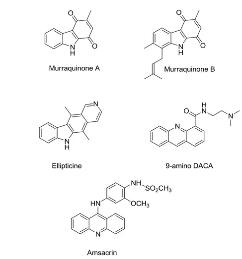
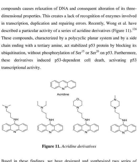
During my PhD thesis work three different structural motives present in natural products have been considered to be suitable scaffold in the design of new antitumoral agents: carbazoles, acridines and spirooxindole derivatives. Carbazoles either in a pure substituted or in an annellated substituted form, represent an important and heterogeneous class of anticancer agents, which has grown considerably over the last two decades. Recently, Wong et al. have described a particular activity of a series of acridine derivatives characterized
III
by a polycyclic planar system and by a side chain ending with a tertiary amine, act stabilized p53 protein by blocking its ubiquitination, without phosphorylation of ser15 or ser20 on p53. Furthermore, these derivatives induced p53-dependent cell death, activating p53 transcriptional activity Based on the structural cytotoxic requirements for these class of products my first PhD project was centered on synthesis of two series of compounds in which a carbazole eskeleton were linked by an alkyl chain to an amine (series 1) or substituted amide (series 2) groups.
In the other hand, small molecule natural products containing spirooxindole derivatives have demonstrated to be invaluable tools in the discovery and characterization of critical events for the progression and the regulation of the cell cycle. Based on the spirotryprostatin-A structure, during my PhD project II I designed, synthesized, and evaluated different series of compounds belonging to the diketopiperazine structural class as potential cell cycle modulators and cytotoxic agents. Starting from the spirooxindolthiazolidine scaffold, amide coupling with Pro derivatives and intramolecular cyclization reactions are suitable synthetic methods to generate chemically diverse diketopiperazine system, such as hexahydropyrrolo[1,2-a][1,3]thiazolo[3,2-d]pyrazine-5,10-dione (structure I), hexahydropyrrolo[1,2-a] [1,3]thiazolo[3,4-d]pyrazine-5,10-dione (structure II) and, spiroindol-2-one[3,30]hexahydro-5,10H-pyrrolo[1,2-a][1,3]thiazolo[3,4-d]pyrazine-5,10-dione (structure III). Some of these compounds, especially those who belong to the series I and II, showed interesting cytotoxic activity.
In the last part of my project, I have designed and synthesized two libraries of compounds based on the spirooxindol-thyazolidine moiety, analogues of spirooxoindol-pyrrolidine template as p53-MDM2 inhibitors.
Compounds (3R,7aR)-6-(4-chlorobenzyl)-1H-spiro[imidazo[1,5-c]thiazole-3,3‘-indoline]-2‘,5,7(6H,7aH)-trione (42c) and (3R,7aR)-50-methyl-6-(3,4,5-
trimethoxybenzyl)-1H-spiro[imidazo-[1,5-c]thiazole-3,3‘-indoline]-IV
2‘,5,7(6H,7aH)-trione (43d) are the most potent compounds of this series, inhibiting cell growth of different human tumor cells at submicromolar and micromolar concentrations, respectively. Compound 42c induces apoptotic cell death in human melanoma cell line M14 at 24 h, while in the same condition, treatment with 43d shows a clear arrest at G2/M phase inducing delay of cell cycle progression. Possibly, these activities may be due to inhibition of p53-MDM2 interaction and subsequent p53 release and activation.
1
CHAPTER 1:
INTRODUCTION
2
1. PROLIFERATION, CELL CYCLE AND APOPTOSIS IN CANCER
1.1 Evolution of cancers
Since its inception, the study of the molecular basis of cancer has carried with it the promise of more refined, more effective cancer therapies. It has generally been assumed that because cancers are derived from numerous tissues with multiple etiologies, and as tumor progression carries with it a bewildering and seemingly endless combination of genetic and epigenetic alterations giving rise to a hugely disparate series of diseases, cures for cancer must be as diverse as the diseases themselves.
Although neoplasia involves many other processes that also present targets for cancer therapy,1 in almost all instances, deregulated cell proliferation and suppressed cell death together provide the underlying platform for neoplastic progression. The challenge before the research community is to identify and understand the molecular anatomy of such pivotal steps in tumor progression and to develop therapies that directly these points of convergence.
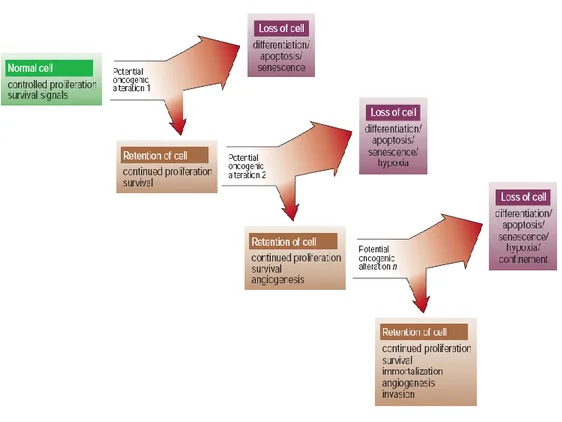
Cancers are diseases in which unremitting clonal expansion of somatic cells kills by invading, subverting and eroding normal tissues. Driving cancer development are stochastic somatic cell mutations in genes that govern and regulate the diverse aspects of metazoan growth control.Evolution of cancer is more complex than the straightforward linear accumulation of oncogenic mutations. Potentially oncogenic proliferative signals are coupled to a variety of growth-inhibitory processes, such as the induction of apoptosis, differentiation or senescence, each of which restricts subsequent clonal expansion and neoplastic evolution. Tumor progression occurs only in the
3
very rare instances where these growth-inhibitory mechanisms are thwarted by compensatory mutations (Figure 1).
\
Figure 1: Evolution of cancer
The processes governing the genesis and progression of cancers are evolutionary ones in which natural selection acts upon the inherent or acquired diversity of various somatic clones, fostering the outgrowth of those with some form of propagative advantage. Metazoans must restrain this tendency of individual somatic cells to establish their own autonomous colonies, yet at the same time sanction sufficient somatic cell proliferation to build and maintain the whole organism.
Unfortunately, long-lived organisms such as vertebrates need substantial and continuous cell proliferation throughout their extended lives, both for
4
development and long-term maintenance and repair. In teleological terms, the evolutionary imperative of vertebrates has been to find a way to allow cell proliferation when needed, while at the same time efficiently suppressing the genesis of mutated cells leading to deregulated growth. When such measures fail, cancer is the inevitable consequence.
Awareness of the evolutionary nature of cancer offers a number of important insights into the malignant process. First, and perhaps most striking, is the rarity of the cancer cell. With an estimated mutation rate of some 1 in 22107 per gene cell division2, some 1014 target cells in the average human, and an abundant repertoire of genes regulating all aspects of cell expansion, it is remarkable that cancers arise in only 1 in 3 lifetimes. This is even more striking when one considers that oncogenic mutations, by their nature, foster clonal expansion of the affected cell, so propagating the initial mutation and thereby increasing the number of target cells available for (and hence the probability of) further oncogenic mutation. The rarity of cancer highlights the efficacy of potent anti-tumorigenic mechanisms presiding over somatic cells. Cancers prevail only when these mechanisms have failed.3
Second, cancers ‗progress‘ for the same reason organisms seem to — we see only the successes, not the failures. This distorts our statistical view of cancer progression. No matter how rare the genesis and evolution of a cancer cell or how effective the anti-cancer therapy administered, our perception is only of the rare surviving clones that beat all the odds and appear as clinical disease. Our inability to discern the mechanisms that thwart the vast majority of inchoate tumors deprives us of great insight into how these mechanisms break down in cancer and, correspondingly, how we might best reactivate them. Third, evolutionary trajectories of cancers are shaped by the selective pressures they encounter. Tumors evolve within differing somatic environments, each of which imposes its own unique constraints.
5
Fourth, evolution is an ongoing process. As a neoplasm progresses, expands and spreads, it confronts shifting selective pressures. The heterogeneity and diversity seen in cancers are vestiges of a dynamic and stochastic evolutionary force that varies with differing somatic environments.
1.2 The commonality of cancers 4
Tumors are diverse and heterogeneous, but all share the ability to proliferate beyond the constraints limiting growth in normal tissue. Aberrations in the regulation of a restricted number of key pathways that control cell proliferation and cell survival are mandatory for establishment of all tumors. Deregulated cell proliferation together with suppressed apoptosis constitute the minimal common platform upon which all neoplastic evolution occurs. The critical issue is to identify how tumor cells differ from normal cells and how those differences can be exploited therapeutically.
1.2.1. Carcinogenesis
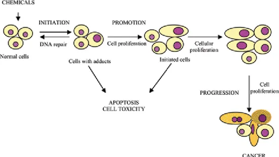
The transformation of a normal cell into a cancerous cell is a complex and multi-step process that include initiation, promotion, and progression (Figure 2). A number of factors such as diet, toxins, smoking, alcohol, obesity and infections are attributed to the development of cancer.
These factors help in the carcinogenesis process by inducing oxidative damage to the DNA. If a cell containing damaged DNA divides before the DNA can be repaired, the result is likely to be a permanent genetic alteration constituting a first step in carcinogenesis. Body cells that divide rapidly are more susceptible to carcinogenesis because there is less opportunity for DNA repair before cell division. Factors as the variation in epigenetic code or aberrations in tumor suppressor genes, oncogenes, and genes involved in repair machinery leads to uncontrolled growth by recessive loss of tumor
6
suppressor genes or dominant gain of oncogens and/or aneuploidy. Viruses, or mutagenic spontaneous changes in the components of signaling pathways lead also to cellular transformation and have also been implicated in the cause of cancer.
Figure 2. Carcinogenesis is initiated with the transformation of the normal
cell into a cancer cell (initiated cell).
Causative factors may vary among different cancers but the basic hypothesis that a normal human cell has to acquire at least six essential alterations to become a malignant cancerous cell in case of solid tumors. These six alterations include: the ability to sustain autonomous growth; the ability to
avoid growth inhibitory signals; the evasion of apoptosis; the potential to replicate infinitely; sustained angiogenesis; and finally, for the malignant tissue to be invasive and metastatic.1
Cancer cells contain many altered and/or mutated genes. These almost always include: mutations in genes that are involved in mitosis; that is, in genes that
7
control the cell cycle. Their mutated or over-expressed products stimulate mitosis even though normal growth signals are absent. Example: many
tyrosine kinase receptors including epidermal growth factor receptor (EGFR),
the gene encoding the receptor for epidermal growth factor (EGF) (EGFR is also known as HER1). Moreover, for altering cell homeostasis in favor of cellular immortality cell has to by bass cell signals, which inhibit uncontrolled cell proliferation, they are tumor suppressor genes, and these genes normally inhibit mitosis. Example: the p53 gene product normally senses DNA damage and either halts the cell cycle until it can be repaired or, if the damage is too massive; triggers apoptosis. During the course of cellular immortalization different cell signaling pathways are altered in favor of tumorogenesis and understanding of these altered cell signaling pathways in detail helps to design anticancer therapies
1.3 The cell cycle and its regulation
With advancements in our understanding of the basic mechanisms of oncogenesis, we have gained a greater appreciation for the critical role that cell cycle regulation plays in malignant transformation and in the development of resistance to chemotherapy. Perturbations in the cell cycle are described commonly in carcinogenesis. Furthermore, with our improved understanding of the effects of chemotherapy on healthy and cancerous cells, it is increasingly apparent that the cell cycle also plays a critical role in the development of resistance to chemotherapy. These observations have led to the development of a new class of anticancer therapeutics in clinical development today.5
The fundamental task of the cell cycle is to ensure that DNA is faithfully replicated once during S phase and that identical chromosomal copies are distributed equally to two daughter cells during M phase.6 The machinery for
8
DNA replication and chromosome segregation is insulated from interruption by extracellular signals, and its essential and autonomous nature implies that damage to the pivotal components would be highly debilitating, if not fatal, to cells. Therefore, genes commanding these processes should not be frequent targets of mutation, deletion, or amplification in cancer. Oncogenic processes exert their greatest effect by targeting particular regulators of GI phase progression.7 During the G, phase, cells respond to extracellular signals by either advancing toward another division or withdrawing from the cycle into a resting state (Go).8 Unlike transit through the S, G2, and M phases, G progression normally relies on stimulation by mitogens and can be blocked by antiproliferative cytokines. Cancer cells abandon these controls and tend to remain in cycle, and because cell cycle exit can facilitate maturation and terminal differentiation, these processes are subverted as well. The decision to divide occurs as cells pass a restriction point late in GI, after which they become refractory to extracellular growth regulatory signals and instead commit to the autonomous program that carries them through to division. An appreciation of restriction point control is central to our understanding of how and why cancer cells continuously cycle.
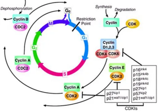
The cell cycle is a critical regulator of the processes of cell proliferation and growth as well as of cell division after DNA damage. It governs the transition from quiescence (G0) to cell proliferation, and through its checkpoints, ensures the fidelity of the genetic transcript. It is the mechanism by which cells reproduce, and is typically divided into four phases. The periods associated with DNA synthesis (S phase) and mitosis (M phase) are separated by gaps of varying length called G1 and G2 (Figure 3). Progression of a cell through the cell cycle is promoted by a number of CDKs which, when complexed with specific regulatory proteins called cyclins, drive the cell forward through the cell cycle. There exist corresponding cell cycle inhibitory proteins (CDK inhibitors [CDKIs]) that serve as negative regulators of the cell
9
cycle and stop the cell from proceeding to the next phase of the cell cycle (Fig 1). The INK4 (for inhibitor of cdk4) class of CDKIs, notably p16lnk4a, p15lnk4b, p18lnk4c, and p191nk4 days, bind and inhibit cyclin D–associated kinases (CDK2, -4, and -6). The kinase inhibitor protein (KIP) group of CDK inhibitors, p21waf1, p27kip1, and p57kip2, negatively regulate cyclin E/CDK2 and cyclin A/CDK2 complexes.9
Figure 3. The cell cycle. The cell cycle is divided into four phases (G1, S, G2,
and M). Progression through the cell cycle is promoted by cyclin-dependent kinases (CDKs), which are regulated positively by cyclins and negatively by CDK inhibitors (CDKIs). The restriction point is the point at which cells progress through the cell cycle independent of external stimuli.
10
The pattern of cyclin expression varies with a cell‘s progression through the cell cycle, and this specific cyclin expression pattern defines the relative position of the cell within the cell cycle.10 At least nine structurally related CDKs (CDK1- CDK9) have been identified, though not all have clearly defined cell cycle regulatory roles. A considerable number of cyclins have been identified to date (cyclin A–cyclin T). CDK/cyclin complexes themselves become activated by phosphorylation at specific sites on the CDKby cdk7/cyclin H, also referred to as CDK-activating kinase (CAK).11 Cyclin D isoforms (cyclin D1-D3) interact with CDK2, -4, and -6 and drive a cell‘s progression through G1. The association of cyclin E with CDK2 is active at the G1/S transition and directs entry into S phase. S phase progression is directed by the cyclin A/CDK2 complex, and the complex of cyclin A with CDK1 (also known as cdc2) is important in G2. CDK1/cyclin B is necessary for mitosis to occur.
1.3.1 Cancer as a disease of deregulated cell proliferation
Each of the pathways that constrains the proliferative response in normal cells is perturbed in most cancers. One class of mutations required for tumor development acts by short circuiting the normally obligate requirement of somatic cells for external mitogenic signals.12 Such mutations may involve autocrine production of a normally limiting mitogen, activating mutations of the mitogen RTKs or G-protein signal transducers such as Ras, or mutations affecting one of the many intermediary signal transducing molecules that convey mitogenic information to its intracellular targets. A second class of growth-deregulating mutations comprises those that target the principal late-G1 cell-cycle checkpoint regulated by pRB.13 Defects in this pathway, which may be universal in human cancers, include deletion of the RBgene itself and deregulation of the CDKs that phosphorylate and functionally inactivate pRB,
11
either through direct over-activation of CDKs or through genetic loss of their inhibitors.14 Another frequent proliferative lesion that has the effect of deregulating the cell cycle is uncontrolled expression of Myc.15 Myc expression is tightly controlled by mitogen availability in normal cells, but it is usually expressed in a deregulated or elevated manner in tumor cells. Myc seems to be a strategic controller of cell proliferation that acts pleiotropically to coordinate both cell growth 16 and concomitant progression through the cell cycle.17
The presence in individual tumors of multiple mutations that affect each of the pathways discussed above suggests that each pathway contributes a discrete type of proliferative function to the neoplastic phenotype. But precisely what such functions are and how and why they interact, remains unknown.
In addition to driving aberrant cell division, mutations in the various proliferative control pathways have a profound impact on other cell functions. For example, many of the proliferative lesions in tumor cells also contribute to the inhibition of differentiation, thereby preventing the elimination of progeny cells from the proliferative compartment of many types of tissue. pRB, for example, is essential in differentiation of several tissue types through interactions with factors such as the helix–loop–helix proteins MyoD2618 and Id2. Loss or inhibition of pRB function prevents normal differentiation, a contribution to tumor development distinct from the direct deregulation of cell-cycle progression. Deregulated Myc expression also inhibits differentiation, in part by activation of Id2 expression.19
1.4. Apoptosis in health and disease 20
Apoptosis, or programmed cell death, is a normal component of the development and health of multicellular organisms. Cells die in response to a
12
variety of stimuli and during apoptosis they do so in a controlled, regulated fashion. This makes apoptosis distinct from another form of cell death called necrosis in which uncontrolled cell death leads to lysis of cells, inflammatory responses and, potentially, to serious health problems. Apoptosis, by contrast, is a process in which cells play an active role in their own death (which is why apoptosis is often referred to as cell suicide).
Apoptosis occurs during the normal development of multicellular organisms and continues throughout adult life. The combination of apoptosis and cell proliferation is responsible for shaping tissues and organs in developing embryos. For example the apoptosis of cells located in-between the toes allows for their separation. Apoptosis is also an important part of the regulation of the immune system. T lymphocytes are cells of the immune system that are responsible for destroying infected or damaged cells in the body. They mature in the thymus, but before they can enter the bloodstream they are tested to ensure that they are effective against foreign antigens and are also not reactive against normal, healthy cells. Any ineffective or self-reactive T-cells are removed through the induction of apoptosis. Problems with the regulation of apoptosis have been implicated in a number of diseases. Cancer is a disease that is often characterized by too little apoptosis. Cancer cells typically possess a number of mutations that have allowed them to ignore normal cellular signals regulating their growth and become more proliferative than normal. Under normal circumstances damaged cells will undergo apoptosis, but in the case of cancer cells mutations may have occurred that prevent cells from undergoing apoptosis. In these cases there is no check on the cellular proliferation and consequently the disease can progress to the formation of tumors. In many cases these tumors can be difficult to kill as many cancer treatments rely on damaging the cells with radiation or chemicals and mutations in the apoptotic pathway often produce cells that are resistant to this type of attack.
13 1.4.1 The apoptotic process 20
Upon receiving specific signals instructing the cells to undergo apoptosis a number of distinctive changes occur in the cell. A family of proteins known as caspases are typically activated in the early stages of apoptosis. These proteins breakdown or cleave key cellular components that are required for normal cellular function including structural proteins in the cytoskeleton and nuclear proteins such as DNA repair enzymes. The caspases can also activate other degradative enzymes such as DNases, which begin to cleave the DNA in the nucleus.
There are a number of mechanisms through which apoptosis can be induced in cells. The sensitivity of cells to any of these stimuli can vary depending on a number of factors such as the expression of pro- and anti-apoptotic proteins (eg. the Bcl-2 proteins or the Inhibitor of Apoptosis Proteins), the severity of the stimulus and the stage of the cell cycle. Some of the major stimuli that can induce apoptosis include virus infection, cell stress and DNA damage.
In some cases the apoptotic stimuli comprise extrinsic signals such as the binding of death inducing ligands to cell surface receptors called death receptors. These ligands can either be soluble factors or can be expressed on the surface of cells such as cytotoxic T lymphocytes. The latter occurs when T-cells recognize damaged or virus infected cells and initiate apoptosis in order to prevent damaged cells from becoming neoplastic (cancerous) or virus-infected cells from spreading the infection. Apoptosis can also be induced by cytotoxic T-lymphocytes using the enzyme granzyme. In other cases apoptosis can be initiated following intrinsic signals that are produced following cellular stress. Cellular stress may occur from exposure to radiation or chemicals or to viral infection. It might also be a consequence of growth factor deprivation or oxidative stress caused by free radicals. In general intrinsic signals initiate apoptosis via the involvement of the mitochondria. The relative ratios of the
14
various bcl-2 proteins can often determine how much cellular stress is necessary to induce apoptosis.
1.4.2 Cancer as a disease of deregulated survival
Survival of all somatic cells requires the continuous input of survival and trophic signals to suppress apoptosis. The central engines of apoptosis are the caspases, cascades of cysteine aspartyl proteases that implement cell death by cleaving a variety of intracellular substrates that trigger cell dissolution. Caspases are synthesized as latent zymogens that are activated by proteolytic cleavage: typically through the action of upstream apical caspases. One such pathway is mediated by transmembrane death receptors of the CD95 (Apo-1 or Fas)/TRAIL/tumor-necrosis factor (TNF) receptor 1 family, whose ligation triggers recruitment and assembly of multiprotein complexes that activate apical caspase 8.21 The other principal death-signaling pathway involves the mitochondrion, which acts as an integrating sensor of multiple death insults by releasing cytochrome c into the cytosol where it triggers caspase activation. The mitochondrial pathway is thought to be the principal target of survival signaling pathways, which act by stabilizing mitochondrial function and integrity and suppressing release of cytochrome c.22 Once cytochrome c has been released from the mitochondrion, it orchestrates assembly of an intracellular apoptosome complex that recruits apical caspase 9 via the adaptor protein Apaf-1.23 The anti-apoptotic oncoproteins Bcl-2 and Bcl-xL, which exert their principal effects through stabilization of the mitochondrion, are found to be overexpressed in several tumor types and recent analyses have indicated that loss of Apaf-1 is a relatively frequent event in malignant melanoma that presumably confers resistance to apoptosis.24 A particularly potent driving force for the suppression of apoptosis in tumor cells is the coupled relationship between cell proliferation and cell death, a phenomenon
15
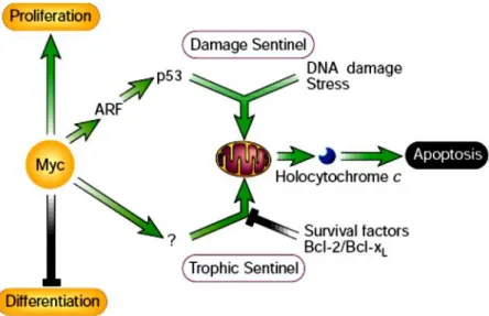
exemplified by the Myc protein. In addition to its well documented growth-promoting property, Myc was found to be a powerful inducer of apoptosis, especially under conditions of stress, genotoxic damage or depleted survival factors.25 Consideration of such observations led to the proposal that the innate apoptotic potential of Myc serves as an inbuilt foil to its oncogenic capacity (Figure 4).
Figure 4 Activation of growth-deregulating lesions triggers ‘sentinel’ functions that guard the cell against acquiring mutations or propagating into an inappropriate somatic compartment.
In this example, the oncoprotein Myc is shown activating a p53 damage sentinel through the ARF/MDM-2 pathway, thereby sensitizing the cell to any DNA damage. Myc also promotes release of holocytochrome c from the mitochondrion into the cytosol where it triggers apoptosis. Release of holocytochrome c is inhibited by paracrine ‗survival‘ signals that are typically restricted both in supply and location. Clonal outgrowth driven by relentless Myc expression outstrips survival factor availability, triggering the ‗trophic sentinel‘ to kill the cell.
16
Another common pathway through which a wide variety of proliferative signals influence the apoptotic program is through induction of ARF, an alternate product of the INK4alocus, one of whose functions is to trigger upregulation of p53 through its inhibitory action on MDM-2.26
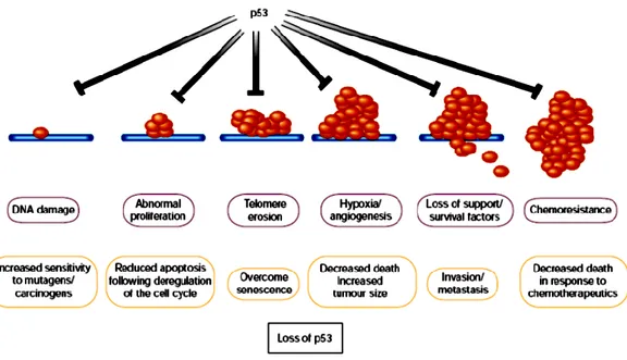
Another potent selective pressure in cancers to suppress apoptosis arises from the fact that programmed cell death is the typical response of somatic cells to many forms of stress and damage; in particular damage to cell DNA (a fact exploited by most classical cancer therapeutics). Stress-associated signals that activate apoptosis include many of those encountered by the incipient tumor cell, including hypoxia and nutrient deprivation, as well as DNA damage arising from telomere erosion, defective repair, oncogene deregulation and therapy. The p53 protein is important in transducing such diverse signals into tumor-suppressive apoptotic or growth-arresting responses, which implies that there is strong selection for tumor cells to loose p53 function.27 Importantly, differing p53-activating stresses tend to arise at different stages of carcinogenic progression. For example, oncogene deregulation occurs early, as it is a prerequisite for clonal expansion, whereas hypoxia is significant only after the tumor reaches macroscopic size. Consequently, p53 exerts a tumor-suppressive role at multiple stages of carcinogenic progression (Figure 5), offering an explanation for why loss of p53 has such a profound effect on tumor development.
17
Figure 5. Many stress signals encountered during tumor progression activate
p53, resulting in apoptosis or growth arrest.
Loss either of the ability to activate p53 or of p53 function itself has considerable impact on the ‗success‘ of the carcinogenic process, as it increases the chances of a tumor cell surviving progressively adverse conditions. Inability to activate p53 in response to stress signals encountered early during tumor development, such as deregulated proliferation, may to be sufficient to allow the formation of preneoplastic lesions. However, lesions that suppress activation of p53 in response to such oncogene-associated stress signals do not necessarily block activation of p53 by subsequent events encountered during malignant progression, such as DNA damage. Consequently, additional alterations in pathways that activate or respond to p53, or loss of p53 by direct mutation of the gene itself, may be selected during progression to more malignant cancers.
18
1.5 Therapeutic targeting of cell proliferation and apoptosis
Because deregulated proliferation and inhibition of apoptosis lie at the heart of all tumor development, they present two obvious targets for therapeutic intervention in all cancers. Clearly there are numerous mechanisms through which these two defects can occur, and the success of targeted therapy will depend to a large part on the molecular fingerprinting of individual tumours.28 Although most existing cancer drugs are anti-mitotic, they act not by targeting the specific lesions responsible for deregulated tumor growth, but by crudely interfering with the basic machinery of DNA synthesis and cell division. Moreover, we now know that the surprising selectivity of such crude agents results largely from the increased sensitivity to apoptosis afforded to tumor cells by their oncogenic lesions. Drugs designed to specifically inhibit growth-deregulating lesions are currently being tested in clinical trials, and include inhibitors of RTKs, Ras, downstream signaling kinases such as the mitogen-activate protein kinase and Akt pathway, and CDKs.
At first glance, targeted inhibition of growth-deregulating lesions in cancer would be seem to have limited therapeutic efficacy, as they would at best be cytostatic. However, unexpected therapeutic bonuses may emerge from such an approach because growth deregulation induces a plethora of downstream activities in affected cells and their adjacent tissues. Therapeutic inhibition of the offending oncoprotein in tumors arising from cell lineages where terminal differentiation has been blocked could be sufficient to trigger a resumption of that differentiation program, permanently expelling the tumor cell from the proliferating compartment.29
The second obvious strategy for cancer therapy is to target the lesions that suppress apoptosis in tumor cells. The potent proapoptotic effects of growth-deregulating mutations mean that tumors are peculiarly dependent upon their particular suite of antiapoptotic mutations for continued survival. Thus,
19
although apoptosis in tumor cells is sufficiently suppressed to below a critical threshold to enable them to survive, they remain acutely sensitized to apoptosis. In most, if not all, cancer, this ability to survive results in part from inhibition of the p53 pathway, either by inactivating mutations in p53 itself, perturbation of the signaling pathways that allow activation of p53 in response to stress, or defects in the downstream mediators of p53-induced apoptosis. Reintroduction of p53 function is sufficient to induce apoptosis in many tumor cells, and several mechanisms to reactivate p53 are being considered as therapeutic strategies. These include introduction of wild-type p53 into tumors expressing a mutant protein, or inhibition of negative regulators of p53, such as MDM-2, in those tumors that retain wild-type p53.30
Regardless of efficiency in cell killing, the success of repairing the apoptotic response in tumor cells depends on the extent to which such therapies confine death to the cancer cells, and allow survival of normal tissue. Many conventional chemotherapies induce significant toxicity, particularly in tissues that normally maintain a proliferative compartment, such as gut epithelium and the hematopoietic system. This DNA damage-induced toxicity is mediated in part through p53, leading to the suggestion that inhibition of p53 in these normal tissues may protect against drug-induced toxicity, thereby improving the tolerance of conventional cancer therapies. However, implicit in the development of drugs that target specific lesions responsible for tumor cell growth is the prediction that these approaches will show significantly more specificity for tumor cell killing than conventional therapies.
Although activation of apoptotic pathways can lead to the death of untransformed cells, a process that is essential in normal development, a fundamental difference exists between tumor cells and their normal counterparts, as normal cells neither have to sustain the pro-apoptotic onslaught that is inherent in deregulated proliferation, nor survive away from
20
their usual environment in the absence of requisite survival signals. Repair or replacement of a single apoptotic signal, be it reactivation of p53 or removal of a survival signal, could well prove too much for a tumor cell already burdened with a heavy apoptotic load. By contrast, the same perturbation may scarcely ruffle the equilibrium of a normal cell, safely buffered in its appropriate soma and enjoying the full gamut of trophic support that ensures normal cell survival.
21
CHAPTER 2:
INTRODUCTION
22
2. p53: TARGET FOR CANCER THERAPY
2.1 p53 network
p53, (53KD) also known as tumor protein 53 (TP53), is a transcription factor that regulates the cell cycle and apoptosis, in case of cellular insults, and hence functions as a tumor suppressor. p53 has been described as "the guardian of the genome", "the guardian angel gene", or the "master watchman", referring to its role in conserving stability by preventing genome mutation. The transcription factor p53 responds to diverse cellular stresses to regulate target genes that induce cell cycle arrest, apoptosis, senescence, DNA repair, or changes in metabolism.31 In addition, p53 appears to induce apoptosis through nontranscriptional cytoplasmic processes.32 In unstressed cells, p53 is kept inactive essentially through the actions of the ubiquitin ligase MDM2, which inhibits p53 transcriptional activity and ubiquitinates p53 to promote its degradation.32Numerous posttranslational modifications modulate p53 activity, most notably phosphorylation and acetylation. Several less abundant p53 isoforms also modulate p53 activity. Activity of p53 is ubiquitously lost in human cancer either by mutation of the p53 gene itself or by loss of cell signaling upstream or downstream of p53
2.1.1 Role of p53: normal versus cancer cells
Activation of p53 can result in a number of cellular responses, and it is possible that different responses are induced by different stress signals. There is evidence that p53 can play a part in determining which response is induced through differential activation of target-gene expression. Although the importance of these responses to tumor suppression is clear, previously unanticipated contributions of these responses to other aspects of human
23
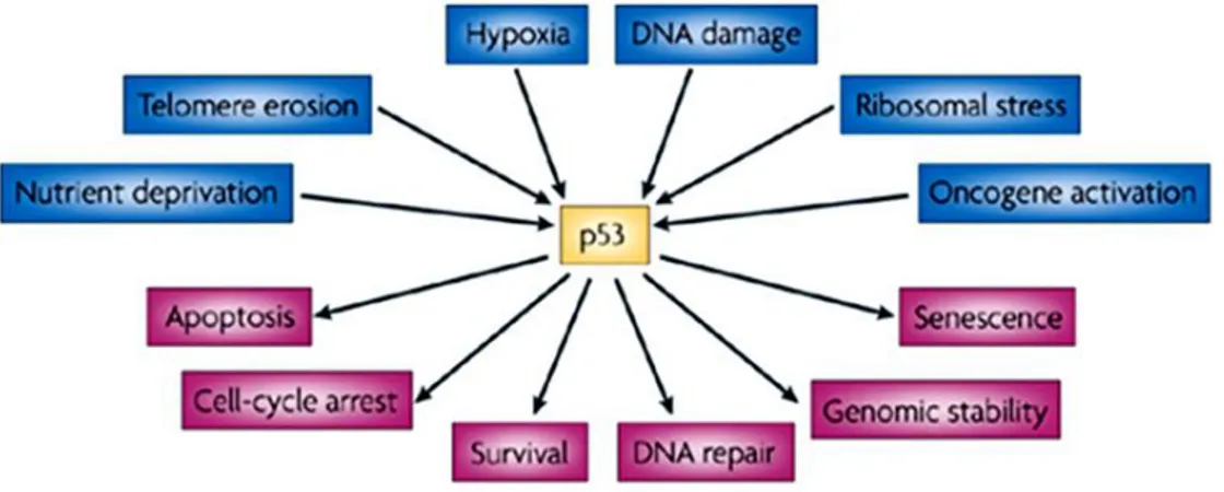
health and disease are being uncovered. The role of p53 in tumor suppression, development and ageing is likely to depend on which cellular response is activated and on the context in which the activation occurs. p53 is an intensively studied protein, its fame stemming mainly from its clear role as a tumor suppressor in humans and other mammals.33 Loss or mutation of p53 is strongly associated with an increased susceptibility to cancer, and most functions of p53 have been considered in the light of how p53 might protect from malignant progression.34 Some p53-null mice can develop normally 35 an observation that has been taken to rule out major functions for p53 in normal physiology. But recent studies are questioning whether p53 is truly such a single-minded protein, and other functions of p53 that might be profoundly important during normal life are being uncovered. These include roles for p53 in regulating longevity and ageing, glycolytic pathways that might determine endurance and overall fitness, and apoptotic responses during ischaemic and other types of stress. Evidence for genetic variations in the activity of the p53 pathway in humans gives these ideas extra relevance.36 One of the major mechanisms by which p53 functions is as a transcription factor that both positively and negatively regulates the expression of a large and disparate group of responsive genes (Figure 6).37 Although some of these p53- responsive genes have an important role in mediating cell-cycle arrest, senescence and apoptosis (the best understood activities of p53), it is now evident that the ability of p53 to influence gene expression has wider reaching effects. Numerous studies have identified p53-regulated genes that could have a role in a number of different and sometimes unexpected responses.38 Although some of these still need to be fully validated, there is now clear evidence for a role of p53 in the regulation of glycolysis,39 and autophagy, 40 the repair of genotoxic damage, 41 cell survival and regulation of oxidative stress,42 invasion and motility,43 cellular senescence,44 angiogenesis,45 differentiation,46 and bone remodeling.47 The cellular pathways in which p53
24
is involved, are schematically represented in Figure 6. In these aspects, its worthy to analyze there is any cancer cells are expressing wild type p53, and if they are expressing, its role in cancer cells has to be studied before clinical use of p53 mediated gene therapy as an anticancer therapy.
Figure 6. Activation and functions of p53. p53 has a key role in integrating
the cellular responses (pink arrows) to different types of stress (blue arrows).
2.2 The role of p53 in human cancer
In the two decades since its original discovery, p53 has found a singularly prominent place in our understanding of human cancer. Although the biochemistry of p53 has been worked out in some detail, our knowledge of the biologic consequences of p53 dysfunction is still quite rudimentary. p53 dysfunction in cancer cells are mainly due to its mutation (50%), epigenetic modulation at expressional level and low persistence of p53 protein level due to its enhanced turnover. Most p53 mutations found in human cancers are not null mutations but rather encode mutant version of the p53 protein that may have unwanted activities such as a gain-of-function or be dominant negative inhibitors of wt p53 activity. In this regard, it will be important to determine how best to harness the complex properties of p53‘s ability to induce cellular growth arrest and cell death to generate novel, effective approaches to cancer
25
therapy. Furthermore, a clearer appreciation of the direct interaction epigenetic factors with p53 will lead to development of strategies to inhibit tumor initiation and progression.
2.2.1 p53; the guardian of the genome
DNA damage was the first type of stress found to activate p53 and, based on this, p53 has been widely regarded as ―the guardian of the genome‖.48 Extensive characterization of the signaling routes that connect DNA damage with p53 have identified a cascade of Ser/Thr kinases that includes ATM, ATR, Chk1 and Chk2, which phosphorylate p53.49 This signaling cascade is permanently activated in human cancer, suggesting that the cancerous state is intrinsically associated to the generation of DNA damage .50 The constitutive DNA damage present in cancer cells is thought to emanate primarily from the strong generation of reactive oxygen species, 51 as well as, from the aberrant firing of DNA replication origins.52
.Recent characterization of mice genetically manipulated with a knocked-in p53 that cannot be phosphorylated at two of the main residues targeted by ATM/ATR/Chk1/Chk2, namely, Ser18 and Ser23 (Ser15 and Ser20 in human p53), indicates an important role of these phosphorylation sites in some, but not all, the DNA damage induced and p53-dependent responses.53 In agreement with this, mice carrying p53S18A/S23A alleles are tumor prone,54 although this phenotype is considerably milder than in the case of p53-null mice.55
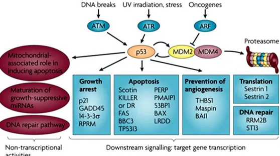
26
Figure 7. p53 is at the center of a complex web of biological interactions that
translates stress signals into cell cycle arrest or apoptosis26. 53BP1, p53 binding protein 1; ATM, ataxia telangiectasia mutated; ATR, ataxia telangiectasia and Rad3 related; BAI1, brain-specific angiogenesis inhibitor 1; BAX, BCL2-associated X protein; BBC3, BCL2 binding component 3 (also known as PUMA); DR, death receptor; GADD45, growth arrest and DNA-damage-inducible 45; KILLER, p53-regulated DNA DNA-damage-inducible cell death receptor (also known as TNFRSF10B); LRDD, leucine-rich repeats and death domain containing; mRNA, microRNA; PMAIP1, phorbol-12-myristate-13-acetate-induced protein 1 (also known as NOXA); RPRM, reprimo; RRM2B, ribonucleotide reductase M2 B; ST13, suppression of tumorigenicity 13 (also known as p48); TP53I3, tumor protein p53 inducible protein 3; THBS1, thrombospondin 1; UV, ultraviolet.
These data suggest that the activation of p53 in response to DNA damage occurs through multiple pathways, which in addition to the well-established kinase cascade of ATM/ATR/Chk1/Chk2, probably include other kinases such as p38, JNK/SAPK and c-Abl (Figure 7).56 Regarding human cancer, the
27
available information gathered from the analysis of epigenetic aberrations indicates that the aforementioned DNA damage signaling kinases are not, in general, significant targets of genetic and epi-genetic inactivation.57 The only exception to this is found in hematological malignancies, which present a high incidence of mutations in ATM (13–40% depending on the particular type of malignancy).58 In line with this, a recent large-scale sequencing effort of 210 diverse human cancers has identified ATM among the three most frequently mutated kinases (5% incidence).59 Based on the above genetic evidence, it can be concluded that DNA damage is conveyed to p53 through multiple redundant pathways in which many transducers participate, but none of them plays a critical role and, therefore, alteration of a single component does not have a significant impact on p53 function.
According, upstream signaling to p53 increases its level and activates its function as a transcription factor in response to a wide variety of stresses, whereas downstream components execute the appropriate cellular response (see below). The principal sensors seem to be MDM2 and MDM4 and their interaction with p53.60 In non-stressed conditions these proteins bind p53, ubiquitylate it and target it for degradation by the proteasome. In stressed conditions the function of the MDM2–MDM4 complex is blocked by phosphorylation, protein-binding events and/or enhanced degradation. Hence, phosphorylation of MDM4 is essential for the p53 response to ionizing radiation, and the response to oncogene activation depends on the binding of ARF to MDM2. Many p53-activating small molecules function by causing the release of ribosomal proteins from the nucleolus to the nucleoplasm, where they bind to MDM2 and MDM4 and inhibit their function. Molecules that activate wild-type p53 in tumors by disrupting MDM2 activity can compensate for any missing upstream components of the p53 pathway, for example the loss of ARF expression that is frequent in cancer cells142. However, defective downstream p53 signaling might substantially decrease
28
their effectiveness. Therefore, the ability to identify tumors in which downstream p53 signaling is unaffected is important. The development of strategies to ensure that the desired p53 response is initiated when it is reactivated might be necessary and could require the judicious use of drug combinations.
2.2.1.1. p53; The policeman of the oncogenes
Among the many and varied stimuli that have been reported to activate p53, oncogenic signaling 61 has gained much attention because, as DNA damage, is also universally present in cancer. Therefore, analogous to the title of ―guardian of the genome‖, we can also assign to p53 the function of ―policeman of the oncogenes‖. Oncogenic signaling activates p53 through ARF,62 which, in turn, interacts with MDM2 inhibiting its p53-ubiquitin ligase activity.
In this manner, ARF-dependent stabilization of p53 results in a dramatic increase in p53 activity. Many transcription factors activate ARF in response to oncogenic signaling, most notably, Dentin matrix acidic phosphoprotein (DMP1).63 Mice lacking ARF have a remarkable tumor-prone phenotype, 64 although not as severe as p53-deficient mice,65 and there is good genetic evidence in mice supporting the relevance of the ARF/MDM2/p53 axis in tumor suppression.66 Importantly, mice deficient in ARF present a normal DNA damage response, indistinguishable from ARF-proficient mice.67 Regarding human cancer, the analysis of (epi)genetic alterations indicate that ARF is indeed inactivated with an extraordinary high frequency (~30%).68 However, inactivation of ARF almost invariably occurs in combination with the loss of p16INK4a, the cell cycle inhibitor, thus generating an ambiguity about which is the key targeted tumor suppressor. In this regard, it should be mentioned the existence of germline point mutations that inactivate ARF alone (i.e., sparing p16INK4a) in human kindreds predisposed to cancer.69
29
Nonetheless, the number of germline mutations that inactivate p16INK4a only (i.e., sparing ARF) outnumbers by a factor of ~20 those that inactivate ARF alone.70 Together, currently available evidence indicates that ARF is an important upstream regulator of p53, whose lack of activity has a significant impact on cancer.
2.2 p53 co-activators
The interaction between p53 and transcriptional co-activators also influences its affinity for promoters. It is therefore plausible that the specific co-factors expressed in a particular cellular context determine the repertoire of p53-target genes induced, and consequently whether the cell undergoes growth arrest or apoptosis, or even a particular apoptotic pathway, may be subject to the availability of co-activators.71
The Myc protein has been implicated as an important determinant of the choice between p53-induced growth arrest or apoptosis. Myc inhibits growth arrest in response to UV light, g-irradiation and DNA damage inflicted by reactive oxygen species.72 In the absence of Myc, cells that are exposed to UV light arrest in a p53- and Miz-1 (DNA-binding Myc-interacting zinc-finger 1)-dependent manner through activation of p21. However, when Myc is present, exposure to UV triggers its recruitment by Miz-1 to the proximal promoter region of p21. This interaction effectively represses p21 induction by p53 and other activators.73 Intriguingly, this repression appears to be specific for p21, because other p53-target genes, such as p53 upregulated modulator of apoptosis (PUMA) and PIG3, are induced. This block in p21 induction shifts the balance away from growth arrest towards apoptosis.73b It should be noted, however, that arrested cells are not necessarily protected from apoptosis. For example, normal thymocytes and mature lymphocytes undergo p53-mediated apoptosis under certain stress conditions.74 Interaction of p53 with several
30
other proteins specifically enhances the induction of apoptotic target genes. The apoptosis stimulating proteins of p53 (ASPP), for example, increases the DNA binding and transactivation activity of p53 on the promoters of apoptotic genes (e.g. Bax and PIG3), while failing to promote binding to the p21 promoter by a mechanism that remains to be defined.75 A novel insight into the interplay between p53 and its family members, p63 and p73, in the induction of apoptosis has been recently revealed by Flores et al.76
Their study of the effect of p63 and p73 on p53 transcriptional activity, using a selection of knockout mouse embryo fibroblasts (MEFs), defined two distinct classes of target gene. Whereas p53 alone is sufficient for the induction of p21 and Mdm2, the induction of the apoptotic genes PERP, Bax and Noxa requires p53 together with p63 and p73. This finding demonstrates an essential role for both p63 and p73 in the efficient induction of apoptotic target genes by p53. The mechanism of this cooperation is currently unknown, but it may involve an enhanced binding to and/or stabilization of the transcription complex on the promoters of p53 apoptotic target genes by the cooperative action of all three members.77 In addition to the contribution of p63 and p73 to the apoptotic function of p53, they play an important role in the precise control of cell death during normal mouse development. p73 also plays a role in the induction of cell death in response to DNA damage, a process involving cooperation between the Abl tyrosine kinase and p73.78
2.3 Downstream events of the p53 pathway
Once the p53 protein is activated, it initiates a transcriptional program that reflects the nature of the stress signal, the protein modifications and proteins associated with the p53 protein. The p53 protein binds to a specific DNA
31
sequence, termed the p53- responsive element (RE),79 and induces the expression of downstream genes. An algorithm that identifies p53-responsive genes in the human and mouse genome has been utilized to detect a number of new genes regulated by the p53 protein.80 The genes in this p53 network mainly initiate one of three programs that result in cell cycle arrest, DNA repair or apoptosis.
2.3.1 Growth arrest
p21WAF1/CIP1 is known to be a p53-downstream gene, and has been suggested to mediate p53-induced growth arrest triggered by DNA damage. The p21 protein is a cyclin-dependent kinase inhibitor that associates with a class of CDKs and inhibits their kinase activities. This will facilitate the accumulation of hypophosphorylated form of pRB that in turn associates with E2F inhibiting its transcriptional activity, leading to cell cycle arrest. As long as pRb is bound to E2F, the cell is prevented from entering into S phase. This G1 arrest affords the cell time to repair the DNA damage. Should repair be unsuccessful, P53 levels drop and CDK-cyclin protein kinase activity resumes, leading to entry into S phase. In the event that the DNA is not repair, p53 triggers apoptosis.81
2.3.2 DNA repair
Soon after having established TP53 as the most frequently altered gene in human tumors in the 1990s, 82 p53 was understood as a major component of the DNA damage response pathway.48,83 After the introduction of DNA injuries the level of p53 protein rises, which in turn induces a transient cell cycle arrest or apoptotic cell death. DNA damage activates p53 through
post-32
translational modifications by specific kinases, such as the strand break sensor ataxia telangiectasia mutated protein (Atm), by acetyltransferases like CREB-binding protein (Cbp)/p300, and by the poly (ADPribose) polymerase 1 (Parp-1), which prevent proteolysis via the Arf-mouse double minute 2 (Mdm2) pathway and/or enhance binding of p53 to consensus sequences within the genome.84 Initially, investigations on a direct participation of p53 in DNA repair were spurred by a number of biochemical observations. Thus, the C-terminal 30 amino acids of p53 were shown to recognize several DNA damage-related structures, such as DNA ends, gaps, and insertion/deletion mismatches.85 p53 was also demonstrated to catalyze reannealing of short stretches of single- and double-stranded DNA and to promote strand exchange between them .86 Further, p53 binds to three-stranded heteroduplex joints and four-stranded Holliday junction DNA structures with localization specifically at the junction, suggesting that p53 directly participates in recombinational repair.87 Moreover, several groups demonstrated a Mg2þ-dependent 3‘– 5‘exonuclease activity intrinsic to p53.88
Noticeably, the same central region within p53, where tumorigenic mutations are clustered, recognizes DNA sequence specifically, is required for junction specific binding of heteroduplex joints and is necessary and sufficient for the 3‘–5‘ exonuclease activity on DNA.89 In addition to p53‘s biochemical activities, numerous reports on physical and functional protein interactions further strengthened the proposal of a direct role of p53 in nucleotide excision repair (NER), base excision repair (BER), and double-strand break (DSB) repair.90
2.3.3 Apoptosis
Pivotal to the tumor-suppressor activity of p53 is its ability to activate apoptosis via multiple different pathways. Since the most-studied function of p53 is its role as a transcription factor that can activate transcription of an ever-increasing number of target genes, its transcriptional activation of
pro-33
apoptotic genes, as well as its transcriptional repression of anti-apoptotic genes, has been widely analyzed..91 However, although a large number of genes regulated by p53 during induction of apoptosis are known no single target gene has been identified whose altered expression alone can sufficiently explain p53 mediated transcription dependent apoptosis, and whose genetic deficiency phenocopies p53 deficiency in vivo. As an additional mode of p53‘s pro-apoptotic activity, recent studies have placed non transcriptional pro- apoptotic activities of p53 at the center of an active debate that aims to establish a comprehensive understanding of p53- mediated apoptosis.92
2.3.3.1 Cell cycle arrest or apoptosis?
The exact criteria that influence p53 to stimulate cell cycle arrest or apoptosis are only partially understood and are the subject of intense study. Several general factors that influence this decision include p53 expression levels, the type of stress signal, the cell type and the cellular context at the time of exposure to stress. Several intriguing observations have recently provided insight into the apparent intricacies of such cell fate determination. The examples described below involve the binding of p53 to its canonical binding sequence in target genes. Note, however, that p53 can also activate target genes through a non-canonical sequence. The first such example is in the p53-induced gene 3 (PIG3), which has been implicated in the accumulation of reactive oxygen species and apoptosis induction. PIG3 can be induced by p53 through a microsatellite sequence within its untranslated region.93 Another recently described example is the gene encoding the pro-apoptotic phosphatase PAC1, which is induced through binding of p53 to a novel palindromic binding site.94 This might represent a new mechanism for transcriptional regulation of apoptotic genes by p53, which differs from that already described (see below). Exacting discrimination between p53 arrest and apoptotic functions has been critical to the identification of the importance of
34
the latter in tumor suppression. A strong link between the apoptotic function of p53 and tumor suppression has been demonstrated using transgenic mice bearing an SV40 large T antigen (LT) mutant, which inhibited pRb function without directly compromising p53 activity.95 p53-mediated growth arrest is however impaired in these mice owing to pRb loss of function, but the apoptotic activity is functional. These mice develop choroid plexus tumors, but at a slow rate owing to continuous p53-dependent apoptosis. Elimination of p53 from these mice, by crossbreeding with p53-null mice, resulted in aggressive tumor development. This finding suggested that tumor suppression is primarily due to p53-mediated apoptosis. The most compelling insight into this fundamental question came from a recent study using the Em-myc-mediated lymphoma mouse model.96 The apoptotic pathway of the lymphoma cells was blocked either by retroviral expression of bcl-2 or a dominant negative caspase-9. The effect of this block on the growth of these lymphomas was then tested in recipient mice. In the apoptosis-impaired cells there was no selection for p53 mutations, in contrast to cells that had intact apoptotic pathways. Remarkably, in the apoptosis-impaired lymphoma cells expressing functional p53, the integrity of the genome and the cell cycle checkpoints were maintained. Taken together these studies support the notion that apoptosis is the critical function of p53 in tumor suppression.
2.3.4 p53 role in transcription dependent apoptosis
The past twenty-five years have seen intensive and varied investigations to better understand the functions that p53 uses to mediate apoptosis. The first indication of the role of p53 in apoptosis was obtained using the M1 mouse myeloid leukemia cell line lacking endogenous p53. Using M1 cells stably transfected with a temperature-sensitive mutant that acquires the conformation of wild-type p53 at permissive temperature (32°C), it was observed that upon downshift to the permissive temperature, the transfectants underwent rapid
35
loss of viability with the characteristics of apoptosis.97 Several mechanisms have been implicated in p53-mediated apoptosis. One is p53 activation to up-regulation of pro-apoptotic Bax and down-up-regulation of pro-survival Bcl-2.92b,98 More recently its determined that p53-mediated apoptosis of M1 cells involves rapid activation of the pro-apoptotic Fas/CD95 death pathway-via up-regulation of membrane bound Fas99 and the intrinsic mitochondrial pathway, which results in activation of caspases 8, 9 and 10. Either Fas blocking antibody or inhibition of the apical caspases 8 and 10, were each almost as effective as IL-6 in abrogating p53 mediated apoptosis. These observations argue that p53 regulation of the bcl-2 members Bax and BcI-2, associated with the intrinsic mitochondrial apoptotic pathway, is ancillary to the extrinsic Fas/CD95 apoptotic pathway in mediating p53 induced apoptosis of M1 myeloid leukemia cells.100
36
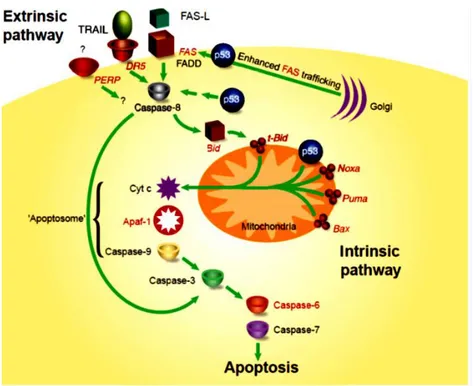
Figure 8. A model for p53-mediated apoptosis. This model depicts the
involvement of p53 in the extrinsic and intrinsic apoptotic pathways. p53 target genes are shown in red. The convergence of the two pathways through Bid is shown.
In other cell types up-regulation IGF-BP3,101 which sequesters the cell survival factor insulin-like growth factor-1 has been associated with p53 mediated apoptosis.102 The gene encoding for the cathepsin-D protease, 103 PAG608 which encodes a nuclear zinc finger protein104 and the human homolog of the Drosophilasina gene105 have also been implicated as mediators of p53 induced apoptosis in various cell types. Furthermore, a series of p53-induced genes (PIG genes) were documented to encode proteins that respond to oxidative stress, suggesting that p53-mediated apoptosis involves activation of redox- controlling targets followed by increase in ROS, oxidative damage to mitochondria and caspase activation.93, 106 Along this research line it was recently observed that p53 suppresses Nrf2-dependent transcription of