Abstract - Monoclonal gammopathy of undetermined significance (MGUS) is a non malignant plasma cell disorder with a relatively low risk of progression to Multiple Myeloma (MM) and to related Plasma cells disordes (lymphoplasmacellular neoplasms, Waldenstrom Macroglobulinemia or light chain amyloidosis). It is a quite common finding, especially in the population above the age of 50 and it can also present in association with many non malignant conditions. Differential diagnosis of symptomatic and asymptomatic forms is the determinant for starting therapy. Over the last few years many advances in the understanding of the biology of MGUS, together with large epidemiological studies, allowed to define risk models to estimate the risk of progression to MM according to MGUS isotype and, more recently, to peculiar flow cytometry findings. The goal of many recent studies aims at evaluating individual patients and their overall risk of progression, the detection of early signs of progression and the development of timely treatment strategies.
Keywords: MGUS, Monoclonal gammopathy of undetrmined significance, non malignant haematological disorders, pre-malignant haematological neoplasms
I. INTRODUCTION
Monoclonal gammopathy of undetermined significance (MGUS) is an asymptomatic plasma cell disorder; it is a non malignant common condition affecting at least 3% of the population above the age of 50, with an average 1% annual risk of progression to Multiple Myeloma (MM) [1].
A monoclonal gammopathy can be associated with many non malignant conditions, frequently observed in common clinical practice.
MGUS virtually precedes the development of MM and related disorders:, lymphoplasmacellular neoplasms, Waldenstrom Macroglobulinemia or light chain amyloidosis [2,3].
Differential diagnosis of symptomatic and asymptomatic monoclonal gammopathies is the determinant for starting therapy [4].
Over the last few years, advances in the understanding of the pathogenesis of this disease and large
epidemiological studies [5], allowed the design of risk models to estimate the individual risk of progression to MM. The development of individualized risk profiles, through the use of flow cytometry [6], free light chain analyses and risk models [5] represent an interesting ongoing challenge since the distinction of patients in low- and high-risk would allow a tailored clinical management of MGUS patients [7]. An early detection of the signs of progression could lead to the development of early treatment strategies. The aim of this report is to provide current information on the diagnosis, biology, risk stratification and follow-up of patients with MGUS.
II. DEFINITIONS AND CLINICAL ASPECTS Most monoclonal proteins (M-proteins) are detected incidentally during routine checks, especially when investigating an increased erythrocyte sedimentation rate in older people.
When a spike-like peak is first found on serum protein electrophoresis (SPEP), serum and urine immunefixation electrophoresis (IFE) should be performed additionally and the class specific immunoglobulins should be quantitatively determined to confirm the diagnosis of monoclonal gammopathy [8,9]. Quantitative measuring of free light chains in the serum is a new, highly sensitive, method that may be helpful in assessing the prognosis and controlling the course of the disease [5].
The typical laboratory investigations necessary to differentiate MGUS from other related plasma cell (PC) disorders are a complete blood cell count (CBC), serum creatinine measurement, serum calcium measurement, and a complete radiographic bone survey.
Among B cells disorders, MGUS is, by definition, characterized by a serum M protein concentration of less than 30 g/L, fewer than 10% clonal PCs in the bone marrow, and the absence of end-organ damage defined by hypercalcemia, renal insufficiency, anemia, or bone lesions (CRAB) [10] (Table 1). A bone marrow aspirate and biopsy are required in case of abnormalities in the blood/urine tests, when the M protein level is greater than or equal to 15 g/L, in patients with
Discovering the meaning of monoclonal gammopathy of undetermined
significance: current knowledge, future challenges
C.
Palladino
1, B. Bruno
1, M. Boccadoro
11
Division of Hematology, University of Torino, Azienda Ospedaliera Città della Salute e della Scienza di Torino, Torino, Italy.
TABLE I. DIAGNOSTIC CRITERIA FOR PLASMA CELL DISORDERS
DISORDER DISEASE DEFINITION
MGUS
Serum monolonal protein level< 30 g/L, bone marrow plasma cells<10% and absence of end-organ damage, such as lytic bone lesions, hypercalcemia, or renal failure, that can be attributed to a plasmacell proliferative disorder.
SMM (also referred to asymptomatic multiple myeloma)
Serum monoclonal protein (IgG or IgA) level ≥30 g/L an or bone marrow plasma cells ≥10%, absence of end-organ damage such as lytic bone lesions, hypercalcemia, or renal failure, that can be attributed to a plasmacell proliferative disorder.
Multiple Myeloma Bone marrow plasma cells ≥10%, presence of serum and/or urinary monoclonal protein (except in patients with
true nonsecretory multiple myeloma, plus evidence of lytic bone lesions, anemia, hypercalcemia, or renal failure, that can be attributed to the underlying plasma cell proliferative disorder.
Waldenström Macroglobulinemia
IgM monoclonal gammopathy (regardless of the size of the M protein) with > 10% bone marrow lymphoplasmacytic infiltration (usually intertrabecoular) by small lymphocytes that exhibit plasmacytoid or plasma cell differentiation and a typical immunophenotype (eg, surface IgM+, CD5+/-, CD10-, CD19+, CD20+,CD23-) that satisfactorily excludes other lymphoproliferative disorders, including chronic lymphocytic leukemia and mantle cell lymphoma. Note: IgM MGUS is defined is defined as a serum IgM monoclonal protein level <30 g/L, bone marrow lymphoplasmocytic infiltration <10%, and no evidence of anemia, constitutional symptoms, hyperviscosity, lymphadenopathy, or hepatosplenomegaly. Smolderin Waldenström macroglobulinemia (also referred to as indolent or asymptomatic Waldenström macroglobulinemia) is defined as serum IgM monoclonal protein level ≥30 g/L and/or bone marrow lymphoplasmocytic infiltration ≥10% and no evidence of end-organ damage, such as anemia, constitutional symptoms, hyperviscosity, lymphadenopathy, or hepatosplenomegaly, that can be attributed to a plasma cell proliferative disorder.
Solitary Plasmocitoma
Biopsy-proven solitary lesion of bone or soft tissue with evidence of clonal plasma cells, normal bone marrow with no evidence of clonal plasma cells, normal skeletal survey and MRI of spine and pelvis, and absence of end-organ damage such as anemia, hypercalcemia, renal failure, that can be attributed to a plasma cell proliferative disorder.
Systemic AL amyloidosis
Presence of an amyloid-related systemic syndrome (such as renal, liver, heart, gastrointestinal tract, or peripheral nerve involvement) with positive amyloid staining by Congo red in any tissue (eg fat aspirate, bone marrow, or organ biopsy), plus evidence that amyloid is light chain related established by direct examination of the amyloid (immunoperoxidase staining, direct sequencing, etc) plus evidence of a monoclonal plasma cell proliferative disorder (serum or urine M protein, abnormal free light chain ratio, or clonal plasma cells in the bone marrow).
POEMS
Presence of a monoclonal plasma cell disorder, peripheral neuropathy, and at least one of the following 7 features: osteosclerotic myeloma; Castleman disease, Organomegaly, endocrinopathy (excluding diabetes mellitus or hypothyroydism), edema, typical skin changes, and papilledema.
Abbreviations. MGUS: monoclonal Gammopathy of undetermined significance; SMM: ; POEMS: Polyneuropathy, Organomegaly, Endocrinopathy,
M (protein) and Skin changes.
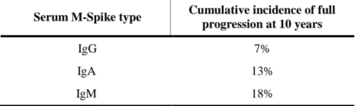
non-IgG MGUS, and/or an abnormal serum free light chain (FLC) ratio, and in any other patient with presumed MGUS in whom there is doubt about the diagnosis [11]. Large epidemiological and clinical studies have led Mayo Clinic investigators to define 3 distinct clinical subtypes of MGUS: non-IgM MGUS, by far the most common; IgM-MGUS; and light-chain MGUS [12], each with a different mode and cumulative risk of progression to MM at 10 years (Table 2).
TABLE II. PROGNOSIS OF MGUS: TYPE OF M-SPIKE AND ABSOLUTE RISK OF
PROGRESSION
Serum M-Spike type Cumulative incidence of full progression at 10 years
IgG 7%
IgA 13%
IgM 18%
Source: see ref. 11.
The most well-characterized subtype is non-IgM MGUS, in which the M-component isotype can be IgG (69%), IgA (11%), or biclonal (3%); IgD and IgE are rare [1].
Malignant transformation of non-IgM MGUS approximates 1% per year and typically develops into multiple myeloma rather than lymphoproliferative disorders (LPDs) [8].
IgM MGUS occurs in approximately 11% of patients [13], it has a predilection for developing Waldenstrom macroglobulinemia or other lymphomas while rarely progressing to IgM MM [14].
People with MGUS can occasionally present numbness or tingling in their hands and feet, or problems with their balance, due to peripheral nerves damage caused by paraproteinemia.
This situation configures a disorder called Paraproteinaemic Demyelinating Neuropathy (PDN) or MGUS - associated neuropathy. The higher prevalence of neuropathy in patients with IgM MGUS may be related to the frequent reactivity of IgM M-proteins with myelin-associated glycoprotein (MAG) [15]. Almost 50% of patients with IgM PDN have high titres of antibodies to
myelin- associated glycoprotein (MAG) , and this is the best defined syndrome of PDN, some other patients present IgM antibodies against different gangliosides. Most patients with IgM PDN have the “distal acquired demyelinating symmetrical” (DADS) clinical phenotype of predominantly distal, chronic (duration over 6 months), slowly progressive, symmetric, predominantly sensory impairment, with ataxia and relatively mild or no weakness, and often tremor. Testing for MAG-antibodies should be considered in all patients with IgM PDN. If negative, then testing for IgM antibodies against other neural antigens, including gangliosides GQ1b, GM1, GD1a and GD1b, and SGPG, may be considered [16].
Patients with IgG or IgA PDN usually have both proximal and distal weakness, with motor and sensory impairment. No specific antibody has been consistently associated with demyelinating neuropathy in patients with IgG or IgA paraprotein, so there is no need to test for serum antibodies to known neural epitopes in routine practice.
Many patients are initially thought to have an ordinary PDN, until POEMS (Polyneuropathy, Organomegaly, Endocrinopathy, Monoclonal band and Skin changes) syndrome (Table 1) is suggested by the presence of systemic features such as sclerotic bone lesions, hepatosplenomegaly, lymphadenopathy, endocrinopathy, papilloedema, skin changes (hypertrichosis, hyperpigmentation, diffuse skin thickening, finger clubbing, dermal haemangiomas, white nail beds) and edema.
POEMS usually has an underlying osteosclerotic myeloma, with IgA or IgG lambda paraprotein, or sometimes Castleman’s disease.
Patients with PDN must be referred to the neurologist for the specific treatment (plasma exchange, intravenous immunoglobulin, corticosteroids, immunosoppressive therapies, interpheron-alpha, Rituximab) [17].
MGUS has confirmed and reported associations with numerous diseases that are commonly encountered in clinical practice, such as osteoporosis [18], dermatological diseases (Lichen myxoedematosus, scleroderma, pyoderma gangrenosum, necrobiotic xanthogranuloma, discoid lupus erythematosus, psoriasis, cutaneous lymphoma), rheumatologic diseases (rheumatoid arthritis, inflammatory seronegative polyarthritis, polymyositis, polymyalgia rheumatica, myasthenia gravis, angioneurotic edema) and liver diseases (chronic hepatitis, cirrhosis, primary biliary cirrhosis) [19]. AIDS and HIV infection, renal transplantation, bone marrow transplantation and many other conditions determining immunosuppression can be associated to MGUS. Recent observations suggest to also consider a form of MGUS of renal significance (MGRS) [18] as a growing number of kidney diseases which present with renal impairment and a nephrotic range proteinuria are associated to a MGUS-like clonal pasma cell disorder. The renal impairment seems to be misdiagnosed in this subgroup of patients, leading to a great deal of morbidity and even mortality [18]. From a
practical point of view these data suggest in case of M-protein detection and renal failure and/or M-proteinuria, not only a bone marrow aspirate to exclude MM, but also a kidney biopsy [18]. Among rare disorders, Gaucher disease can debut with the presence of a monoclonal gammopathy and anemia and it should be considered in the differential diagnosis of MGUS [21].
Venous thrombosis can also be associated to MGUS [19]. MGUS patients just like MM patients present an increased risk of Venous thromboembolic disease (VTD) and arterial thrombosis. There is no current evidence to explain this pre-thrombotic state; a high concentration of M-protein is linked to an increased risk for thrombosis, especially in patients with IgG or IgA MGUS [13,22,23].
While guidelines for prophylactic interventions in MM patients have been published [24] there is lack of a consensus about the management of MGUS patients. Studies published to date suggest to consider antithrombotic therapy for patients with IgG/IgA MGUS plus the presence of additional risk factors for thrombosis (age, obesity, inherited thrombophilia, history of VTE, comorbidities, surgical procedures ) [23].
III. PATHOPHYSYOLOGY
The etiology of MGUS remains unclear and is a current topic of investigation. Race and ethnicity seem to play a role in the pathogenesis as the prevalence of MGUS is 2- to 3-fold higher in African-Americans and blacks from Africa compared to whites. Advancing age, male sex, family history of haematologic malignancies, immunosuppression, and exposure to certain pesticides all increase the risk of MGUS [1,25- 29].
MGUS and smoldering multiple myeloma (SMM) are premalignant precursor tumors of MM that are stable and not associated with the presence of secondary clinical manifestations [30,31]. They both are derived from activated B- cells that have undergone several rounds of hypermutation and antigen selection in Germinal centers (GCs) and immunoglobulin heavy chain (IgH) switch recombination before differentiating into plasmoblasts (PBs). PBs from the GC migrate back to the bone marrow where they become terminally differentiated long-lived PCs. MGUS, SMM, and MM are monoclonal tumors that retain many of the phenotypic properties of healthy PBs/PCs but in contrast to their normal counterpart they maintain low proliferation rates that can increase markedly in late stages of MM [32,33].
The pathophysiology of the transition from normal PCs to MGUS to multiple myeloma involves many overlapping oncogenic events [34] .
The first step in the pathogenesis is usually an abnormal response to antigenic stimulation, possibly mediated by aberrant expression of toll-like receptors, overexpression of interleukin (IL) 6 receptors and IL-1β, and dysregulation of the cyclin D gene [4,35,36].
The development of primary cytogenetic abnormalities (the most common are t(4;14), t(14;16), t(6;14), t(11;14), and t(14;20), hyperdiploidy or immunoglobulin heavy chain (IgH) translocations are likely followed by a random second hit such as Ras and p53 mutations, p16 methylation, myc abnormalities, and induction of angiogenesis. Finally, in most advanced stages, there is increased osteoblast RANKL (receptor activator of nuclear factor κB ligand) expression and reduction in the level of its decoy receptor, osteoprotegerin, which results in osteoclast activation and increased bone resorption and turnover [37]. This is accompanied by increased levels of IL-3, IL-7, and dickkopf 1 that simultaneously inhibit osteoblast differentiation, leading to the pure lytic lesions typical of myeloma [38-40].
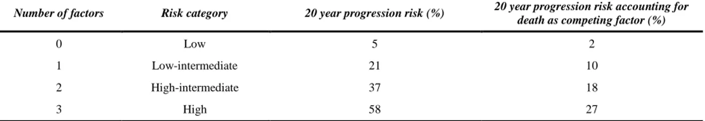
IV. RISK STRATIFICATION MODELS At present, there are no reliable biologic markers that predict which individual with MGUS will progress to MM or related conditions. In the absence of such markers, MGUS is currently risk stratified based on clinical variables identified through epidemiological studies.
Two predictive risk models for MGUS to MM have been developed by the Mayo Clinic and the Spanish study group investigators (PETHEMA).
The Mayo Clinic model is centered on serum protein abnormalities and identifies 3 major risk factors for progression: non-IgG isotype, serum M-component concentration > 1.5 g/dL, and an abnormal FLC ratio. The FLC ratio is measured using a highly sensitive serum free light chain assay that quantitates free kappa (κ) and lambda (λ) chains secreted by PCs (normal range for free κ, 0.33 to 1.94 mg/dL; normal range for free λ, 0.57 to 2.63 mg/dL).(41-42). The normal FLC ratio (free κ /free λ) is 0.26 to 1.65. (33) Patients with a serum FLC ratio < 0.26 are defined as having monoclonal λ free light chain and those with ratios > 1.65 are defined as having a monoclonal κ free light chain (Table 1) [5].
Patients with an abnormal serum FLC ratio, non-IgG MGUS, and a high serum M protein level (≥1.5 g/dL) had a risk of progression of 58% at 20 years (high-risk MGUS), compared to 5% when none of the risk factors were present (low-risk MGUS) (Table 2,3) [6].
The Spanish model uses multiparametric flow cytometry of BM aspirates to differentiate aberrant (aPCs) from normal PCs. PCs characteristically express CD138 and intense (bright) CD38. The features of aPCs include decreased CD38 expression, expression of CD56, and the absence of CD19 and/or CD45. Risk factors for progression are ≥ 95% aPCs/BMPC and DNA aneuploidy [43].
Despite the existence of these significant predictive factors for progression, there are insufficient data on the preventative treatment of high-risk patients. In the future, additional markers may be added to risk stratification models to better define high-risk patients [44]. A small study, showed that PC phenotype (CD138/38/45 expression) and sFLCs provide independent and complementary prognostic information on the risk of progression [45].
V. THERAPEUTICAL STRATEGIES
Although most people with MGUS die from unrelated illnesses, MGUS may transform into malignant monoclonal gammopathies. Patients should therefore be monitored on a regular basis to identify early signs of progression.
Once MGUS is diagnosed primary care physicians will attend patients with M-protein < 15 g/L if IgG and patients with M-protein < 10 g/L if IgA or IgM, without end-organ damage and without signs and symptoms of LPD (lymphocitosis, thrombocytopenia, lymphadenopaty, hepatosplenomegaly, constitutional symptoms, hyperviscosity, unexplained heart failure, polyneuropathy). In case of IgM MGUS, the execution of a chest X-ray and an abdomen Echo tomography may be indicated.
In June 2010, the International Myeloma Working Group (IMWG) released consensus guidelines for monitoring and managing patients with MGUS and smoldering myeloma. Patients with MGUS are divided into different categories based on low risk, intermediate risk, and high risk. If the serum monoclonal protein is <1.5 g/dL, IgG type, and the free light chain ratio is normal, then the risk of eventual progression to multiple myeloma or related malignancy is low [5].
TABLE III. RISK STRATIFICATION MODEL
Number of factors Risk category 20 year progression risk (%) 20 year progression risk accounting for death as competing factor (%)
0 Low 5 2
1 Low-intermediate 21 10
2 High-intermediate 37 18
3 High 58 27
In this low-risk setting, a baseline bone marrow examination or skeletal survey is not routinely indicated if the clinical evaluation and laboratory values suggest MGUS. Patients should be followed with SPEP 6 months after diagnosis and if stable can be followed every 2-3 years (or sooner if symptoms suggestive of disease progression arise). Patients in the intermediate and high risk MGUS category are managed differently. They usually have a serum monoclonal protein >1.5 g/dL, IgA or IgM type, and/or an abnormal free light chain ratio. In this situation, a bone marrow biopsy should be carried out at baseline. Both conventional cytogenetics and fluorescence in situ hybridization should be performed. These patients are followed with SPEP, complete blood count, serum calcium and creatinine levels 6 months after diagnosis and then yearly for life. A bone marrow biopsy and skeletal survey is always indicated in patients with presumed MGUS with unexplained anemia, renal insufficiency, hypercalcemia, skeletal lesion or in case of increase of the M-protein by more than 25% (a minimum absolute increase of 5 g/L) [45].
VI. CONCLUSION
MGUS represents an interesting pre-malignant condition. Novel biomarkers, molecular profiles, and microenvironmental interactions of interest in myelomagenesis are being investigated to better understand the underlying mechanisms of its transformation to MM.
Currently, in clinical practice, MGUS patients are followed without treatment until progression. In recent years, efforts to design models based on laboratory findings to evaluate the individual risk of progression have aimed to identify high- and low-risk patients for whom a tailored clinical management may let detect early signs of progression, and, possibly, its prevention or delay through the development of early treatment strategies.
REFERENCES
[1] Kyle RA, Therneau TM, Rajkumar SV, Larson DR, Plevak MF, Offord JR, Dispenzieri A, Katzmann JA, Melton LJ 3rd. Prevalence of monoclonal gammopathy of undetermined significance. N Engl J Med 2006; 354(13): 1362-1369.
[2] Landgren O, Kyle RA, Pfeiffer RM, Katzmann JA, Caporaso NE, Hayes RB,Dispenzieri A, Kumar S, Clark RJ, Baris D, Hoover R, Rajkumar SV. Monoclonal gammopathy of undetermined significance (MGUS) consistently precedes multiple myeloma: a prospective study. Blood 2009;113(22):5412-5417.
[3] Weiss BM, Abadie J, Verma P, Howard RS, Kuehl WM. A monoclonal gammopathy precedes multiple
myeloma in most patients. Blood. 2009;113(22):5418-5422.
[4] Merlini G, Palladini G. Differential diagnosis of monoclonal gammopathy of undetermined significance. Hematology Am Soc Hematol Educ Program. 2012;2012:595-603.
[5] Rajkumar SV, Kyle RA, Therneau TM, Melton LJ 3rd, Bradwell AR, Clark RJ, Larson DR, Plevak MF, Dispenzieri A, Katzmann JA. Serum free light chain ratio is an independent risk factor for progression in monoclonal gammopathy of undetermined significance. Blood 2005;106(3):812-817.
[6] Raja KR, Kovarova L, Hajek R. Review of phenotypic markers used in flow cytometric analysis of MGUS and MM, and applicability of flow cytometry in other plasma cell disorders. Br J Haematol 2010;149(3):334-351. [7] Hübel K, Hallek M. Monoclonal gammopathy of undetermined significance and monoclonal B-lymphocytosis. Internist (Berl) 2013;54(6):709-714. [8] Manfred Hensel, Peter Dreger, Anthony D. Ho Monoclonal IgM Gammopathy. Differential Diagnosis, Clinical Presentation, and Therapy. Medicine Dtsch Arztebl 2007;104(26): 1907–1913.
[9] Katzmann JA, Kyle RA, Benson J, Larson DR, Snyder MR, Lust JA, Rajkumar SV, Dispenzieri A. Screening panels for detection of monoclonal gammopathies. Clin Chem 2009;55(8):1517-1522.
[10] International Myeloma Working Group. Criteria for the classification of monoclonal gammopathies, multiple myeloma and related disorders: a report of the International Myeloma Working Group. Br J Haematol 2003;121(5):749-757.
[11] Rajkumar SV, Dispenzieri A, Kyle RA. Monoclonal gammopathy of undetermined significance, Waldenström macroglobulinemia, AL amyloidosis, and related plasma cell disorders: diagnosis and treatment. Mayo Clin Proc 2006;81(5):693-703. Review. Erratum in: Mayo Clin Proc 2006;81(11):1509.
[12] S. Vincent Rajkumar, MD, Robert A. Kyle, MD, and Francis K. Buadi, MD Advances in the diagnosis, classification, risk stratification, and management of monoclonal gammopathy of undetermined significance: implications for recategorizing disease entities in the presence of evolving scientific evidence. Mayo Clin Proc 2010; 85(10): 945–948.
[13] Kyle RA, Therneau TM, Rajkumar SV, Offord JR, Larson DR, Plevak MF, Melton LJ 3rd. A long-term study of prognosis in monoclonal gammopathy of undetermined significance. N Engl J Med 2002;346(8): 564-569.
[14] Kyle RA, Therneau TM, Rajkumar SV, Offord JR, Larson DR, Plevak MF, Melton LJ 3rd. Long-term follow-up of IgM monoclonal gammopathy of undetermined significance. Semin Oncol 2003;30(2):169-171.
[15] Nobile-Orazio E, Barbieri S, Baldini L, Marmiroli P, Carpo M, Premoselli S, Manfredini E, Scarlato G. Peripheral neuropathy in monoclonal gammopathy of undetermined significance: prevalence and
immunopathogenetic studies. Acta Neurol Scand 1992;85(6):383-390.
[16] Van den Berg L, Hays AP, Nobile-Orazio E, Kinsella LJ, Manfredini E, Corbo M, Rosoklija G, Younger DS, Lovelace RE, Trojaborg W, Lange DE, Goldstein S, Delfiner JS, Sadiq SA, Sherman WH, Latov N. Anti-MAG and anti-SGPG antibodies in neuropathy. Muscle Nerve 1996;19(5):637-643.
[17] European Federation of Neurological Societies; Peripheral Nerve Society, Hadden RD, Nobile-Orazio E, Sommer C, Hahn A, Illa I, Morra E, Pollard J, Hughes RA, Bouche P, Cornblath D, Evers E, Koski CL, Léger JM, Van den Bergh P, van Doorn P, van Schaik IN. European Federation of Neurological Societies/Peripheral Nerve Society guideline on management of paraproteinaemic demyelinating neuropathies: report of a joint task force of the European Federation of Neurological Societies and the Peripheral Nerve Society. Eur J Neurol 2006;13(8):809-818.
[18] Leung N, Bridoux F, Hutchinson CA, Nasr SH, Cockwell P, Fermand JP, Dispenzieri A, Kevin W, Song KW, Kyle RA. Monoclonal gammpathy of nal significance: when MGUS is no longer undetermined or insignificant. Blood 2012; 120(22):4292-4295.
[19] Bida JP, Kyle RA, Therneau TM, Melton LJ 3rd, Plevak MF, Larson DR, Dispenzieri A, Katzmann JA, Rajkumar SV. Disease associations with monoclonal gammopathy of undetermined significance: a population-based study of 17,398 patients. Mayo Clin Proc 2009;84(8):685-693.
[20] Brown LM, Gridley G, Check D, Landgren O. Risk of multiple myeloma and monoclonal gammopathy of undetermined significance among white and black male United States veterans with prior autoimmune, infectious, inflammatory, and allergic disorders. Blood 2008; 111(7):3388-3394.
[21] Hughes D, Cappellini MD, Berger M, Van Droogenbroeck J, de Fost M, anic D, Marinakis T, Rosenbaum H, Villarubia J, Zhukovskaya E, Hollak C. Recommendations for the management of the haematological and onco-haematological aspects of Gaucher disease. Br J Haematol 2007; 138(6): 676–686. [22] Sallah S, Husain A, Wan J, Vos P, Nguyen NP. The risk of venous thromboembolic disease in patients with monoclonal gammopathy of undetermined significance. Ann Oncol 2004;15(10):1490-1494.
[23] Heinz L, Delforge M. Threatening clots in MGUS and myeloma. Blood 2010;115(24):4975-4976.
[24] Palumbo A, Rajkumar SV, Dimopoulos MA, Richardson PG, San Miguel J, Barlogie B, Harousseau J, Zonder JA, Cavo M, Zangari M, Attal M, Belch A, Knop S, Joshua D, Sezer O, Ludwig H, Vesole D, Bladé J, Kyle R, Westin J, Weber D, Bringhen S, Niesvizky R, Waage A, von Lilienfeld-Toal M, Lonial S, Morgan GJ, Orlowski RZ, Shimizu K, Anderson KC, Boccadoro M, Durie BG, Sonneveld P, Hussein MA. International Myeloma Working Group. Prevention of thalidomide- and lenalidomide-associated thrombosis in myeloma. Leukemia 2008;22(2):414-423.
[25] Landgren O, Gridley G, Turesson I, Caporaso NE, R. Goldin LR, Baris D, Fears TR, Hoover RN, Linet MS. Risk of monoclonal gammopathy of undetermined significance (MGUS) and subsequent multiple myeloma among African American and white veterans in the United States. Blood. 2006; 107(3): 904–906.
[26] Landgren O, Katzmann JA, Hsing AW, Pfeiffer RM, Kyle RA, Yeboah ED, Biritwum RB, Tettey Y, Adjei AA, Larson DR, Dispenzieri A, Melton LJ 3rd, Goldin LR, McMaster ML, Caporaso NE, Rajkumar SV. Prevalence of Monoclonal Gammopathy of Undetermined Significance Among Men in Ghana. Mayo Clin Proc 2007; 82(12):1468-1473.
[27] Landgren O, Kyle RA, Hoppin JA, Beane Freeman LE, Cerhan JR, Katzmann JA, Rajkumar SV, Alavanja MC. Pesticide exposure and risk of monoclonal gammopathy of undetermined significance in the Agricultural Health Study. Blood 2009;113(25):6386-6391.
[28] Vachon CM, Kyle RA, Therneau TM, Foreman BJ, Larson DR, Colby CL, Phelps TK, Dispenzieri A, Kumar SK, Katzmann JA, Rajkumar SV. Increased risk of monoclonal gammopathy in first-degree relatives of patients with multiple myeloma or monoclonal gammopathy of undetermined significance. Blood 2009;114(4):785-790.
[29] Kristinsson SY, Goldin LR, Björkholm M, Koshiol J, Turesson I, Landgren O. Genetic and immune-related factors in the pathogenesis of lymphoproliferative and plasma cell malignancies. Haematologica 2009; 94(11):1581-1589.
[30] Landgren O, Kyle RA, Pfeiffer RM, Katzmann JA, Caporaso NE, Hayes RB, Dispenzieri A, Kumar S, Clark RJ, Baris D, Hoover R, Rajkumar SV. Monoclonal gammopathy of undetermined significance (MGUS) consistently precedes multiple myeloma: a prospective study. Blood 2009;113(22):5412-5417.
[31] Kyle RA, Durie BG, Rajkumar SV, Landgren O, Blade J, Merlini G, Kröger N, Einsele H, Vesole DH, Dimopoulos M, San Miguel J, Avet-Loiseau H, Hajek R, Chen WM, Anderson KC, Ludwig H, Sonneveld P, Pavlovsky S, Palumbo A, Richardson PG, Barlogie B, Greipp P, Vescio R, Turesson I, Westin J, Boccadoro M; International Myeloma Working Group. Monoclonal gammopathy of undetermined significance (MGUS) and smoldering (asymptomatic) multiple myeloma: IMWG consensus perspectives risk factors for progression and guidelines for monitoring and management. Leukemia 2010;24(6):1121-1127.
[32] Shapiro-Shelef M, Calame K. Regulation of plasma-cell development. Nat Rev Immunol 2005;5(3):230-242. [33] Kuehl WM, Bergsagel PL. Multiple myeloma: evolving genetic events and host interactions. Nat Rev Cancer 2002;2(3):175-187.
[34] Chng WJ, Glebov O, Bergsagel PL, Kuehl WM. Genetic events in the pathogenesis of multiple myeloma. Best Pract Res Clin Haematol 2007;20(4):571-596. [35] Jego G, Bataille R, Geffroy-Luseau A, Descamps G, Pellat-Deceunynck C. Pathogen-associated molecular
patterns are growth and survival factors for human myeloma cells through Toll-like receptors. Leukemia 2006;20(6):1130-1137.
[36] Dinarello CA. Targeting the pathogenic role of interleukin 1β in the progression of smoldering/indolent myeloma to active disease Mayo Clin Proc 2009; 84(2): 105–107.
[37] Roodman GD. Pathogenesis of myeloma bone disease. J Cell Biochem 2010;109(2):283-291.
[38] Drake MT. Bone disease in multiple myeloma. Oncology (Williston Park) 2009;23(14, Suppl 5):28-32. [39] Tian E, Zhan F, Walker R, Rasmussen E, Ma Y, Barlogie B, Shaughnessy JD Jr. The role of the Wnt-signaling antagonist DKK1 in the development of osteolytic lesions in multiple myeloma. N Engl J Med 2003;349(26):2483-2494.
[40] Rajkumar SV, Kyle RA, Buadi FK. Advances in the diagnosis, classification, risk stratification, and management of monoclonal gammopathy of undetermined significance: implications for recategorizing disease entities in the presence of evolving scientific evidence. Mayo Clin Proc 2010 Oct;85(10):945-948.
[41] Drayson M, Tang LX, Drew R, Mead GP, Carr-Smith H, Bradwell AR. Serum free light-chain measurements for identifying and monitoring patients with nonsecretory multiple myeloma. Blood 2001;97(9):2900-2902.
[42] Roshini S. Abraham, Raynell J. Clark, Sandra C. Bryant, James F. Lymp, Timothy Larson, Robert A. Kyle, and Jerry A. Katzmann, Correlation of Serum Immunoglobulin Free Light Chain Quantification with Urinary Bence Jones Protein in Light Chain Myeloma. Clinical Chemistry 2002;48(4): 655-657.
[43] Pérez-Persona E, Vidriales MB, Mateo G, García-Sanz R, Mateos MV, de Coca AG, Galende J, Martín-Nuñez G, Alonso JM, de Las Heras N, Hernández JM, Martín A, López-Berges C, Orfao A, San Miguel JF. New criteria to identify risk of progression in monoclonal gammopathy of uncertain significance and smoldering multiple myeloma based on multiparameter flow cytometry analysis of bone marrow plasma cells. Blood 2007;110(7):2586-2592.
[44] Rawstron AC, Davis B, D. DS, de Tute RM, Kerr MA, Owen RG, Ashcroft AJ. Plasma cell phenotype and SFLC provide independent prognostic information in MGUS. Haematologica 2007;92:907a.
[45] Kyle RA, Durie BG, Rajkumar SV, Landgren O, Blade J, Merlini G, Kröger N, Einsele H, Vesole DH, Dimopoulos M, San Miguel J, Avet-Loiseau H, Hajek R, Chen WM, Anderson KC, Ludwig H, Sonneveld P, Pavlovsky S, Palumbo A, Richardson PG, Barlogie B, Greipp P, Vescio R, Turesson I, Westin J, Boccadoro M; International Myeloma Working Group. Monoclonal gammopathy of undetermined significance (MGUS and smoldering (asymptomatic) multiple myeloma: IMWG consensus perspectives risk factors for progression and guidelines for monitoring and management. Leukemia 2010;24(6):1121-1127.