Università degli Studi di Salerno
Dipartimento di Medicina, Chirurgia e Odontoiatria
“Scuola Medica Salernitana”
Dottorato di Ricerca in
“Medicina traslazionale dello sviluppo e dell’invecchiamento attivo”
Curriculum: tecnologie innovative in medicina traslazionale
TESI DI DOTTORATO
“Transcutaneous laryngeal ultrasonography: a reliable, non-invasive
and inexpensive preoperative method in the evaluation of vocal cords
motility. A prospective multicentric analysis on a large series and an
analysis of Literature”
TUTOR
CANDIDATA
Prof. Alessandro Puzziello
Dott.ssa Marica Grasso
COORDINATORE
Prof. Palmiero Monteleone
Table of contents
List of abbreviations ... 3
PART I ... 4
Introduction ... 4
PART II ... 6
Materials and methods ... 6
Transcutaneous laryngeal ultrasonography ... 7
Flexible Fiberoptic laryngoscopy ... 8
Statistical Analysis ... 9 PART III ... 10 Results ... 10 PART IV ... 14 Discussion ... 14 PART V ... 19 References ... 19 Ringraziamenti ... 23
List of abbreviations
Total Thyroidectomy (TT)
Recurrent Laryngeal Nerve (RLN)
Vocal Cord Paralysis (VCP)
Flexible Fiberoptic Laryngoscopy (FFL)
American Thyroid Association (ATA)
Transcutaneous Laryngeal UltraSonography (TLUS)
Body Mass Index (BMI)
Marica Grasso (MG)
Giovanni Docimo (GD)
Maurizio De Palma (MDP)
Positive Predictive Value (PPV)
Negative Predictive Value (NPV)
PART I
Introduction
Benign and malignant thyroid diseases affect a large population worldwide. [1] Total Thyroidectomy (TT) is one of the most commonly performed intervention in general surgery and, since the turn of the century, its rate has sharply increased, along with a general increase of differentiated thyroid cancer incidence. [2] The most feared and dangerous complication of thyroidectomy is the paresis or paralysis of the recurrent laryngeal nerve (RLN), with a wide range of incidence reported in Literature (0.5-20% of cases). [3]
Several causes have been advocated as apparent or inapparent mechanism of nerve damage. Among them the most frequent are compression, crush, stretch, laceration, thermal or vascular injuries. [3] Unilateral vocal cord paralysis (VCP) generally does not affect voice. Nevertheless, in 30% of patients undergoing TT, without nervous injury, vocal subjective alterations are reported. [4] Conversely, a bilateral VCP can be cause of an acute respiratory distress, requiring temporary or permanent tracheotomy. VCP has become the most frequent cause of medicolegal litigations after thyroid surgery. [3] Therefore, endocrine surgeons have been prompted to include, among the preoperative examinations of patients addressed to TT, the evaluation of vocal cords function through flexible fiberoptic laryngoscopy (FFL).
RLN injuries have a low incidence in referral center with experienced surgeons, and according to several Authors, a routine FFL could be uncomfortable for patients and leads to unjustifiable increase of health care costs. [5] According to the American Thyroid Association (ATA) guidelines, only in patients with voice alterations, with extrathyroidal cancer extension, in patients already undergone neck surgery with suspicious or certified RLN or vagus nerve lesion is required a preoperative laryngeal evaluation. [6]
Transcutaneous laryngeal ultrasonography (TLUS) has been proposed as a non-invasive and painless indirect examination of vocal cords function as alternative to direct FFL. TLUS is an easy and feasible technique that can be performed by the endocrinologist or the surgeon during the preoperative ultrasound evaluation; it is a non-invasive, inexpensive, rapid, painless, repeatable and well tolerated by the patient. [7, 8] TLUS allows to clearly evaluate vocal cords and arytenoids vibration on real time during the examination. TLUS is not considered an innovative method to evaluate vocal cord movement, but in clinical practice its use is very limited.
The aim of this study is to assess TLUS reliability as an alternative method to direct FFL in the evaluation vocal folds function in patients candidate to thyroid surgery.
PART II
Materials and methods
We conducted a prospective observational multicentric cohort study on 396 consecutive patients diagnosed with benign and malignant thyroid disease referred to the Thyroid Surgery Division of the University of Campania "Luigi Vanvitelli" and to the General and Specialistic Surgery Division of the “Antonio Cardarelli Hospital”, from 1 January 2018 to 31 December 2018.
Exclusion criteria were age <16 years, previous neck surgery and irradiation, pre-existing diagnosis of vocal cord nodules. Clinical data were collected in a prospective maintain electronic database, including patients’ age, sex, body mass index (BMI), previous cervical surgery, preoperative diagnosis, ability of visualizing both vocal cords, mobility or paralysis of both vocal cords and preoperative FFL findings. In addition, definitive pathology was recorded. [Table1-2]
Patients were stratified into 2 groups according to BMI in a non-overweight group (BMI <25) and in an overweight or obese group (BMI ≥25).
All patients have a diagnosis of benign or malignant thyroid disease and preoperatively, underwent TLUS and subsequently all ultrasonographic findings were validated via FFL by a blinded otolaryngologist. TLUS was performed by an experienced investigator (MG), skilled in neck ultrasound, in order to minimize variability in vocal cord assessment. All patients, in both the divisions, underwent total thyroidectomy (TT) performed by two experienced surgeons (GD and MDP).
The ethical committee of Campania “Luigi Vanvitelli” University approveD the study. All selected patients received an information leaflet on the aims of the study and signed a written informed consent.
Transcutaneous laryngeal ultrasonography
Cervical and thyroid ultrasound have been performed using a 7-13 MHz linear probe (Esaote, MyLabTM X5). The patients were placed in a supine position with neck hyperextension. In
overweight patients or in patients with a short neck, we placed a pillow under the shoulder in order to hyperextend the neck. The ultrasonography was carried out identifying vascular neck bundle, both lobes of thyroid gland and isthmus.
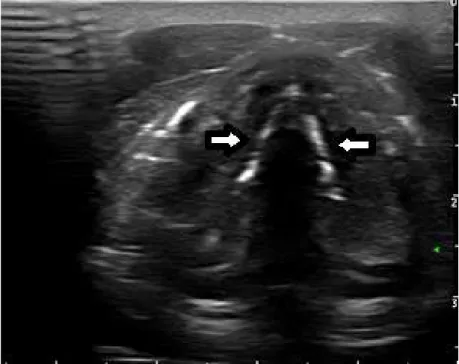
The probe was firstly placed transversely over the thyroid cartilage when we proceeded to vocal cords evaluation. Craniocaudal scans allowed visualization of the movement of the vocal cords during spontaneous breathing. Thyroid cartilage was used as acoustic window. In case of hard vocal folds visualizations for the presence of a very represented thyroid cartilage, the ultrasonographic probe was placed in a different acoustic window, laterally to the cartilage. [9] When possible, true vocal cords, false vocal cords and arytenoids were identified. The movement of the true vocal cords has been visualized by the patient pronunciation of the letter I, as a symmetrical vibration in adduction and abduction. The paretic vocal cords appeared as immobile or with a sign of passive movement.
True vocal cords are hypoechoic structures, while false vocal cords appear as hyperechoic structures.
Real-time TLUS lasted less than one minute.
After TLUS, all patients underwent routine preoperative FFL by a blinded experienced otolaryngologist, and findings were classified as normal or impaired vocal cord function. Vocal cord impairment includes weakness, asymmetry and paralysis. Prospectively, TLUS findings and FFL have been compared as concordant or discordant. [10]
Flexible Fiberoptic laryngoscopy
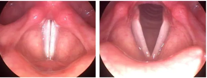
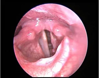
FFL was performed with the patient in a semi-recumbent position (the head of the examining chair raised 30 degrees). Before the procedure, the patient was instructed to close his or her mouth and breathe gently through the nose. All patients received topical anesthetic and decongestant before the examination. The fiberoptic laryngoscope (3.6 mm diameter, Olympus, Japan) was then advanced along the floor of the nose, avoiding the nasal septum. As the endoscope was advanced to the nasopharynx, the patient was instructed to breathe through the nose.
Then, the instrument was introduced past to the soft palate and subsequently the vocal cords were visualized. The patient was invited to phonate "I" to allow visualization of vocal cords mobility, making note of incomplete abduction, adduction.
Statistical Analysis
Age and BMI were indicated as mean, while patients’ sex, visualization through TLUS and benign and malign diseases were described as number of cases. Sensitivity, specificity, Positive Predictive Value (PPV), Negative Predictive Value (NPV) were evaluated according to the FFL’s results. [Table 3] Moreover Authors have calculated the prevalence, the probability to be affected by the vocal cord impairment either if test results were positive or negative and the concordance with relative Cohen’s K, by the use of Excel. Statistical significance was defined as p<0.05 with a Confidence Interval (CI) at 95%. Database has been realized through the use of Excel and P-value has been calculated with Fisher exact test (IBM SPSS-23).
PART III
Results
The current study included 396 patients who underwent a total of 792 vocal cord assessments. The overall mean age was 56.4 years (18-82 years), 262 patients (66.16%) were female and 134 (33.84%) patients were male. The female-male ratio was 1.9/1.0. In population studied, there is a statistically significant difference (p <0.001) between males and females as regards difficult visualization; in detail all the cases of hard visualization (14/396) were male. Conversely, no significant difference between the two groups was reported in terms of age (p=0.436), BMI (p=0.99) and definitive diagnosis (p=0.52). One hundred and sixty-nine patients (42.7%) had a BMI <25 and 227 patients (57.3%) were overweight (BMI ≥25). All patients underwent TT. Preoperatively, 316 patients (79.8%) received the diagnosis of benign thyroid disease, 50 patients (12.6%) a diagnosis of suspicion and 30 patients (7.6%) a diagnosis of malignant thyroid disease. In details, preoperative diagnosis was a non-toxic multinodular goiter in 255 patients (64.39%), a toxic multinodular goiter in 12 patients (3.03%); a toxic adenoma in 49 patients (12.37%); an indefinite cytology nodule in 50 patients (12.62%); a positive cytology for thyroid carcinoma in 26 patients (6.56%) with and a diagnosis of medullary thyroid carcinoma in 4 patients (1.01%). Definitive pathology showed 308 cases (77.8%) of benign disease and 88 cases (22.2%) of thyroid carcinoma. Twenty-two patients (5.55%) had a diagnosis of follicular thyroid carcinoma, 52 patients (13.13%) a diagnosis of papillary thyroid carcinoma, 4 patients (1.01%) with a medullary thyroid carcinoma and 10 patients (2.52%) with a microcarcinoma.
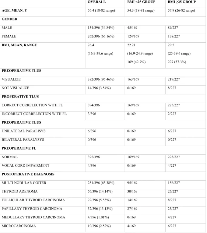
Table 1. Patients’ demographic and clinical informations.
OVERALL BMI <25 GROUP BMI ≥25 GROUP
AGE, MEAN, Y 56.4 (18-82 range) 54.3 (18-81 range) 57.9 (26-82 range)
GENDER
MALE 134/396 (34.84%) 45/169 89/227
FEMALE 262/396 (66.16%) 124/169 138/227
BMI, MEAN, RANGE 26.4
(16.9-39.6 range) 22.21 (16.9-24.9 range) 169 (42.7%) 29.5 (25-39.6 range) 227 (57.3%) PREOPERATIVE TLUS VISUALIZE 382/396 (96.46%) 163/169 219/227 NOT VISUALIZE 14/396 (3.54%) 6/169 8/227 PROPERATIVE TLUS
CORRECT CORRELECTION WITH FL 394/396 169/169 225/227
INCORRECT CORRELECTION WITH FL 3/396 0/169 2/227
PREOPERATIVE TLUS
UNILATERAL PARALISYS 6/396 0/169 6/227
BILATERAL PARALYSYS 0/396 0/169 0/227
PREOPERATIVE FL
NORMAL 392/396 169/169 223/227
VOCAL CORD IMPAIRMENT 4/396 0/169 4/227
POSTOPERATIVE DIAGNOSIS
MULTI NODULAR GOITER 251/396 (63.38%) 95/169 156/227
THYROID ADENOMA 56/396 (14.14%) 30/169 26/227
FOLLICULAR THYROID CARCINOMA 22/396 (5.55%) 14/169 8/227
PAPILLARY THYROID CARCINOMA 52/396 (13.13%) 27/169 25/227
MEDULLARY THYROID CARCINOMA 4/396 (1.01%) 0/169 4/227
MICROCARCINOMA 10/396 (2.52%) 4/169 6/227
*BMI, body mass index; TLUS, transcutaneous laryngeal ultrasound; FL, fiberoptic laryngoscopy.
Patients diagnosed with malignancy were all patients affected by thyroid-limited cancer (T1-T2), without lymph node involvement and metastasis. Patients’ demographic and pathological data were collected in Table 1 and in Table 2.
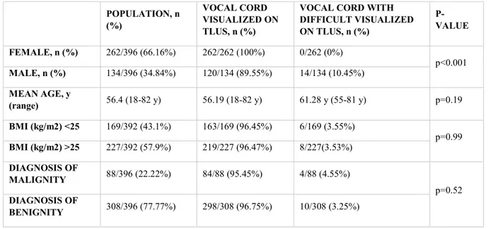
Table 2. Demographics and clinical variables according to vocal cord visualization on TLUS
POPULATION, n (%)
VOCAL CORD VISUALIZED ON TLUS, n (%)
VOCAL CORD WITH DIFFICULT VISUALIZED ON TLUS, n (%) P-VALUE FEMALE, n (%) 262/396 (66.16%) 262/262 (100%) 0/262 (0%) p<0.001 MALE, n (%) 134/396 (34.84%) 120/134 (89.55%) 14/134 (10.45%) MEAN AGE, y (range) 56.4 (18-82 y) 56.19 (18-82 y) 61.28 y (55-81 y) p=0.19 BMI (kg/m2) <25 169/392 (43.1%) 163/169 (96.45%) 6/169 (3.55%) p=0.99 BMI (kg/m2) >25 227/392 (57.9%) 219/227 (96.47%) 8/227(3.53%) DIAGNOSIS OF MALIGNITY 88/396 (22.22%) 84/88 (95.45%) 4/88 (4.55%) p=0.52 DIAGNOSIS OF BENIGNITY 308/396 (77.77%) 298/308 (96.75%) 10/308 (3.25%)
*BMI, body mass index; TLUS, transcutaneous laryngeal ultrasonography.
All patients underwent TLUS and subsequently ultrasonographic findings were validated through FFL. TLUS identified unilateral paralysis in 6 patients (1.51%). FFL diagnosed unilateral paralysis in 4 cases (1.01%) with a PPV of 66.7%. The overall assessability rate through TLUS was 96.46% (382/396). In the remaining 14 patients (3.54%), the investigator reported a hard visualization of vocal cords through ultrasonography. The latter patients were all male with a very represented thyroid cartilage, 6 of non-overweight group and 8 of overweight group (p-value 0.99). In these 14 out it was not possible to visualize the vocal cords with transverse front visualization, and the ultrasonographic probe was placed in a different acoustic window, laterally to the cartilage, allowing a correct identification.
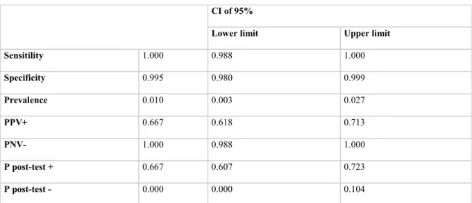
Sensitivity was equal to 100% (98 – 100%), specificity was 99,5% (98 – 99,9%), positive predictive value 66,7 % (61,8 – 71,3%), negative predictive value 100% (98 – 100%). The
resulted positive was 66,7% (60,7 – 72,3%). In our series, no False Negative have been observed.
The prevalence of VCP in our series was 1% (0,3 – 2,7%). The results showed a concordance between TLUS and FL of 99,5%, with a Cohen’s K value of 0,798. Statistical data were collected in Table 3.
Table 3. Statistical data
CI of 95%
Lower limit Upper limit
Sensitility 1.000 0.988 1.000 Specificity 0.995 0.980 0.999 Prevalence 0.010 0.003 0.027 PPV+ 0.667 0.618 0.713 PNV- 1.000 0.988 1.000 P post-test + 0.667 0.607 0.723 P post-test - 0.000 0.000 0.104
*PPV+, predictive positive value; PNV-, predictive negative value; P post-test+, probability of alteration if the test is positive;
PART IV
Discussion
Vocal cords are tendon flaps that vibrate, abduct and adduce with air passage, generating sounds. RLN controls vocal cords movement. Preoperative evaluation of bilateral nerve function in patients addressed to thyroid surgery is recommended. FFL is the widespread technique used to directly evaluate vocal cords movement. Unfortunately, surgeons are not experienced in FFL that moreover causes considerable discomfort to patients, whom often refuse or complain to undergo the investigation. Nevertheless, the study of the motility of the vocal cords is a primary information that guide the surgeon in the planning of surgical strategy. In preoperative work-up, neck ultrasound plays a primary role for the diagnosis of thyroid disease and it is irreplaceable for the execution of fine-needle aspiration.
American Academy of Otolaryngology and Head and Neck Surgery guideline suggests that all patients who have to undergo thyroid and parathyroid surgery should perform the preoperative and postoperative FFL. [11] Conversely, ATA guideline suggests that only high-risk patients for vocal cord paralysis (extracapsular thyroid carcinoma, with a large thyroid goiter, patients with voice changes and previous cervical surgery) should undergo preoperative FFL. [6] American Academy of Otolaryngology and Head and Neck Surgery guideline suggests preoperative FFL because the postoperative vocal outcome is a primary target in thyroid surgery.
Recently, in all medical fields, research has been turned towards less invasive, rapid, painless and less discomfort techniques. Since 1964, several Authors have hypostasized and studied the use of ultrasound in the assessment of vocal cord motility. [12] After its first description, the feasibility of this technique has been described in numerous studies with a sensitivity and
patients, reported an excellent assessability rate >94% and considered TLUS a reproducible, noninvasive tool with high capacity on evaluating VCP before and after thyroidectomy. Moreover, the Authors, in a subsequent paper, underlined the importance of identifying sonographic landmarks during the procedure. [14,18] These promising data have proposed TLUS as a less invasive and less expensive technique alternative to FFL. Even more satisfactory results were reported by the same Wong on the 2014. [16] The Authors, in fact, reported an assessability rate in the 95% of their cohort. This surprising result was explained with the demographic features of their 581 assessed patients, which were mostly women (80%), young and with low BMI. More conclusive results were reported by Wong, in the largest published series on 1000 patients undergone TLUS. [17] The Authors, assessed that vocal cords were evaluable in 92.4% of the studied patients, achieving a high sensitivity of 87.5% and concluding that over 87% of the patients could be saved from a FFL exam.
On the other hand, several Authors have reported a low sensitivity of the method equal to 62% and a specificity of 97%. [19] Borel et al, reported mediocre efficiency of TLUS, with a sensitivity of 33%, a specificity of 95% when used as tool for VCP screening in patients already undergone thyroidectomy. [20] Probably, these poor results were linked to the presence of thyroid lodge hematoma and major postoperative laryngeal edema in the early postoperative days. Nevertheless, the Authors recognize the role of TLUS in select patients candidate to FFL. Moreover, Borel reported better results in premenopausal women, which represent a significant proportion of patients requiring total thyroidectomy. [20] Kandil et al reported, in a study on 250 patients, a harder visualization of vocal cords in patients with BMI > 25, considering TLUS not suitable for replace FFL. [21]
A mild sensitivity value compared with a sensitivity value of 100% of fiberoptic laryngoscopy, the actual gold standard technique, is not acceptable to consider TLUS a valid alternative method. In fact, a low sensitivity of a test would lead to underestimate the presence of
pathology. Nevertheless, several biases were recognizable in the published studies reporting a low sensitivity value: TLUS was performed by surgeons who usually perform only preoperative diagnostic ultrasound but who were not skilled in ultrasonographic visualization of the vocal cords, the use of multiple investigators and the lack of use a different acoustic window in patients with hard visualizations due for example to the prominence of the thyroid cartilage. [9]
In our protocol we enrolled an investigator (MG) who has undergone specific training in thyroid and laryngeal ultrasound, and we used the lateral approach in patients with difficult visualization due to thyroid cartilage hypertrophy. Several Authors, such as Carneiro-Pla et al confirmed that the ability to visualize both vocal cords on ultrasonography depends on the skill of the surgeon performing the examination. [22] Thanks to the standardization of the ultrasound technique, we registered a high overall assessability rate was 96.46%, a sensitivity of 100%, a specificity of 99.5%, a positive predictive value of 66.7% and a negative predictive value of 100% in the identification of vocal cords alterations. Our results showed a concordance between TLUS and FL of 99.5%, with a Cohen’s K value of 0.798. These encouraging data allowed us to consider TLUS as part of the routine preoperative screening, as it is absolutely reliable in identifying healthy patients without paresis of the vocal cords. In case of doubts on the motility of the vocal cords, however, TLUS allowed to select patients that should be addressed to FFL. Furthermore, Carneiro-Pla et al showed that in 24% of the population studied with ultrasound it was not possible to visualize the vocal cords. In details, the 82% of the hard visualizable patients were male, with hypertrophy and/or calcification of the thyroid cartilage; these data were consistent with the previously reported Literature. [8, 13, 19] Our study confirmed some difficulty in identifying the vocal cords in 14 male patients with hypertrophy of the thyroid cartilage without calcification. This difficulty was solved thanks to adoption of a different acoustic window in lateral approach, as our investigator had undergone specific training in ultrasound of the cervical region. Therefore, the limit of this technique is the low confidence of
the adequate skills in performing TLUS, that could potentially replace FFL in low-risk patients. The TLUS, in turn, being an operator-dependent method and has a not-standardized learning curve. Therefore, in many centers the FFL is still used as standard of care for the preoperative assessment of the vocal cords motility, leading patients to undergo an invasive method, requesting another specialist (otolaryngologist) and another outpatient appointment.
The current study showed that visualization of both vocal cords depended on patients’ characteristics, especially sex. In fact, our work showed a statistically significant difference (p <0.001) between males and females. It showed that all the 14 patients in whom there was a difficult identification were males with hypertrophy of the thyroid cartilage. A hypertrophy of the thyroid cartilage acts as an acoustic barrier in the visualization of the movement of the vocal cords and in these cases the only solution is the lateral approach, which sacrifices the simultaneous display of both ropes. [9,22-23]
A BMI ≥25, related to an increase in subcutaneous fat and, therefore, to a greater distance between skin and thyroid, did not show a difficulty in visualizing the vocal cords (p-value 0.99). This data was consistent with Literature. [21-22]
The learning curve of the procedure is not still standardized, but surely is directly linked to the physician ultrasonographic skills. Wong et al, reported an analysis on 80 patients undergone TLUS by a group of inexperienced ultrasound assessors, residents in surgery. [14] The Authors concluded that the assessors became competent in TLUS after seven examinations and provided proficient and accurate vocal cords assessments after 40 examinations, attesting a short learning curve.
ATA guidelines suggest undergoing to a preoperative FFL patient with a high risk of vocal cord paralysis (extracapsular thyroid carcinoma, with a large thyroid goiter, patients with voice changes and previous cervical surgery). Our data did not show a statistically significant difference between patients with benign disease and those with malignant diseases (p=value
0.52). Nevertheless, it should not be neglected that in our series, patients with malignancies were staged as T1 and T2. The latter could be considered a limitation of the current study along with the performance of the procedure by one single person, determining a questionable reproducibility of our results. Further studies comparing TLUS with FL in a population with high stage thyroid tumors are needed to achieve definitive results.
TLUS is a valid non-invasive and painless alternative method in the preoperative assessment of vocal cords for a selected population, such as pediatric patients, cardiopathic patients, patients who do not tolerate invasive exams, patients with no diagnosis or suspicion diagnosis of malignancy and patients who do not have voice changes. It could save a high percentage of patients from FFL and in the same time could accurately select patients candidate to second level examinations. In our series, we reported extremely encouraging results with TLUS showing an assessability rate of over 96%, a sensitivity of 100%, a specificity of 99.5% in the identification of vocal cords alterations and a concordance between TLUS and FL of 99.5%. TLUS can be easily performed by the surgeon itself during the preoperative routine diagnostic ultrasound, lasts only 1-2 minutes, not require another specialist, is inexpensive, not require a dedicated instrumentation and is not an invasive method.
PART V
References
1. Marotta V, Sciammarella C, Capasso M, Testori A, Pivonello C, Chiofalo MG et al. Germline Polymorphisms of the VEGF Pathway Predict Recurrence in Nonadvanced Differentiated Thyroid Cancer. J ClinEndocrinolMetab. 2017 Feb 1;102(2):661-671. 2. Calò PG, Conzo G, Raffaelli M, Medas F, Gambardella C, De Crea C et al. Total
thyroidectomy alone versus ipsilateral versus bilateral prophylactic central neck dissection in clinically node-negative differentiated thyroid carcinoma. A retrospective multicenter study. Eur J SurgOncol. 2017 Jan;43(1):126-132.
3. Gambardella C, Polistena A, Sanguinetti A, Patrone R, Napolitano S, Esposito D et al. Unintentional recurrent laryngeal nerve injuries following thyroidectomy: Is it the surgeon who pays the bill? Int J Surg. 2017;41 Suppl1:S55-S59.
4. Young VN, Smith LJ, Rosen C. Voice outcome following acute unilateral vocal fold paralysis. Ann OtolRhinolLaryngol. 2013 Mar;122(3):197-204.
5. Schlosser K, Zeuner M, Wagner M, Slater EP, DomínguezFernández E, Rothmund M et al. Laryngoscopy in thyroid surgery--essential standard or unnecessary routine? Surgery. 2007 Dec;142(6):858-864; discussion 864.e1-2. Epub 2007 Nov 5.
6. Haugen BR, Alexander EK, Bible KC, Doherty GM, Mandel SJ, Nikiforov YE et al. 2015 American Thyroid Association Management Guidelines for Adult Patients with Thyroid Nodules and Differentiated Thyroid Cancer: The American Thyroid Association Guidelines Task Force on Thyroid Nodules and Differentiated Thyroid Cancer. Thyroid. 2016 Jan;26(1):1-133.
7. Wang CP, Chen TC, Yang TL, Chen CN, Lin CF, Lou PJ et al. Transcutaneous ultrasound for evaluation of vocal fold movement in patients with thyroid disease. Eur J Radiol. 2012 Mar;81(3):e288-291. Epub 2011 Oct 22.
8. Cheng SP, Lee JJ, Liu TP, Lee KS, Liu CL. Preoperative ultrasonography assessment of vocal cord movement during thyroid and parathyroid surgery. World J Surg. 2012 Oct;36(10):2509-2515.
9. Woo JW, Suh H, Song RY, Lee JH, Yu HW, Kim SJ et al. A novel lateral-approach laryngeal ultrasonography for vocal cord evaluation. Surgery. 2016 Jan;159(1):52-56. Epub 2015 Sep 28.
10. Cheng SP, Liu CL. Preoperative ultrasonography assessment of vocal cord movement during thyroid and parathyroid surgery. World J Surg. 2013 Jul;37(7):1741-1742. 11. Chandrasekhar SS, Randolph GW, Seidman MD, Rosenfeld RM, Angelos P,
Barkmeier-Kraemer J et al. Clinical practice guideline: improving voice outcomes after thyroid surgery. Otolaryngol Head Neck Surg. 2013 Jun;148(6 Suppl):S1-37.
12. Menschi B. [Analysis by ultrasonic exploration of the movement of isolated vocal cords]. C R Seances Soc Biol Fil. 1964;158:2295-2296.
13. Wong KP, Lang BH, Ng SH, Cheung CY, Chan CT, Lo CY. A prospective, assessor-blind evaluation of surgeon-performed transcutaneous laryngeal ultrasonography in vocal cord examination before and after thyroidectomy. Surgery. 2013 Dec;154(6):1158-64; discussion 1164-1165. Epub 2013 Aug 19.
14. Wong KP, Woo JW, Youn YK, Chow FC, Lee KE, Lang BH. The importance of sonographic landmarks by transcutaneous laryngeal ultrasonography in post-thyroidectomy vocal cord assessment. Surgery. 2014 Dec;156(6):1590-1596; discussion 1596. Epub 2014 Nov 11.
15. Shah MK, Ghai B, Bhatia N, Verma RK, Panda NK. Comparison of transcutaneous laryngeal ultrasound with video laryngoscope for assessing the vocal cord mobility in patients undergoing thyroid surgery. Auris Nasus Larynx. 2018 Dec 18. pii: S0385-8146(18)30800-30809.
16. Wong KP, Lang BH, Chang YK, Wong KC, Chow FC. Assessing the Validity of Transcutaneous Laryngeal Ultrasonography (TLUSG) After Thyroidectomy: What Factors Matter? Ann Surg Oncol. 2015;22(6):1774-1780. Epub 2014 Oct 17.
17. Wong KP, Au KP, Lam S, Lang BH. Lessons Learned After 1000 Cases of Transcutaneous Laryngeal Ultrasound (TLUSG) with Laryngoscopic Validation: Is There a Role of TLUSG in Patients Indicated for Laryngoscopic Examination Before Thyroidectomy? Thyroid. 2017 Jan;27(1):88-94.
18. Wong KP, Lang BH, Lam S, Au KP, Chan DT, Kotewall NC. Determining the Learning Curve of Transcutaneous Laryngeal Ultrasound in Vocal Cord Assessment by CUSUM Analysis of Eight Surgical Residents: When to Abandon Laryngoscopy. World J Surg. 2016 Mar;40(3):659-664.
19. Sidhu S, Stanton R, Shahidi S, Chu J, Chew S, Campbell P. Initial experience of vocal cord evaluation using grey-scale, real-time, B-mode ultrasound. ANZ J Surg. 2001;71(12):737-739.
20. Borel F, Delemazure AS, Espitalier F, Spiers A, Mirallie E, Blanchard C. Transcutaneous Ultrasonography in Early Postoperative Diagnosis of Vocal Cord Palsy After Total Thyroidectomy. World J Surg. 2016 Mar;40(3):665-71.
21. Kandil E, Deniwar A, Noureldine SI, Hammad AY, Mohamed H, Al-Qurayshi Z et al. Assessment of Vocal Fold Function Using Transcutaneous Laryngeal Ultrasonography and Flexible Laryngoscopy. JAMA Otolaryngol Head Neck Surg. 2016 Jan;142(1):74-78.
22. Carneiro-Pla D, Miller BS, Wilhelm SM, Milas M, Gauger PG, Cohen MS et al. Feasibility of surgeon-performed transcutaneous vocal cord ultrasonography in identifying vocal cord mobility: A multi-institutional experience. Surgery. 2014 Dec;156(6):1597-602; discussion 1602-1604. Epub 2014 Oct 8.
23. Carneiro-Pla D, Solorzano CC, Wilhelm SM. Impact of vocal cord ultrasonography on endocrine surgery practices. Surgery. 2016 Jan;159(1):58-63. Epub 2015 Oct 23.