DOI: 10.14601/Phytopathol_Mediterr-14302
Corresponding author: L. Schena E-mail: [email protected]
RESEARCH PAPERS
Olive leachates affect germination of Colletotrichum godetiae conidia
and the development of appressoria
Giovanni Enrico aGoSTEo1, Simona marianna SanZani2, carmEla macrÌ1, SanTa olGa cacciola3, maria Giulia li
DESTri nicoSia1 and lEonarDo ScHEna1
1 Dipartimento di Agraria, Università degli Studi Mediterranea, Località Feo di Vito, 89122 Reggio Calabria, Italy
2 Dipartimento di Scienze del Suolo, della Pianta e degli Alimenti, Università degli Studi “Aldo Moro”, Via Amendola 165/A, 70126 Bari, Italy
3 Dipartimento di Agricoltura, Alimentazione e Ambiente, Università degli Studi, Via Santa Sofia 100, 95123 Catania, Italy
Summary. The effects of nutrients and microorganisms from olive carpospheres on germination of conidia and the
development of appressoria of Colletotrichum godetiae were investigated. The final germination ratio was the result of a dynamic equilibrium between the positive action of nutrients and the negative competition of indigenous microorganisms. In contrast, formation of appressoria was greatly increased by microorganisms and reduced by nutrients. Removal of the microbial fraction from olive leachates rich in natural nutrients amplified the vegetative phase after conidium germination, resulting in increased germtube length, and delayed and reduced production of appressoria. Three exogenous nutrients (sucrose, asparagine and glycine) increased germination of conidia and reduced the formation of appressoria. These results provide evidence that nutrients and microorganisms of the olive carposphere play important roles in the infection processes of C. godetiae. Since appressoria are necessary for successful host infection, microorganisms may favour the penetration of C. godetiae by stimulating the production of appressoria, reducing saprophyitic pre-infectional mycelial growth and reducing duration of the critical moist period required for host penetration.
Key words: olive anthracnose, microbial fraction, nutrients, penetration, pre-infection process.
Introduction
Olive anthracnose is the most damaging disease of olive fruit worldwide severely affecting both fruit yield and quality of oil (Cacciola et al., 2012). Two complexes of species showing high pheno-typic and genopheno-typic diversity, Colletotrichum gloe-osporioides sensu lato (s.l.) and C. acutatum s.l., have been associated with olive anthracnose (Moral et al., 2008; Cacciola et al., 2011; Talhinhas et al., 2011). In particular, among taxa of the C. acutatum complex, C. acutatum sensu stricto (s.s.) and five new species defined as C. simmondsii, C. fioriniae, C. godetiae (syn. C. clavatum), C. nymphaeae, and C. rhombiforme have
been reported as causal agents of olive anthracnose (Shivas and Tan, 2009; Faedda et al., 2011; Damm et al., 2012). Furthermore, six different species (C. aenigma, C. gloeosporioides s.s., C. kahawae, C. queens-landicum, C. siamense and C. theobromicola) of the C. gloeosporioides complex, and a species (C. karstii) of the C. boninense complex were associated to the disease (Schena et al., 2014). However, currently available data indicate C. acutatum s.l. species as the most important causal agents of olive anthracnose worldwide (Cacciola et al., 2012). Among them, C. godetiae, formerly identified as C. acutatum group B or A4, is one of the most common in the Mediter-ranean basin and the prevalent species in Southern Italy (Agosteo et al., 2002; Sreenivasaprasad and Talhinhas, 2005; Talhinhas et al., 2009, 2011; Cacciola et al., 2012; Mosca et al., 2014).
Although no specific information is available about the life style of C. godetiae, this pathogen is ex-pected to be a hemibiotroph like most Colletotrichum species (Peres et al., 2005; Gomes et al., 2009). Early events in the Colletotrichum spp. infection process are the recognition of host surface followed by conidium germination and appressoria formation, which are the first steps in host cuticle penetration. Appresso-ria are thick-walled, melanised infection structures that adhere to host surfaces achieving penetration. Moreover, the appressoria of Colletotrichum may act as short-term survival structures, since they possess the capacity to endure conditions of high light inten-sity, desiccation or antagonism by other microorgan-isms that can be lethal to germ tubes (Emmet and Parberry, 1975; Fernando et al., 1994).
Conidium germination, germ tube elongation and formation of appressoria are different steps of the pre-infection processes for plant anthracnose in-citing fungi, which are stimulated or inhibited in re-sponse to appropriate physical (surface hardness, to-pography, hydrophobicity) and chemical exogenous factors. Both the type and amounts of inorganic and organic compounds play major roles in determining the pre-penetration steps of the infection process by Colletotrichum spp. (Podila et al., 1993; Manandhar et al., 1995). These substances can be exuded from in-ternal plant tissues and leached by rain from leaves and fruit surfaces.
Another important factor greatly influencing early infection processes by Colletotrichum spp. are microorganisms. In particular, bacteria are by far the most abundant inhabitants of the olive phyllosphere (Ercolani, 1991), and were effective in controlling Colletotrichum sp. on olive drupes, probably through production of unknown antimycotic substances (Balestra et al., 1997). However, other evidence indi-cates that phylloplane bacteria may favour the infec-tion process by promoting formainfec-tion of appressoria (Lenne and Parbery, 1976; Blackeman and Parbery, 1977; Koomen and Jeffries, 1993). Differentiation of appressoria has been reported to occur on inductive surfaces when apical growth of germ tubes is inhib-ited (Perfect et al., 1999; Apoga et al., 2004). Fernando et al. (1994) observed increased numbers of appres-soria and increased disease severity on velvetleaf when C. coccodes was co-inoculated with phyllo-plane bacteria.
The aim of the present study was to investigate the effect of nutrients and microorganisms from
ol-ive leachates on germination of conidia, elongation of germ tubes and development of appressoria of C. godetiae in order to determine the roles of these fac-tors in the infection process.
Materials and methods
Olive leachate and nutrients
Olive leachates for different experiments were prepared using different olive stocks. In order to standardize the procedure all leachates were pre-pared with olive drupes cv. Ottobratica, collected from an organic farm of the Gioia Tauro plain (South-ern Italy). Ripe olives were uniformly black drupes collected in mid-November, while “breaker” olives were in the initial phase of colour turning from green to black and were harvested at the beginning of Oc-tober. In all experiments, five olives were stirred in 10 mL of sterile distilled water for 2 min at 250 rpm. The leachates were filtered through a 0.22 μm cel-lulose acetate/nitrate membrane filter (Millipore, Bedford, USA), to obtain sterile solutions without altering their chemical composition. Alternatively, solutions were sterilized by autoclaving at 120°C for 20 min (Table 1).
Exogenous nutrients to be tested, and their con-centrations were decided according to previous re-ports (Manandhar et al., 1995; Mahuku and Good-win, 1998). Substances were obtained from Sigma-Aldrich, and were diluted with sterile water to ob-tain stock solutions of the desired concentration of 0.1 or 10 mM for sucrose, 0.02 M for asparagine, and 0.02 M for glycine. Stock solutions were sterilised at 120°C for 20 min and stored at 5°C.
Conidium suspensions of Colletotrichum godetiae and cell suspensions of Bacillus subtilis
An isolate (OL10 = IMI 398854) of C. godetiae, previously characterised using biochemical (electro-phoretic patterns of isozymes) and molecular analy-ses (Agosteo et al., 1997; Faedda et al., 2011) was uti-lised in Experiments 1 and 2 (Table 1). Since produc-tion of appressoria may vary for different strains, a second isolate of C. godetiae (GEA5) was utilised in Experiment 3, in order to investigate the effect of olive leachates on a different target (Table 1). Both isolates were obtained from olive drupes with typical symptoms of anthracnose, and were
identi-fied by means of morphological features and gene sequencing of internal transcribed spacer (ITS) re-gions and β-tubulin 2 gene, as described by Schena et al. (2014). To prepare a spore suspension, isolates were grown on potato dextrose agar (PDA) for 6 d at 24°C. Conidia were collected by adding sterile dis-tilled water to the dishes and brushing the colony surfaces with a razor blade. Water suspensions of conidia were filtered through a double layer of ster-ile cheesecloth to remove most of the hyphae, and were diluted to a concentration of 105 conidia mL-1.
An isolate of B. subtilis obtained from olive fruits was grown overnight at 37°C on Luria-Bertani (LB) agar plates. Cells were harvested with a spatula and directly added to a sterile leachate solution to obtain a final concentration of approx. 2 × 10⁶ cells mL-1. The actual concentration of living B. subtilis cells was es-timated by plating serial dilutions of the suspension on LB plates.
Experiments
Three different experiments were conducted as outlined in Table 1. Experiments were consequential to each other i.e. later experiments were designed ac-cording to results of previous ones. The experiments included the use of sterile and non-sterile leachates obtained from ripe (all experiments) or breaker (ex-periment 2) olives, and sucrose, asparagine or gly-cine (Experiment 1) as representative nutrients (Ta-ble 1). Furthermore, in Experiment 3, a sterile lea-chate solution obtained by filtration was amended with cell suspension of B. subtilis. In one experiment (Experiment 3), sterile olive leachates were also ob-tained by autoclaving.
In all of the experiments, 10 μL of C. godetiae co-nidial suspension were added to an equal volume of each tested solution/suspension or sterile distilled water (experimental control), in order to halve the leachate concentration and to have a final
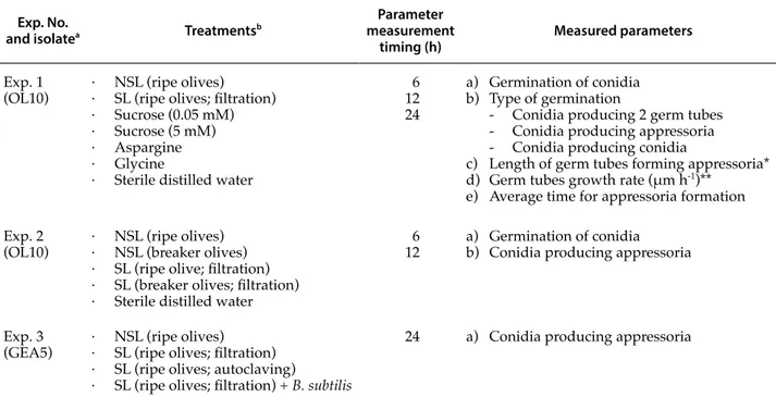
concentra-Table 1. Summary of the trials conducted to evaluate the effects of non-sterile (NSL) and sterile (SL) olive leachates on
conidium germination of different isolates of Colletotrichum godetiae. The effects of three exogenous nutrients (sucrose, aspargine and glycine) and the addition of Bacillus subtilis cells to sterile olive leachates were also investigated.
Exp. No.
and isolatea Treatmentsb
Parameter measurement
timing (h) Measured parameters
Exp. 1
(OL10) · NSL (ripe olives)· SL (ripe olives; filtration) · Sucrose (0.05 mM) · Sucrose (5 mM) · Aspargine · Glycine
· Sterile distilled water
6 12 24
a) Germination of conidia b) Type of germination
- Conidia producing 2 germ tubes - Conidia producing appressoria - Conidia producing conidia
c) Length of germ tubes forming appressoria* d) Germ tubes growth rate (μm h-1)**
e) Average time for appressoria formation Exp. 2
(OL10) · NSL (ripe olives)· NSL (breaker olives) · SL (ripe olive; filtration) · SL (breaker olives; filtration) · Sterile distilled water
6
12 a) Germination of conidia b) Conidia producing appressoria
Exp. 3
(GEA5) · NSL (ripe olives)· SL (ripe olives; filtration) · SL (ripe olives; autoclaving)
· SL (ripe olives; filtration) + B. subtilis
24 a) Conidia producing appressoria
a Isolates of Colletotrichum godetiae utilized in experiments.
b Olive leachates were obtained from ripe (uniformly black) or breaker (initial turning from green to black) olive drupes, and were steri-lized by filtration or by autoclaving.
* Parameter recorded only at 24 h. ** Parameter evaluated between 6 and 12 h.
tion of 5 × 10⁴ conidia mL-1 of the pathogen and 0.05 or 5 mM sucrose, 0.01 M asparagine and 0.01 M gly-cine. Conidial suspensions were incubated at 24°C in the dark for 6, 12, or 24 h according to different experimental schemes (Table 1).
All assays were conducted on sterile micro-scope hanging-drop slides with 13 mm diam. wells of depth 0.5–0.6 mm. Before use, the slides were washed by immersion in concentrated HCl (37%), thoroughly rinsed with distilled water and then sterilised at 120°C for 20 min. Slides with conidial suspensions were incubated in Petri dishes on a moistened filter paper.
Morphological measurements
Different morphological measurements were per-formed at 6, 12 and/or 24 h using a light microspore set up for a ×1000 magnification, as summarised in Table 1. Before measurements, the germination pro-cess was stopped by adding a drop (5 μL) of lacto-phenol cotton blue (Sigma-Aldrich).
In Experiments 1 and 2, conidia with clearly vis-ible germ tube initials were considered germinat-ed. Furthermore, those producing appressoria (all experiments) and secondary conidia or two germ tubes (Experiment 1) were specifically recorded (Table 1). The lengths of germ tubes producing ap-pressoria, the germ tube growth rates and the aver-age time needed for the formation of appressoria were estimated during Experiment 1. The germ tube growth rates were determined between 6 and 12 h for all incubation media by analysing actively growing conidia: growth rate = (average length at 12 h – average length at 6 h)/6 h. This parameter was then used to estimate the average time for ap-pressorium formation, assuming that the growth rate was constant during the entire incubation peri-od: average time for appressoria formation = length of germ tubes with appressoria recorded at 24 h/ growth ratio.
In all measurements, three sets of 40 conidia each were randomly selected from three different slides and observed. Mean data from the experiments were analyzed for homogeneity of variance with the software package Statistics for Windows (StatSoft), and standard errors of the means (SEM) were cal-culated.
Results
Experiment 1
All treatments increased the germination of co-nidia compared with the water controls, at the three assessment times (Table 2). In particular, after 6 h of incubation the greatest proportions of germinated conidia was recorded for those in sterile leachate (98.6%), 5 mM sucrose and asparagine (94.6%), and glycine (94.3%), followed by non-sterile leachate (78.3%) and 0.05 mM sucrose (77.6%). Only 68.3% of the conidia germinated in sterile water. The propor-tions of germinated conidia increased slightly for all treatments after 12 and 24 h, but these later results largely confirmed those obtained at 6 h (Table 2).
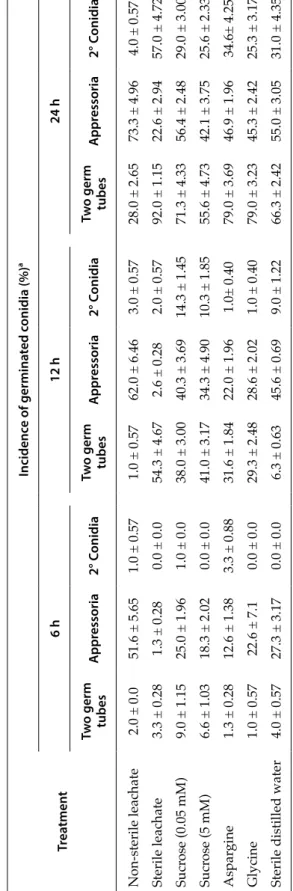
Incubation media modified the morphology of ger-minated conidia. Sterile leachate increased the differ-entiation of two germ tubes from individual conidia at 12 and 24 h, and the incidence of conidia producing secondary conidia at 24 h (Table 3). In contrast, sterile leachate almost completely inhibited the formation of appressoria at 6 and 12 h and strongly inhibited ap-pressorium formation at 24 h. Completely different results were obtained with the non-sterile leachate, which gave greater incidence of conidia producing appressoria at all assessment times, and reduced the percentage of conidia producing secondary conidia or two germ tubes at 12 and 24 h.
Table 2. Results of Experiment 1a (Table 1). Mean
propor-tions of germinated Colletotrichum godetiae conidia in non-sterile olive leachate, non-sterile olive leachate, in solutions containing three different nutrient elements, and sterile distilled water (control), after 6, 12 and 24 h of incubation.
Treatment Germination of conidia (%)
a 6 h 12 h 24 h Non-sterile leachate 78.3 ± 6.69 85.6 ± 4.33 88.3 ± 3.75 Sterile leachate 98.6 ± 0.28 99.0 ± 0.57 99.3 ± 0.34 Sucrose (0.05 mM) 77.6 ± 0.63 84.6 ± 1.73 87.3 ± 2.48 Sucrose (5 mM) 94,6 ± 3.34 97.0 ± 0.57 97.6 ± 0.75 Aspargine 94.6 ± 0.92 97.3 ± 0.86 97.6 ± 0.92 Glycine 94.3 ± 0.63 95.6 ± 1.32 96.6 ± 1.32 Sterile distilled water 68.3 ± 4.67 77.6 ± 5.77 80.6 ± 3.63 a Means ± standard error of the mean (SEM).
Except for sucrose at the lower concentration (0.05 mM), a significant reduction of appressoria was recorded for all tested exogenous nutrients as com-pared to the water control (Table 3). In contrast, the production of two germ tubes per conidium was in-creased by most treatments at 12 and 24 h, and there was also a minor influence on production of second-ary conidia.
After 24 h of incubation, the lengths of germ tubes from conidia producing appressoria were increased in sterile leachates, but reduced in non-sterile leachates (Table 4). These parameters were not significantly influenced, however, by the tested exogenous nutrients. Both sterile and non-sterile lea-chates as well as the three tested nutrients caused increased germ tube growth rates as compared to the water controls. Non-sterile leachate gave the shortest length of germ tubes producing appresso-ria and the greatest growth ratio. The combination of these two parameters resulted in the estimation of reduced time needed for the production of appres-soria as compared to the water control and all other incubation media. In contrast, the greatest estimated time for appressorium formation was recorded for the sterile leachate. Similar to the estimated time for appressorium formation, after 6 hours of incubation, 51.6% of germinated conidia in non-sterile leachates, 1.3% in sterile leachate, and 27.3% sterile distilled water produced appressoria (Table 3).
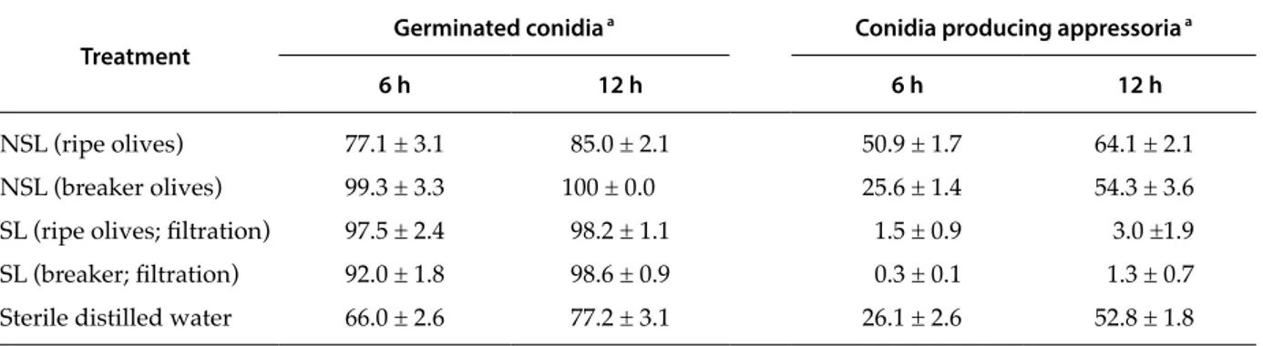
Experiment 2
In Experiment 2, olive leachates from ripe and breaker olives were compared by determining their effects on conidium germination and appressorium formation. After 6 h of incubation, the percentage of germinated conidia was greater in non-sterile chates from breaker olives than in non-sterile lea-chates from ripe olives (Table 5). However, an op-posite trend was recorded for sterile leachates, i.e. conidium germination was greater in leachates from ripe olives compared to those from breaker olives. Results obtained at 6 h were generally confirmed at 12 h, although differences in the treatments were less.
Non-sterile olive leachates from ripe olives in-creased the production of appressoria as compared to the water controls after both 6 and 12 h of incuba-tion. In contrast, non-sterile leachates from breaker olives did not modify the production of appressoria.
Sterile leachates from both ripe and breaker olives almost completely inhibited the production of ap-pressoria after 6 and 12 h.
Experiment 3
The influence of the procedure utilised to sterilize olive leachates and the role of bacteria on the appres-sorium formation was evaluated in this experiment. Overall the isolate utilised in this experiment (GEA5) produced many fewer appressoria compared with isolate OL10, used Experiments 1 and 2. After 24 h of incubation, 9% of the germinated conidia in water and 42.3% of those in non-sterile leachates produced appressoria (Table 6). The smallest percentage of co-nidia producing appressoria was recorded in sterile leachate obtained by filtration (3.6 %) followed by sterile leachate obtained by autoclaving (11.0%) and sterile leachates obtained by filtration and amended with B. subtilis (13.6%).
Discussion
Experiments conducted in this study with two different isolates of C. godetiae have clearly demon-strate that the germination of conidia, in terms of both percentage and mode of germination, is likely to be greatly influenced by the nutrient substances and microorganisms of the olive carposphere. The same trends in measured parameters were detected for conidia of two different isolates, although the isolates differed in capability to produce appressoria after germination.
Olive leachates increased the percentage of conid-ium germination in both Experiments 1 and 2, con-ducted with isolate OL10 of C. godetiae, compared to germination in sterile distilled water. This effect was more marked with sterile leachates compared to the non-sterile leachates. The early recognition events that take place in host-pathogen interactions are complex phenomena, which are conditioned by host wax surfaces and the quality and quantity of solutes present on plant surfaces. Some of these many com-pounds may be regarded as nutrients (e.g. sugars, amino acids, inorganic ions) or fungitoxic molecules (e.g. phenols), and have been reported to have a stimulating action on germination and/or appresso-rium formation (Emmett and Parbery, 1975). In our study, the presence of different nutrients in both ster-ile and non-sterster-ile olive leachates stimulated the
ger-Table 3. Results of Experiment 1b (T able 1). Mean pr oportions of germinated conidia of Colletotrichum godetiae pr oducing appr essoria, secondary (2°) conidia or two germ tubes in non-sterile olive leachate, sterile olive leachate, solutions containing thr ee differ ent nutrient elements and sterile distilled water (contr
ol), after 6, 12 and 24 h of incubation.
Tr ea tmen t Incidenc e of germina ted c onidia (%) a 6 h 12 h 24 h Tw o germ tubes A ppr essoria 2° C onidia Tw o germ tubes A ppr essoria 2° C onidia Tw o germ tubes A ppr essoria 2° C onidia Non-sterile leachate 2.0 ± 0.0 51.6 ± 5.65 1.0 ± 0.57 1.0 ± 0.57 62.0 ± 6.46 3.0 ± 0.57 28.0 ± 2.65 73.3 ± 4.96 4.0 ± 0.57 Sterile leachate 3.3 ± 0.28 1.3 ± 0.28 0.0 ± 0.0 54.3 ± 4.67 2.6 ± 0.28 2.0 ± 0.57 92.0 ± 1.15 22.6 ± 2.94 57.0 ± 4.72 Sucr ose (0.05 mM) 9.0 ± 1.15 25.0 ± 1.96 1.0 ± 0.0 38.0 ± 3.00 40.3 ± 3.69 14.3 ± 1.45 71.3 ± 4.33 56.4 ± 2.48 29.0 ± 3.00 Sucr ose (5 mM) 6.6 ± 1.03 18.3 ± 2.02 0.0 ± 0.0 41.0 ± 3.17 34.3 ± 4.90 10.3 ± 1.85 55.6 ± 4.73 42.1 ± 3.75 25.6 ± 2.33 Aspar gine 1.3 ± 0.28 12.6 ± 1.38 3.3 ± 0.88 31.6 ± 1.84 22.0 ± 1.96 1.0± 0.40 79.0 ± 3.69 46.9 ± 1.96 34.6± 4.25 Glycine 1.0 ± 0.57 22.6 ± 7.1 0.0 ± 0.0 29.3 ± 2.48 28.6 ± 2.02 1.0 ± 0.40 79.0 ± 3.23 45.3 ± 2.42 25.3 ± 3.17
Sterile distilled water
4.0 ± 0.57 27.3 ± 3.17 0.0 ± 0.0 6.3 ± 0.63 45.6 ± 0.69 9.0 ± 1.22 66.3 ± 2.42 55.0 ± 3.05 31.0 ± 4.35 a Means ± standar d err
Table 4. Results of Experiments 1c, d and e (Table 1). Mean length (μm) of germ tubes of Colletotricum godetiae conidia forming appressoria after 24 h of incubation, and estimated mean germ tube growth rates (μm h-1) and average time for
the production of appressoria in sterile and non-sterile olive leachate, solutions containing three different nutrients, and sterile distilled water (control).
Treatment Length of germtubes (µm) a
Germ-tube growth rate (µm h-1) a Average time for appressorium formation (h) a
Non sterile leachate 19.9 ± 3.56 5.7 ± 0.7 3.5 ± 0.5
Sterile leachate 59.8 ± 4.20 4.4 ± 0.7 13.6 ± 1.6
Sucrose (0.05 mM) 30.0 ± 2.56 4.7 ± 0.9 6.4 ± 0.4
Sucrose (5 mM) 33.0 ± 2.69 4.9 ± 0.6 6.7 ± 0.9
Aspargine 37.3 ± 3.73 3.8 ± 0.1 9.8 ± 0.8
Glycine 22.5 ± 5.42 4.2 ± 0.4 5.4 ± 0.6
Sterile distilled water 32.6 ± 4.24 3.3 ± 0.5 9.9 ± 0.7
a Means ± standard error of the mean (SEM).
Table 5. Results of Experiments 2a and b (Table 1). Mean percentage of germinated Colletotricum godetiae conidia and of
conidia producing appressoria after 6 and 12 h incubation in sterile (SL) and non-sterile (NSL) olive leachates from ripe and breaker olives, or in sterile distilled water (control).
Treatment Germinated conidia
a Conidia producing appressoria a
6 h 12 h 6 h 12 h
NSL (ripe olives) 77.1 ± 3.1 85.0 ± 2.1 50.9 ± 1.7 64.1 ± 2.1
NSL (breaker olives) 99.3 ± 3.3 100 ± 0.0 25.6 ± 1.4 54.3 ± 3.6
SL (ripe olives; filtration) 97.5 ± 2.4 98.2 ± 1.1 1.5 ± 0.9 3.0 ±1.9
SL (breaker; filtration) 92.0 ± 1.8 98.6 ± 0.9 0.3 ± 0.1 1.3 ± 0.7
Sterile distilled water 66.0 ± 2.6 77.2 ± 3.1 26.1 ± 2.6 52.8 ± 1.8
a Means ± standard error of the mean (SEM).
Table 6. Results of Experiment 3a (Table 1). Mean percentage of Colletotrichum godetiae conidia forming appressoria after
germination in sterile distilled water, non-sterile olive leachate, sterile olive leachate obtained by filtration or autoclaving, sterile leachate obtained by filtration and amended with cells of Bacillus subtilis and sterile distilled water (control), after 24 h of incubation.
Treatments Conidia producing appressoria (%)a
Non-sterile leachate 42.3 ± 1.17
Sterile leachate (filtration) 3.6 ± 0.66
Sterile leachate (autoclaving) 11.0 ± 1.50 Sterile leachate + B. subtilis 13.6 ± 1.71
Sterile distilled water 9.0 ± 0.57
mination of conidia, but this effect was partially re-duced in non-sterile leachates due to the presence of natural competing microorganisms. The non-sterile olive leachate from breaker olives was more effective for increasing conidium germination than the non-sterile leachate from ripe olives, probably because of lower microbial concentrations in the breaker olive leachate. The total microbial population in the car-posphere, as well as that associated with other plant organs, is mainly represented by bacteria, and these populations are likely to increase with fruit ripening as more nutrient elements are available (Teixidó et al., 1999; Fürnkranz et al., 2012).
In agreement with these considerations, sterile leachates from ripe olives stimulated germination to a greater extent than those from breaker olives. Ripe fruits are likely to have greater content of nutrients than unripe fruit, and induce effects on germination of conidia, but this was not observed when more nu-trients were also associated with rich microbial pop-ulations. In other words, nutrients and microorgan-isms both influenced the germination of conidia. The final germination ratio was the result of a dynamic equilibrium between the positive effects of nutrients the negative competition of indigenous microorgan-isms. This conclusion was also supported by data ob-tained with the three different exogenous nutrients utilised in the present study. Sucrose, asparagine and glycine significantly increased the germination of co-nidia as compared to the water control.
The germination ratio of conidia is an important aspect that can have significant impacts on the se-verity of a disease. However, other events are also necessary for successful infection by hemibiotroph pathogens like C. godetiae. Development of appresso-ria is one of the most important of these (Gomes et al., 2009, 2012). Data obtained in the present study sug-gest a primary role of natural microbial populations in olive carpospheres in the formation of appresso-ria. Non-sterile olive leachates greatly increased the formation of appressoria compared to the water con-trols in all experiments, conducted with two differ-ent C. godetiae isolates. In contrast, the formation of appressoria was almost completely inhibited when conidia were incubated in olive leachates sterilized by filtration. More precisely, the elimination of the microbial fraction in combination with the high con-tent in nutrients of the fruit leachate amplified the vegetative phase (saprophytic growth) after conidi-um germination, induced longer germ tubes, greater
average time for appressorium formation and re-duced production of appressoria. In agreement with these results, minor incidence of appressoria was also recorded when the nutrients sucrose, aspara-gine and glycine were added to the incubation me-dia. Similarly, reduced production of appressoria has been reported for C. graminicola grown in nutritively rich media (Apoga et al., 2004).
When leachates were sterilized by autoclaving greater incidence of appressoria was recorded, com-pared with leachate sterilized by filtration. In both cases, however, the incidence of appressoria was much less compared to non-sterile leachate. These data confirm that the microbial fraction of leachate is likely to be a major factor inducing appressoria, but particles suspended in the non-sterile leachate and removed during filtration may also play also a role in appressorium formation. It has been suggested that toxic substances, such as the anthranilic acid found in banana, can initiate appressorium formation by C. musae (Emmett and Parbery, 1975; Swinburne, 1976). It is also possible that olive leachates contain sub-stances that are toxic to fungi. However, their role in appressorium formation are likely to be of minor relevance considering that putative toxic substances should not be lost during filtration.
Further confirmation of the role of microorgan-isms in appressorium formation was obtained by the lower number of appressoria differentiated in the sterile leachate as compared to same medium amended with cells of B. subtilis. This bacterium is one of the most abundant saprophytic genera in olive phylloplanes, and is a well-known bacterial antago-nist (Ercolani, 1978). Furthermore, a few strains of B. subtilis are registered and used as biocontrol agents of plant pathogens, including Colletotrichum spp. caus-ing anthracnose on leaves and fruits of various crops (Cawoy et al., 2011). Although the amendment of B. subtilis to the sterile leachate significantly increased the production of appressoria, the incidence of these structures remained much less compared to the non-sterile leachate, and was similar for conidia incu-bated in the leachate sterilized by autoclaving. This suggests that microorganisms may not be the only factors playing a role in the formation of appressoria, which are lost/changed during filtration. However, it is important to note that cells of only one bacteri-um were added to the sterile leachate, while a rich natural microbial population colonizes natural olive carpospheres. Although the olive microbiota was not
evaluated in the present study, very complex indig-enous populations are known to colonize the carpo-sphere (Janisiewicz et al., 2010; Janisiewicz and Buyer, 2010). Both quality and quantity of nutrients and mi-croorganisms seem to be important. It is possible to suppose a strict relationship between the microbial composition of the infection drop and the quality and quantity of plant exudates that, varying in relation to different plant and environmental condition, could modify the conidia response (Tukey, 1971).
In conclusion, results from the present study pro-vide strong epro-vidence that nutrients and microorgan-isms of the olive carposphere can play important roles in the infection processes of C. godetiae. On one hand, nutrients enhanced the germination of conid-ia, while on the other, indigenous microorganisms established a hostile environment that led to a high-ly increased appressorium differentiation, probabhigh-ly related to micobial competition (Lenne and Parbery, 1976). Since appressoria are necessary for successful infection of host tissues (Deising et al., 2000), signals that reduce their differentiation may enhance disease control and vice versa (Gomes et al., 2009). In particu-lar, bacterial species present on host surfaces may ac-celerate the penetration of C. godetiae by stimulating a rapid production of appressoria, and reducing the saprophytic pre-infectional mycelial growth of the pathogen. This will result in reduced duration of the critical moisture period required by the fungus to produce infection in the field (Fernando et al., 1994).
Literature cited
Agosteo G.E., S.O. Cacciola, A. Pane and S. Frisullo, 1997. Veg-etative compatibility groups of Colletotrichum
gloeospori-oides from olive in Italy. In: Proceedings, 10th Congress of the
Mediterranean Phytopathological Union (Société Française
de Phytopathologie, ed.), Le Corum, Montpellier, France, 95–99.
Agosteo G.E., G. Magnano di San Lio, S.O. Cacciola and S. Frisullo, 2002. Characterization of the causal agent of ol-ive anthracnose in southern Italy. Acta Horticulturae 586, 713–716.
Apoga D., J. Barnard, H.G. Craighead and H.C. Hocha, 2004. Quantification of substratum contact required for initia-tion of Colletotrichum graminicola appressoria. Fungal
Ge-netics and Biology 41, 1–12.
Balestra G.M., F. Pugliese and L. Varvaro, 1997. Biological con-trol of Colletotrichum gloeosporioides by epiphytic bacteria from olive phylloplane. In: Proceedings, 10th Congress of the
Mediterranean Phytopathological Union (Société Française
de Phytopathologie, ed.), Le Corum, Montpellier, France, 657–659.
Blackeman J.P. and D.G. Parbery, 1977. Stimulation of appres-sorium formation in Colletotrichum acutatum by phyllo-plane bacteria. Physiological Plant Pathology 11, 313–325. Cacciola S.O., R. Faedda and G.E. Agosteo, 2011. Olive
an-thracnose. In: Olive diseases and disorders (L. Schena, G.E. Agosteo, S.O. Cacciola, ed.), Transworld Research Net-work, Kerala, India, 223–246.
Cacciola S.O., R. Faedda, F. Sinatra, G.E. Agosteo, L. Schena, S. Frisullo and G. Magnano di San Lio, 2012. Olive Anthrac-nose. Journal of Plant Pathology 94, 29–44.
Cawoy H., W. Bettiol, P. Fickers and M. Ongena, 2011. Bacil-lus-based biological control of plant diseases. In: Pesticides
in the Modern World - Pesticides Use and Management (M.
Stoytcheva, ed.), 31 pp., <http://cdn.intechopen.com/ pdfs-wm/21989.pdf>, InTech. Accessed on March 28, 2014.
Damm U., P.F. Cannon, J.H.C. Woudenberg and P.W.Crous, 2012. The Colletotrichum acutatum species complex. Studies
in Mycology 73, 37–113.
Deising H.B., S. Werner and M. Wernitz, 2000. The role of fun-gal appressoria in plant infection. Microbes and Infection 13, 1631–1641.
Emmett R.W. and D.G. Parbery, 1975. Appressoria. Annual
Re-view of Phytopathology 13, 147–167.
Ercolani G.L., 1978. Pseudomonas savastanoi and other bacteria colonizing the surface of olive leaves in the field. Journal of
General Microbiology 109, 245–257.
Ercolani G.L., 1991. Distribution of epiphytic bacteria on olive leaves and the influence of leaf age and sampling time.
Microbial Ecology 21, 35–48.
Faedda R., G.E. Agosteo, L. Schena, S. Mosca, S. Frisullo, G. Magnano Di San Lio and S.O. Cacciola, 2011.
Colletotri-chum clavatum sp. nov. identified as the causal agent of
olive anthracnose in Italy. Phytopathologia Mediterranea 50, 283–302.
Fernando W.G., A.K. Watson and T.C. Paulitz, 1994. Phyl-loplane Pseudomonas spp. enhance disease caused by
Colletotrichum coccodes on velvetleaf. Biological Control 4,
125–131.
Fürnkranz M., B. Lukesch, H. Müller, H. Huss, M. Grube and G. Berg, 2012. Microbial Diversity Inside Pumpkins: Mi-crohabitat-Specific Communities Display a High Antago-nistic Potential Against Phytopathogens. Microbial Ecology 63, 418–428.
Gomes S., P. Prieto, P. Martins-Lopes, T. Carvalho, A. Martin and E. Guedes-Pinto, 2009. Development of Colletotrichum
acutatum on Tolerant and Susceptible Olea europaea L.
culti-vars: A Microscopic Analysis. Mycopathology 168, 203–211. Gomes S., P. Prieto, T. Carvalho, H. Guedes-Pinto and P.
Martins-Lopes, 2012. Olive - Colletotrichum acutatum: An example of fruit-fungal interaction. In: Plant Breeding (I. Abdurakhmonov, ed.), <http://www.intechopen.com/ books/plant-breeding/olive-colletotrichum-acutatum-an-example-of-fruit-fungalinteraction>. InTech. Accessed on December 9, 2013.
Janisiewicz W.J. and J.S. Buyer, 2010. Culturable bacterial mi-croflora associated with nectarine fruit and their potential for control of brown rot. Canadian Journal of Microbiology 56, 480–486.
Janisiewicz W.J., C.P. Kurtzman and J.S. Buyer, 2010. Yeasts as-sociated with nectarines and their potential for biological control of brown rot. Yeast 27, 389–398.
Koomen I. and P. Jeffries, 1993. Effects of antagonistic micro-organisms on the post-harvest development of
Colletotri-chum gloeosporioides on mango. Plant Pathology 42, 230–237.
Lenne J.M. and D.G. Parbery, 1976. Phyllosphere antagonists and appressorium formation in Colletotrichum
gloeospori-oides. Transactions of the British Mycological Society 66, 334–
336.
Manandhar J.B., G.L. Hartman and T.C. Wang, 1995. Conidial germination and appressorial formation of Colletotrichum
capsici and C. gloeosporioides isolates from pepper. Plant Disease 79, 361–366.
Mahuku G.S. and P.H. Goodwin, 1998. Influence of sucrose, mucin and xanthan gum on spore germination of ten dif-ferent fungi. European Journal of Plant Pathology 104, 849– 852.
Moral J., K. Bouhmidi and A. Trapero, 2008. Influence of fruit maturity, cultivar susceptibility, and inoculation method on infection of olive fruit by Colletotrichum acutatum. Plant
Disease 92, 1421–1426.
Mosca S., M.G. Li Destri Nicosia, S.O. Cacciola and L. Sche-na, 2014. Molecular analysis of Colletotrichum species in the carposphere and phyllosphere of olive. PLoS One 9, e114031.
Peres N.A., L.W. Timmer, J.E. Adaskaveg and J.C. Correll, 2005. Lifestyles of Colletotrichum acutatum. Plant Disease 89, 784–796.
Perfect S.E., H. Bleddyn Hughes, R.J. O’Connel and J.R. Green, 1999. Colletotrichum: A model genus for studies on pathol-ogy and fungal-plant interactions. Fungal Genetics and
Biol-ogy 27, 186–198.
Podila G.K., L.M. Rogers and P.E. Kolattukudy, 1993.
Chemi-cal signals from avocado surface wax trigger germination and appressorium formation in Colletotrichum
gloeospori-oides. Plant Physiology 103, 267–272.
Schena L., S. Mosca, S.O. Cacciola, R. Faedda, S.M. Sanzani, G.E. Agosteo, V. Sergeeva and G. Magnano di San Lio. 2014. Species of the Colletotrichum gloeosporioides and C.
boninense complexes associated to olive antrachnose. Plant Pathology 63, 437–446.
Shivas R.G. and Y.P. Tan, 2009. A taxonomic re-assessment of
C. acutatum introducing C. fioriniae comb. et stat. nov. and C. simmondsii sp. nov. Fungal Diversity 39, 111–122.
Sreenivasaprasad S. and P. Talhinhas, 2005. Genotypic and phenotypic diversity in Colletotrichum acutatum, a cosmo-politan pathogen causing anthracnose on a wide range of hosts. Molecular Plant Pathology 6, 361–378.
Talhinhas P., J. Neves-Martins, H. Oliveira and S. Sreenivasaprasad. 2009. The distinctive population struc-ture of Colletotrichum species associated with olive an-thracnose in the Algarve region of Portugal reflects a host-pathogen diversity hot spot. FEMS Microbiology Letters 296, 31–38.
Talhinhas P., C. Mota-Capitão, S. Martins, A.P. Ramos, J. Ne-ves-Martins, L. Guerra-Guimarães, V. Várzea, M.C. Silva, S. Sreenivasaprasad and H. Oliveira, 2011. Epidemiology, histopathology, and aetiology of olive anthracnose caused by Colletotrichum acutatum and C. gloeosporioides in Portu-gal. Plant Pathology 60, 483–495.
Teixidó N., J. Usall, N. Magan and I. Viñas, 1999. Microbial population dynamics on golden delicious apples from bud to harvest and effect of fungicide applications. Annals
of Applied Biology 134, 109–116.
Tukey H.B., 1971. Leaching of substance from plants. In:
Ecolo-gy of leaf surface microorganism (T.F. Preece, C.H. Dickinson,
ed.), Academic Press, London, UK, 67–80.
Accepted for publication: August 12, 2014 Published online: April 14, 2015