Decellularization and Delipidation Protocols
of Bovine Bone and Pericardium for Bone
Grafting and Guided Bone Regeneration
Procedures
Chiara Gardin1☯, Sara Ricci2☯, Letizia Ferroni1*, Riccardo Guazzo2, Luca Sbricoli2, Giulia De Benedictis3, Luca Finotti3, Maurizio Isola3, Eriberto Bressan2, Barbara Zavan1* 1 Department of Biomedical Sciences, University of Padova, Padova, Italy, 2 Department of Neurosciences, University of Padova, Padova, Italy, 3 Department of Animal Medicine, Productions and Health, University of Padova, Legnaro, Padova, Italy
☯ These authors contributed equally to this work.
*[email protected](BZ);[email protected](LF)
Abstract
The combination of bone grafting materials with guided bone regeneration (GBR) mem-branes seems to provide promising results to restore bone defects in dental clinical practice. In the first part of this work, a novel protocol for decellularization and delipidation of bovine bone, based on multiple steps of thermal shock, washes with detergent and dehydration with alcohol, is described. This protocol is more effective in removal of cellular materials, and shows superior biocompatibility compared to other three methods tested in this study. Furthermore, histological and morphological analyses confirm the maintenance of an intact bone extracellular matrix (ECM).In vitro and in vivo experiments evidence osteoinductive and osteoconductive properties of the produced scaffold, respectively. In the second part of this study, two methods of bovine pericardium decellularization are compared. The osmotic shock-based protocol gives better results in terms of removal of cell components, biocom-patibility, maintenance of native ECM structure, and host tissue reaction, in respect to the freeze/thaw method. Overall, the results of this study demonstrate the characterization of a novel protocol for the decellularization of bovine bone to be used as bone graft, and the acquisition of a method to produce a pericardium membrane suitable for GBR applications.
Introduction
In the field of oral surgery and dental implantology, bone deficiency represents the main prob-lem that clinicians have to overcome in order to ensure the implant stability and the complete functional restoration. Bone grafting has emerged as a surgical procedure to make up for the bone deficiency [1]. Bone graft not only replaces the missing bone, but also helps regrowth of lost bone by acting as a scaffold for osteoconduction and as a source of osteogenic and a11111
OPEN ACCESS
Citation: Gardin C, Ricci S, Ferroni L, Guazzo R, Sbricoli L, De Benedictis G, et al. (2015)
Decellularization and Delipidation Protocols of Bovine Bone and Pericardium for Bone Grafting and Guided Bone Regeneration Procedures. PLoS ONE 10(7): e0132344. doi:10.1371/journal.pone.0132344 Editor: Xiaohua Liu, Texas A&M University Baylor College of Dentistry, UNITED STATES Received: April 21, 2015
Accepted: June 14, 2015 Published: July 20, 2015
Copyright: © 2015 Gardin et al. This is an open access article distributed under the terms of the
Creative Commons Attribution License, which permits unrestricted use, distribution, and reproduction in any medium, provided the original author and source are credited.
Data Availability Statement: All relevant data are within the paper and its Supporting Information files. Funding: This research was supported by a University of Padova (progetto di Ateneo) grant to BZ. The funders had no role in study design, data collection and analysis, decision to publish, or preparation of the manuscript.
Competing Interests: The authors have declared that no competing interests exist.
osteoinductive molecules for bone formation [2]. Osteoinduction refers to the ability of the scaffold to recruit multipotent mesenchymal stem cells (MSCs) from the surrounding tissue, and to induce their differentiation into bone-forming osteoblasts. Osteoconduction is a charac-teristic whereby the scaffold acts as a permanent or resorbable matrix that mechanically sup-port the ingrowth of vessels and new bone from the borders of the defect into and onto its surfaces. Osteogenesis is the synthesis of new bone by cells derived from either the graft or the host [3–5]. An ideal bone graft should function through all three mechanisms by providing a substrate that directs three-dimensional (3D) bone growth, recruits and induces differentiation of resident bone-forming cells, and supplies more bone-forming cells to the recipient site. Another fundamental property of an ideal bone graft is osseointegration, which is the ability to bind to the surrounding bone without an intervening layer of fibrous tissue, allowing incorpo-ration of the graft at the host site. Osseointegincorpo-ration is not an isolated phenomenon but depends on previous osteoinduction and osteoconduction [3].
Bone grafts may result from the patient’s bone (autograft), from human donors (allograft) or from animals (xenograft) [6]. Autogenous bone is still considered the“gold standard”, because of its osteoinductive, osteoconductive and osteogenic properties, and absence of immune response. On the contrary, allografts and xenografts have osteoinductive and osteo-conductive characteristics but lack the osteogenic properties of autografts [7,8]. Nevertheless, some restrictions in using autograft in clinical practice exist because of donor site morbidity observed with harvesting procedures and the limited amount of bone available [9]. Allograft is a possible alternative to bone autograft with major limitations associated to rejection, transmis-sion of diseases, and cost [10]. With progression in biomaterials technology, the use of xeno-graft for human tissue reconstruction is increasing [11]. These animal-derived tissues represent an unlimited supply of available material if they could be processed to be safe for transplanta-tion in humans. Indeed, the disadvantage associated to xenografts is that they may trigger unwanted immunological and inflammatory host reactions [12].
To increase efficiency of bone repair, especially in the management of large osseous defects, bone graft areas often require covering with guided bone regeneration (GBR) membranes [13,14]. The underlying concept of GBR was first introduced more than 50 years ago, when cel-lulose acetate filters were experimentally used for the regeneration of nerves and tendons [15]. Later, a series of animal studies showed that GBR can predictably facilitate bone regeneration in critical-sized osseous defects, as well as healing of bone defects around dental implants [16– 18]. The GBR procedure uses membranes that act as physical barriers to epithelial and connec-tive cell invasion from the surrounding soft tissues, thus providing osteogenic cells, which exhibit slower migration rate, better conditions to perform bone regeneration [19]. The use of a barrier membrane is advantageous to facilitate augmentation of alveolar ridge defects, induce bone regeneration, improve bone-grafting results, and treat failing implants. Several barrier membranes have been developed to serve a variety of functions in clinical applications; all of them must satisfy five main design criteria, as described by Scantlebury [20]: biocompatibility, space-making, cell-occlusiveness, tissue integration and clinical manageability.
Although different bone graft materials and barrier membranes have been developed and their use has been extensively investigated, research is ongoing to generate bone grafts and membranes suitable for dental clinical applications. In order to make xenografts an acceptable alternative to autografts and allografts, several processing and storage methods have been stud-ied. Typically, these scaffolds are produced by the process of decellularization of naturally derived tissues. Decellularization has the finality to completely eliminate the cellular compo-nent of the native tissue, while maintaining as much of the structure and composition as possi-ble of the original extracellular matrix (ECM) [21]. The ECM has been shown to modulate the behavior of cells that contact the scaffold either by regulating cell migration, influencing
tissue-specific phenotypic differentiation, and inducing constructive host tissue remodeling
responses. It is likely that the 3D ultrastructure, surface topology, and composition of the ECM all contribute to these effects [22]. Decellularization can be achieved by a variety of agents and techniques. It typically involves exposure to chemical and biologic agents such as detergents and enzymes, and physical forces that unavoidably cause disruption of the associated ECM. Delipidation is another factor to consider, especially for bone grafts [11]. Indeed, the remaining lipids in bone may represent a barrier to cell invasion, negatively influencing its biocompatibil-ity and osseointegration [23]. Moreover, they can induce giant cell reactions which can increase bone resorption and encapsulating fibrosis [24].
The aim of the present work was to develop new methods for decellularization and delipida-tion of bovine bone and pericardium for the generadelipida-tion of bone substitutes and membranes to be used in bone grafting and GBR procedures, respectively. Histological, morphological, and molecular in vitro analyses have been performed in order to test the structural features and the biocompatibility of the produced scaffolds. Additionally, in vivo experiments were carried out to evaluate the biological properties and the host tissue reactions to the implanted bovine biomaterials.
Materials and Methods
Source of bovine bone and pericardium
Fresh bone samples were harvested from epiphysis of bovine femur (18–24 month old), imme-diately after slaughter from a local slaughterhouse (Macello Pubblico Comunale, Udine, Italy). In the laboratory, bone blocks (3 x 3 x 2 cm) were stored at -80°C until use.
Bovine pericardium samples were obtained from the same animals and delivered to the lab-oratory in cold PBS plus. PBS plus was made of phosphate buffered saline (PBS) (EuroClone, Milan, Italy), containing 1% Penicillin/Streptomycin (P/S) (EuroClone) and 1% Gentamycin (Invitrogen, Paisley, UK). External fat and adherences were removed; then pericardium strips (3 x 1 cm) were obtained, and stored at -80°C until use.
Bone and pericardium samples were either immediately analyzed—hereafter named as native bone sample (NBS) or native pericardium sample (NPS)—or further processed as described below.
Decellularization protocols of bovine bone
Decellularization of bovine bone blocks started with a wash in PBS plus. Then, four cycles of thermal shock, each comprising a step at 121°C for 20 min, followed by freezing in liquid nitro-gen (-196°C) for 16 h, were carried out. During these passages, bone blocks were immersed in bidistilled water (ddH2O); the solution was changed at every cycle. Cellular debris was then
removed by washing bone blocks in 1% Triton X-100 (Promega, Madison, WI, USA) for 8 h, followed by a second wash in 0.1% Triton X-100 for 16 h. Triton X-100 was dissolved in ddH2O. To remove residual detergent, bone samples were washed two times in ddH2O for 24
h. All these steps were conducted under continuous shaking at room temperature (RT) with the rotatory shaker VDRL 711 (Asal Srl, Cernusco s/N, Milan, Italy).
At this point, decellularized bone blocks were reduced to granules with dimensions of 0.25– 1 mm using Cutting Mill SM 300 (Retsch GmbH, Haan, Germany), then divided into four groups and processed as described inTable 1.
MTT assay
To determine the presence of viable cells in bone samples after decellularization, the MTT based proliferation assay was performed according to the method of Denizot and Lang with
minor modifications [25]. Briefly, tissue samples were incubated for 3 h at 37°C in 1 mL of 0.5 mg mL-1MTT solution prepared in PBS. After removal of the MTT solution by pipette, 0.5 mL of 10% DMSO in isopropanol was added to extract the formazan in the samples for 30 min at 37°C. For each sample, optical density (O.D.) values at 570 nm were recorded in duplicate on 200μL aliquots deposited in microwell plates using a multilabel plate reader (Victor 3, Perkin Elmer, Milano, Italy).
DNA and RNA content
The DNA content was determined using a DNeasy Blood and Tissue kit (Qiagen) to isolate total DNA from known masses of bone samples following the manufacturer’s protocol for tis-sue isolation, using overnight incubation in proteinase K (Qiagen).
Total RNA from either native or decellularized tissue samples of known masses was isolated using RNeasy Mini Kit (Qiagen), including DNase digestion with the RNase-Free DNase Set (Qiagen).
The DNA and RNA quality and concentration was measured using the NanoDrop ND-1000 (Thermo Scientific, Waltham, MA, USA) and expressed as nanograms per milligram of dry tissue.
Lipid content
Oil Red O quantification was used to evaluate the residual lipid content in decellularized bone samples. Oil Red O stock solution was prepared by dissolving 3.5 mg mL-1Oil Red O (Sigma-Aldrich) in isopropanol. Working solution consisted of 3:2 Oil Red O stock in ddH2O. About 100 mg of dry bovine bone was incubated in 0.5 mL of Oil Red O working
solution for 15 min at RT. After four washing in ddH2O, Oil Red O was extracted with 0.25
mL 100% isopropanol. For each sample, O.D. values at 490 nm were recorded using a multi-label plate reader (Victor 3).
Table 1. Description of the processing methods after decellularization of bone samples. protocol
number
sample acronym
steps
B#1 DBSa1 Bone granules were washed in 0.5 M sodium bicarbonate (Sigma-Aldrich, St. Louis, MO, USA) in ddH2O for 2 days under continuous shaking at RT; the solution was changed twice a day. Residual
bicarbonate was removed with two washes of 24 h each in ddH2O. Bone granules were dehydrated with a graded ethanol series (50%, 70%, 96%, 100%) for 2 h each in slow agitation at RT. Bone granules were transferred to cell culture dishes and allowed to dry at RT under a sterile laminarflow hood.
B#2 DBS2 Bone granules were washed in 0.5 M sodium bicarbonate (Sigma-Aldrich, St. Louis, MO, USA) in ddH2O for 2 days under continuous shaking at RT; the solution was changed twice a day. Residual
bicarbonate was removed with two washes of 24 h each in ddH2O. Bone granules were freeze-dried with ScanVac Cool Safe freeze-dryer (LaboGene, Lynge, Denmark) for 8h.
B#3 DBS3 Bone granules were dehydrated with a graded ethanol series (50%, 70%, 96%, 100%) for 2 h each in slow agitation at RT. Bone granules were transferred to cell culture dishes and allowed to dry at RT under a sterile laminarflow hood.
B#4 DBS4 Bone granules were freeze-dried with ScanVac Cool Safe freeze-dryer (LaboGene) for 8 h.
aDecellularized Bone Sample
In vitro cytotoxicity test on bovine bone granules
The cytotoxicity of the decellularized bovine bone was evaluated in vitro using a mouse-derived established cell line of L929 fibroblasts (Cell bank Interlab Cell Line Collection, Genova, Italy). L929 cells were seeded at a density of 4 × 10⁴/well in 24-well plates for 24 h in cDMEM medium. cDMEM was made of Dulbecco’s Modified Eagle Medium (DMEM) (Lonza S.r.l., Milano, Italy), supplemented with 10% Fetal Bovine Serum (FBS) (Bidachem S.p.A., Milano, Italy) and 1% P/S. Cytotoxicity was assessed with the direct cell contact method, by placing about ten bone granules of the tests materials DBS1, DBS2, DBS3, and DBS4 in each well. The negative control consisted of fibroblasts seeded in presence of a titanium (Ti) disc; blank was obtained seeding fibroblasts in cDMEM with no test material added. Three samples were pre-pared for each group. The cytotoxicity produced for each different group was assessed with a 48 h cell exposure. After removing the test materials and medium, 1 mL of 0.5 mg mL-1MTT solution was placed in each well. The MTT assay was then performed as explained in the MTT assay paragraph.
Scanning Electron Microscopy (SEM)
For SEM imaging, bone samples were fixed in 2.5% glutaraldehyde in 0.1 M cacodylate buffer for 1 h, progressively dehydrated in ethanol, then critical point-dried followed by gold-palla-dium coating. The SEM analysis was carried out at the Interdepartmental Service Center C.U. G.A.S. (University of Padova, Italy) by JSM 6490 JEOL SEM.
Histological analysis
For histological examinations, bone samples were fixed in 4% paraformaldehyde solution in PBS overnight, decalcified with 10% EDTA (Sigma-Aldrich) pH 7.2 for 7 days, then paraffin-embedded and cut into 7μm thick sections. For Hematoxylin&Eosin (H&E) staining, bone sec-tions were stained with the nuclear dye Hematoxylin (Sigma-Aldrich) and the counterstain Eosin (Sigma-Aldrich).
Seeding of human Adipose-Derived Stem Cells (ADSCs) onto bovine
bone granules
Adipose tissue was digested and the cells isolated and expanded as previously described [26]. At confluence, ADSCs were harvested by trypsin treatment, then cultivated up to passage 3 (p3). Cells at p4 were seeded at density of 1 x 106cm-2onto DBS3 granules in a 24-well culture plate. The 3D cultures were incubated at 37°C and 5% CO2for up to 28 days in cDMEM,
changing the medium every 2 days.
Cell proliferation rate was then evaluated after 3, 7, 14 and 28 days from seeding with the MTT assay. Expression of osteoblast-specific markers in ADSCs cultured 7 and 28 days onto DBS3 granules was measured by Real-time PCR.
Real time PCR
For the first-strand cDNA synthesis, 1000 ng of total RNA of each sample was reverse tran-scribed with M-MLV Reverse Transcriptase (Invitrogen, Carlsbad, CA, USA), following the manufacturer’s protocol. Human primers were selected for each target gene with Primer 3 soft-ware (S1 Table). Real-time PCRs were carried out using the designed primers at a concentra-tion of 300 nM and FastStart SYBR Green Master (Roche Diagnostics, Mannheim, Germany) on a Rotor-Gene 3000 (Corbett Research, Sydney, Australia). Thermal cycling conditions were as follows: 15 min denaturation at 95°C; followed by 40 cycles of denaturation for 15 sec at
95°C; annealing for 30 sec at 60°C; and elongation for 20 sec at 72°C. Differences in gene expression were evaluated by the 2ΔΔCt method [27]. ADSCs cultured for 7 and 28 days onto tissue culture polystyrene (TCP) in cDMEM were used as control condition. Values were nor-malized to the expression of the glyceraldehyde-3-phosphate dehydrogenase (GAPDH) inter-nal reference, whose abundance did not change under our experimental conditions.
Sheep sinus augmentation surgical procedure
The in vivo study here described was approved by the Institutional Animal Care and Use Com-mittee of Padova University. Four adult sheep, two years old and 40–50 Kg of weight, were bred according to the European community guidelines (E.D. 2010/63/UE) before performing bilateral sinus augmentation [28]. DBS3 bovine granules were inserted in the inferior osseum septum of the sinuses. Animals were quarantined for 2 weeks to check the general healthy sta-tus. Surgical procedures were then carried out in the authorized Veterinary Hospital of Padova University. The animals were euthanized to explant grafted sinuses at 15 and 30 days post intervention (p.i.). For histological analyses, bone grafts were processed as described in the His-tological analysis paragraph and stained with H&E.
Decellularization protocols of bovine pericardium
Bovine pericardium was decellularized using two protocols based on either osmotic shock or freeze/thaw associated with enzymes. These passages are detailed inTable 2. Bovine pericar-dium strips obtained from both protocols were then subjected to a decontamination step, con-sisting in an incubation with 30% isopropanol for 24 h, changing the solution after 8 h. Pericardium samples were then washed twice with PBS for 24 h at RT, and stored in PBS at +4°C until being analyzed.
MTT assay, DNA and RNA content
To determine the presence of viable cells in pericardium samples after decellularization, the MTT assay was performed as described for bone in the MTT assay paragraph.
DNA and RNA were isolated from pericardium samples as reported above in the DNA and RNA content paragraph. The only difference was that the nucleic acids concentration was expressed as micrograms per milligram of dry tissue.
Seeding of human fibroblasts onto bovine pericardium
Before seeding the cells, squares (1 x 1 cm) of decellularized pericardium samples were cut and laid down on a 24-well culture plate using sterile forceps and scissors. Human fibroblasts were then seeded at a density of 1 x 106cm-2in presence of cDMEM for 7 days.
SEM and Histological analyses
For SEM imaging, pericardium samples were processed and analyzed as described for bone in the Scanning Electron Microscopy (SEM) paragraph.
For histological examinations, formalin-fixed and paraffin-embedded pericardium samples were cut into 7μm thick sections. Pericardium sections were then stained with H&E, or with Weigert’s solution (Sigma-Aldrich) in order to visualize elastic fibers.
Rat subcutaneous implantation
The protocol for the in vivo study was performed as approved by the Institutional Animal Care and Use Committee of Padova University. Decellularized pericardium samples were implanted
in the abdominal area of three adult female Lewis rats (Charles River Laboratories), weighing 150–200 g, as described by Silva-Correia et al. [29]. After 7 days of implantation, the animals were sacrificed by an overdose of gaseous anesthetic, and the implants with surrounding subcu-taneous tissue were retrieved for histological examinations after H&E staining.
Statistical analysis
Results are reported as mean ± standard deviation. One-way analysis of variance (ANOVA) Bonferroni’s post hoc test was applied to identify any significant changes between groups in the MTT, DNA/RNA, Oil Red O, and real time PCR data. Differences were considered statisti-cally significant at P<0.05Statistical analyses were performed using the SPSS 16.0 software package (SPSS Inc., Chicago, IL, USA).
Results and Discussion
Decellularization protocols of bovine bone
The use of bone grafting materials associated with the GBR technique seems to provide the most promising result to restore bone defects in dental clinical practice. A variety of decellular-ized tissues have been used as scaffold for bone regeneration applications [11–14].
In the first part of our work, we compared four different methods of bovine bone decellular-ization and delipidation. Epiphysis of bovine femur was chosen as source of starting material, due to its composition. Epiphysis is made almost entirely of spongy cancellous bone, which is considered more osteogenic than cortical bone [9]. Indeed, the presence of spaces between the structure of cancellous bone, and the large surface area, makes it very attractive at sites where new bone formation is desired. Although cancellous bone lacks mechanical strength, it is a good space filler and mostly preferred for repairing dental defects [2].
The optimal decellularization strategy would involve the use of the mildest protocol possible that yields an acellular material without disruption of the structural and functional component of the ECM. The physical and chemical methods described in this study have been already applied to a variety of tissues and organs, as widely reported in the literature [21,22,30].
Table 2. Description of the processing methods of pericardium decellularization. protocol
number sampleacronym steps
P#1 DPSa1 Strips of bovine pericardium were treated with an hypotonic buffer (ten times diluted PBS) containing 10% dimethyl sulfoxide (DMSO) (Sigma-Aldrich) and 10 mM ascorbic acid (Sigma-Aldrich), in presence of protease inhibitors (PI) (Roche, Basel, Switzerland) for thefirst 8 h, and without PI for the subsequent 16 h; all these steps were conducted at 4°C with agitation. Pericardium samples were washed in 1% Triton X-100 for 8 h, then in 0.1% Triton X-100 for 16 h at 4°C with agitation. Triton X-100 was prepared in hypotonic buffer. Strips of bovine pericardium were then treated with an hypertonic solution made of 0.5 M sodium chloride (Sigma-Aldrich) in PBS for 24 h at RT with agitation; the solution was changed twice. Pericardium samples were washed two times with 10 mM sodium deoxycholate (SD) (Sigma-Aldrich) dissolved in hypotonic buffer for 24 h at RT with agitation. Residual detergent was removed with two washes of 24 h each in hypotonic buffer.
P#2 DPS2 Strips of bovine pericardium were exposed to two cycles of dry freeze/thaw, followed by two cycles of freeze/thaw in hypotonic solution; in both cases, freezing was at -80°C for 2 h, then samples were left to thaw on the bench for 2–4 h. Pericardium samples were then treated with hypotonic buffer containing 0.1% SD, 0.1% ethylenediaminetetraacetic acid (EDTA) (Sigma-Aldrich), in presence of PI for 8 h at RT under continuous shaking. This was followed by a wash in hypotonic buffer for 16 h. Bovine pericardium strips were incubated in an enzymatic solution made of 50 U mL-1 DNase (Qiagen GmbH, Hilden, Germany) and 1 U/mL RNase (Sigma-Aldrich) in PBS containing 10 mM magnesium chloride (Sigma-Aldrich) and 50 ug/mL bovine serum albumin (BSA) (Sigma-Aldrich) for 3 h at 37°C. Pericardium samples were washed twice with PBS for 24 h at RT under slow agitation.
aDecellularized Pericardium Sample
However, the specific combination of these treatments and the duration of each step, as well as the proper concentration of the reagents used, have been conceived, designed and developed in our work. All the bone decellularization protocols started with four cycles of thermal shock (from -196°C to 121°C) in ddH2O. This step, generally used at the beginning of the
decellulari-zation protocol, is useful for disrupting cellular membranes and causing cell lysis [30]. Cellular debris was then removed from all the bone samples washing with Triton X-100. Triton X-100 is a non-ionic detergent, that can disrupt lipid-lipid and lipid-protein interactions, leaving pro-tein-protein interactions intact [31]. This passage was conducted under mechanical agitation in order to increase the effectiveness of the detergent diffusion through the bone. Decellularized bone samples were then reduced to granules of dimensions ranging from 0.25 to 1 mm. It is well accepted that the particle size may interfere with the success of the regenerative therapy [32]. In particular, it has been reported that particle sizes ranging from 0.125 to 1 mm possess a higher osteoinductive effect than do particles below 0.125 mm [33]. After grinding, bovine bone granules were divided into four groups and subjected to different additional steps: wash-ing with sodium bicarbonate and dehydration with graded ethanol for DBS1; washwash-ing with sodium bicarbonate and lyophilization for DBS2; dehydration with graded ethanol for DBS3; lyophilization for DBS4. The sterility of the produced materials was achieved by exposing bone samples to a 25 kGy dose of gamma irradiation. In order to identify the best decellularization protocol, we evaluated their efficiency in terms of removal of all cellular materials, including nucleic acids and lipids, as well as their in vitro cytotoxicity.
Evaluation of cell removal, residual nucleic acids and lipid content in
decellularized bovine bone granules
The MTT assay was performed for assessing the residual cell vitality in the four decellularized bone samples. Native bone (NBS) was used as positive control. As shown inFig 1, protocols B#3 (DBS3) and B#4 (DBS4) resulted in a significant (P<0.01) removal of cells when compared to the first two decellularization methods. The additional washing step with sodium bicarbon-ate may explain the slightly worst results obtained with protocols B#1 and B#2 (P<0.05 and P<0.01, respectively).
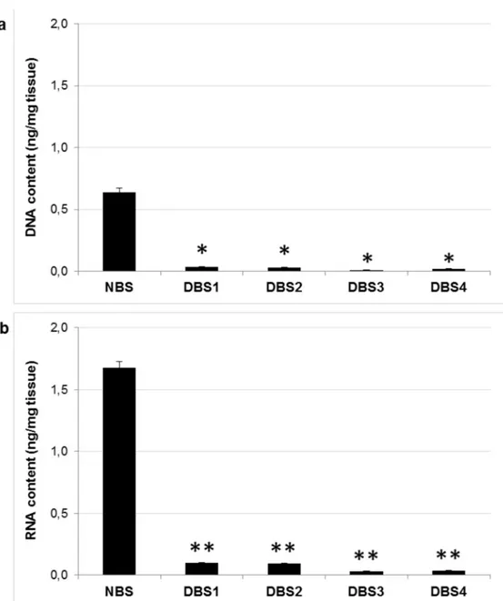
The quantification of nucleic acids in all the four samples is shown inFig 2. In detail, the average DNA content was: 0.037 ± 0.005 ng/mg for DBS1; 0.031 ± 0.006 ng/mg for DBS2; 0.011 ± 0.003 ng/mg for DBS3; 0.019 ± 0.002 ng/mg for DBS4. The average RNA content was: 0.098 ± 0.004 ng/mg for DBS1; 0.093 ± 0.007 ng/mg for DBS2; 0.029 ± 0.003 ng/mg for DBS3; 0.036 ± 0.004 ng/mg for DBS4. The reduction in both DNA and RNA content was significant (P<0.05 and P<0.01, respectively) and higher than 90% compared to that of native bone (0.639 ± 0.048 ng DNA/mg; 1.678 ± 0.058 ng RNA/mg), indicating that cells and their nuclear materials have been effectively removed. This result is particularly important because it would ensure that the bone tissue is essentially devoid of immunogenic active molecules [33].
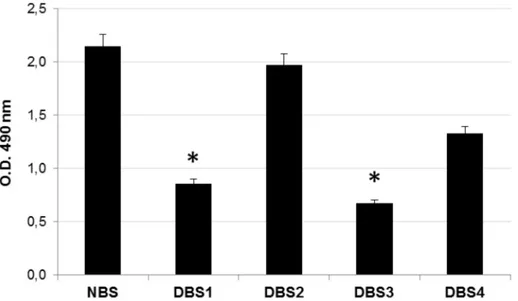
Oil red O staining was then used to evaluate the residual lipid content in bone samples. As an azo dye, Oil Red O can combine with triacylglycerol to give out jacinth lipid droplets, which can be spectrophotometrically quantified [34]. As evident fromFig 3, protocols B#1 and B#3 produced a significant (P<0.05) removal of lipids from bone granules. On the contrary, bovine bone samples processed with protocols B#2 and B#4 did not achieve an appropriate lipid elimi-nation. It is reasonable to think that treatment with ethanol positively contributed to removal of lipids from bone. This result should be taken strongly into account, since delipidation is an important procedure for the preparation of bone grafts because residual lipids negatively affect the osseointegration [11].
Evaluation of cytotoxicity of decellularized bovine bone granules
When a material has to be used in living tissues, excellent biocompatibility is essential in order to avoid any adverse effect [35]. In vitro cytotoxicity represents an initial and critical step to evaluate the biocompatibility of the ideal material. The cytotoxicity of bone samples produced in this work was quantitatively measured with MTT assay, where a higher cell proliferation rate is expected for biocompatible candidate. The cytotoxicity was estimated by directly expos-ing L929 fibroblasts to DBS1, DBS2, DBS3 and DBS4 granules. Cells cultivated in absence of bone granules or with a Ti disc were used as blank and negative control condition, respectively. The MTT results reported inFig 4ashow that L929 cells grew well in presence of DBS3 or DBS1 granules, with absorbance values quite similar to those of blank and negative control. On the contrary, cell proliferation rate was negatively affected by the presence of DBS2 and DBS4 (P<0.05) granules.
In accordance with the MTT results, L929 cells were visualized to grow directly underneath DBS3 granules without necrosis or detachment, presenting a morphology similar to both nega-tive control and blank samples (Fig 4b). On the contrary, the cell morphology was compro-mised for L929 cells cultured with DBS2 and DBS4 granules, indicating the inhibition of cell growth and a certain degree of cytotoxicity. It is likely that treatment with ethanol provided for DBS1 and DBS3 promoted the total removal of cellular debris, conferring them superior biocompatibility.
Morphological and histological analyses of decellularized bovine bone
In the light of the results presented so far, in our opinion bovine bone samples decellularized with protocol B#3 gave better results compared to the other protocols in terms of native cells elimination, nucleic acids and lipid removal, and lack of cytotoxicity. For this reason, further analyses were conducted exclusively on DBS3. SEM images demonstrate that the morphology of DBS3 is not altered by the decellularization protocol. Indeed, the typical structure of cancel-lous bone with bone trabeculae is well preserved, whereas the lipid component has been
Fig 1. MTT assay on decellularized bovine bone. Quantification analysis of amount of residual cells in decellularized bone samples DBS1, DBS2, DBS3, and DBS4 compared to NBS, estimated with MTT assay. Values are expressed as mean± standard deviation (n = 3 per group). Statistically significant differences are indicated as*P<0.05 and **P<0.01 and compared with NBS.
completely removed (Fig 5a). This is in contrast with the morphology of NBS, whose structure is hidden by a layer of fatty material, which makes its surface smoother. These observations are confirmed by histological analyses (Fig 5b). H&E staining of NBS clearly shows the presence of fat cells between bone trabeculae. Conversely, the lipid component is absent in the decellular-ized bone sample, but retains an intact ECM. These results seem to confirm what previously suggested, namely that the final step in graded ethanol, in addition to dry bone granules, strongly contributed to the elimination of any contaminating material. Such a result is very encouraging since one of the fundamental challenges of this work was to find an effective
Fig 2. Nucleic acids quantification in decellularized bovine bone. Quantification analysis of amount of residual (a) DNA and (b) RNA in decellularized bone samples DBS1, DBS2, DBS3, and DBS4 compared to that of NBS. Content of residual DNA and RNA was normalized by dry weight of each specimen. Values are expressed as mean± standard deviation (n = 3 per group). Statistically significant differences are indicated as*P<0.05 and **P<0.01 and compared with NBS.
method to defat the produced bone granules. Different chemical or physical treatments are generally used to carry out this step. Chemical organic solvents, such as chloroform, dichloro-methane or acetone, may leave toxic residues in the grafts as well as being harmful to the health of operators. On the other hand, physical methods of lipid removal such as supercritical CO2,
although efficient in defatting bone samples, need expensive equipment and solvents [36]. Recently, Zhang and co-workers demonstrated that the use of lipase could eliminate fat from porcine bone similarly to acetone, but in a shorter time and without toxic effects [11]. Although lipase is extensively used in various applications in many fields, its activity is strongly pH and temperature dependent, and thus more difficult to control. Furthermore, the influence of treat-ment with lipase in bone formation has not yet been investigated in vivo. The results reported in this study would indicate that the use of graded ethanol could be strongly considered as an alternative treatment for removing lipids from bone samples.
Evaluation of the osteoinductivity of decellularized bovine bone in vitro
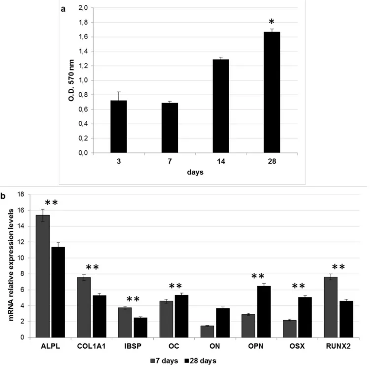
As described in the Introduction, a key property of an ideal bone graft is its osteoinductivity [2,4,5]. The osteoinductivity of DBS3 was assessed evaluating the ability of ADSCs seeded onto these granules to express osteogenic markers in absence of osteogenic differentiation factors. A preliminary MTT assay indicated that DBS3 granules are able to support the growth of ADSCs, whose proliferation rate increases during the culturing time, with a maximum value at 28 days (P<0.05) (Fig 6a). When ADSCs are cultured on the DBS3 granules, the gene expression of alkaline phosphatase liver/bone/kidney (ALPL), collagen type I alpha 1 (COL1A1), integrin-binding sialoprotein (IBSP), and runt-related transcription factor 2 (RUNX2) is found to be significantly (P<0.01) upregulated after 7 days from seeding, then slightly decreases at 28 days (Fig 6b). ALPL is a marker of early osteogenic development, and has probably an initiator and regulator role in calcification [37]. The elevated ALPL expression observed in this work sup-ports the success of the osteogenic differentiation of ADSCs and might be an indication of the osteoinductive properties of the bone granules used. RUNX2 is one of the transcription factors
Fig 3. Lipids quantification in decellularized bovine bone. Quantification analysis of residual lipid content in bone samples DBS1, DBS2, DBS3, and DBS4 compared to NBS, estimated with Oil red O quantification. Values are expressed as mean± standard deviation (n = 3 per group). Statistically significant differences are indicated as*P<0.05 and compared with NBS.
Fig 4. In vitro cytotoxicity test on decellularized bovine bone granules. (a) O.D. values at 570 nm representing proliferation rates of L929 fibroblasts cultured in direct contact with DBS1, DBS2, DBS3, DBS4 granules, titanium (Ti) disc, and without granules (blank) (n = 3 per group) Statistically significant differences are indicated as*P<0.05 and compared with blank. (b) Morphology of L929 fibroblasts cultured in presence of the test materials cited above. Images were acquired after removal of the bone granules. Cells (blue
required for the differentiation of MSCs into preosteoblasts [38]. Several studies report that RUNX2 controls the expression of several bone ECM protein genes, including COL1A1, IBSP, osteocalcin (OC), and osteopontin (OPN), through binding to their promoters [39,40]. The high expression found for COL1A1 at 7 days is very interesting since collagen synthesis is known to be a prerequisite for ECM formation and mineralization in bone [41]. IBSP and OPN are ECM glycoproteins implicated in the regulation of mineralized nodule nucleation [42]. The results presented in this study might confirm the role of RUNX2 in stimulating the expression of both IBSP and OPN [43]. RUNX2 is essential for the commitment of MSCs to the osteoblast lineage, but its expression has to be downregulated for bone maturation [44]. Osterix (OSX) is considered a downstream gene of RUNX2, and plays a fundamental role in the commitment of preosteoblastic cell differentiation into mature osteoblasts [45]. In our long-term cultures, OSX expression becomes predominant on that of RUNX2 (P<0.01). The acquisition of a mature osteoblast phenotype could also be explained by the significant (P<0.01) increase in the expression of OC over time. OC is the most abundant non-collage-nous protein in bone ECM after collagens and a marker of mature osteoblasts [46]. Finally, osteonectin (ON) is a glycoprotein that binds calcium [47]. It is secreted by osteoblasts during bone formation, initiating mineralization and promoting mineral crystal deposition. ON also shows affinity for collagen in addition to bone mineral calcium. In this study, the expression levels of ON increases at 28 days of culture. Taken together, our results seem to demonstrate that the upregulation of several osteogenic genes in ADSCs may be dependent on the DBS3 granules surface characteristics.
Evaluation of the osteoconductivity of decellularized bovine bone in vivo
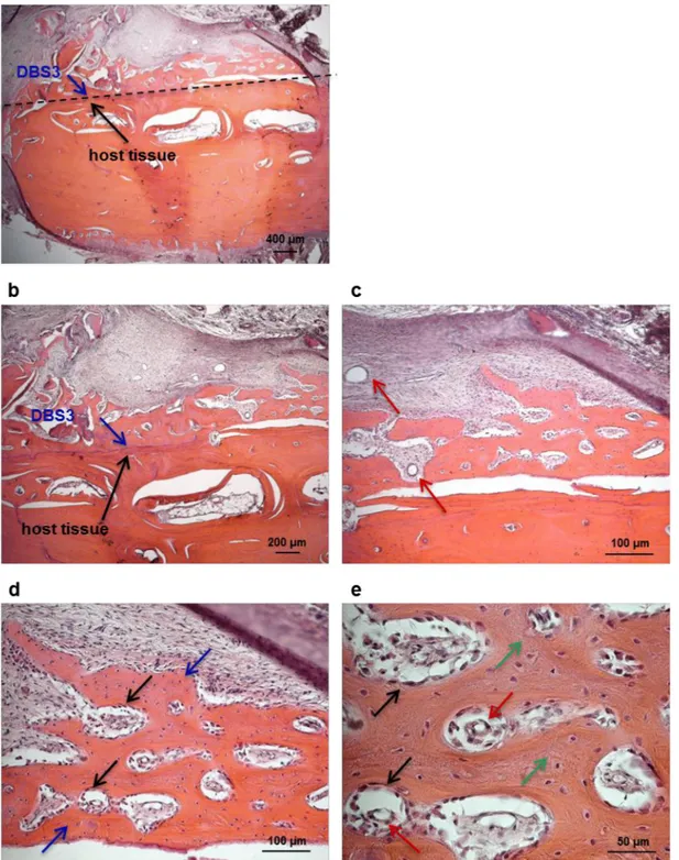
An essential goal for the development of a bone graft material to be used in a clinical setting is osteoconductivity [2,4,5]. The osteoconductivity of DBS3 granules was evaluated in a maxil-lary sinus augmentation model in sheep. Previous investigations have demonstrated that such a procedure in sheep is a reliable model to evaluate bone formation in humans [48,49]. The surgical procedure was uneventful for all animals. All sheeps did not show any p.i. complica-tions nor clinical symptoms of maxillary sinusitis. Sinus explants evaluated macroscopically 15 and 30 days after augmentation reveal that DBS3 bovine granules are inserted in the sur-rounding host tissue, where no signs of inflammation or adverse tissue reactions are present. From an histological point of view, the interaction between the biomaterial particles and the host bone tissue is limited at 15 days (Fig 7a and 7b); however, their integration in the aug-mentation area increases over time (Fig 8a and 8b). This is also demonstrated by the presence of ovine connective tissue that completely surrounds the implanted biomaterial. The presence of blood vessels is already detectable in sinus explants at 15 days (Fig 7c), and they become more defined after 30 days from biomaterial implantation (Fig 8c). This result is very encour-aging since vascularization is a prerequisite for the production of new bone tissue [4,50]. In the process of new bone formation, osteoblasts play a pivotal role since they are the cells responsible for new ECM deposition. Ovine osteoblasts start to colonize the implanted DBS3 granules at 15 days (Fig 7); the adhesion of osteoblasts to the biomaterial is clearly visible at a higher magnification (Fig 7). The biomaterial appears fully infiltrated by these cells after an implantation period of 30 days (Fig 8d). At this stage, the presence of newly synthesized bone is significant. This new woven bone appears as vital bone tissue, containing osteocytes inside
arrows) grown in contact with DBS1 and DBS3 granules show a morphology similar to that of cells (black arrows) contacting the Ti disc or blank. The morphology of cells (red arrows) in contact with DBS2 and DBS4 granules is altered.
Fig 5. Morphological analyses of bovine bone. (a) Representative SEM micrographs of native (NBS) and decellularized (DBS3) bone samples. In DBS3, the typical structure of cancellous bone with bone trabeculae (red arrow) is well evident; whereas it is hidden by a layer of fatty material (blue arrows) in NBS. (b) H&E staining of NBS and DBS3. Adipose tissue (blue arrow) between bone trabeculae (red arrows) is visible in NBS but it has been completely removed after decellularization in DBS3.
bone lacunae, and blood vessels in between the bone voids (Fig 8e). Based on the histological results of this study, we can conclude that the DBS3 bovine granules determine a substantial amount of new bone formation and a proper blood supply, thus representing a suitable bone graft candidate material.
Fig 6. Biological responses of ADSCs seeded onto DBS3 granules. (a) Proliferation rate of ADSCs seeded onto DBS3 granules for 3, 7, 14 and 28 days calculated with MTT assay. Statistically significant differences are indicated as*P<0.05 and compared with the first time point. (b) Expression of osteoblast specific markers after in vitro ADSCs seeding onto DBS3 granules for 7 and 28 days in basal medium. The results are reported as ratio with respect to the mRNA expression of ADSCs seeded on TCP. Statistically significant differences are indicated as**P<0.01 and compared with the control condition. doi:10.1371/journal.pone.0132344.g006
Fig 7. Morphological analysis of sinus explants 15 days p.i.. (a) Histological overview of the sinus area showing a scarce integration of the biomaterial (DBS3 granules, blue arrow) within the host bone tissue (black arrow). (b) Higher magnification of the implantation area, displaying scarce contact between DBS3 granules (blue arrow) and host bone tissue (black arrow). (c) Presence of blood vessels (red arrows) in the augmentation area. (d) Ovine osteoblats (black arrows) adhering to DBS3 granules (blue arrows). (e) Higher magnification of osteoblasts (black arrows) producing new bone ECM (green arrows). doi:10.1371/journal.pone.0132344.g007
Fig 8. Morphological analysis of sinus explants 30 days p. i.. (a) Histological overview of the sinus area, showing the direct contact between the biomaterial (blue arrow) and host bone (black arrow). (b) Higher magnification of the implantation area, demonstrating that DBS3 granules (blue arrow) are well integrated within the host bone tissue (black arrow). (c) Presence of blood vessels (red arrows) in the augmentation area. (d) Ovine osteoblasts (black arrows) have colonized the bovine granules (blue arrows) and started to lay down new bone ECM. (e) Higher magnification of the new bone ECM (green arrows) synthesized by ovine osteoblasts (black arrows) containing blood vessels (red arrows).
Decellularization protocols of bovine pericardium
The use of bone grafting material alone seems to be less effective for an optimal bone regenera-tion than the combinaregenera-tion of a supporting material and a barrier [51]. As explained in the Introduction, the GBR technique relies on the presence of a membrane that acts as a barrier for non-osteogenic cell infiltration. The choice of a barrier membrane is a critical step in GBR pro-cedures. Resorbable membranes are generally preferred to non-resorbable membranes because they avoid the need for membrane removal, they have greater cost-effectiveness and decreased patient morbidity [52]. Currently used resorbable membranes are polymeric or collagen derived from different animal sources. Collagen membranes derived from bovine pericardium were shown to be efficient for GBR in rabbit mandibular defects [53]. The structure of pericar-dium, consisting of a network of collagen and elastic fibers embedded in an amorphous matrix, is unique, and this results not only in a smooth yet porous surface for cellular attachment and proliferation, but also in sufficient density for soft tissue exclusion [54]. In order to generate a resorbable membrane to be used in the GBR technique, in the second part of this study we developed a method of bovine pericardium decellularization. Most of the methods described in the literature for decellularization of bovine pericardium are based on treatments with hypo-tonic buffer, sodium dodecyl sulfate (SDS), and nuclease solution; or alkaline treatment fol-lowed by phosphoric acid washing; or enzymatic digestion [55–57]. In our work, two different protocols were tested and compared: the first (protocol P#1) was based on osmotic shock asso-ciated with detergents, the second one (protocol P#2) implied multiple steps of freeze/thaw fol-lowed by use of enzymes. The single physical, chemical or enzymatic methods here proposed have been already described previously [21,22,30]. Nevertheless, the appropriate combination and the duration of each treatment, as well as the time of exposure to the various reagents, have been developed and optimized in this study. Osmotic shock was obtained by placing bovine pericardium strips in hypotonic and hypertonic solutions, alternated by washing in non-ionic (Triton X-100) and ionic (SD) detergents. It has been reported that multiple steps in hypotonic/hypertonic solutions achieve the maximum osmotic effect [22]. The hypotonic solu-tion was enriched with PI in order to prevent degradasolu-tion by enzymes released from disrupted cell compartments. The use of Triton X-100 and SD were then preferred over other detergents in the decellularization process of pericardium because they did not affect the structural integ-rity of either collagen and elastin, as previously reported [58]. The second decellularization protocol was based on the method described by Stapleton et al. [59] for porcine meniscus with some modifications. Briefly, bovine pericardium samples were exposed to two cycles of dry freeze/thaw, followed by two cycles of freeze/thaw in hypotonic solution. The freeze/thaw tech-nique was adopted to allow ice crystal formation, potentially opening up the membrane to facilitate diffusion of the subsequent solutions. Bovine pericardium strips were then incubated in SD and hypotonic buffer, followed by a treatment with nucleases (DNase and RNase) in order to digest residual nucleic acids. Following decellularization with the two protocols, DPS1 and DPS2 were disinfected with isopropanol. Sterilization was achieved by exposing pericardia to a 25 kGy dose of gamma irradiation.
Evaluation of cell removal and residual nucleic acids content in
decellularized bovine pericardium
At this point, the efficiency of decellularization protocols in terms of cell removal, and reduc-tion of DNA and RNA content, was analyzed and compared. Both protocols produced an equivalent and good (P<0.05) removal of native cells (Fig 9a), and a reduction in the content of DNA (Fig 9b) and RNA (Fig 9c) higher than 90% when compared to that of the native peri-cardium (NPS) (P<0.01). In detail, the average DNA content was: 3.730 ± 0.178 μg/mg for
Fig 9. MTT assay and nucleic acids content in decellularized bovine pericardium. Quantification analyses of (a) residual cells, (b) DNA content, and (c) RNA content in pericardium samples DPS1 and DPS2 compared to NPS. Values are expressed as mean± standard deviation (n = 3 per group). Statistically significant differences are indicated as*P<0.05 **P<0.01 and compared with NPS.
NPS; 0.160 ± 0.006μg/mg for DPS1; 0.142 ± 0.007 μg/mg for DPS2. The average RNA content was: 10.676 ± 0.434μg/mg for NPS; 0.368 ± 0.016 μg/mg for DPS1; 0.276 ± 0.012 ng/mg for DPS2.
Biological properties of decellularized bovine pericardium
With the aim to identify the protocol that generates the most suitable membrane for the GBR technique, the histological, morphological and the biocompatibility properties of the two acel-lular bovine pericardia were investigated. As described by Scantlebury [20], one of the design criteria to consider in the development of a GBR membrane is its biocompatibility. The biocompatibility of DPS1 and DPS2 was evaluated by seeding directly human fibroblasts onto these membranes, and maintaining them in culture for 7 days. H&E stained sections of the seeded scaffolds at 7 days reveal that cells grew up to and in contact with both the acellular scaf-folds, but showing a better distribution on DPS1 (Fig 10a). In agreement with histological anal-yses, the results of SEM indicate that human fibroblasts were attached and well distributed on the surface of the pericardium decellularized with protocol P#1; on the contrary, DPS2 was col-onized by a smaller number of cells (Fig 10b). These outcomes seem to indicate that decellulari-zation protocol P#1 removed any residual or potentially cytotoxic reagents, thus resulting in a more biocompatible membrane. Apart from being biocompatible, an optimal barrier should be also sufficiently occlusive to avoid fibrous tissue formation [20]. The observation that the seeded fibroblasts do not penetrate the bovine ECM matrix suggests that the decellularized pericardia are able to block soft-tissue ingrowth, allowing the infiltration and activity of bone-forming cells.
The fundamental goal of any decellularization protocol is to remove all cellular material without adversely affecting the composition integrity, mechanical property, and eventual bio-logical activity of the remaining ECM [60]. Apart from collagen, pericardium ECM is com-posed of a network of elastic fibers, which confers elasticity to the tissue [61,62]. Weigert’s staining of decellularized bovine pericardium samples showed well preserved elastin fibers, indicating the integrity of the ECM, in particular for DPS1 (Fig 10c). Indeed, the elastin fibers maintained wavelike structure in DPS1, whereas they appeared more sparse and partly dis-torted in DPS2. Based on these observations, the first decellularization method has resulted in a much better preservation of the pericardium ECM integrity compared to the freeze/thaw pro-tocol. The results of the morphological analyses thus suggest that the osmotic shock and deter-gents treatment did not affect the flexibility of the bovine pericardium. The product obtained with the decellularization protocol P#1 is very promising, since clinical manageability of a GBR membrane is another essential property to consider, in particular in the dental field [63]. A too malleable membrane, or a stiff one, is difficult to use; by contrast, a membrane that maintains a certain degree of flexibility, at least during the insertion phase, is preferred [64,65].
Evaluation of the host tissue reaction after subcutaneous implantation in
rats
Appropriate integration with the surrounding tissue is the ultimate objective of all tissue regen-eration techniques, as it is essential that the membrane integrates with the host tissue [63]. A rat subcutaneous implantation model was used to evaluate local tissue reactions following implantation of the two decellularized pericardium membranes intended for GBR. After 7 days from implantation, no signs of rejection were observed for both DPS1 and DPS2 membranes. Nevertheless, different responses of rat tissue to the two decellularized pericardia can be dis-played in the histological overviews ofFig 11. In the DPS1 explants, no inflammatory event has been revealed confirming the high tolerability of the material. On the contrary, in the case of
DPS2, mononuclear and multinucleated giant cells were found deep into the body of the explants, as well as the appearance of large blood vessels. Based on these observations, it may be speculated that the acellular pericardium scaffold produced with protocol P#1 may be of potential utility for clinical implantation as a GBR membrane.
Fig 10. Decellularized bovine pericardium seeded with human fibroblasts. (a) H&E staining at 7 days post-seeding shows that cells are present in the upper layer of both tissues, with a more uniform distribution on DPS1 (white asterisks) with respect to DPS2 (yellow asterisks). (b) The elastin fibers of the sections are clearly defined and continuous in DPS1 (blue arrows), whereas they appear partly distorted and discontinuous in DPS2 (red arrows), as shown by Weigert’s staining. (c) SEM images show that cells (blue asterisks) have colonized the whole surface of DPS1; on the contrary, the DPS2 membrane (red asterisks) is still visible under the cell layer (blue asterisks).
Conclusions
The findings of our study are twofold. Driven by the goal of providing an alternative to bone autografts and allografts, we firstly developed a bovine bone graft substitute which closely mimics the natural structure and properties of natural bone ECM. The proposed protocol for bovine bone decellularization consisting in multiple steps of thermal shock, followed by wash-ing with detergent and dehydration with alcohol resulted in a biocompatible, osteoinductive and osteoconductive product. Subsequently, we identified an efficient protocol for decellulariz-ing bovine pericardium. The osmotic shock method seemed superior to the freeze/thaw method for preparing decellularized bovine membranes, as it achieved not only the complete removal of cellular materials but also the preservation of the ECM structure of the bovine peri-cardium tissue. In addition, the absence of any inflammatory reaction in the host tissue repre-sents an advantage of the method proposed.
In conclusion, we believe that the application of protocols B#3 and P#1 could be considered as a suitable approach to produce decellularized bovine bone and pericardium scaffolds for generating bone substitutes and GBR membranes intended for dental clinical use.
Fig 11. Histological evaluation of host tissue reaction to DPS1 and DPS2 implants. (a) H&E staining performed 7 days after implantation shows that implanted pericardia (white asterisks) are intact, and there is little sign of capsular formation. (b) H&E staining at higher magnification showing a certain degree of inflammation (blue arrows) in DPS2.
Supporting Information
S1 Table. Human primer sequences. (DOCX)
Author Contributions
Conceived and designed the experiments: BZ EB MI. Performed the experiments: CG L. Fer-roni RG LS SR GDB L. Finotti. Analyzed the data: CG L. FerFer-roni BZ EB. Contributed reagents/ materials/analysis tools: BZ EB MI. Wrote the paper: CG L. Ferroni BZ. Obtained permission for use of animal: RG LS.
References
1. Elsalanty ME, Genecov DG. Bone grafts in craniofacial surgery. Craniomaxillofac Trauma Reconstr. 2009; 2: 125–134. doi:10.1055/s-0029-1215875PMID:22110806
2. Oryan A, Alidadi S, Moshiri A, Maffulli N. Bone regenerative medicine: classic options, novel strategies, and future directions. J Orthop Surg Res. 2014; 9: 18. doi:10.1186/1749-799X-9-18PMID:24628910
3. Albrektsson T, Johansson C. Osteoinduction, osteoconduction and osseointegration. Eur Spine J. 2001; 10: S96–101. PMID:11716023
4. Hsiong SX, Mooney DJ. Regeneration of vascularized bone. Periodontol. 2000. 2006; 41: 109–122. PMID:16686929
5. da Cruz GA, de Toledo S, Sallum EA, de Lima AF. Morphological and chemical analysis of bone substi-tutes by scanning electron microscopy and microanalysis by spectroscopy of dispersion energy. Braz Dent J. 2007; 18: 129–133. PMID:17982552
6. Bressan E, Favero V, Gardin C, Ferroni L, Iacobellis L, Favero L, et al. Biopolymers for Hard and Soft Engineered Tissues: Application in Odontoiatric and Plastic Surgery Field. Polymers. 2011; 3: 509–526.
7. Keskin D, Gundogdu C, Atac AC. Experimental comparison of bovine derived xenograft, xenograft-autologous bone marrow and autogenous bone graft for the treatment of bony defects in the rabbit ulna. Med Princ Pract. 2007, 16: 299–305. PMID:17541296
8. Dimitriou R, Jones E, McGonagle D, Giannoudis PV. Bone regeneration: current concepts and future directions. BMC Med. 2011, 9: 66. doi:10.1186/1741-7015-9-66PMID:21627784
9. Khan SN, Cammisa FP Jr, Sandhu HS, Diwan AD, Girardi FP, Lane JM. The biology of bone grafting. J Am Acad Orthop Surg. 2005; 13: 77–86. PMID:15712985
10. Tadic D, Epple M. A thorough physicochemical characterisation of 14 calcium phosphate-based bone substitution materials in comparison to natural bone. Biomaterials. 2004; 25: 987–994. PMID:
14615163
11. Zhang N, Zhou M, Zhang Y, Wang X, Ma S, Dong L, et al. Porcine bone grafts defatted by lipase: effi-cacy of defatting and assessment of cytocompatibility. Cell Tissue Bank. 2014; 15: 357–367. doi:10. 1007/s10561-013-9391-zPMID:23955020
12. Katz J, Mukherjee N, Cobb RR, Bursac P, York-Ely A. Incorporation and immunogenicity of cleaned bovine bone in a sheep model. J Biomater Appl. 2009; 24: 159–174. doi:10.1177/0885328208095174
PMID:18987022
13. Liu J, Kerns DG. Mechanisms of guided bone regeneration: a review. Open Dent J. 2014; 8: 56–65. doi:
10.2174/1874210601408010056PMID:24894890
14. Ereno C, Guimarães SA, Pasetto S, Herculano RD, Silva CP, Graeff CF, et al. Latex use as an occlu-sive membrane for guided bone regeneration. Biomed Mater Res A. 2010; 95: 932–939.
15. Ashley FL, Stone RS, Alonsoartieda M, Syverud JM, Edwards JW, Sloan RF, et al. Experimental and clinical studies on the application of monomolecular cellulose filter tubes to create artificial tendon sheaths in digits. Plast Reconstr Surg Transplant Bull. 1959; 23: 526–534. PMID:13657726
16. Salata LA, Hatton PV, Devlin AJ, Craig GT, Brook IM. In vitro and in vivo evaluation of e-PTFE and alkali-cellulose membranes for guided bone regeneration. Clin Oral Implants Res. 2001; 12: 62–68. PMID:11168272
17. Donos N, Lang NP, Karoussis IK, Bosshardt D, Tonetti M, Kostopoulos L. Effect of GBR in combination with deproteinized bovine bone mineral and/or enamel matrix proteins on the healing of critical-size defects. Clin Oral Implants Res. 2004; 15: 101–111. PMID:14731183
18. Kim JY, Yang BE, Ahn JH, Park SO, Shim HW. Comparable efficacy of silk fibroin with the collagen membranes for guided bone regeneration in rat calvarial defects. J Adv Prosthodont. 2014; 6: 539–546. doi:10.4047/jap.2014.6.6.539PMID:25551015
19. Cucchi A, Ghensi P. Vertical Guided Bone Regeneration using Titanium-reinforced d-PTFE Membrane and Prehydrated Corticocancellous Bone Graft. Open Dent J. 2014; 8: 194–200. doi:10.2174/ 1874210601408010194PMID:25419250
20. Scantlebury TV. 1982–1992: a decade of technology development for guided tissue regeneration. J Periodontol. 1993; 64: 1129–1137. PMID:8295101
21. Badylak SF, Taylor D, Uygun K. Whole-organ tissue engineering: decellularization and recellularization of three-dimensional matrix scaffolds. Annu Rev Biomed Eng. 2011; 13: 27–53. doi: 10.1146/annurev-bioeng-071910-124743PMID:21417722
22. Crapo PM, Gilbert TW, Badylak SF. An overview of tissue and whole organ decellularization processes. Biomaterials. 2011; 32: 3233–3243. doi:10.1016/j.biomaterials.2011.01.057PMID:21296410
23. Chappard D, Fressonnet C, Genty C, Baslé MF, Rebel A. Fat in bone xenografts: importance of the purification procedures on cleanliness, wettability and biocompatibility. Biomaterials. 1993; 14: 507– 512. PMID:8329523
24. Moreau MF, Gallois Y, Baslé MF, Chappard D. Gamma irradiation of human bone allografts alters med-ullary lipids and releases toxic compounds for osteoblast-like cells. Biomaterials. 2000; 21: 369–376. PMID:10656318
25. Denizot F, Lang R. Rapid colorimetric assay for cell growth and survival. Modifications to the tetrazo-lium dye procedure giving improved sensitivity and reliability. J Immunol Methods. 1986; 89: 271–277. PMID:3486233
26. Gardin C, Bressan E, Ferroni L, Nalesso E, Vindigni V, Stellini E, et al. In vitro concurrent endothelial and osteogenic commitment of adipose-derived stem cells and their genomical analyses through com-parative genomic hybridization array: novel strategies to increase the successful engraftment of tissue-engineered bone grafts. Stem Cells Dev. 2012; 21: 767–777. doi:10.1089/scd.2011.0147PMID:
21521013
27. Pfaffl MW. A new mathematical model for relative quantification in real-time RT-PCR. Nucleic Acids Res. 2001; 29: e45. PMID:11328886
28. Berardinelli P, Valbonetti L, Muttini A, Martelli A, Peli R, Zizzari V, et al. Role of amniotic fluid mesenchy-mal cells engineered on MgHA/collagen-based scaffold allotransplanted on an experimental animesenchy-mal study of sinus augmentation. Clin Oral Investig. 2013; 17: 1661–1675. doi: 10.1007/s00784-012-0857-3PMID:23064983
29. Silva-Correia J, Zavan B, Vindigni V, Silva TH, Oliveira JM, Abatangelo G, et al. Biocompatibility evalu-ation of ionic- and photo-crosslinked methacrylated gellan gum hydrogels: in vitro and in vivo study. Adv Healthc Mater. 2013; 2: 568–575. doi:10.1002/adhm.201200256PMID:23184642
30. Gilbert TW. Strategies for tissue and organ decellularization. J Cell Biochem. 2012; 113: 2217–2222. doi:10.1002/jcb.24130PMID:22415903
31. Gilbert TW, Sellaro TL, Badylak SF. Decellularization of tissues and organs. Biomaterials 2006; 27: 3675–3683. PMID:16519932
32. Shapoff CA, Bowers GM, Levy B, Mellonig JT, Yukna RA. The effect of particle size on the osteogenic activity of composite grafts of allogeneic freeze-dried bone and autogenous marrow. J Periodontol. 1980; 51: 625–630. PMID:7007609
33. Dong J, Li Y, Mo X. The study of a new detergent (octyl-glucopyranoside) for decellularizing porcine pericardium as tissue engineering scaffold. Surg Res. 2013; 183: 56–67.
34. Guo T, Zhu L, Tan J, Zhou X, Xiao L, Liu X, et al. Promoting effect of triterpenoid compound from Agri-monia pilosa Ledeb on preadipocytes differentiation via up-regulation of PPARγ expression. Pharma-cogn Mag. 2015; 11: 219–225. doi:10.4103/0973-1296.149741PMID:25709235
35. Wang YB, Li HF, Cheng Y, Zheng YF, Ruan LQ. In vitro and in vivo studies on Ti-based bulk metallic glass as potential dental implant material. Mater Sci Eng C Mater Biol Appl. 2013; 33: 3489–3497. doi:
10.1016/j.msec.2013.04.038PMID:23706238
36. Fages J, Marty A, Delga C, Condoret JS, Combes D, Frayssinet P. Use of supercritical CO2 for bone delipidation. Biomaterials. 1994; 15: 650–656. PMID:7948586
37. Peng F, Yu X, Wei M. In vitro cell performance on hydroxyapatite particles/poly(L-lactic acid) nanofi-brous scaffolds with an excellent particle along nanofiber orientation. Acta Biomater. 2011; 7: 2585– 2592. doi:10.1016/j.actbio.2011.02.021PMID:21333762
38. Komori T, Yagi H, Nomura S, Yamaguchi A, Sasaki K, Deguchi K, et al. Targeted disruption of Cbfa1 results in a complete lack of bone formation owing to maturational arrest of osteoblasts. Cell. 1997; 89: 755–764. PMID:9182763
39. Ducy P, Zhang R, Geoffroy V, Ridall AL, Karsenty G. Osf2/Cbfa1: a transcriptional activator of osteo-blast differentiation. Cell. 1997; 89: 747–754. PMID:9182762
40. Kern B, Shen J, Starbuck M, Karsenty G. Cbfa1 contributes to the osteoblast-specific expression of type I collagen genes. J Biol Chem. 2001; 276: 7101–7107. PMID:11106645
41. Franceschi RT, Iyer BS, Cui Y. Effects of ascorbic acid on collagen matrix formation and osteoblast dif-ferentiation in murine MC3T3-E1 cells. J Bone Miner Res. 1994; 9: 843–854. PMID:8079660
42. Seibel MJ. Molecular markers of bone turnover: biochemical, technical and analytical aspects. Osteo-poros Int. 2000; 11: S18–29. PMID:11193236
43. Phillips JE, Gersbach CA, Wojtowicz AM, García AJ. Glucocorticoid-induced osteogenesis is nega-tively regulated by Runx2/Cbfa1 serine phosphorylation. J Cell Sci. 2006; 119: 581–591. PMID:
16443755
44. Liu W, Toyosawa S, Furuichi T, Kanatani N, Yoshida C, Liu Y, et al. Overexpression of Cbfa1 in osteo-blasts inhibits osteoblast maturation and causes osteopenia with multiple fractures. J Cell Biol. 2001; 155: 157–166. PMID:11581292
45. Nakashima K, Zhou X, Kunkel G, Zhang Z, Deng JM, Behringer RR, et al. The novel zinc finger-contain-ing transcription factor osterix is required for osteoblast differentiation and bone formation. Cell. 2002; 108: 17–29. PMID:11792318
46. Hauschka PV, Lian JB, Cole DE, Gundberg CM. Osteocalcin and matrix Gla protein: vitamin K-depen-dent proteins in bone. Physiol Rev. 1989; 69: 990–1047. PMID:2664828
47. Termine JD, Kleinman HK, Whitson SW, Conn KM, McGarvey ML, Martin GR. Osteonectin, a bone-specific protein linking mineral to collagen. Cell. 1981; 26: 99–105. PMID:7034958
48. Aral A, Yalçin S, Karabuda ZC, Anil A, Jansen JA, Mutlu Z. Injectable calcium phosphate cement as a graft material for maxillary sinus augmentation: an experimental pilot study. Clin Oral Implants Res. 2008; 19: 612–617. doi:10.1111/j.1600-0501.2007.01518.xPMID:18474064
49. Estaca E, Cabezas J, Usón J, Sánchez-Margallo F, Morell E, Latorre R. Maxillary sinus-floor elevation: an animal model. Clin Oral Implants Res. 2008; 19: 1044–1048. doi:10.1111/j.1600-0501.2008.01557. xPMID:18828821
50. Kanczler JM, Oreffo RO. Osteogenesis and angiogenesis: the potential for engineering bone. Eur Cell Mater. 2008; 15: 100–114. PMID:18454418
51. Donos N, Kostopoulos L, Karring T. Alveolar ridge augmentation using a resorbable copolymer mem-brane and autogenous bone grafts. An experimental study in the rat. Clin Oral Implants Res. 2002; 13: 203–213. PMID:11952741
52. Hammerle CH, Jung RE. Bone augmentation by means of barrier membranes. Periodontol. 2000. 2003; 33: 36–53. PMID:12950840
53. Thomaidis V, Kazakos K, Lyras DN, Dimitrakopoulos I, Lazaridis N, Karakasis D, et al. Comparative study of 5 different membranes for guided bone regeneration of rabbit mandibular defects beyond criti-cal size. Med Sci Monit. 2008; 14: BR67–73. PMID:18376341
54. Jimbo R, Marin C, Witek L, Suzuki M, Tovar N, Chesnoiu-Matei I, et al. Bone Morphometric Evaluation around Immediately Placed Implants Covered with Porcine-Derived Pericardium Membrane: An Exper-imental Study in Dogs. Int J Biomater. 2012; 2012: 279167. doi:10.1155/2012/279167PMID:
23227052
55. Mirsadraee S, Wilcox HE, Watterson KG, Kearney JN, Hunt J, Fisher J, Ingham E. Biocompatibility of acellular human pericardium. J Surg Res. 2007; 143: 407–414. PMID:17574597
56. Hirata HH, Munhoz MA, Plepis AM, Martins VC, Santos GR, Galdeano EA, Cunha MR. Feasibility study of collagen membranes derived from bovine pericardium and intestinal serosa for the repair of cranial defects in ovariectomised rats. Injury. 2015: doi:10.1016/j.injury.2015.03.039
57. Bai M, Zhang T, Ling T, Zhou Z, Xie H, Zhang W, Hu G, Jiang C, Li M, Feng B, Wu H. Guided bone regeneration using acellular bovine pericardium in a rabbit mandibular model: in-vitro and in-vivo stud-ies. J Periodontal Res. 2014; 49: 499–507. doi:10.1111/jre.12129PMID:24024647
58. Samouillan V, Dandurand-Lods J, Lamure A, Maurel E, Lacabanne C, Gerosa G, et al. Thermal analy-sis characterization of aortic tissues for cardiac valve bioprostheses. J Biomed Mater Res. 1999; 46: 531–538. PMID:10398014
59. Stapleton TW, Ingram J, Katta J, Knight R, Korossis S, Fisher J, et al. Development and characteriza-tion of an acellular porcine medial meniscus for use in tissue engineering. Tissue Eng Part A. 2008; 14: 505–518. doi:10.1089/tea.2007.0233PMID:18370607
60. Badylak SF, Freytes DO, Gilbert TW. Extracellular matrix as a biological scaffold material: Structure and function. Acta Biomater. 2009; 5: 1–13. doi:10.1016/j.actbio.2008.09.013PMID:18938117
61. Lai PH, Chang Y, Chen SC, Wang CC, Liang HC, Chang WC, et al. Acellular biological tissues contain-ing inherent glycosaminoglycans for loadcontain-ing basic fibroblast growth factor promote angiogenesis and tissue regeneration. Tissue Eng. 2006; 12: 2499–2508. PMID:16995783
62. Frantz C, Stewart KM, Weaver VM. The extracellular matrix at a glance. Cell Sci. 2010; 123: 4195–4200. 63. Rakhmatia YD, Ayukawa Y, Furuhashi A, Koyano K. Current barrier membranes: titanium mesh and
other membranes for guided bone regeneration in dental applications. J Prosthodont Res. 2013; 57: 3– 14. doi:10.1016/j.jpor.2012.12.001PMID:23347794
64. Heinze J. A space-maintaining resorbable membrane for guided tissue regeneration. In: Annual confer-ence of the International Association of Dental Research; Honolulu, USA, 2004.
65. Becker W, Becker B, Mellonig J. A prospective multicenter study evaluating periodontal regeneration for class II furcation invasions and infrabony defects after treatment with a bioabsorbable barrier mem-brane: 1-year results. J Periodontol. 1996; 67: 641–649. PMID:8832474


