Involvement of the P2X7-NLRP3
axis in leukemic cell proliferation
and death
Erica Salaro
1, Alessia Rambaldi
1, Simonetta Falzoni
1, Francesca Saveria Amoroso
1,
Alessia Franceschini
1, Alba Clara Sarti
1, Massimo Bonora
1, Francesco Cavazzini
2,
Gian Matteo Rigolin
2, Maria Ciccone
2, Valentina Audrito
3, Silvia Deaglio
3, Pablo Pelegrin
4,
Paolo Pinton
1, Antonio Cuneo
2& Francesco Di Virgilio
1Lymphocyte growth and differentiation are modulated by extracellular nucleotides and P2 receptors. We previously showed that the P2X7 receptor (P2X7R or P2RX7) is overexpressed in circulating lymphocytes from chronic lymphocytic leukemia (CLL) patients. In the present study we investigated the P2X7R/NLRP3 inflammasome axis in lymphocytes from a cohort of 23 CLL patients. P2X7R, ASC and NLRP3 were investigated by Western blot, PCR and transfection techniques. P2X7R was overexpressed and correlated with chromosome 12 trisomy in CLL patients. ASC mRNA and protein were also overexpressed. On the contrary, NLRP3 was dramatically down-modulated in CLL lymphocytes relative to lymphocytes from healthy donors. To further investigate the correlation between P2X7R, NLRP3 and cell growth, NLRP3 was silenced in THP-1 cells, a leukemic cell line that natively expresses both NLRP3 and P2X7R. NLRP3 silencing enhanced P2X7R expression and promoted growth. On the contrary, NLRP3 overexpression caused accelerated apoptosis. The P2X7R was also up-modulated in hematopoietic cells from NLRP3-KO mice. In conclusion, we show that NLRP3 down-modulation stimulates P2X7R expression and promotes growth, while NLRP3 overexpression inhibits cell proliferation and stimulates apoptosis. These findings suggest that NLRP3 is a negative regulator of growth and point to a role of the P2X7R/NLRP3 axis in CLL.
The NLRP3 inflammasome has become a focus of hot interest for its ability to modulate cell responses to a wide array of exogenous or endogenous injurious agents1. The most important plasma membrane receptor responsible
for NLRP3 activation is P2X7R2. P2X7R drives NLRP3 recruitment at discrete intracellular sites and activation
via an as yet poorly understood mechanism3. The most important pathophysiological response triggered by
acti-vation of the P2X7R/NLRP3 inflammasome axis is release of the pro-inflammatory cytokine IL-1β4. However,
over the last few years it has been clearly shown that P2X7R can also support cell growth5,6,7,8. In vitro and in
vivo observations show that P2X7R expression increases the endoplasmic reticulum Ca2+ content and the
mito-chondrial potential, enhances intracellular ATP levels, activates NFATc1, prevents apoptosis and promotes cell proliferation5,9.
We previously showed that P2X7R is upregulated in peripheral lymphocytes from patients affected by chronic lymphocytic leukemia (CLL)10. In addition, in vivo studies show that P2X7R overexpression accelerates, while
on the contrary its down-modulation inhibits tumor growth6. NLRP3 is known to cause “pyroptosis”11, a type of
caspase-1-dependent programmed necrosis often observed following infection by intracellular pathogens (e.g. Salmonella), or “pyronecrosis”12, a caspase-1-independent cell death. It has been proposed that NLRP3 might
function as a negative regulator of tumorigenesis in colitis-associated cancer13,14, or as a contributing factor
to melanoma spreading due to autoinflammation15, and a recent study shows that the NLRP3 protein is either
down-regulated or completely lost in hepato-cellular carcinoma16. However, an in depth investigation of the
con-tribution of NLRP3 to cancer cell growth has never been carried out17,18. While it is clear that NLRP3 might 1Department of Morphology, Surgery and Experimental Medicine, Section of Pathology, Oncology and Experimental
Biology, University of Ferrara, Italy. 2Department of Medical Sciences, Section of Hematology, University of Ferrara,
Italy. 3Department of Medical Sciences, Immunogenetics Unit, Human Genetics Foundation, Torino, Italy. 4Murcia
Biomedical Research Institute (IMIB), Hospital Virgen de la Arrixaca, 30120 Murcia, Spain. Correspondence and requests for materials should be addressed to F.D.V. (email: [email protected])
received: 30 July 2015 accepted: 25 April 2016 Published: 25 May 2016
www.nature.com/scientificreports/
interfere with cancer cell growth by modulating the inflammatory state of the tumor microenvironment, it cannot be excluded that NLRP3 itself has a direct role, acting on intracellular pathways responsible for the modulation of cell growth or apoptosis.
In this study, we investigated P2X7R and NLRP3 expression and function in B lymphocytes from CLL patients and their role in cell growth. Our data show that P2X7R and ASC are overexpressed while NLRP3 is strongly down-modulated in CLL lymphocytes. P2X7R expression closely correlates with chromosome 12 trisomy, a known adverse prognostic factor in CLL. Furthermore, NLRP3 down-modulation drives P2X7R expression and promotes growth. On the contrary, NLRP3 overexpression stimulates apoptotic caspase activity and cell death. In conclusion, our findings identify NLRP3 as a novel growth-inhibiting factor of potential relevance in CLL.
Results
NLRP3 is down-modulated in CLL lymphocytes.
We previously showed that peripheral blood mono-cytes (PBMCs) from CLL patients up-modulate P2X7R mRNA and protein10. This observation suggested thatthe P2X7R might be involved in leukemogenesis, and might even be a marker of the disease7. In full agreement
with our previous findings10 Fig. 1a shows that leukemic lymphocytes expressed P2X7R mRNA to a level at least
six fold higher than PBMCs from healthy donors (HD), and almost 15 fold higher than purified HD peripheral blood B lymphocytes. Over-expression of P2X7R mRNA was paralleled by overexpression of the P2X7R protein,
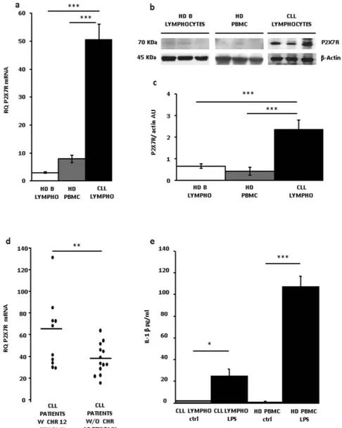
Figure 1. P2X7R expression in CLL lymphocytes. (a) P2X7R mRNA level was evaluated by Real-Time PCR
as described in Methods. P2X7R expression was normalized on G3PDH internal control and displayed as fold increase. Data are shown as mean ± SEM, n = 3, ***p < 0.001. (b) Representative Western blot of P2X7R from three subjects representative of each population. (c) Densitometry of P2X7R normalized on actin level (AU, arbitrary Units). N = 3, ***p < 0.001. (d) P2X7R mRNA expression in lymphocytes from CLL patients with or without chromosome 12 trisomy; **p < 0.01. (e) IL-1β release (see Methods) was evaluated by ELISA from CLL lymphocytes and HD PBMCs. Cells were overnight (12 h) incubated in the presence of LPS. Data are averages ± SEM, *p < 0.05; ***p < 0.001.
as shown by an exemplificative Western blot, and corresponding densitometry, of CLL lymphocytes, HD PBMCs and peripheral blood B lymphocytes (Fig. 1b,c). Next we asked whether the increased P2X7R expression level might correlate with known CLL prognostic factors. The P2RX7 gene is located on chromosome 12q24, that is duplicated in about 16% of CLL patients (chromosome 12 trisomy), and is a known adverse prognostic factor. Therefore, we analyzed whether P2X7R overexpression was associated with chromosome 12 trisomy. As shown in Fig. 1d, there was a statistically significant (**p < 0.01) higher P2X7R mRNA level in patients with versus patients without chromosome 12 trisomy, suggesting a gene-dosage effect.
The P2X7R is a main switch for processing and release of the potent pro-inflammatory cytokine IL-1β, thus we asked whether CLL lymphocytes, due to increased P2X7R expression might be endowed with IL-1β-releasing activity. Previous literature reports suggested that this might not be the case as IL-1β plasma levels are lower in CLL than in healthy controls19. Figure 1e, shows that IL-1β release from quiescent lymphocytes was negligible,
whether from HD or CLL subjects. Notably, LPS-stimulated release was about four fold lower in CLL lympho-cytes than in HD PBMCs despite much higher P2X7R expression in the former cells. The P2X7R is tightly func-tionally associated to the NLRP3 inflammasome, the major intracellular apparatus responsible for IL-β processing and release2, therefore, we investigated whether ASC and NLRP3 expression paralleled P2X7R expression level,
and thus was also increased in CLL lymphocytes.
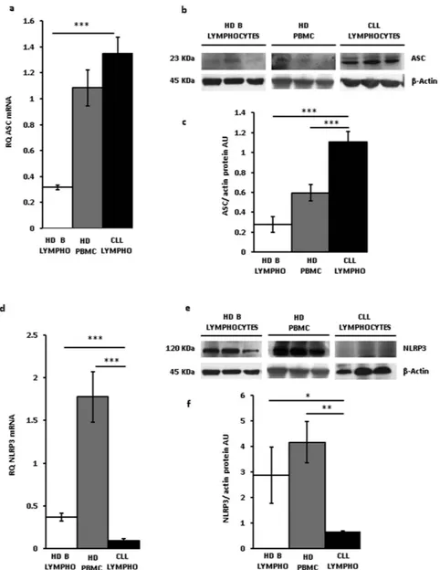
Figure 2a shows that ASC mRNA expression in CLL lymphocytes was slightly and not significantly higher than in HD PBMCs, but several fold higher relative to HD B lymphocytes. ASC protein was also overexpressed in CLL lymphocytes (Fig. 2b,c). On the contrary, and to our surprise, NLRP3 mRNA levels of CLL lymphocytes were at least 10 and 3 fold lower compared to HD PBMCs and HD B lymphocytes, respectively. NLRP3 protein levels were also significantly lower in CLL lymphocytes compared to HD lymphocytes or HD PBMC (*p < 0.05 and **<0.01 versus PBMCs and B lymphocytes, respectively, Fig. 2e,f). These data show that leukocytes from HDs, whether PBMCs or peripheral blood B lymphocytes, are characterized by low P2X7R and ASC expression and high NLRP3 levels, while CLL lymphocytes on the contrary are characterized by high P2X7R and ASC expression and low NLRP3 levels. A regression plot highlighted the inverse correlation between NLRP3 and P2X7R mRNA levels in CLL lymphocytes, although regression coefficient was low (0.2441, Figure S1). Our previous findings showed that the P2X7R promotes growth in vitro and in vivo, and in a number of experimental tumors its expression positively correlates with proliferation. Contrary to P2X7R, present data show that NLRP3 is down-modulated in leukemia cells and negatively correlates with proliferation. To properly investigate the role of NLRP3 in cell proliferation silencing experiments would be needed. However, modulation of NLRP3 expression by silencing in CLL lympho-cytes is impractical as these cells cannot be easily transfected, thus we took advantage of two human leukemic cell lines, THP-1 and Ramos cells, that can be transfected and have a fast in vitro growth rate.
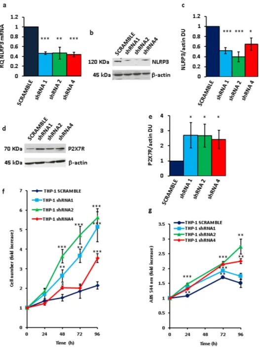
NLRP3 modulates THP-1 cell growth.
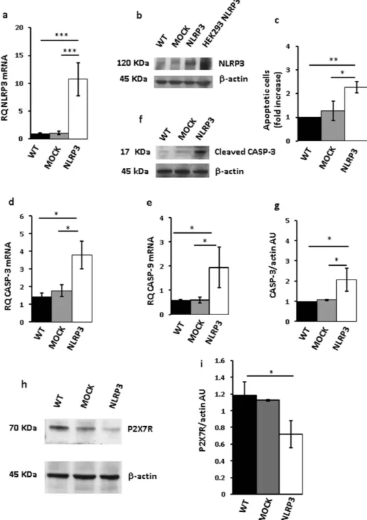
To down-modulate NLRP3 mRNA and protein, THP-1 cells were transfected with three different NLRP3 shRNAs (Fig. 3a–c). Very interestingly, P2X7R expression in these NLRP3- silenced cell clones was strongly enhanced (Fig. 3d,e). Our anticipation was that growth rate of NLRP3-silenced, P2X7R-overexpressing stable THP-1 clones should be faster relative to wt or mock-transfected cells. As shown in Fig. 3f,g, all NLRP3-silenced clones showed a faster growth rate relative to scramble shRNA-transfected clone over a 5 d culture assay, whether measured by counting cell number (Fig. 3f) or by the MTT assay (Fig. 3g). These observations on the one hand showed that inhibition of NLRP3 expression promoted growth, and on the other suggested that NLRP3 overexpression might negatively correlate with and even inhibit growth. To test this pre-diction we made several attempts to generate stable THP-1 or Ramos cell clones overexpressing NLRP3, however we were consistently unable to expand these NLRP3-overexpressing cell cultures as they systematically stopped proliferating and died few days after transfection.Figure 4a,b, show that both NLRP3 protein and mRNA were strongly up-regulated in THP-1 cells 48 h after transfection. However, these cells almost at the same time underwent accelerated apoptosis, as shown by increased annexin-V staining (Fig. 4c). Expression of both caspase-9 and -3 was stimulated in NLRP3-overexpressing cells, and caspase-3 was also activated (Fig. 4d–g). Quite interestingly, P2X7R expression in these NLRP3-overexpressing cells was dramatically downmodulated (Fig. 4h,i). Transfection with the NLRP3 plasmid increased NLRP3 mRNA and protein (Fig. 5a,b), stimulated caspase-3 and -9 expression, caspase-3 acti-vation (Fig. 5c–f), and apoptotic cell death (Fig. 5g) also in Ramos cells.
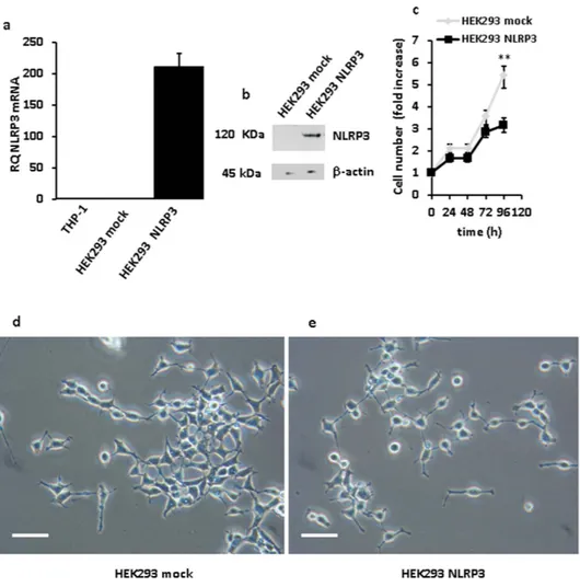
Since both THP-1 and Ramos cells proved to be extremely sensitive to the adverse effects of NLRP3 overex-pression, long-term effects of NLRP3 transfection were investigated in HEK293 cells. We were able to select sev-eral stable NLRP3-expressing HEK293 clones, one shown in Fig. 6. Growth rate of these stable NLRP3-expressing clones, as shown in Fig. 6c, was significantly slower (**p < 0.01) than that of HEK293 cells transfected with the empty vector. Interestingly, not only NLRP3-transfected HEK293 cells had a slower growth rate, but also showed substantial shape changes relative to mock-transfected cells. In fact, mock-transfected cells showed the spindle-like morphology typical of wt HEK293 cells, while on the contrary HEK293 overexpressing NLRP3 were rounded with numerous dendrite-like protrusions (Fig. 6d,e).
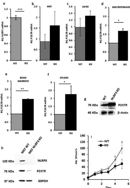
These findings strongly suggested that NLRP3 might participate in the control of cell growth and that this might involve the P2X7R. However, NLRP3 and P2X7R are mainly, although by any means not exclusively, expressed by immune cells and HEK293 are a cell line of uncertain histologic origin, thus it is possible that growth-modulatory effects of NLRP3 are restricted to lymphocytes or myelomonocytic cells. Thus, we inves-tigated P2X7R expression in different cell populations isolated from the NLRP3-KO mouse. Figure 7a shows NLRP3 mRNA amplification from NLRP3-wt and –KO mice. Figure 7d–f, show that bone marrow-derived mac-rophages, bone marrow cells and spleen cells from NLRP3-KO mice expressed at least twice as much P2X7R mRNA levels compared to wt mice. Slightly higher, but not statistically significant, P2X7R levels were also present in mouse embryo fibroblasts (MEFs) and liver cells from NLRP3-deleted mice (Fig. 7b,c). However, while higher P2X7R mRNA level translated into higher P2X7R protein level in spleen lymphocytes (Fig. 7g), this was not the
www.nature.com/scientificreports/
case in MEF cells, as in these latter cells P2X7R was strongly down-modulated (Fig. 7h). These data show that up-modulation of P2X7R in NLRP3-null cells is a feature of cells of hematopoietic origin, while P2X7R levels are much lower in NLRP3-KO than in NLRP3-WT fibroblasts. On the basis of the hypothesized growth-promoting role of the P2X7R, we anticipated that, due to P2X7R down-modulation and despite lack of NLRP3, NLRP3-KO MEFs should have a slower growth rate compared to NLRP3-WT MEFs. This anticipation was fulfilled as indeed NLRP3-KO MEFs showed a growth rate in culture which was about 30% to 50% than that of wt MEFs (Fig. 7i). These data suggest that NLRP3 effects on P2X7R expression and growth are cell type specific and likely restricted to cells of hematopoietic origin, and that anyway P2X7R is a key modulator of cell growth.
Discussion
Inflammasomes have become a focus of hot interest in cell biology and immunology for their crucial role in several cell functions, most notably cytokine secretion and host-pathogen interaction20. Among the different
subtypes so far described, the NLRP3 inflammasome is attracting increasing attention as it is a main target of DAMPs and the most active intracellular organelle responsible for maturation of cytokines of the IL-1β family. NLRP3 is functionally closely associated to the P2X7R, and in fact P2X7R is the most potent NLRP3-activating plasma membrane receptor2. P2X7R makes the NLRP3 inflammasome sensitive to extracellular ATP, one of
the major DAMPs released during inflammation21. Furthermore, other early pro-inflammatory agents, some
Figure 2. ASC and NLRP3 expression in CLL lymphocytes. Peripheral CLL lymphocytes and B lymphocytes
and PBMCs from healthy donors were isolated and qRT-PCR and Western blot analysis performed as described in Methods. ASC (a–c) and NLRP3 (d–f) protein (b,c,e,f) and mRNA expression (a,d) from lymphocytes from 23 CLL patients (see Table 1), PBMCs from 12 HDs, and B lymphocytes from 5 HDs is shown. Representative Western blot (b,e)and densitometry (c,f) of CLL lymphocytes, HD PBMC and HD peripheral blood B lymphocytes from three subjects (determinations from each subject in triplicate) representative of each population are shown. Data are averages ± SEM. *p < 0.05; **p < 0.01; ***p < 0.001.
themselves considered DAMPs such as the cathelicidin LL37 and amyloid-β, may directly interact with and stim-ulate P2X7R22,23, and by consequence activate the NLRP3 inflammasome.
Besides its crucial role in IL-1β processing and release, P2X7R is also well known for its cytotoxic activity and for its counterintuitive trophic activity. We and others have extensively shown that tonic, low-level, P2X7R stim-ulation, far from being cytotoxic, is beneficial for cell growth and proliferation5,24,6. In fact, and not surprisingly,
most human malignant tumors overexpress P2X7R, and targeting this receptor with selective blockers slows down in vivo growth of several transplanted tumors. Overexpression of P2X7R in CLL is of particular interest since a known adverse prognostic factor in this leukemia is chromosome 12 trisomy25. Albeit chromosome 12
is known to host several genes related to cancer26, a pathogenetic explanation for the adverse prognostic
signif-icance of chromosome 12 trisomy in CLL is lacking. Present and previous reports documenting an increased expression and activity of the P2X7R in CLL provide a pathophysiological explanation given the compelling
Figure 3. NLRP3 silencing in THP-1 cells triggers P2X7R up-regulation and stimulates growth. THP-1
cells were cultured and analyzed for NLRP3 mRNA (a) and protein (b,c) expression as described in Methods. NLRP3 (a–c) was silenced by HuSH-29 transfection and selected with puromycin as described in Methods. Densitometry of NLRP3 protein bands is shown in panel (c) (n = 3). Up-regulation of P2X7R protein in NLRP3-silenced THP-1 cells (d–e). Densitometry of P2X7R protein bands is shown in panel (e) (n = 3). For cell proliferation, 5 × 10⁴ cells were plated in 6-well plates in complete RPMI 1640 medium in a 5% CO
2
incubator (37 °C) and counted (f) or analyzed by MTT assay (g) at the indicated time points. Data from triplicate determinations are shown in panels (a,c,e–g). Data are averages ± SEM. *p < 0.05; **p < 0.01; ***p < 0.001.
www.nature.com/scientificreports/
evidence supporting a role of this receptor in cell proliferation and metastatic spreading. The close functional association between P2X7R and NLRP3 and the known, albeit controversial, role of IL-1β in tumor progression, initially prompted us to hypothesize that NLRP3 followed in CLL lymphocytes an expression pattern similar to P2X7R, and thus was overexpressed. However, contrary to this anticipation, our present data show that NLRP3 was strikingly down-regulated at the mRNA and protein level in CLL lymphocytes compared to HD PBMCs or peripheral blood B lymphocytes. The ASC protein on the other hand, like P2X7R, was up-regulated in cells from CLL patients.
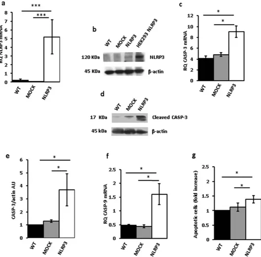
Figure 4. NLRP3 overexpression in THP-1 cells triggers cell death. THP-1 cells were transfected with a
plasmid encoding NLRP3 as described in Methods, and then analyzed for NLRP3 mRNA and protein expression (a,b). Empty vector (mock)-transfected cells and a HEK293 clone stably transfected with NLRP3 (HEK293 NLRP3) are shown as control. Apoptotic cell number (annexin V-positive cells) is shown in panel (c). Caspase-3 and -9 expression is shown in panels (d,e). Effect of NLRP3 transfection on caspase-3 activation (cleavage) is shown in panels (f,g). Down-modulation of P2X7R protein by NLRP3 overexpression is shown in panels (h,i). Data are averages ± SEM from triplicate independent determinations. *p < 0.05; **p < 0.01; ***p < 0.001.
The inverse P2X7R/NLRP3 correlation suggested that high NLRP3 levels might negatively affect cell pro-liferation, contrary to high P2X7R levels that support proliferation. Since primary human B lymphocytes are not amenable to transfection studies, we turned as a model for transfection to THP-1 cells, a human leukemic cell line that expresses both P2X7R and NLRP3. In agreement with our prediction, NLRP3 down-modulation caused a strong acceleration of cell proliferation paralleled by an increase in P2X7R expression. Given the known growth-promoting effect of P2X7R, it is highly likely that the proliferative advantage conferred by NLRP3 sup-pression was due to the enhanced P2X7R exsup-pression. Identification of P2X7R overexsup-pression as a mechanism by which NLRP3 down-modulation supports growth provides also hints as to the metabolic pathways involved as we have previously demonstrated that P2X7R overexpression increases mitochondrial matrix Ca2+ and potential,
intracellular ATP stores, activates NFATc1 and protects from apoptosis5,9.
We then investigated the effect of NLRP3 overexpression. NLRP3 overexpression caused P2X7R down-modulation in THP-1 cells, but it was rather difficult to prove that NLRP3 overexpression had an adverse effect on cell growth as its mere transfection proved to be cytotoxic on most THP-1 and Ramos cells, making it impossible to select stably-transfected clones, as we did for the NLRP3-silenced cells. Thus, we were forced to investigate a few and more important apoptotic markers in the 48 h following transfection. During this time, apoptosis was accelerated in both THP-1 and Ramos cells, and apoptotic caspases-3 and -9 were overexpressed and activated. After 48–72 h very few transfected cells survived, thus making additional studies impossible. However, we were able to verify the long term effects of NLRP3 transfection in HEK293 cells. This cell type turned out to be much more resistant to the adverse effects of NLRP3 transfection compared to THP-1 or Ramos cells. A few stably-transfected clones were selected and their growth kinetics could be investigated. In agreement with our prediction of the anti-proliferative role of NLRP3, NLRP3-transfected cells showed a much slower growth rate compared to empty vector- transfected or wt cells, and a strikingly different morphology.
Figure 5. NLRP3 overexpression in Ramos cells triggers cell death. Ramos cells were transfected with
a plasmid encoding NLRP3 as described in Methods, and then analyzed for NLRP3 mRNA and protein expression (a,b) Empty vector (mock)-transfected cells and a HEK293 clone stably transfected with NLRP3(HEK293NLRP3) are shown as control. Effect of NLRP3 over- expression on caspase-3 mRNA (c) and protein (e) expression and caspase-3 activation (d) is shown. Effect of NLRP3 transfection on caspase-9 expression (f) and apoptotic cell number (annexin V-positive cells) is shown in panel (g). Data are averages ± SEM from 3 to 4 independent determinations. *p < 0.05; ***p < 0.001.
www.nature.com/scientificreports/
Our findings raise the provocative issue of the possible direct participation of the NLRP3 inflammasome in the control of cell proliferation. In principle, it is not surprising that NLRP3 overexpression stimulates cell death as it is well known that inflammasome activation may trigger pyroptosis, a form of lytic cell death mediated by inflammasome activation and IL-1β release11. However, it is indeed surprising that NLRP3 down-modulation is
growth-promoting in a cell- autonomous fashion, as shown by our results. It was very recently shown that NLRP3 is repressed in human hepatocellular carcinoma compared to healthy liver cells, and that NLRP3 expression inversely correlates with liver cancer progression16. This finding was interpreted by the Authors as a proof that low
NLRP3 levels impaired liver cell ability to sense cytoplasmic danger signals and react accordingly, thus enhancing cell susceptibility to undergo malignant transformation. In the present study we confirm that tumor cells (CLL B lymphocytes) have low NLRP3 levels, but contrary to the interpretation put forward by Wei et al.16 we show that
NLRP3 expression directly affects cell growth, in a cell autonomous fashion. However, this observation cannot be generalized to cells of non-hematopoietic origin since data from NLRP3-KO mice show that the P2X7R is up-regulated in bone marrow cells, in macrophages and in spleen cells, but not in liver cells or in fibroblasts. The mechanism by which NLRP3 modulates growth might involve the P2X7R, a receptor that is known to have a dual role as cytotoxic and a growth-promoting receptor27, but other mechanisms might also be involved as NLRP3
transfection also down-modulate growth in HEK293 cells, which lack endogenous P2X7R expression.
These findings help place the NLRP3 inflammasome in an entirely novel context in cancer. Current view holds that NLRP3 interferes with tumor progression in virtue of its ability to modulate cytokine release and cell death (pyroptosis), and thus control inflammation. In this context, the outcome can then be different since in certain experimental settings this activity is protective against cancer development, while in other settings NLRP3 acti-vation and IL-1β secretion are associated to a more malignant tumor phenotype. Our data expand the role of NLRP3 in B cell leukemia as a factor that is directly controlling growth.
Figure 6. NLRP3 overexpression inhibits proliferation of HEK293 cells. HEK293 cells were transfected with
a NLRP3-encoding plasmid as described in Methods, cultured in complete DMEM-F12 medium and assayed for proliferation. Panels (a,b) show level of mRNA and protein expression, respectively, in mock- and NLRP3-transfected cells. THP-1 cells (a) are shown as a control for baseline NLRP3 expression. Panel (c) shows growth kinetics of NLRP3- and mock-transfected HEK293 cells, respectively. **p < 0.01. Morphology of mock- (d) and NLRP3-transfected (e) HEK293 cells. **p < 0.01 Bar = 40 μm.
In conclusion, the P2X7R-NLRP3 axis appears to have an increasing importance at multiple steps involved in tumor progression, from to modulation of inflammation to control of cell proliferation and cell death. These findings might be of potential interest for the identification of novel therapies against cancer.
Methods
Animals.
C57BL/6 wild type and C57BL/6 Nlrp3−/− mice, all 4 to 8-week-old males were used for primarycultures. All procedures were performed in accordance with the Italian institutional guidelines in compliance with national and international laws and policies and were approved by the Italian Ministry of Health (n 76/2013-B).
Figure 7. P2X7R expression in tissues and cells from Nlrp3−/− mouse. NLRP3 mRNA expression in bone
marrow-derived macrophages from wt and Nlrp3−/− mice (a). P2X7R mRNA expression in MEF (b), liver (c),
macrophages (d), bone marrow (e) and spleen (f) from wt and Nlrp3−/− mice. P2X7R protein expression in
spleen (g) and MEF (h) from wt and Nlrp3−/− mice. Growth rate of MEFs from wt and Nlrp3−/− mice (i). Cell
were isolated and processed as described in Methods. Data are averages ± SEM from triplicate independent determinations. *p < 0.05; **p < 0.01; ***p < 0.001.
www.nature.com/scientificreports/
Cell cultures and transfections.
Ramos and THP-1 cells were purchased from American Type Culture Collection (ATCC, Rockville, MD, USA), and were maintained in RPMI 1640 medium supplemented with 10% heat-inactivated fetal bovine serum, L-glutamine, 100 U/ml penicillin and 100 mg/ml streptomycin (all from Celbio, Euroclone, Milano, Italy). NLRP3-silenced THP-1cell clones were transfected by electroporation with Neon®
Transfection System for Electroporation (Life Technologies, Grand Island, NY, USA) with prede-signed HuSH-29 shRNA targeting the following sequences: AGAGAAGGCAGACCATGTGGATCTAGCCA (shRNA1, c.n. GI321521), CAGTCTGATTCAGGAGAACGAGGTCCTCT (shRNA2,c.n. GI321522); GTACGTGAGAAGCAGATTCCAGTGCATTG (shRNA4, c.n. GI321524), or with scrambled RNA cassette (c.n. TR30013), all from OriGene (Rockville, MD, USA). Stably transfected cell lines were obtained by selection with puromycin (0.5–1 μml/L, Sigma-Aldrich, Milan, Italy). HEK293 were cultured in DMEM-F12 (Sigma-Aldrich) supplemented with 10% FBS, 100 U/ml penicillin and 100 mg/ml streptomycin. HEK293NLRP3 cells were obtained by transfection with lipofectamin 2000 (Life Technologies) as per manufacturer’ s instructions. NLRP3 plasmid was provided by OriGene (c.n. RG220952). Stably-transfected cell lines were obtained by selection with G418 sulphate (0.8 mg/ml) (Calbiochem, La Jolla, CA, USA). Single cell-derived clones, were obtained by limiting dilution. For transient expression experiments, 6 × 10⁶ cells were plated in 6- well plates and used 48 h later.Patients.
Chronic Lymphocitic Leukemia patients were recruited at the Hematology Clinic of Ferrara University Hospital according to the guidelines issued by the local Ethical Committee and in compliance with International Regulations. Patient clinical features are shown in Table 1. All patients gave their informed consent. All experimental protocols were approved by the Ethical Committee of the Ferrara University Hospital and all experiments were performed in accordance with relevant guidelines and regulations.PBMC and lymphocytes isolation from CLL patients and healthy donors.
Blood samples from CLL patients and healthy donors were obtained from the Hematology Clinic of Ferrara University Hospital, or from the local blood bank. Blood was diluted 1:2 with PBS (Celbio), and centrifuged on Ficoll-Hypaque (GE Healthcare Life Sciences). Cells were then rinsed twice with PBS, and resuspended in 10% FBS-supplemented RPMI 1640 medium prior to experiments.B lymphocyte isolation from healthy donor samples.
Blood samples of healthy individuals were obtained through the blood bank of the University of Torino. PBMCs were obtained by Ficoll‐Hypaque (GE Healthcare Life Sciences, Milan, Italy) centrifugation. B lymphocytes were purified through a first step of negative selection, using anti-CD3, anti-CD16, and anti-CD14 monoclonal antibodies (produced locally) and immuno-magnetic bead separation (Dynabeads Sheep anti-Mouse IgG, Life Technologies) followed by positive purifi-cation using the CD19 microbeads kit [Miltenyi Biotec S.r.l., Calderara di Reno (BO), Italy]. Flow cytometric analysis showed that these cells were more than 95% CD19+.ID PATIENT SEX AGE CD 38 ZAP 70 IgVH WBC/mm3 LYMPHOCYTES/mm3 β2-microglobulin mg/L TRISOMY 12
CLL 01 M 62 + + unmutated 12050 5330 5,6 + CLL 02 F 70 + − unmutated 19360 14470 3,6 − CLL 03 M 84 − − unmutated 74040 66640 4,04 − CLL 04 F 58 + + unmutated 14160 8830 5,6 + CLL 05 F 70 + + unmutated 79430 65550 4,1 − CLL 06 M 62 − − mutated 98780 94400 2,04 − CLL 07 M 61 − + unmutated 13470 8690 4,6 − CLL 08 F 77 + + unmutated 15900 9920 6,6 + CLL 09 F 68 + + unmutated 21950 18440 9 + CLL 10 M 54 + − mutated 13700 7110 2,7 + CLL 11 M 64 − + unmutated 14000 10220 3,1 + CLL 12 M 75 + + unmutated 34310 22490 4 − CLL 13 F 79 + + unmutated 14800 7460 2,3 + CLL 14 F 74 − − mutated 39000 29220 2,5 − CLL 15 F 77 + − unmutated 14160 8300 5,3 + CLL 16 M 54 − + unmutated 12452 108330 2,2 + CLL 17 M 72 − − mutated 36850 27070 6,2 − CLL 18 M 79 − + unmutated 17477 16401 11 − CLL 19 F 69 + + unmutated 7520 5930 2 − CLL 20 M 70 − + unmutated 20810 12960 2,2 − CLL 21 F 65 + + unmutated 39360 31060 2 − CLL 22 M 46 − + unmutated 15770 10450 3,2 − CLL 23 M 84 + + unmutated 11956 9627 5 +
RNA isolation and qRT PCR.
Total RNA was extracted with Trizol reagent (Life Technologies), and then purified with the Pure link RNA Mini Kit (Invitrogen) as per manufacturer’s instructions. RNA content was quantitated using a Nanodrop 2000 spectrophotometer (Thermo Fisher Scientific, Milan, Italy). Reverse tran-scription was performed by using 1 μg of total RNA extract, using the High Capacity cDNA Reverse Trantran-scription kit (Applied Biosystem, Carlsbad, CA, USA). qRT PCR was performed in a Step One Real Time PCR System (Applied Biosystem). Two μl of cDNA were used as a template. Amplification was performed using predesigned Taqman probes (Applied Biosystem) for P2X7R, NLRP3, ASC, casp-3, casp-9 and G3PDH as a housekeeping gene. A comparative analysis using the ΔΔCt method was used to quantitate fold increase of target cDNA relative to THP-1 wild type reference sample.MTT assay.
Growth rate of control and NLRP3-silenced cells was measured by direct cell number count and MTT assay (Invitrogen). For MTT assay, 5 × 10⁴ cells were plated into each well of a 96-well plate. Twenty four,48, 72 and 96 h after plating 10 μl of MTT was added to each well and incubated at 37 °C for additional 4 h. Finally, 100 μl of SDS-HCl was added to each well to solubilize the precipitate. Optical density was measured at 544 nm and data were expressed as a fold increase of THP-1 scrambled control.
Interleukin-1β measurement.
PBMCs from CLL patients or HDs (5 × 10⁵ cells/ml) were plated ina 24-well plates in complete RPMI medium, and left untreated or primed overnight with 1 μg/ml of LPS (Sigma-Aldrich). IL-1β release was measured in the supernatants of PBMCs by ELISA (R&D Systems, Minneapolis, MI, USA).
Western blot analysis.
Total cell lysates were prepared in a lysis buffer (300 μM sucrose, 1 mM K2HPO4,5.5 mM D-glucose, 20 mM Hepes) supplemented with protease inhibitors (1 mM phenylmethylsulfonyl fluoride, 1 mM benzamidine) (all by Sigma-Aldrich). Proteins were quantified using the Bradford method. Samples were loaded into 4–12% Bolt-SDS pre cast-gel (Life Technologies), and transferred onto nitrocellulose membranes (GE Healthcare Life Sciences). Membranes were incubated overnight with the primary Abs at 4 °C. The anti-P2X7R Ab (Sigma-Aldrich) was used at a dilution of 1:200 in TBS-t (50 mM Tris, 150 mM NaCl, 0.1% Tween 20, pH 7.6) supplemented with 3% non- fat milk (Biorad, Copenhagen, Denmark) and 0.5% BSA (Sigma-Aldrich). The anti-ASC Ab (MBL, Woburn, MA,USA) was diluted 1:200 in PBS supplemented with 1% skimmed milk. The anti-NLRP3 Ab (Abcam, Cambridge, UK) was diluited 1:500 in PBS. The anti-caspase-3 Ab (Cell Signalling, Milano, Italy) was diluited 1:1000 in TBS-t supplemented with 5% non-fat milk (Biorad). The anti-actin Ab (Sigma-Aldrich) was diluited 1:1000 in TBS-t. Membranes were incubated with secondary goat anti-rabbit or anti-mouse HRP-conjugated Abs (Biorad) at a 1:3000 dilution in TBS-t buffer, or in PBS-t (PBS supplemented with 0.1% Tween 20) for 1 h at room temperature. Detection was performed with the Luminata
™
Western chemi-luminescent HRP kit (Merck S.p.A., Milan, Italy). Densitometric analysis of the protein bands was perfomed with ImageJ software. Band density was normalized over the actin band.Measurement of Apoptosis.
Forty eight hours after transfection cells were centrifuged, washed with PBS (Celbio), and resuspended in 500 μl of labelling medium containing 5 μl of annexin V-Cy3 solution (Biovision, Milpitas,CA,USA). After 10 min of incubation cell fluorescence was measured with a Tali™
image-based cytom-eter (Invitrogen).Isolation and differentiation of murine bone marrow cells and bone marrow-derived
mac-rophage.
Bone marrow cells were isolated by flushing femurs and tibia of 4-to 8-week old C57Bl/6 mice with complete RPMI medium. The cell suspension was collected, centrifuged for 10 min at 150xg and re-suspended for 5 minutes at room temperature in red blood cell lysis buffer (Sigma-Aldrich). Reaction was stopped by adding RPMI, cells were centrifuged, and resuspended in Trizol for mRNA analysis. Alternatively, after incubation in red blood cell lysis buffer, bone marrow cells were re-suspended in M-CSF-supplemented (200 ng/ml) RPMI for macrophage differentiation, and seeded in Petri dishes at a concentration of 2.5 × 10⁵ cells/ml. After 7 days cellswere detached using cold PBS supplemented with 2 mM EDTA and counted.
Isolation of spleen cells.
Spleens were isolated, homogenized by careful pulping and treated with red blood cell lysis buffer for 5 minutes at room temperature to remove erythrocytes. The cell suspension was then supplemented with RPMI, centrifuged for 10 minutes at 150xg, filtered through at 70 μm-cell strainer (Becton Dickinson, Franklin Lakes, NJ, USA), rinsed twice with RPMI and finally re-suspended in the same medium at a concentration of 20×106 cells/ml.Isolation of mouse embryo fibroblasts (MEFs) and analysis of proliferation.
WT and Nlrp3−/−MEFs were isolated from pregnant mice at 13 or 14 day post coitum. Mice were sacrificed by cervical disloca-tion. Embryos were harvested, MEFs were isolated as previously described and cultured in DMEM medium supplemented with 10% FBS, penicillin/streptomycin, and 2 mM L-glutamine28. Growth curves were generated
by seeding 104 cells per well in 12-well plates. Plates were stained with crystal violet at each indicated time point.
Each experiment was performed in triplicate. Crystal violet was extracted with 10% acetic acid followed by plate reading at 590 nm.
Statistical analysis.
All data are shown as means ± standard error of the mean (SEM). Test of significance was performed by Student’s t-test using GraphPad InStat software (GraphPad, San Diego, Ca, USA).www.nature.com/scientificreports/
References
1. Lamkanfi, M. & Dixit V. M. Mechanisms and Functions of Inflammasomes. Cell. 157, 1013–1022 (2014).
2. Di Virgilio, F. The therapeutic potential of modifying inflammasomes and NOD-like receptors. Pharmacol. Rev. 65, 872–905 (2013). 3. Franceschini, A. et al. The P2X7 receptor directly interacts with the NLRP3 inflammasome scaffold protein. FASEB J. 29, 2450–2461
(2015).
4. Dinarello, C. A. Immunological and inflammatory functions of the interleukin-1 family. Annu. Rev. Immunol. 27, 519–550 (2009). 5. Adinolfi, E. et al. Basal Activation of the P2X7 ATP Receptor Elevates Mitochondrial Calcium and Potential, Increases Cellular ATP
Levels, and Promotes Serum-independent Growth. Mol. Biol. Cell. 16, 3260–3272 (2005).
6. Adinolfi, E. et al. Expression of P2X7 receptor increases in vivo tumor growth. Cancer Res. 72, 2957–2969 (2012). 7. Di Virgilio, F. Purines, purinergic receptors, and cancer. Cancer Res. 72, 5441–5447 (2012).
8. Rigato, C. et al. Microglia proliferation is controlled by P2X7 receptors in a Pannexin-1-independent manner during early embryonic spinal cord invasion. J. Neurosci. 32, 11559–11573 (2012).
9. Adinolfi, E. et al. Expression of the P2X7 receptor increases the Ca2+ content of the endoplasmic reticulum, activates NFATc1 and
protects from apoptosis. J. Biol. Chem. 284, 10120–10128 (2009).
10. Adinolfi, E. et al. P2X7 receptor expression in evolutive and indolent forms of chronic B lymphocytic leukemia. Blood. 99, 706–708 (2002).
11. Miao, E. A., Rajan, J. V. & Aderem, A. Caspase-1-induced pyroptotic cell death. Immunol. Rev. 243, 206–214 (2011).
12. Willingham, S. B. et al. Microbial pathogen-induced necrotic cell death mediated by the inflammasome components CIAS1/ cryopyrin/NLRP3 and ASC. Cell Host Microbe. 2, 147–159 (2007).
13. Allen, I. C. et al. The NLRP3 inflammasome functions as a negative regulator of tumorigenesis during colitis-associated cancer. J.
Exp. Med. 207, 1045–1056 (2010).
14. Allen, I. C. et al. NLRP12 suppresses colon inflammation and tumorigenesis through the negative regulation of noncanonical NF-kappaB signaling. Immunity. 36, 742–754 (2012).
15. Okamoto, M. et al. Constitutively active inflammasome in human melanoma cells mediating autoinflammation via caspase-1 processing and secretion of interleukin-1beta. J. Biol. Chem. 285, 6477–6488 (2010).
16. Wei, Q. et al. Deregulation of the NLRP3 inflammasome in hepatic parenchymal cells during liver cancer progression. Lab. Invest.
94, 52–62 (2014).
17. Zaki, M. H. et al. The NLRP3 inflammasome protects against loss of epithelial integrity and mortality during experimental colitis.
Immunity. 32, 379–391 (2010).
18. Fan, S. H. et al. Luteoloside suppresses proliferation and metastasis of hepatocellular carcinoma cells by inhibition of NLRP3 inflammasome. PLos One. 9, e89961 (2014).
19. Hulkkonen, J., Vilpo, J., Vilpo, L., Koski, T. & Hurme, M. Interleukin-1 beta, interleukin-1 receptor antagonist and interleukin-6 plasma levels and cytokine gene polymorphisms in chronic lymphocytic leukemia: correlation with prognostic parameters.
Haematologica. 85, 600–606 (2000).
20. Strowig, T., Henao-Mejia, J., Elinav, E. & Flavell, R. Inflammasomes in health and disease. Nature. 481, 278–286 (2012).
21. Bours, M. J., Dagnelie, P. C., Giuliani, A. L., Wesselius, A. & Di Virgilio, F. P2 receptors and extracellular ATP: a novel homeostatic pathway in inflammation. Front. Biosci. (Schol. Ed.) 3, 1443–1456 (2011).
22. Tomasinsig, L. et al. The human cathelicidin LL-37 modulates the activities of the P2X7 receptor in a structure-dependent manner.
J. Biol. Chem. 283, 30471–30481 (2008).
23. Sanz, J. M. et al. Activation of microglia by amyloid {beta} requires P2X7 receptor expression. J. Immunol. 182, 4378–4385 (2009). 24. Bianco, F. et al. A role for P2X7 in microglial proliferation. J. Neurochem. 99, 745–758 (2006).
25. Zenz, T., Mertens, D., Dohner, H. & Stilgenbauer, S. Molecular diagnostics in chronic lymphocytic leukemia - pathogenetic and clinical implications. Leuk. Lymphoma. 49, 864–873 (2008).
26. Scherer, S. E. et al. The finished DNA sequence of human chromosome 12. Nature. 440, 346–351 (2006). 27. Di Virgilio, F. Dr. Jekyll/Mr. Hyde: the dual role of extracellular ATP. J. Auton. Nerv. Syst. 81, 59–63 (2000).
28. Giorgi, C. et al. PML regulates apoptosis at endoplasmic reticulum by modulating calcium release. Science. 330, 1247–1251 (2010).
Acknowledgements
FDV is supported by grants from AIRC (n. IG 13025), Telethon (n. GGP 11014), ERA-NET Neuron “Nanostroke”, EU COST Program n. BM1406, the Ministry of Health of Italy (n. RF-2011-02348435), the Italian Ministry of Education, University and Research (n. RBAP11FXBC_001) and funds from the University of Ferrara. FDV was also supported by the 7th Framework Program HEALTH-F2-2007-202231 “ATPBone”. P. Pinton is supported by AIRC (IG 14442), and the Italian Ministry of Education, University and Research (COFIN n. 20129JLHSY_002, FIRB n. RBAP11FXBC_002, and Futuro in Ricerca n. RBFR10EGVP_001). P. Pelegrin is supported by European Research Council (ERC-2013-CoG 614578) and Instituto Salud Carlos III-Fondo Europeo de Desarrollo Regional (PI13/00174). The Authors also wish to acknowledge the help of Drs Fabio Casciano Flow Cytometry and Sorting Facility, LTTA Laboratory, University of Ferrara, Elenora Gallerani and Federica Poletti (University of Ferrara) for FACS analysis and help with transfections, and of Prof. Antonio Rosato (University of Padova) for providing irradiated control mouse spleen cells.
Author Contributions
E.S. performed most of the experiments, analyzed data and wrote the M.S. draft. A.R., F.C., M.R. and M.C. collected clinical samples, did some of the experiments and helped in data analysis. S.F. did some of the experiments, helped in data analysis and revised the M.S. F.S.A. and A.F. participated in the experimental work in support of E.S. and S.F. V.A. and S.D. helped in the characterization of B lymphocytes from healthy donors and revised the M.S. A.C.S. and M.B. performed experiments with MEFs. A.C., P. Pelegrin and P. Pinton supervised the study and helped in writing the M.S. F.D.V. was responsible for the overall organization of the study, planned the experiments and wrote the M.S.
Additional Information
Supplementary information accompanies this paper at http://www.nature.com/srep Competing financial interests: The authors declare no competing financial interests.
How to cite this article: Salaro, E. et al. Involvement of the P2X7-NLRP3 axis in leukemic cell proliferation and
This work is licensed under a Creative Commons Attribution 4.0 International License. The images or other third party material in this article are included in the article’s Creative Commons license, unless indicated otherwise in the credit line; if the material is not included under the Creative Commons license, users will need to obtain permission from the license holder to reproduce the material. To view a copy of this license, visit http://creativecommons.org/licenses/by/4.0/