RESEARCH
Italian legumes: effect of sourdough
fermentation on lunasin-like polypeptides
Carlo Giuseppe Rizzello
1*, Blanca Hernández‑Ledesma
2, Samuel Fernández‑Tomé
2, José Antonio Curiel
1,
Daniela Pinto
3, Barbara Marzani
3, Rossana Coda
4and Marco Gobbetti
1Abstract
Background: There is an increasing interest toward the use of legumes in food industry, mainly due to the qual‑ ity of their protein fraction. Many legumes are cultivated and consumed around the world, but few data is available regarding the chemical or technological characteristics, and especially on their suitability to be fermented. Never‑ theless, sourdough fermentation with selected lactic acid bacteria has been recognized as the most efficient tool to improve some nutritional and functional properties. This study investigated the presence of lunasin‑like polypeptides in nineteen traditional Italian legumes, exploiting the potential of the fermentation with selected lactic acid bacteria to increase the native concentration. An integrated approach based on chemical, immunological and ex vivo (human adenocarcinoma Caco‑2 cell cultures) analyses was used to show the physiological potential of the lunasin‑like polypeptides.
Results: Italian legume varieties, belonging to Phaseulus vulgaris, Cicer arietinum, Lathyrus sativus, Lens culinaris and
Pisum sativum species, were milled and flours were chemically characterized and subjected to sourdough fermenta‑ tion with selected Lactobacillus plantarum C48 and Lactobacillus brevis AM7, expressing different peptidase activities. Extracts from legume doughs (unfermented) and sourdoughs were subjected to western blot analysis, using an anti‑ lunasin primary antibody. Despite the absence of lunasin, different immunoreactive polypeptide bands were found. The number and the intensity of lunasin‑like polypeptides increased during sourdough fermentation, as the conse‑ quence of the proteolysis of the native proteins carried out by the selected lactic acid bacteria. A marked inhibitory effect on the proliferation of human adenocarcinoma Caco‑2 cells was observed using extracts from legume sour‑ doughs. In particular, sourdoughs from Fagiolo di Lamon, Cece dell’Alta Valle di Misa, and Pisello riccio di Sannicola flours were the most active, showing a decrease of Caco‑2 cells viability up to 70 %. The over‑expression of Caco‑2 filaggrin and involucrin genes was also induced. Nine lunasin‑like polypeptides, having similarity to lunasin, were identified.
Conclusions: The features of the sourdough fermented legume flours suggested the use for the manufacture of novel functional foods and/or pharmaceuticals preparations.
Keywords: Legumes, Sourdough, Lactic acid bacteria, Lunasin
© 2015 Rizzello et al. This article is distributed under the terms of the Creative Commons Attribution 4.0 International License (http://creativecommons.org/licenses/by/4.0/), which permits unrestricted use, distribution, and reproduction in any medium, provided you give appropriate credit to the original author(s) and the source, provide a link to the Creative Commons license, and indicate if changes were made. The Creative Commons Public Domain Dedication waiver (http://creativecommons.org/ publicdomain/zero/1.0/) applies to the data made available in this article, unless otherwise stated.
Background
According to the Food and Agricultural Organization (FAO) pulses are dry seeds of annual legume plants, belonging to the Fabaceae (also known as Leguminosae) family. FAO classifies the large number of legume species
and varieties employed as food or feed into eleven main groups (dry beans, dry broad beans, dry peas, chick-peas, dry cowchick-peas, pigeon chick-peas, lentils, bambara beans, vetches and lupins) and minor pulses [1]. Nutritionally, pulses are an important source of proteins, which, in spite of being deficient in sulfur-containing amino acids and tryptophan, possess higher amounts of lysine, argi-nine, glutamic, and aspartic acid compared to cereal grains [2]. Beyond the nutritional benefits, consumption
Open Access
*Correspondence: [email protected]
1 Department of Soil, Plant and Food Science, University of Bari Aldo Moro, 70126 Bari, Italy
of pulses is recently associated with protective or thera-peutic effects on chronic health conditions, such as car-diovascular diseases, diabetes, cancer, overweight, and obesity [2]. Owing to the low cost and easy adaptation to grow under poor conditions, pulses are used as staple foods in several low-income countries, serving as main
source of both protein and calories [2, 3]. Otherwise, in
high-income countries of America and Europe, pulses consumption is low and efforts are done to promote their healthy intake.
Several health organizations recommend pulse con-sumption as a part of a healthy diet and initiatives are addressed to increase the cultivation, intake, and food
processing uses [2, 4]. The protein content of pulses
ranges from 20 to 40 % of dry weight. Within this inter-val, the most abundant are the seed storage proteins. The remaining part are minor or housekeeping proteins, which include enzymes, protease, amylase inhibitors,
lec-tins, lipoxygenases, defense proteins, and others [3, 5].
Legume seeds also contain proteins, which are consid-ered as anti-nutritional compounds due to the effect on the quality of the diet. Nevertheless, the harmful effects of such compounds is easily inactivated after cooking or processes like fermentation, germination and dehull-ing [6]. Once inactivated, lectins or protease inhibitors may present potential health benefits. Protease inhibitors are potential anti-inflammatory and anticancer agents, whereas lectins have demonstrated to play a key role in preventing certain cancers and activating innate defense mechanisms. Besides, lectins are also proposed as thera-peutic agents to prevent or control obesity [5].
Many of the physiological and functional properties of proteins are attributed to biologically active peptides which are often encrypted in the native sequence [7, 8]. Biogenic or bioactive peptides are released from their precursor proteins either by enzymes during gastroin-testinal digestion or through proteolysis (e.g., microbial fermentation), which occurs during food processing [8]. In particular, legume hydrolysates and bioactive peptides had in vitro activities towards cancer and cardiovascular diseases or their physiological manifestations like oxi-dative damage, inflammation, hypertension, and high cholesterol [2]. Lunasin is a 43-amino acid peptide with anticancer, anti-inflammatory, antioxidant and choles-terol lowering activities [9]. It is purified from soybean and commercialized as an ingredient or dietary supple-ment. In the quest for readily available natural sources of lunasin, the identification and purification of lunasin from different vegetable sources deserve a marked
inter-est [10, 11]. Moreover, the potential of sourdough
fer-mentation for increasing the concentration of lunasin in food matrices was recently investigated [12]. Sour-dough is the natural starter traditionally used for making
leavened baked goods, harboring a rich lactic acid bacte-ria and yeast microbiota. A large number of studies [13] showed that the fermentative and proteolytic activities of sourdough lactic acid bacteria not only determined opti-mal sensory, technology and nutritional characteristics, but also increased the functional value of leavened baked goods. Compared to unfermented soybean, amaranth, barley and wheat flours, the concentration of lunasin and related fragments increased up to four times during fer-mentation with lactic acid bacteria, which were selected based on proteolytic activities [12].
This study reported the presence of lunasin-like poly-peptides in nineteen traditional Italian legumes, and exploited the potential of the fermentation with selected lactic acid bacteria to increase the native concentration. An integrated approach based on chemical, immuno-logical and ex vivo (human adenocarcinoma Caco-2 cell cultures) analyses was used to show the physiological potential of the lunasin-like polypeptides.
Results
Chemical and microbiological characteristics of the flours
All the traditional Italian legumes used in this study have specific certifications (names, abbreviations,
geo-graphical origin and certification are listed in Table 1).
Legume grains were milled to obtain the correspond-ing flours. The proximate composition of the flours is
reported in Table 2. Moisture ranged from 7.2 ± 0.4 to
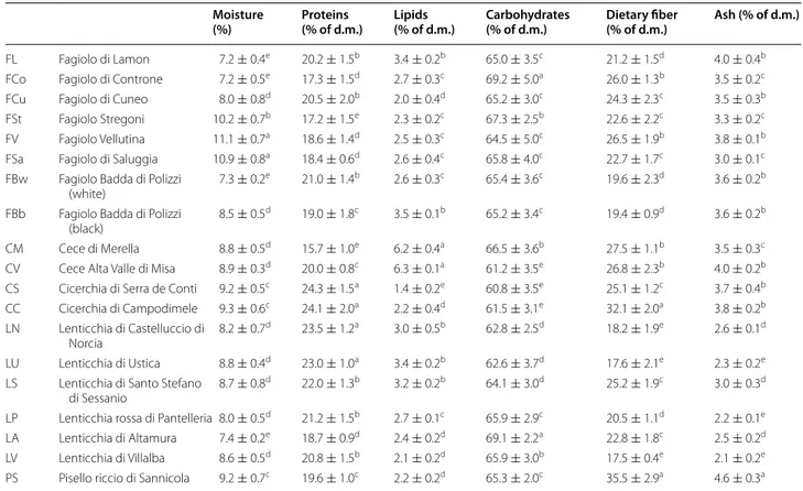
11.1 ± 0.7 %. The protein concentration of all the flours was higher than 15 % of dry matter (d.m.). In particular, grass pea flours (CS and CC) showed the highest values (>24.0 %). The lipid concentration varied from 1.4 ± 0.2 to 3.5 ± 0.1 % of d.m., with the exception of chickpea flours (CM and CV), showing a significantly (P < 0.05) higher concentration. All flours had an amount of car-bohydrates higher than 60 % of d.m. The highest concen-tration was found for FCo and LA flours, produced by milling Fagiolo di Controne kidney beans and Lenticchia di Altamura lentils, respectively. Legume flours had con-centrations of total dietary fiber higher than 17 % of d.m. PS and CC flours had the highest values (35.5 ± 2.9 and 32.1 ± 2.0 %, respectively), whereas the lentils group con-tained the lowest value of total dietary fiber. Ash ranged from 2.1 ± 0.2 to 4.6 ± 0.3 %, being the lowest values for lentil flours.
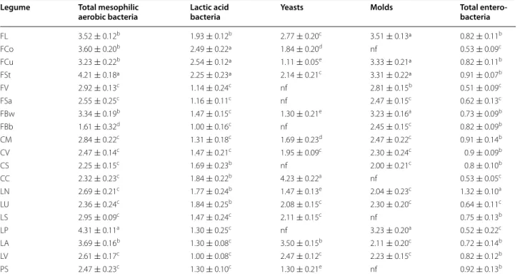
Total mesophilic aerobic bacteria ranged from
1.61 ± 0.32 to 4.31 ± 0.11 log cfu/g (Table 3).
Entero-bacteria were below 2.0 log cfu/g in all the samples. The number of presumptive lactic acid bacteria var-ied from 1.0 ± 0.08 to 2.54 ± 0.12 log cfu/g. Except for grass pea flour CC (4.23 ± 0.22 log cfu/g), yeasts were 1.11 ± 0.05–2.77 ± 0.20 log cfu/g. Yeasts were not found in 10 g of LP, FV, FSa, and FBb flours. Molds were found
at 2.0 ± 0.21–3.51 ± 0.13 log cfu/g in most of the flours and were absent in 10 g of LS, FCo, CC, and PS flours.
Lactic acid bacteria fermentation
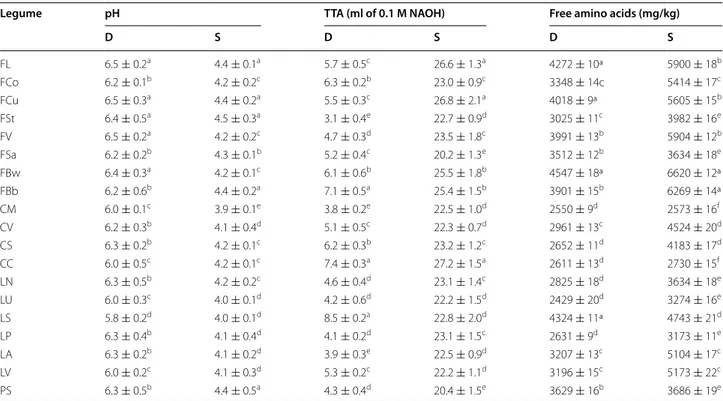
After incubation for 24 h at 30 °C, control doughs (D), without bacterial inoculum, showed values of pH that ranged from 5.8 ± 0.2 to 6.5 ± 0.3, corresponding to val-ues of TTA of 3.8 ± 0.2 to 8.5 ± 0.5 ml 0.1 M NaOH/10 g of dough (Table 4). When L. plantarum C48 and L. bre-vis AM7 were used as starters for sourdough (S) fer-mentation, the cell density of presumptive lactic acid bacteria was 9.8–10.2 log cfu/g. All S had values of pH significantly (P < 0.05) lower (3.9 ± 0.1–4.5 ± 0.3) than the corresponding D. TTA ranged from 20.2 ± 1.3 to 27.2 ± 1.5 ml NaOH/10 g of dough.
The concentration of total free amino acids (FAA)
of D and S is reported in Table 4. Before incubation,
doughs had concentrations of total FAA varying from 2429 ± 20 (D made with LU flour) to 4547 ± 18 mg/kg (D made with FBw flour). In several cases, the concen-tration of total free amino acids (FAA) of S was signifi-cantly (P < 0.05) higher than that of the corresponding D. The average increase was ca. 28 % for kidney bean and pea S. Almost the same average increases (23–26 %) were
found for grass pea, chickpea, and lentil S. Compared to D, slight increases of total FAA were shown by FSa, CM, CC, LS, and PS sourdoughs.
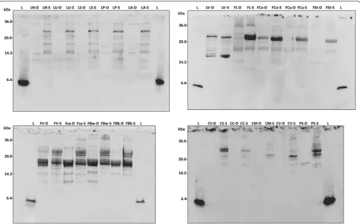
Western blot
The water/salt soluble extracts (WSE) of D and S were analyzed by SDS PAGE, and the proteins were elec-troblotted and detected after incubation with luna-sin polyclonal primary antibody (Fig. 1). A polyclonal primary antibody was already used for identification and quantification of lunasin and its use proposed for optimized methods of detection [14]. Lunasin peptide was not found in any of the samples analyzed. Nev-ertheless, different immunoreactive bands, having molecular masses higher than lunasin, were found. Among D, grass pea and chickpea doughs did not shown any immunoreactive bands, while pea and all the lentil doughs (with the exception of LV) showed very weak signals, mainly distributed below 15 kDa. A large variability was found among the doughs made with bean flours. Very weak bands were found for Fl, FCo, FCu, and Fst, while a large protein band (molec-ular mass of ca. 17 kDa) was found for LV, FV, FSa, FBw, and FBb.
Table 1 List and abbreviations of the Italian legumes
Product certifications and origin are also reported
a IGP (Indicazione Geografica Protetta, Protected Geographical Indication) and DOP (Denominazione d’Origine Protetta, Designation of Protected Origin) are regulated by Reg. (CE) N. 510/2006 (20.03.2006); PAT (Prodotti Agroalimentari Tradizionali, Traditional Food Products) are included in the list of the Italian Ministry of Agriculture, Food and Forestry (D.M. 07/06/2012); SFP (Slow Food Presidia) are listed at http://www.slowfoodfoundation.org
Legume Name Abbreviation Product certificationa Origin
Kidney bean (Phaseolus vulgaris) Fagiolo di Lamon FL IGP Veneto
Fagiolo di Controne FCo DOP Campania
Fagiolo di Cuneo FCu PAT Piedmont
Fagiolo Stregoni FSt PAT Piedmont
Fagiolo Vellutina FV PAT Sicily
Fagiolo di Saluggia FSa PAT Piedmont
Fagiolo Badda di Polizzi (white) FBw SFP Sicily
Fagiolo Badda di Polizzi (black) FBb SFP Sicily
Chickpea (Cicer arietinum) Cece di Merella CM PAT Piedmont
Cece dell’Alta Valle del Misa CV SFP Marche
Grass pea (Lathyrus sativus) Cicerchia di Serra de Conti CS SFP Marche
Cicerchia di Campodimele CC PAT Lazio
Lentil (Lens culinaris) Lenticchia di Castelluccio di Norcia LN IGP Umbria
Lenticchia di Ustica LU SFP Sicily
Lenticchia di Santo Stefano di Sessanio LS SFP Abruzzo
Lenticchia rossa di Pantelleria LP PAT Sicily
Lenticchia di Altamura LA PAT Apulia
Lenticchia di Villalba LV PAT Sicily
Lactic acid bacteria fermentation induced a large modi-fication of the profiles of immunoreactive protein of all the legume flours. A band of ca. 30 kDa, which was absent in the corresponding D, was found for LN, LU, LS, LP, and LA sourdoughs. The intensity of the 17 kDa band of fermented LV increased compared to D. The same sig-nal became evident in all bean sourdoughs, especially for FL. Two protein bands were detected at ca. 30 kDa in FV, FSa, FBw and FBb. The same was found for CS and PS. Moreover, FV, FSa, FBw and FBb showed reactive bands also having molecular masses of 14–17 kDa.
Effect on proliferation of Caco‑2 cells
Aiming at determining the cytotoxicity effect of the freeze-dried WSE from D and S towards human colon adenocarcinoma cells (Caco-2), five samples (FL, CV, CC, LN, and PS) with different immunoreactive protein profiles, were chosen as representatives of the legume species considered in this study, and were subjected to further characterization. On the basis of the results obtained from the western blot analysis, WSE were par-tially purified by ultra-filtration, collecting the fractions
containing the molecules with molecular mass lower than 30 kDa. In particular, the MTT assay was performed after treatment of Caco-2 cells with 0.1, 1, and 10 mg/ml of proteins for 24, 48, or 72 h.
Overall, all the WSE from D and S allowed a significant (P < 0.05) decrease of the cell proliferation compared to
control (Fig. 2). Compared to the results obtained after
24 h-treatment, the inhibitory effect increased after prolonging the incubation to 48 h, and in many cases, to 72 h. Exceptions were the treatments with 10 mg/ ml of proteins, which did not cause a further significant decrease of proliferation. The cytotoxic effect of WSE from S was in all the cases higher than the corresponding D. Besides the effect of the treatment duration, cytotoxic-ity increased proportionally to the concentration of pro-teins tested.
After 24 h of treatment (Fig. 2a), a weak anti-prolifer-ative effect was found for WSE from D and S at 0.1 mg/ ml of proteins. The strongest effect corresponded to LN: (72.25 ± 2.12 and 69.54 ± 1.88 % of vitality for D and S, respectively. When WSE from S were used at concentra-tion of 1 mg/ml, the vitality of Caco-2 cells decreased by
Table 2 Proximate composition of the Italian legume flours
The data are the means of three independent experiments ± standard deviations (n = 3) d.m. dry matter
a–e Values in the same column with different superscript letters differ significantly (P < 0.05) Moisture
(%) Proteins (% of d.m.) Lipids (% of d.m.) Carbohydrates (% of d.m.) Dietary fiber (% of d.m.) Ash (% of d.m.)
FL Fagiolo di Lamon 7.2 ± 0.4e 20.2 ± 1.5b 3.4 ± 0.2b 65.0 ± 3.5c 21.2 ± 1.5d 4.0 ± 0.4b
FCo Fagiolo di Controne 7.2 ± 0.5e 17.3 ± 1.5d 2.7 ± 0.3c 69.2 ± 5.0a 26.0 ± 1.3b 3.5 ± 0.2c
FCu Fagiolo di Cuneo 8.0 ± 0.8d 20.5 ± 2.0b 2.0 ± 0.4d 65.2 ± 3.0c 24.3 ± 2.3c 3.5 ± 0.3b
FSt Fagiolo Stregoni 10.2 ± 0.7b 17.2 ± 1.5e 2.3 ± 0.2c 67.3 ± 2.5b 22.6 ± 2.2c 3.3 ± 0.2c
FV Fagiolo Vellutina 11.1 ± 0.7a 18.6 ± 1.4d 2.5 ± 0.3c 64.5 ± 5.0c 26.5 ± 1.9b 3.8 ± 0.1b
FSa Fagiolo di Saluggia 10.9 ± 0.8a 18.4 ± 0.6d 2.6 ± 0.4c 65.8 ± 4.0c 22.7 ± 1.7c 3.0 ± 0.1c
FBw Fagiolo Badda di Polizzi
(white) 7.3 ± 0.2
e 21.0 ± 1.4b 2.6 ± 0.3c 65.4 ± 3.6c 19.6 ± 2.3d 3.6 ± 0.2b
FBb Fagiolo Badda di Polizzi
(black) 8.5 ± 0.5
d 19.0 ± 1.8c 3.5 ± 0.1b 65.2 ± 3.4c 19.4 ± 0.9d 3.6 ± 0.2b
CM Cece di Merella 8.8 ± 0.5d 15.7 ± 1.0e 6.2 ± 0.4a 66.5 ± 3.6b 27.5 ± 1.1b 3.5 ± 0.3c
CV Cece Alta Valle di Misa 8.9 ± 0.3d 20.0 ± 0.8c 6.3 ± 0.1a 61.2 ± 3.5e 26.8 ± 2.3b 4.0 ± 0.2b
CS Cicerchia di Serra de Conti 9.2 ± 0.5c 24.3 ± 1.5a 1.4 ± 0.2e 60.8 ± 3.5e 25.1 ± 1.2c 3.7 ± 0.4b
CC Cicerchia di Campodimele 9.3 ± 0.6c 24.1 ± 2.0a 2.2 ± 0.4d 61.5 ± 3.1e 32.1 ± 2.0a 3.8 ± 0.2b
LN Lenticchia di Castelluccio di
Norcia 8.2 ± 0.7
d 23.5 ± 1.2a 3.0 ± 0.5b 62.8 ± 2.5d 18.2 ± 1.9e 2.6 ± 0.1d
LU Lenticchia di Ustica 8.8 ± 0.4d 23.0 ± 1.0a 3.4 ± 0.2b 62.6 ± 3.7d 17.6 ± 2.1e 2.3 ± 0.2e
LS Lenticchia di Santo Stefano
di Sessanio 8.7 ± 0.8
d 22.0 ± 1.3b 3.2 ± 0.2b 64.1 ± 3.0d 25.2 ± 1.9c 3.0 ± 0.3d
LP Lenticchia rossa di Pantelleria 8.0 ± 0.5d 21.2 ± 1.5b 2.7 ± 0.1c 65.9 ± 2.9c 20.5 ± 1.1d 2.2 ± 0.1e
LA Lenticchia di Altamura 7.4 ± 0.2e 18.7 ± 0.9d 2.4 ± 0.2d 69.1 ± 2.2a 22.8 ± 1.8c 2.5 ± 0.2d
LV Lenticchia di Villalba 8.6 ± 0.5d 20.8 ± 1.5b 2.1 ± 0.2d 65.9 ± 3.0b 17.5 ± 0.4e 2.1 ± 0.2e
ca. 10 %. In particular, WSE from CC sourdough caused the highest decrease of vitality (58.47 ± 2.08 %). At the highest concentration assayed, the most remarkable cytotoxicity was found for CC, LN, and PS sourdoughs (30.49 ± 1.13, 36.97 ± 1.01, and 35.13 ± 0.95 %, respec-tively). The same trend was found after 48 h of
treat-ment (Fig. 2b), although lower values of vitality were
observed. The lowest proliferation was found for CC sourdough, when treatments were done with 1 mg/ml (39.59 ± 1.05 %), and for CC, LN, and PS sourdoughs, when 10 mg/ml of proteins (30.67 ± 0.87, 29.32 ± 1.11, and 37.75 ± 1.23 %, respectively) were used.
After 72 h of treatment (Fig. 2c), the vitality of Caco-2
cells was lower than that found after 24 or 48 h for treat-ments performed with 0.1 and 1 mg/ml of proteins, while the use of 10 mg/ml of proteins was less effective than 48 h-treatments, with the only exception of FL sourdough.
Transcriptional regulation of filaggrin (FLG) and involucrin (IVL)
Human colon adenocarcinoma cells (Caco-2) were treated with the partially purified and freeze dried WSE from leg-ume D and S aiming at investigating filaggrin (FLG) and involucrin (IVL) gene expressions through RT-PCR. Treat-ments were lasting 4, 8, and 24 h, After 4 h of exposure
(Fig. 3A), a significant (P < 0.05 %) over-expression of FLG was found only for fermented PS. All the other WSE, both from D and S, did not cause significant (P < 0.05) variations compared to LPS (positive control). After 8 h of exposure, a significant (P < 0.05) up-regulation of the FLG gene was also found for FL dough and FL, CV, and PS sourdoughs (Fig. 3B). The same trend was found for treatment lasting 24 h (Fig. 3C) with FL, CV, CC, and PS sourdoughs.
Regarding IVL gene, none of the extracts, except for PS sourdough, induced an expression after 4 h of treatment
higher than that of LPS after 4 h of treatment (Fig. 4A).
After 8 h of treatment, WSE extracted from S caused significant (P < 0.05) increases of the expression com-pared to the corresponding D, especially FL, CV, and PS (Fig. 4B). A longer treatment with the WSE (24 h) did not favour a further increase of the gene expression. Only PS sourdough caused the same expression of IVL gene under all the conditions assayed (Fig. 4C).
Identification of lunasin‑like polypeptides
Based on the results from the MTT assay on Caco-2 cell, the immunoreactive protein bands of FL, CV, and PS sourdoughs were recovered from Tris-Tricine gels, and subjected to tryptic digestion, and HPLC coupled to nanoESI-MS/MS analysis.
Table 3 Microbiological analyses of the Italian legume flours
Total mesophilic aerobic bacteria were estimated on Plate Count Agar (PCA), lactic acid bacteria on agar MRS; yeasts and molds on Yeast extract Peptone Dextrose Agar (YPD-y and YPD-m, respectively); and total enterobacteria on Violet Red Bile Glucose Agar (VRBGA). Details are reported in “Methods” section
The data are the means of three independent experiments ± standard deviations (n = 3) a–e Values in the same column with different superscript letters differ significantly (P < 0.05)
Legume Total mesophilic
aerobic bacteria Lactic acid bacteria Yeasts Molds Total entero‑bacteria
FL 3.52 ± 0.12b 1.93 ± 0.12b 2.77 ± 0.20c 3.51 ± 0.13ª 0.82 ± 0.11b FCo 3.60 ± 0.20b 2.49 ± 0.22ª 1.84 ± 0.20d nf 0.53 ± 0.09c FCu 3.23 ± 0.22b 2.54 ± 0.12ª 1.11 ± 0.05e 3.33 ± 0.21ª 0.82 ± 0.11b FSt 4.21 ± 0.18ª 2.25 ± 0.23ª 2.14 ± 0.21c 3.31 ± 0.22ª 0.91 ± 0.07b FV 2.92 ± 0.13c 1.14 ± 0.24c nf 2.81 ± 0.15b 0.51 ± 0.09c FSa 2.55 ± 0.25c 1.16 ± 0.11c nf 2.47 ± 0.15c 0.62 ± 0.13c FBw 3.34 ± 0.19b 1.47 ± 0.15c 1.30 ± 0.21e 3.23 ± 0.16a 0.73 ± 0.09b FBb 1.61 ± 0.32d 1.00 ± 0.16c nf 2.45 ± 0.15c 0.82 ± 0.09b CM 2.84 ± 0.22c 1.31 ± 0.18c 1.69 ± 0.23d 2.47 ± 0.22c 0.91 ± 0.14b CV 2.47 ± 0.14c 1.47 ± 0.21c 1.95 ± 0.09c 2.30 ± 0.24c 0.9 ± 0.09b CS 2.25 ± 0.15c 1.69 ± 0.23b nf 2.00 ± 0.21c 0.8 ± 0.10b CC 2.32 ± 0.23c 1.84 ± 0.22b 4.23 ± 0.22a nf 0.53 ± 0.05c LN 2.69 ± 0.21c 1.77 ± 0.24b 1.47 ± 0.13e 2.04 ± 0.23c 1.32 ± 0.10a LU 2.36 ± 0.24c 1.84 ± 0.25b 2.08 ± 0.15c 2.30 ± 0.20c 0.64 ± 0.11c LS 2.95 ± 0.09c 1.47 ± 0.24c 2.11 ± 0.15c nf 0.75 ± 0.13b LP 4.31 ± 0.11a 1.30 ± 0.25c nf 3.23 ± 0.20a 0.52 ± 0.22c LA 3.69 ± 0.16b 1.30 ± 0.08c 3.50 ± 0.15b 2.11 ± 0.20c 0.72 ± 0.14b LV 2.61 ± 0.17c 1.00 ± 0.08c 2.47 ± 0.12c 2.23 ± 0.15c 0.82 ± 0.12b PS 2.47 ± 0.23c 1.30 ± 0.10c 1.30 ± 0.21e nf 0.92 ± 0.13b
Ten different proteins were identified through the
Mascot research on the NCBI database (Table 5). It can
be hypothesized that lunasin-like polypeptides were released from native legume proteins via proteolysis dur-ing sourdough fermentation. This should explain the lack of correspondence between the molecular masses of the proteins identified and the polypeptide bands revealed by western blot analyses. The immunodetected polypep-tide bands from FL sourdough corresponded to the fol-lowing matches: Subtilisin inhibitor 1 (P16064), Legumin A2 (P15838) and Phaseolin (P02853). The protein bands from CV sourdough matched with: leucoagglutinating phytohemagglutinin (P05087), phatogenesis related pro-tein (CAA56142) and seed linoleate 9S-lipoxygenase-3 (P09918). The proteins from PS sourdough were identified as: Provicilin (P02855), seed linoleate 9S-lipoxygenase-2 (P14856), seed biotin-containing protein SBP65 (Q41060) and Albumin-1 C (P62928). All the database matches cor-responded to proteins previously identified from legumes. For five proteins, the exact correspondence between the species analyzed and the database matches was not found, especially for CV. Probably, this was due to the limited number of legume protein sequences previously identified and included in the NCBI database.
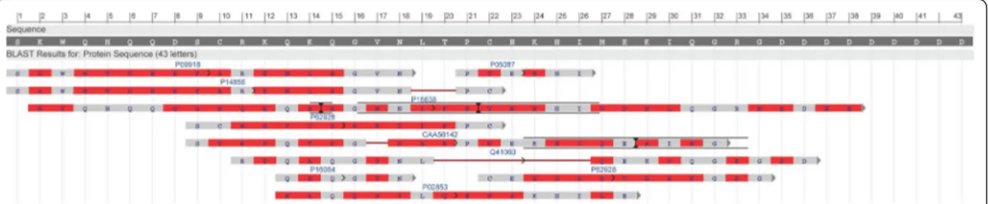
The protein sequences were compared to soy lunasin sequence (deposited at the National Center for Biotech-nology Information, NCBI database with the accession number AAP62458) using BLAST. Except for Provici-lin identified that was identified in PS sourdough, all the sequences might be aligned with different lunasin epitopes (Fig. 5). The longest alignments were found for Legumin A2, corresponding to fragments (f) 24–38 and 33–41 of the soy lunasin sequence, and Phaseolin (f13– 28) from FL sourdough; pathogenesis related protein and seed-linoleate 9S lipoxygenate-3 (f9–32 and f1–18, respectively) from CV, and seed-linoleate 9S lipoxygen-ate-2 and seed biotin-containing protein SBP65 (f1–22 and f11–36, respectively) from PS. The alignments showed identities from 26 to 67 % (pathogenesis related protein and subtilisin inhibitor1/leucoagglutinating phy-toemagglutinin, respectively) and positives from 35 to 93 % (Albumin-1C and Legumin A2, respectively).
Peptidase activities
The peptidase activities of the two lactic acid bacte-ria strains used as starters were assayed using relatively specific synthetic substrates. Significant (P < 0.05) dif-ferences were found between strains. Compared to that
Table 4 Biochemical characteristics of doughs
Control doughs (D), without bacterial inoculation, and sourdoughs (S), started with selected lactic acid bacteria, made with the Italian legume flours were incubated at 30 °C for 24 h
The data are the means of three independent experiments ± standard deviations (n = 3) a–f Values in the same column with different superscript letters differ significantly (P < 0.05)
Legume pH TTA (ml of 0.1 M NAOH) Free amino acids (mg/kg)
D S D S D S FL 6.5 ± 0.2a 4.4 ± 0.1a 5.7 ± 0.5c 26.6 ± 1.3a 4272 ± 10ª 5900 ± 18b FCo 6.2 ± 0.1b 4.2 ± 0.2c 6.3 ± 0.2b 23.0 ± 0.9c 3348 ± 14c 5414 ± 17c FCu 6.5 ± 0.3a 4.4 ± 0.2a 5.5 ± 0.3c 26.8 ± 2.1a 4018 ± 9ª 5605 ± 15b FSt 6.4 ± 0.5a 4.5 ± 0.3a 3.1 ± 0.4e 22.7 ± 0.9d 3025 ± 11c 3982 ± 16e FV 6.5 ± 0.2a 4.2 ± 0.2c 4.7 ± 0.3d 23.5 ± 1.8c 3991 ± 13b 5904 ± 12b FSa 6.2 ± 0.2b 4.3 ± 0.1b 5.2 ± 0.4c 20.2 ± 1.3e 3512 ± 12b 3634 ± 18e FBw 6.4 ± 0.3a 4.2 ± 0.1c 6.1 ± 0.6b 25.5 ± 1.8b 4547 ± 18ª 6620 ± 12ª FBb 6.2 ± 0.6b 4.4 ± 0.2a 7.1 ± 0.5a 25.4 ± 1.5b 3901 ± 15b 6269 ± 14ª CM 6.0 ± 0.1c 3.9 ± 0.1e 3.8 ± 0.2e 22.5 ± 1.0d 2550 ± 9d 2573 ± 16f CV 6.2 ± 0.3b 4.1 ± 0.4d 5.1 ± 0.5c 22.3 ± 0.7d 2961 ± 13c 4524 ± 20d CS 6.3 ± 0.2b 4.2 ± 0.1c 6.2 ± 0.3b 23.2 ± 1.2c 2652 ± 11d 4183 ± 17d CC 6.0 ± 0.5c 4.2 ± 0.1c 7.4 ± 0.3a 27.2 ± 1.5a 2611 ± 13d 2730 ± 15f LN 6.3 ± 0.5b 4.2 ± 0.2c 4.6 ± 0.4d 23.1 ± 1.4c 2825 ± 18d 3634 ± 18e LU 6.0 ± 0.3c 4.0 ± 0.1d 4.2 ± 0.6d 22.2 ± 1.5d 2429 ± 20d 3274 ± 16e LS 5.8 ± 0.2d 4.0 ± 0.1d 8.5 ± 0.2a 22.8 ± 2.0d 4324 ± 11ª 4743 ± 21d LP 6.3 ± 0.4b 4.1 ± 0.4d 4.1 ± 0.2d 23.1 ± 1.5c 2631 ± 9d 3173 ± 11e LA 6.3 ± 0.2b 4.1 ± 0.2d 3.9 ± 0.3e 22.5 ± 0.9d 3207 ± 13c 5104 ± 17c LV 6.0 ± 0.2c 4.1 ± 0.3d 5.3 ± 0.2c 22.2 ± 1.1d 3196 ± 15c 5173 ± 22c PS 6.3 ± 0.5b 4.4 ± 0.5a 4.3 ± 0.4d 20.4 ± 1.5e 3629 ± 16b 3686 ± 19e
(See figure on next page.)
Fig. 2 Effect of lunasin‑like polypeptides on the Caco‑2 cells proliferation. Box-plot showing aggregate data for human colon adenocarcinoma (Caco‑2) cells proliferation after treatments of 24 (panel a), 48 (panel b), and 72 h (panel c) with water/salt soluble extracts obtained from control doughs (D), without bacterial inoculum, and sourdoughs (S), started with selected lactic acid bacteria, made with Italian legume flours. Protein concentration of 0.1, 1.0, and 10 mg/ml were assayed. Data were expressed as the mean percentage of viable cells compared to the control culture, grown in basal media without the addition of the WSE. The centre line of the box represents the median (open square), the top and bottom of the box represent the 75th and 25th percentile of the data, respectively. The top and bottom of the bars represent the 5th and 95th percentile of the data, respectively. Outliers (open circle) and extremes (aterisk) are represented
of L. plantarum C48 (5.87 ± 0.02 and 3.52 ± 0.02 U), PepN activity on Leu-p-NA and endopeptidase activ-ity (PepO) of L. brevis AM7 were higher (6.83 ± 0.08 U and 4.31 ± 0.03). Also PepA activity was higher in L. brevis AM7 (14.02 ± 0.04 U). That of L. plantarum C48 resulted 35 % lower. This latter strain showed higher trip-eptidase (PepT) activity (9.12 ± 0.02 vs. 5.61 ± 0.05 U). No significant differences (P > 0.05) were found between the two strains for PepT (7.20 ± 0.04 and 7.12 ± 0.05 U) and PepX (0.95 ± 0.03 and 1.01 ± 0.02 U) activities. Discussion
Despite the beneficial effects on the human diet, the worldwide consumption of legumes is declining [15] and
below the recommended dose [16]. One potential way to increase the consumption of legumes could be the redis-covery of traditional and local varieties, and, especially, the use of legumes into novel and healthy foods, also exploiting the potential of non-conventional processing [17]. The complementation between cereal and legume flours into new formulas may deserve an interest either to increase the levels of biogenic compounds or to fulfill nutritional deficiencies of cereal-based diets [18].
Traditional Italian legumes, all with product certifica-tions and belonging to Phaseulus vulgaris, Cicer arieti-num, Lathyrus sativus, Lens culinaris and Pisum sativum species, were used in this study. Seeds were milled, and flours were subjected to sourdough fermentation, using Fig. 1 Western blot analysis. Water/salt soluble extracts obtained from control doughs (D), without bacterial inoculum, and sourdoughs (S), started with selected lactic acid bacteria, made with Italian legume flours, were used. A lunasin polyclonal primary antibody was used. Before electroblot‑ ting, the electrophoretic separation was obtained by Tris‑Tricine SDS‑PAGE. Image was performed using the VersaDoc Imaging System (Bio‑Rad). Synthetic lunasin (L) was included in the analysis. The correspondence of the legume flour abbreviations is reported in Table 1
the selected Lactobacillus plantarum C48 and Lactoba-cillus brevis AM7.
Compared to cereals, all legume flours showed elevated amount of protein (mainly grass pea and several lentil flours), FAA (>2 g/kg), dietary fibers, and ash. In particular, grass pea varieties had the highest concentration of pro-teins, while pea, chickpea, and grass pea flours contained the highest levels of dietary fibre. The microbiota of leg-ume flours was poorly represented by lactic acid bacteria and yeasts. All the data agreed with previous findings [19].
Recently, the potential of sourdough fermentation was exploited to enhance the nutritional and functional fea-tures of legume flours [19]. Apart from the legume species and variety, sourdough fermentation with selected start-ers is a suitable biotechnology option either to increase the nutritional and functional value or to decrease the levels of anti-nutritional factors [19]. Selected sourdough lactic acid bacteria were able to decrease the concentra-tion of raffinose up to ca. 64 % and a similar trend was found for the concentration of condensed tannins [19]. The sourdough fermentation also increased the concen-tration of GABA, and promoted antioxidant and phytase activities compared to the raw flours [19].
According to protocols used for cereal sourdough fer-mentation [20], legume flours were started with
Lactoba-cillus brevis AM7 and LactobaLactoba-cillus plantarum C48 [21].
As previously shown [19], the conditions of incubation (24 h at 30 °C) allowed the optimal growth and metabo-lism of selected lactic acid bacteria [19, 22]. As expected, fermentation caused a marked decrease of the values of pH as well as an increase of TTA and of the concentra-tion of peptides and FAA, especially when kidney bean flours were used.
Based on their proteolytic activity towards vegetable proteins [20], the use of sourdough lactic acid bacteria for synthesizing bioactive peptides deserves a marked interest [23]. Bioactive peptides derived from food pro-teins may possess physiological properties beyond the role in nutrition. These properties are influenced by the
protein source, enzyme and processing conditions used [24]. Most of the research related to bioactive peptides and cancer was focused on lunasin [25] and, recently, on pulse hydrolysates [2].
To the best of our knowledge, no literature data dealt with the presence of lunasin or lunasin-like polypeptides from native protein sequences of Italian pulses, and with the effect of sourdough fermentation on bioactivity and bioavailability.
Proteinase activity and a large portfolio of peptidases are the pre-requisites to liberate bioactive peptides from
native oligopeptides [7, 26]. Although with some
differ-ences, the two starters used showed different peptidase activities.
Western blot analyses showed that the sequence of lunasin sequence was absent in all the legume flours, and, therefore, in the corresponding sourdoughs. Nev-ertheless, immunoreactive polypeptides with molecu-lar masses lower than 30 kDa were detectable in all the samples. In some cases, immunoreactive polypeptides appeared as multiple bands in western blot, probably due to the presence of multiple fragments differing for few aminoacid residues [12]. Regarding unfermented doughs, proteins with high intensity signals were found for all the bean, followed by lentil varieties. After lactic acid bacteria fermentation, the number and the intensity of the proteins reacting with the anti-lunasin antibody increased for all the legume flours. This was probably due to an acid activation of endogenous proteinases, respon-sible for primary proteolysis, and peptidase activities by lactic acid bacteria, which completed the hydrolysis (sec-ondary proteolysis). All the reactive protein bands had molecular masses higher than that of lunasin. Although some differences were found among varieties, one repre-sentative was chosen for each legume species to assay the effect on Caco-2 cells proliferation. Caco-2 cells derived from a colonic tumor and have a cancerous phenotype, and can be cultivated to become confluent. In this case, they differentiate into enterocyte-like cells [27].
(See figure on next page.)
Fig. 4 Expression of the involucrin (IVL) gene in Caco‑2 cells. The expression of the IVL gene in human colon adenocarcinoma cells (Caco‑2) was determined using RT‑PCR. Caco‑2 cells were treated at 37 °C for 4 (A), 8 (B) and 24 h (C) with basal medium containing 1.0 mg/ml of the freeze‑ dried water/salt soluble extracts obtained from control doughs (D), without bacterial inoculum, and sourdoughs (S), started with selected lactic acid bacteria, made with FL, CV, CC, LN, and PS legume flours. Data are the mean ± SD of three separate experiments, performed in triplicate. a–c Columns with different superscript letters differ significantly (P < 0.05)
(See figure on previous page.)
Fig. 3 Expression of the filaggrin (FLG) gene in Caco‑2 cells. The expression of the FLG gene in human colon adenocarcinoma cells (Caco‑2) was determined using RT‑PCR. Caco‑2 cells were treated at 37 °C for 4 (A), 8 (B) and 24 h (C) with basal medium containing 1.0 mg/ml of the freeze‑ dried water/salt soluble extracts obtained from control doughs (D), without bacterial inoculum, and sourdoughs (S), started with selected lactic acid bacteria, made with FL, CV, CC, LN, and PS legume flours. Data are the mean ± SD of three separate experiments, performed in triplicate. a–c Columns with different superscript letters differ significantly (P < 0.05)
Table 5 L unasin-lik e p olyp eptides sequenc es WS E Pr ot ein Sequenc e a NCBI ac cession number Theor etical mass (kDa) Sequenc e co ver age M asc ot sco re FL ‑S Subtilisin inhibit or 1 ( Vigna angularis ) QEQGTNPSQEQNVPLPRNYK Q ALE TNTPTK TSWPEL VGV TAEQ AE TKI KEEMVD VQIQ VSPHDSFV TAD YNPKR VRL YVDESNK VTR TPSIG P16064 10.38 82 114 Legumin A2 ( Pisum sativum ) M ATKLLALSLSFCFLLL GGCF ALREQPEQNEC QLERLNALEPD NRIESEGGLIE TWNPNNK QFR CA GV ALSRA TL QHNALRRPY YSNAPQEIFIQQGNGYFGMVFPGCPE TFEEPQESEQGEGR RYRDRHQK VNRFREGDIIA VPT GIVFWMYNDQD TP VIA VSL TDIRSSN NQLDQMPRRFYLA GNHEQEFLR YQHQQGGK QEQENEGNNIF SGFKRDFLED AFNVNRHIVDRL QGRNEDEEK GAIVK VK GGLSIISP PEK Q ARHQR GSR QEEDEDEDEER QPRHQR GSR QEEEEDEDEER QPRHQRRR GEEEEEDKKERR GSQK GKSRR QGDNGLEE TV C TAKLRL NIGPSSSPDIYNPEA GRIK TV TSLDLP VLR WLKLSAEHGSLHKNA MFV PHYNLNANSIIY ALK GRARL Q VVNCNGNT VFDGELEA GRAL TVPQN YA VAAKSLSDRFSY VAFK TNDRA GIARLA GT SSVINNLPLD VV AA TFN LQRNEAR QLKSNNPFKFL VP AR QSENRASA P15838 59.63 62 410 Phaseolin ( Phaseolus vulgaris ) MMRAR VPLLLL GILFLASLSASF AT SLREEEESQDNPFYFNSDN SWNTLFKNQ YGHIR VL QRFDQQSKRL QNLED YRL VEFRSKPE TLLL PQQ AD AELLL VVRSGSAIL VL VKPDDRREYFFL TSDNPIFSDHQKIP A GI FYL VNPDPKEDLRIIQLA MP VNNPQIHEFFLSSTEA QQSYL QEFSKH ILEASFNSKFEEINR VLFEEEGQQEGVIVNIDSEQIKELSKHAKSSSRK SLSK QDNTIGNEFGNL TER TDNSLNVLISSIEMEEGALFV PHY YSK AIVIL VVNEGEAHVEL VGPK GNKE TLEYESYRAELSKD D VFVIP AA YP VAIK AT SNVNFT GFGINANNNNRNLLA GK TDNVISSI GRALDGKD VL GL TFSGSGDE VMKLINK QSGSYFVD AHHHQQEQQ KGRK GAFVY P02853 47.540 75 515 C V‑ S Leucoagglutinating ph yt ohemagglutinin ( Phaseolus vulgaris ) M ASSKFFT VLFL VLL THANSSNDIYFNFQRFNE TNLIL QRD ASV SSS GQLRL TNLNGNGEPR VGSL GRAFY SAPIQIWDNT TGT VASF AT SFT FNIQ VPNNA GP ADGLAF AL VP VGSQPKDK GGFL GLFDGSNSNFHT VA VEFD TL YNKD WDPTERHIGID VNSIRSIK TTR WDFVNGENAE V LIT YDSSTNLL VASL VYPSQK TSFIV SD TVDLKSVLPE W VSV GFSA TT GINK GNVE TND VLSW SF ASKLSDGT TSEGLNLANL VLNKIL P05087 29.54 61 506 Pathogenesis r elat ed pr ot ein ( Cic er arietinum ) MGVFTFEQE TAST VPP AKL YK A MVKD AD VIIPK AVD AIK TVE TVEGNGG PGTIKKL TFVEGGQ TL YVLHKIEAIDEANL GYNY SIV GGA GLSE TVER Y HFEAKL CEGPNGGSIGK VSVKY Q TK GD AKPNEKE VQEGK AK GD A LFK AIEGY VLANPNYN CAA56142 16.93 83 408
Table 5 c on tinued WS E Pr ot ein Sequenc e a NCBI ac cession number Theor etical mass (kDa) Sequenc e co ver age M asc ot sco re Seed linoleat e 9S ‑lipo xy genase ‑3 ( Pisum sativum ) MFSGV TGILNR GHKIK GT VVLMRKNVLDINSL TT VGGVIGQGFDIL G ST VDNL TAFL GRSV SL QLISA TKPD AT GK GKL GK ATFLEGIISSLPTL GA GQSAFKIHFE WDDDMGIPGAFYIKNF MQ TEFFL VSL TLDDIPN HGSIYFV CNSWIYNAKHHKIDRIFF ANQ TYLPSE TP APL VHYREEELNN LR GDGT GERKE WERIYD YD VYNDL GNPDSGENHARP VL GGSE TYPYPRR GR TGRKPTRKDPNSESRSD YVYLPRDEAFGHLKSSDFL TY GLK AV SQNV VP ALESVFFDLNFTPNEFDSFDE VHGL YEGGIKLPT NILSQISPLP VLKEIFR TDGENTLKYPPPK VIQ VSRSGWM TDEEF AREM LA GVNPNVIC CL QEFPPRSKLDSQIY GDHT SKISKEHLEPNLEGL T VEEAIQNKKLFLLDHHDSIMPYLRRINST STK AY ATR TILFLNNNQN LKPLAIELSLPHPQGDEHGA VSY VY QP ALEGVESSIWLLAK AY VIVND SCYHQL VSHWLNTHA VVEPFVIA TNRHLSCLHPIYKLL YPHYRD T MNINSLARLSL VNDGGIIEK TFL W GR YSMEMSSK VYKNW VFTEQ AL PADLIKR GM AIEDPSSPC GVKL VVED YPY AVDGLEIW AIIK TW VQD Y VSL YY TSDEKLR QDSEL Q AW WKEL VE VGHGDKKNEP W WPKMQ TREDLIE VCSIVIW TASALHAA VNFGQ YSY GGLILNRPTLSRRF MPEK GSAEFEEL VKSPQK AYLK TITPKFQ TLIDLSVIEILSRHASDEL YL GERD NPNW TSDKRALEAFKKFGNKLAEIEKKL TQRNNDEKLRNRHGP VEM PY TLL YPSSKEGL TFR GIPNSISI P09918 97.57 43 207 PS ‑S Pr ovicilin ( Pisum sativum ) DNAEIEKILLEEHEKE THHRR GLRDKR QQSQEKNVIVK VSKK QIEELSKNAKSSSKKSV SSRSEPFNLKSSDPIY SNQ YGKFFEITPK KNPQL QDLDIFVNY VEIKEGSL WLPHYNSRAIVIV TVNEGK GD FEL VGQRNENQQGLREEDDEEEEQREEE TKNQ VQSYK AKL TPG D VFVIP A GHP VA VRASSNLNLL GFGINAENNQRNFLA GEEDNVIS QIQK Q VKDL TFPGSA QE VDRLLENQK QSYF ANA QPQQRE TRSQEIKE HL YSIL GAF P02855 31.52 53 589
Table 5 c on tinued WS E Pr ot ein Sequenc e a NCBI ac cession number Theor etical mass (kDa) Sequenc e co ver age M asc ot sco re Seed linoleat e 9S ‑lipo xy genase ‑2 ( Pisum sativum ) MFPNV TGLLNK GHKIR GT VVLMRKNVLDFNTIV SIGGGNVHGVIDSGI NIIGSTLDGL TAFL GRSV SL QLISA TKSD ANGK GK VGKD TFLEGV LASLPTL GA GESAFNIHFE WDHEMGIP121GAFYIKNY MQ VEF FLKSL TLED VPNHGTIRFV CNSW VYNSKL YKSPRIFF ANKSYLPSE T PSPL VKYREEEL Q TLR GDGT GERKLHERIYD YD VYNDL GNPDHGE HLARPIL GGSSTHPYPRR GR TGR YPTRKDPNSEKP ATE TY VPRDEN FGHLKSSDFLA YGIKSV SQC VVP AFESAFDLNFTPNEFDSFQD VRN LFEGGIKLPLD VISTLSPLP VVKEIFR TDGEQ VLKFTPPHVIR VSKSA W M TDEEF AREMLA GVNPCMIR GL QEFPPKSNLDP AEY GDHT SKISVD VLNLDGC TIDEALASGRLFILD YHD TFIPFLRRINE TSAK AY ATR TILFLK ENGTLKP VAIELSLPHPDGDKSGFV SK VILP ADEGVESTIWLLAK AY V VVNDSCYHQLMSHWLNTHA VIEPFVIA TNR QLSV VHPINKLLAPHY RD TMMNINALARDSLINANGLIERSFLPSKY AVEMSSA VYKY W VFT DQ ALPNDLIKRNM AVKDSSSPY GLRLLIED YPY AVDGLEIW TAIK T W VQD YV SL YY ATDNDIKNDSEL QHW WKE VVEK GHGDLKDKP W WP KL Q TFDEL VE VC TIIIW TASALHAA VNFGQ YPY GGLILNRPTLSRRLL PEEGT AEYDEMVKSSQK AYLR TITPKFQ TLIDLSVIEILSRHASDE VYL GQRENPHW TSDSK AL Q AFQKFGNKLAEIEAKL TNKNNDPSL YHR VG PV QLPY TLLHPSSKEGL TFR GIPNSISI P14856 97.07 36 178 Seed biotin ‑containing pr ot ein SBP65 ( Pisum sativum ) M ASEQLSRRENIT TERKIQNAEDSVPQR TTHFELRE THEL GPN FQSLPRNENQ AYLDR GARAPLSANV SESYLDRAR VPLNANIP EHR VREKEDFGGVRDMGKFQMESK GGNKSLAEDRE TLD TRSRM VT GTPHIKEASGK GQ VVEERERARERA MEEEEKRL TMEEISKYRN Q A QQSALEALSAA QEKYERAK Q ATNE TLRNT TQ AA QEK GEAA Q AK D ATFEK TQQGYEM TGD TV SNSAR TASEK AA Q AKNT TL GK TQQGYE ATRD TV SNAAR TAAEY ATP AAEK AR C VA VQ AKD VTLE TGK TAAEK AK CAAEIAAK VA VDLKEK AT VA GW TASHY AT QL TVDGTRAAANA VE GA VGY VAPK ASELAAKSVE TVK GLAASA GE TAKEFT ARKKEESWRE YEAKRASQL QEGEEILPST GGIGK VLPSGER TQ A QGTNL QEK VQGK GSDIL GA VTE TV SDIGSSMIKPIDNANTK VKEHGGT TITPK GQD A G GVLD AIGE TIAEIAHT TK VIV VGEDDE VEKSMQKNIGSDSHSLDRAKH EGYRAPKNNV S Q41060 59.52 46 783 Albumin ‑1 C ( Pisum sativum ) M ASVKLASLIVLF ATL GMFL TKNV GAISCNGV CSPFDIPPC GSPL CR CI PA GL VIGNCRNPY GVFLR TNDEHPNL CESD ADCRKK GSGTFC GHYP NPDIEY GW CF ASKSEAED VFSKITPKDLLKSV ST A P62928 13.90 55 333 Iden tifica tion w as car ried out b y nano -L C–ESI–MS –MS. P olypeptides w er e pur ified fr om w at er/salt -soluble e xtr ac ts obtained fr om sour
doughs (-S) made with F
ag iolo di Lamon (FL), C ec e dell ’A lta Valle del M isa ( CV ), and Pisello r ic cio di S annic ola (PS) a Single -lett er amino acid c ode is used
Polypeptides able to regulate cell proliferation and survival were already liberated by enzyme hydrolysis of plant and animal proteins [28]. Such peptides, inhibiting cell growth or promoting apoptosis, could have protec-tive effects on tumor growth at the digesprotec-tive tract level [29, 30]. In particular, it was found that the antitumoral mechanism of soybean lunasin is related to the capacity of the peptide to bind the deacetylated histones, caus-ing the acetylation inhibition [31, 32]. Girón-Calle et al. [28] assessed the cancer cell proliferation after treatment with chickpea hydrolysates made with pepsin/pancreatin. These hydrolysates inhibited the proliferation of human epithelial colorectal adenocarcinoma cells (Caco-2) and monocytics leukemia cells (THP-1) up to 48 and 78 %, respectively. Hydrolysates from the common bean (P. vulgaris) varieties Negro 8025 and Pinto Durango inhib-ited the inflammation in lipopolysaccharide-induced macrophages through suppression of NF-κB pathways [2]. After hydrolysis with Alcalase, proteins extracted from both the above varieties inhibited various markers of inflammation (cyclooxygenase-2 expression, prosta-glandin E2 production, inducible nitric oxide synthase expression, and nitric oxide production) [2]. Inflam-mation and cancer are linked, chronic inflamInflam-mation predisposes individuals to various types of cancer and inflammatory mediators and cells are involved in the migration, invasion, and metastasis of malignant cells [33]. The suppression of pro-inflammatory pathways may provide opportunities for both prevention and treatment of cancer [34].
In this study, extracts from legume doughs regu-lated the proliferation of Caco-2 cells, including those obtained from control doughs, in which very weak sig-nals of immunoreactive bands were found with the west-ern blot analysis. It can be hypothesized that the effect could be related to the contribution of some legume pro-teins or peptides able to act as anticancer compounds in their native form, causing cytotoxicity and apoptosis of tumoral cells [2, 5]. Nevertheless, a strong inhibition of the Caco-2 cell proliferation was found only after lactic
acid fermentation. A marked inhibition was found when cells were treated for 24 h with the fermented legume extracts at concentrations from 1 to 10 mg/ml. Sour-doughs from FL, CV, and PS flours were the most active, showing a decrease of Caco-2 cells vitality up to 70 %. A correlation between the anti-proliferative effect of pro-tein hydrolysates towards Caco-2 cells and the in vivo growth inhibition of tumors in the digestive tract was demonstrated [29, 30].
The nanoESI-MS/MS spectrometer analysis of the immunoreactive protein bands from water/salt-solu-ble extracts of FL, CV and PS sourdoughs allowed the identification of ten legume proteins. According to the molecular masses of the identified proteins, it can be hypothesized that immunoreactive polypeptides are encrypted into the native sequences and released as frag-ments during lactic acid bacteria fermentation. Nine of them showed similarities to soy lunasin sequence, probably related to their recognition by the anti-lunasin antibody.
Using Caco-2 cells, the capacity to induce the expres-sion of human FLG and IVL genes by sourdough fer-mented legumes was investigated. FLG and IVL are important proteins for the formation of the epidermal
skin barrier [35, 36]. FLG aggregates keratin filaments
and provides a cytoskeleton for the cornified envelope [35]. The expression of FLG was markedly induced by PS sourdough, and under some of the assayed condi-tions (e.g., 8 h of treatment), also by CV and FL sour-doughs. Compared to the unfermented dough, the level of IVL gene expression markedly increased when Caco-2 cells were subjected to 24-h treatment with FL, CV, and PS sourdoughs. In particular, the sourdough made with PS was effective under all the conditions likely it was observed for FLG gene expression. IVL serves as a sub-strate for the covalent attachment of ceramides to the cornified envelope [35, 36]. An improvement of the IVL activity may result in an increase of the ceramide binding activity, which, in turn, positively affects the barrier func-tion [37, 38].
Fig. 5 Alignments with lunasin. Alignments of the lunasin‑like polypeptides identified from FL, CV, and PS sourdoughs with the soy lunasin sequence, as obtained through the BLAST alignment on‑line tool (http://blast.ncbi.nlm.nih.gov/)
Conclusions
Nowadays, an unexpected and considerable number of small proteins and peptides are available as drugs [39]. They show high bioactivity, target specificity and wide spectrum of therapeutic actions, with low levels of tox-icity, structural diversity, and absence or low levels of accumulation in body tissues. However, the manufactur-ing protocols (e.g. chemical synthesis and recombinant transgenic approach) are very expensive, representing a hindrance for the use as therapeutic peptides [40]. The alternative option is the exploitation of the potential of bioactive peptides derived from food proteins. Legume flours were already proposed as adjuvant ingredients into a range of baked products and snacks for increasing the nutritional value [41–44], as well as sourdough
fermen-tation was shown to have a functional potential [19, 45,
46]. Despite the need to have an in vivo confirmation, the presence of lunasin-like polypeptides allows to hypoth-esize the use of sourdough fermented legumes in phar-maceuticals preparations (e.g., capsules and powders), protein hydrolysates or as purified peptide mixtures to be incorporated as health-enhancing ingredients in novel functional foods (e.g., leavened baked goods).
Methods
Legumes
Nineteen Italian legume varieties, belonging to the spe-cies Phaseulus vulgaris (Fagiolo di Lamon, Fagiolo di Controne, Fagiolo di Cuneo, Fagiolo Stregoni, Fagiolo Vellutina, Fagiolo di Saluggia, Fagiolo Badda di Polizzi— white, and Fagiolo Badda di Polizzi—black), Cicer ari-etinum (Cece di Merella and Cece dell’Alta Valle del Misa), Lathyrus sativus (Cicerchia di Serra de Conti and Cicerchia di Campodimele), Lens culinaris (Lenticchia di Castelluccio di Norcia, Lenticchia di Ustica, Lentic-chia di Santo Stefano di Sessanio, LenticLentic-chia rossa di Pantelleria, Lenticchia di Altamura and Lenticchia di Villalba), and Pisum sativum (Pisello riccio di Sanni-cola), were used in this study. All legumes were chosen among the Italian pulses that have specific product cer-tification (Table 1). Flours were obtained from whole leg-ume seeds through the laboratory mill Ika-Werke M20 (GMBH, and Co. KG, Staufen, Germany). Protein (total nitrogen ×5.7), ash and moisture contents were deter-mined according to AACC approved methods 46-11A, 08-01 and 44-15A, respectively [47]. Lipids were deter-mined by Soxhlet method. Total carbohydrates were calculated as the difference, using the following formula: [100 − (proteins + lipids + ash + moisture)]. Proteins, lipids, carbohydrates and ash were expressed as % of dry matter (d.m.). The determination of total dietary fiber was carried out by the enzymatic–gravimetric procedure
approved by the Association of Official Analytical Chem-ists [48], as described by Lee et al. [49].
Microbiological analyses of the flours
Ten grams of flour were homogenized with 90 ml of ster-ile peptone water [1 % (wt/vol) of peptone and 0.9 % (wt/ vol) of NaCl] solution. Lactic acid bacteria were enumer-ated using MRS (Oxoid, Basingstoke, Hampshire, UK) agar medium, supplemented with cycloheximide (0.1 g/l). Plates were incubated, under anaerobiosis (AnaeroGen and AnaeroJar, Oxoid), at 30 °C for 48 h. Cell density of yeasts and molds were estimated on Yeast Extract Pep-tone Dextrose Agar (YPD) (Oxoid) medium (pour and spread plate enumeration, respectively), supplemented with chloramphenicol (0.15 g/l), at 30 °C for 72 h. The attribution (yeasts/molds) was confirmed by microscope observation. Total mesophilic aerobic bacteria were determined on Plate Count Agar (PCA, Oxoid) at 30 °C for 48 h, and total enterobacteria were determined on Violet Red Bile Glucose Agar (VRBGA, Oxoid) at 37 °C for 24 h.
Fermentation
Sourdough lactic acid bacteria, belonging to the Culture Collection of the Department of Soil, Plant, and Food Sciences (University of Bari, IT) and previously selected for some biotechnological features, were used. Lactoba-cillus plantarum C48 showed the capacity of synthesiz-ing relevant amount of γ-aminobutyric acid (GABA) [21], and Lactobacillus brevis AM7 was characterized by high proteolytic activities towards cereal and legume flours [12]. Starters were cultivated separately on MRS broth at 30 °C for 24 h. Cells were harvested by centrifuga-tion (10,000×g, 10 min, 4 °C) until the late exponential phase of growth was reached (ca. 10 h), washed twice in 50 mM sterile potassium phosphate buffer (pH 7.0) and re-suspended in tap water at the cell density of ca. 8.0 log cfu/ml. Each legume flour was mixed with tap water, containing the bacterial suspension (initial cell density of 7.0 log cfu/g of sourdough for each strain). Doughs, hav-ing dough yield (DY, dough weight × 100/flour weight) of 160 (corresponding to 62.5 and 37.5 % of flour and water, respectively), were mixed at 60 x g for 5 min with a IM 58 high-speed mixer (Mecnosud, Flumeri, Italy) and incubated at 30 °C for 24 h. The most common DY value
for cereal-based sourdoughs (160) was applied [20, 22,
46, 50]. After fermentation, legume sourdoughs (S) were compared to control doughs (D), without bacterial inoc-ulum, prepared as described above (DY 160) and incu-bated under the same conditions. S and D were stored at −20 °C before the chemical analyses, while microbio-logical analysis was carried out before freezing. All the
doughs were obtained in triplicate and each of them was analyzed twice.
Characterization of fermented flours
The values of pH were determined on-line by a pHme-ter (Model 507, Crison, Milan, Italy) with a food penetra-tion probe. Total titratable acidity (TTA) was determined after homogenization of 10 g of dough with 90 ml of dis-tilled water, and expressed as the amount (ml) of 0.1 M NaOH needed to reach the value of pH of 8.3.
The water/salt-soluble extract (WSE) of D and S was prepared according to Weiss et al. [51], and used to ana-lyze free amino acids (FAA) by a Biochrom 30 series Amino Acid Analyzer (Biochrom Ltd., Cambridge Sci-ence Park, UK) with a Na-cation-exchange column (20 by 0.46 cm internal diameter), as described by Rizzello et al. [50].
Peptidase activities of the starters
General aminopeptidase type N (EC 3.4.11.11; PepN), specific aminopeptidase type A (EC 3.4.11.7; PepA) and endopeptidase (EC 3.4.23; PepO) activities were determined as described by Gobbetti et al. [13], using, respectively, Leu-p-nitroanilides (p-NA), Glu-p-NA and NCBZ-Gly-Gly-Leu-p-NA (Sigma Aldrich Co.) as rela-tively specific substrates. The assay mixture contained 900 μl of 2.0 mM substrate in 0.05 M potassium phos-phate buffer, pH 7.0, and 100 μl of cell suspension (9 log cfu/ml). The mixture was incubated at 30 °C for 1 h and the absorbance was measured at 410 nm. The data obtained were compared to standard curves set up by using p-nitroanilide [52]. One unit (U) of activity was defined as the amount of enzyme required to liberate 1 μmol/min of p-nitroanilide under the assay conditions. Tripeptidase (EC3.4.11.4; PepT), and X-prolyl dipeptidyl aminopeptidase (EC 3.4.14.5; PepX) activities were deter-mined using Leu-Leu-Leu and Gly-Pro-Ala substrates (Sigma Aldrich Co.), respectively. Activities on tripep-tides were determined by the Cd-ninhydrin method [52], [53]. The assay conditions were the same as those described for p-nitroanilide substrates. One unit (U) of activity was defined as the amount of enzyme required to liberate 1 μmol amino acid released per min under the assay conditions. The data obtained were compared to the standard curve set up by using leucine [52].
Western blot analysis
Protein concentration of samples was determined by the bicinchoninic acid method (Thermo Scientific, Rockford, IL, USA), using bovine serum albumin (BSA) as stand-ard protein. SDS-PAGE was performed with samples and synthetic lunasin diluted in tricine sample buffer (Bio-Rad, Richmond, CA, USA), containing 2 % (v/v)
β-mercaptoethanol, and heated at 100 °C for 5 min. Equal amounts of proteins (20–40 µg) were analyzed on Precast Criterion 16.5 % Tris-Tricine gels (Bio-Rad), and elec-trophoretic separations were carried out at 100 V, using Tris-Tricine-SDS as running buffer in the Criterion cell (Bio-Rad). For the attribution of the molecular masses, a Precision Plus Protein Standards mix (BioRad) was used. After SDS-PAGE separation, gels were soaked in transfer buffer (48 mM Tris, 39 mM glycine, 20 % methanol, pH 9.2) for 30 min. Proteins were electroblotted into nitro-cellulose membranes by semidry transfer in a Trans-Blot SD (Bio-Rad) for 30 min at 18 V. Then, the membranes were blocked for 3 h in Tris buffered saline with 0.05 % (v/v) Tween 20 (TBST), containing 1 % (w/v) bovine serum albumin, (TBST-1 % BSA). Afterwards, the mem-brane was washed three times with TBST, and incubated overnight at 4 °C with lunasin polyclonal primary anti-body (diluted 1:2000 in TBST-0.1 % BSA). After washing with TBST, the membrane was incubated overnight at 4 °C with horseradish peroxidase-conjugated mouse anti-rabbit secondary antibody (Santa Cruz Biotechnology, Dallas, TX, USA; 1:3000 in TBST-0.1 % BSA). Finally, the membranes were washed six times with TBST, and visu-alized by chemioluminescence using the detection agent Amersham TM ECL Prime (GE Healthcare, Chalfont St Giles, UK), according to the manufacturer´s recom-mendations. Image acquisition (exposure time 1–4 min) was performed using the VersaDoc Imaging System (Bio-Rad).
MTT assay in Caco‑2 cells
In order to assess the effect of legume D and S on cell pro-liferation, the viability of colon adenocarcinoma Caco-2 cells was measured using the 3-(4,5-dimethyl-2-yl)-2,5-di-phenyltetrazolium bromide (MTT) method [54].
Colon adenocarcinoma Caco-2 cells (ICLC HTL97023), provided by the National Institute for Cancer Research of Genoa (Italy), were cultured in RPMI medium, sup-plemented with 10 % Fetal Bovine Serum (FBS), 1 % 2 mM l-glutamine, 1 % penicillin (10,000 U/ml)/strep-tomycin (10,000 μg/ml) mixture, 0.1 % gentamicin and
0.1 % β-mercaptoethanol, and maintained in 25 cm2
culture flasks at 37 °C, 5 % CO2. Every 2 days, confluent
cultures were washed with PBS 1× (without Ca2+ and
Mg2+), split 1:3–1:6 using Trypsin/EDTA, and seeded at
2–5 × 10⁴ cell/cm2, 37 °C, and 5 % CO
2. For the assay,
Caco-2 cells were harvested at 80 % confluence with
trypsin/EDTA, seeded at the density of 5 × 10⁴ cells/ml
into 96-well plates, and then incubated at 37 °C, 5 % CO2
for 24 h. Before analysis, WSE obtained from legume D and S were partially purified by ultra-filtration, using Vivaspin (GE Healthcare) centrifugal filter units (cut-off 30 kDa), following the manufacturer’s instructions.
Cells were exposed to partially purified and freeze dried WSE at the following concentrations: 0.1, 1.0, and 10.0 mg/ml of proteins. Each WSE was tested in dupli-cate. A control in basal medium, without addition of WSE, was used. Incubation was carried out for 24, 48 and 72 h. After incubation, the medium was removed and replaced by 100 µl/well of MTT solution. Then, plates
were incubated in the dark for 3 h (37 °C, 5 % CO2). MTT
salt was dissolved in PBS (5 mg/ml) and added (1:10) to RPMI, containing 10 % FBS, 1 % 2 mM l-glutamine, 1 % penicillin (10,000 U/ml)/streptomycin (10,000 μg/ml) mixture, 0.1 % gentamicin and 0.1 % β-mercaptoethanol. Then, the medium was removed and 100 µl/well of dime-thyl sulphoxide (DMSO) were added to dissolve the pur-ple formazan product. Plates were shacked for 15 min at room temperature and the absorbance was read at a wavelength of 570 nm, with a Biotek microplate reader (BioTek Instruments Inc., Bad Friedrichshall, Germany), and elaborated with the ELX808 software (BioTek Instru-ments Inc., Bad Friedrichshall, Germany). Data were expressed as the mean percentage of viable cells com-pared to control culture, grown in basal media without addition of WSE.
Identification of the lunasin‑like polypeptides
After Western blot analysis, the immunoreactive bands, separated by Tris-Tricine SDS PAGE, were cut and stored in 20 % ethanol before identification. Protein identifica-tion was performed by Proteome Factory (Proteome Factory AG, Berlin, Germany). Protein bands were in-gel digested by trypsin (Promega, Mannheim, Germany) and analyzed by Nano-Liquid Cromatography-Electro-spray Ionisation-Mass Spectrometry (nanoLC-ESI–MS/ MS). The LC–MS system consisted of an Agilent 1100 nanoHPLC system (Agilent, Waldbronn, Germany), PicoTip electrospray emitter (New Objective, Woburn, MA, USA) and an Orbitrap XL or LTQFT Ultra mass spectrometer (ThermoFisher Scientific, Bremen, Ger-many). Peptides were first trapped and desalted on the enrichment column (Zorbax 300SB-C18, 0.3 × 5 mm, Agilent) for 5 min (solvent: 2.5 % acetonitrile/0.5 % for-mic acid), then separated on a Zorbax 300SB-C18, 75 μm × 150 mm column (Agilent), using a linear gradient from 10 to 32 % B (solvent A: 5 % acetonitrile in water, solvent B: acetonitrile, both with 0.1 % formic acid). Ions of inter-est were data-dependently subjected to MS/MS, accord-ing to the expected charge state distribution of peptide ions. Proteins were identified by database search against the plant sequences of the National Center for Biotech-nology Information, (NCBInr, Bethesda, USA) protein database, using MS/MS ion search of the Mascot search engine (Matrix Science, London, UK). Only peptide matches with a score of 20 or above were accepted. The
sequences of the protein identified were aligned using the BLAST on-line tools (http://blast.ncbi.nlm.nih.gov).
Transcriptional regulation of filaggrin (FLG) and involucrin (IVL) genes
Colon adenocarcinoma Caco-2 cells (ICLC HTL97023) were cultured in RPMI medium supplemented with 10 % FBS, 1 % 2 mM l-glutamine, 1 % penicillin (10,000 U/ ml)/streptomycin (10,000 μg/ml) mixture, 0.1 % gen-tamicin and 0.1 % β-mercaptoethanol and maintained in
25 cm2 culture flasks at 37 °C, 5 % CO
2. Caco-2 cells were
incubated in 25-cm2 culture flasks at 37 °C, under 5 %
CO2 atmosphere [55].
Subconfluent monolayers of Caco-2 cells were sub-jected to treatment with basal medium, containing 2.5 % FBS. The freeze-dried WSE, partially purified by ultra-filtration using Vivaspin (GE Healthcare) centrifu-gal filter units (cut-off 30 kDa), were dissolved in RPMI medium, at the concentration of 10 mg/ml and added to the culture media at the final concentration of 1 mg/ ml. The control was the basal medium, containing 2.5 % FBS. Plates were incubated at 37 °C for 24 h, under 5 %
CO2. Samples were taken after 4, 8, and 24 h of
treat-ment. Each experiment was carried out at least twice in triplicate. For quantitative real-time PCR (RT-PCR), total RNA was extracted from Caco-2 cells using the Ribos-pin Minikit-GeneAll kit. cDNA was synthesized from 2 µg RNA template in a 20-µl reaction volume, using the Prime Script RT reagent kit (perfect Real time) (Takara). Total RNA solution (10 µl) was added to the Master Mix and subjected to reverse transcription in a thermal cycler (Stratagene Mx3000P Real-time PCR System, Agilent Technologies Italia Spa, Milan, Italy). The conditions were 37 °C for 15 min, 85 °C for 5 s, holding the samples at 25 °C.
The cDNA was amplified and detected using the same instrument and the TaqMan assay (Applied Biosystems). The following Taqman gene expression assays were used: FLG Hs00863478_g1 (FLG), IVL Hs00846307_s1 (IVL), and GAPDH Hs99999905_m1 (human glyceraldehyde-3-phosphate dehydrogenase, GAPDH). Human GAPDH was used as the housekeeping gene. PCR amplifications were carried out using 40 ng cDNA in a total volume of 20 µl. The reaction mixture contained 10 μL of Premix Ex Taq, 0.4 μl of RoxTM reference dye II, 1 μL of 20X TaqMan Gene Expression assay and 4 µl of cDNA.
PCR conditions were 95 °C for 30 s (for AmpliTaq acti-vation), followed by 40 amplification cycles (95 °C for 5 s, 60 °C for 20 s). Analyses were carried out in triplicate. Based on preliminary results, the expression of FLG and IVL genes was also assayed by treating Caco-2 cells with lipopolysaccharide (LPS; Sigma Aldrich Co.) at 10 µg/ml in basal medium, containing 2.5 % FBS. Analyses were