UNIVERSITA’ DEGLI STUDI DEL PIEMONTE ORIENTALE
DiSIT-Dipartimento di Scienze e Innovazione Tecnologica
Dottorato di ricerca in Scienze Ambientali (Acque interne e agroecosistemi)
XXVIII Ciclo
Modulation of Ca
2+
signaling by
polyphenols in malignant
mesothelioma cells
Tutor: Ph.D. Student:
Prof. Bruno Burlando Dott.ssa Stefania Ribulla
Prof. Mauro Patrone
Coordinator:
1
Table of contentents
Abbreviations and Acronyms... 3
Introduction………. 6
Disease definitions……… 6
Historical outline……….. 7
Disease etiology……… 8
Asbestos……… 8
SV40………. 10
Genetics……… 10
Radiation……….. 10
Epidemiology………... 12
MPM in Italy……… 14
Molecular pathogenesis of MPM………. 17
Clinical features and diagnosis………. 22
Currently established therapy………... 24
Surgery………. 25
Chemotherapy……….. 26
Radiotherapy……… 26
Multimodal approaches……….... 27
Novel therapies………. 28
2
Therapy targeting cell-surface receptors……… 28
Immune checkpoint inhibitors……… 29
Gene therapy……….. 29
Immunotherapy and vaccines………. 30
Direct physical cytotoxic therapies……… 30
Cancer chemoprevention using natural products………... 32
Natural products as treatment in MPM……….. 34
Aims……….. 35
References……… 38
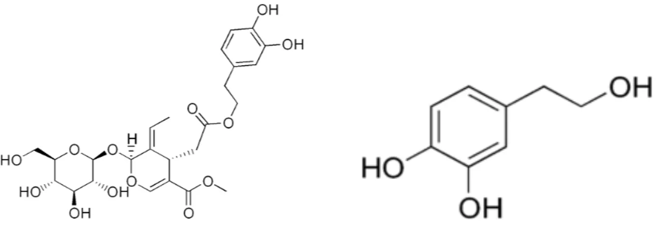
Oleuropein-enriched olive leaf extract affects calcium
dynamics and impairs viability of malignant mesothelioma cells... 50
Resveratrol induces intracellular Ca
2+rise via
T-type Ca
2+channels in a mesothelioma cell line……….. 60
Conclusions………... 85
References………. 87
3
Abbreviations and Acronyms
AA: Ascorbic Acid
Akt: also known as Protein Kinase B (PKB) AND therapy: Active Nutrients/Drug therapy BCA: Bicinchoninic Acid
[Ca2+]i: Cytosolic Ca2+ concentration
CAD: Collision activated dissociation gas Calcein-AM: Calcein-acetoxymethylester CT: Computerized tomography
CTLA-4: Cytotoxic T lymphocyte antigen-4 DM-4: Drug maytansinoids
DMEM: Dulbecco's Modified Eagle's Medium DMSO: DiMethyl SulfOxide
DNA: deoxyribonucleic acid
EDTA: EthyleneDiamineTetraacetic Acid EGCG: EpiGalloCathechin-3-Gallate EGF: Epidermal Growth Factor
EGFR: Epidermal Growth Factor Receptor EPP: Pneumonectomy
ERK: Extracellular signal–regulated kinase FBS: Foetal Bovine Serum
FDG: fludeoxyglucose F 18
Fura-2AM: Fura-2-acetoxymethyl ester H&E stain: Hematoxylin & eosin stain HGF: Hepatocyte growth factor
4 HMGB1: High-mobility group box 1
HT: Hydroxythyrosol
IARC: International Agency for Research on Cancer IC50: half maximal Inhibitory Concentration
ICD: International Classification of Disease IGF: Insulin-like growth factor
IL-4-6-8: Interleukin 4-6-8 INF: Interferon
IPV: inactivated poliovirus vaccine
LC/MS: Liquid chromatography–mass spectrometry
MAP kinase or MAPK: Mitogen-Activated Protein Kinase MM: Malignant Mesothelioma
MPM: Malignant Pleural Mesothelioma MPF: megakaryocyte potentiating factor MRI: Magnetic resonance imaging
MTT: 3-(4,5-Dimethylthiazol-2-yl)-2,5-Diphenyltetrazolium Bromide NF-kβ: Nuclear Factor k beta
OL: Oleuropein
PBS: Phosphate Buffered Saline P/D: pleurectomy/decortication
PDGF: Platelet Derived Growth Factor PD-L1: Programmed death ligand 1 PDT: Photodynamic Therapy PET: Positron emission tomography PMSF: Phenyl-methylsulfonyl fluoride Rb: retinoblastoma
5 Res: Resveratrol
ROS: Reactive Oxygen Species siRNA: Small interfering RNA
SMRP: Soluble mesothelin related protein SV-40: Simian Virus 40
TGF-β: Transforming growth factor beta TIS: Turbo ion spray
TLC: Thin Layer Chromatography TNF-α: Tumor Necrosis Factor-α TNM: Tumor node metastasis TP53: Tumor protein p53
VATS: Video assisted thoracoscopic surgery VEGF: Vascular Endothelial Growth Factor WHO: World Health Organization
6
Introduction
DISEASE DEFINITIONS
Malignant Mesotheliomas (MM) are, by definition, tumors that arise from mesothelial cells of any body cavity: pleura, peritoneum, pericardial sac, and tunica vaginalis testis. Malignant pleural mesothelioma (MPM) is the most common of these, accounting for approximately 90% of disease, and pathologically the best defined one (1). (Fig.1).
Figure 1. Normal and diseased pleura
The risk of MPM is attributable to asbestos exposure, and has been reported to range between 86% and 95% in most recent epidemiological studies (2-4).
The estimated rate of non-asbestos-related MPM varies widely among studies and is allegedly influenced by the different methods of assessing exposure (5).
7
HISTORICAL OUTLINE
There is clear evidence of asbestos use among various cultures since ancient times (6). The word asbestos is derived from a Greek term meaning inextinguishable or unquenchable, a reference to its fire-resistant properties (1).
Heat resistant and insulating properties made asbestos a valuable commodity, particularly as the industrial revolution began. In the USA, mining and subsequent use of asbestos increased steadily during the first half of the 20th century, escalated rapidly following World War II, and
peaked in 1973, after which it precipitously declined.
Asbestos was widely used for the insulation of water and combustion pipes, as material for house construction and shipbuilding, as car brakes and gaskets, or in the manufacture of toys, jewellery, and cigarette filters. At the peak of asbestos use in industrial activities, about 3000 asbestos-containing products were registered (7).
The first report of a pleural tumor occurred in 1767 by Joseph Lieutand, the founder of pathologic anatomy in France, who in a study of 3000 autopsies found two cases of “pleural tumors”. Thereafter, mesothelioma was characterised as a specific disease by Klemperer and Rabin in 1931 (8). It took almost a further 30 years for mesothelioma to become widely accepted as a separate cancer entity. The definitive epidemiological study linking mesothelioma to asbestos came from South Africa, and was published in 1960 by J.C. Wagner, C.A. Sleggs and P. Marchand. These authors showed that mesothelioma was very prevalent in people living or working in crocidolite asbestos mine areas (9). Subsequently, several studies from the USA, Europe, Australia and Japan verified asbestos inhalation as the etiological cause of mesothelioma (10-12).
8
DISEASE ETIOLOGY
Asbestos
The association of mesothelioma with asbestos exposure is well established, with an etiological fraction above 80% (13).
This tumor was once rare, but its incidence has been increasing in several countries because of widespread exposure to asbestos in the past, and it is predicted that it will increase in the next decades, especially in developing countries where asbestos has not yet been banned for use (14).
Asbestos refers to a group of crystalline-hydrated silicate minerals and is classified into two main families, the serpentines and the amphiboles (Tab. 1). The serpentines consist of chrysotile, consisting of short, curly fibres. This mineral is called “white asbestos”, due to its color, and accounts for 95% of marketed asbestos (15). The amphiboles, with straight, longer fibres, include crocidolite or “blue asbestos”, amosite, tremolite, actinolite and anthophyllite (16) (Fig. 2).
Mixtures of chrysotile and amphiboles were used to produce an array of roofing, insulation and fire-proofing materials.
Evidence exists that all types of asbestos fibres induce pulmonary toxicity in a dose-dependent manner. Moreover, all fibre types possess carcinogenic potential; however, exposure to amphibole fibres, rather than to chrysotile, is more likely to cause mesothelioma (17). The association between amphibole asbestos exposure and MPM development is well known. In particular, crocidolite is considered the most carcinogenic type of asbestos. Erionite, an asbestos-like mineral, also causes MPM.
Evidence suggesting a link between asbestos exposure and MPM insurgence has been deemed sufficient by the World Health Organisation (WHO) to conclude that all types of asbestos cause cancer in humans (18).
The median latency between asbestos exposure and disease onset is 44.6 years, and it tends to increase over time in a linear fashion. Therefore, despite asbestos banning, incidence rates in Europe are still rising, with a peak expected around 2020 or beyond (19).
There is some evidence that disease latency has an inverse relationship with the duration or degree of asbestos exposure (20).
Although short or low-level asbestos exposures have been linked to the development of mesothelioma, the risk of disease demonstrates dose dependence (21). However, it has been shown that other etiologic factors have a role in MPM pathogenesis.
9
Table 1. Composition and characteristics of asbestos fibers. Chrysotile is a hydrated magnesium silicate. The amphiboles are a group of hydrated silicates with a wide range of cation substitutions within the silicate backbone of the crystal structure (22).
Figure 2. Scanning electron micrographs contrast curved fibers of chrysotile asbestos (left) with straight fibers of
crocidolite (center) and amosite (right; bar=3 mm). Typically, chrysotile fibers exhibit a curved, curly, or wavy morphology, which is most apparent in fiber bundles exceeding 10 mm in length. In addition, the ends of chrysotile fiber bundles often exhibit a splayed appearance because of the separation of individual fibrillar units. This curly morphology influences the interceptive deposition of chrysotile fibers, which in turn, affects the depth of penetration into the lower respiratory tract. The diameters of individual fibers vary considerably with substantial overlap among members of the amphibole group. However, crocidolite generally has the finest fiber diameters (22).
2
Shinya Toyokuni
deaths predicted from this neoplasm.8) This article briefly reviews the history, epidemiology, and genomic analysis of human DMM, and discusses the possible molecular mechanisms underlying asbestos-induced carcinogenesis.
Asbestos fibers
Asbestos is a naturally occurring mineral conventionally divided into two mineralogic groups. The amphiboles include crocidolite (blue asbestos), amosite (brown asbestos), tremolite, anthophyl-lite, and actinolite. Among the amphiboles, only crocidolite and amosite have widespread commercial utilization. The noncommercial amphiboles (the most commonly occurring and widely distributed amphibole asbestos mineral group) are primarily significant as contaminants of other minerals, such as chrysotile.9) The second group of asbestos minerals is the serpentine group, of which chrysotile (white asbestos) is the sole variety.
The amphibole and serpentine minerals occur both as asbestiform (fibrous) and nonasbestiform (massive) varieties of identical chemical composition. Amphibole crystallization is believed to occur initially as the massive form under conditions of moderate temperature and pressure, with transformation into the fibrous form occurring when the unstable massive form is submitted to rock stresses. Similarly, serpentine minerals first crystallize as the massive form, with chrysotile being subsequently formed by recrystallization.10)
Chrysotile is a hydrated magnesium silicate with the chemical composition indicated in Table 1. Individual fibrils of chrysotile have diameters of 20–40 nm. Crushing of chrysotile ore produces fiber bundles consisting of variable numbers of aggregated individual fibrils. These
Table 1 Composition and characteristics of asbestos fibers
Name Composition Source Morphology
Chrysotile Mg6Si4O10(OH)8 U.S. and Canada Curly, pliable
Crocidolite Na2(Fe3+)2(Fe2+)3Si8O22(OH)2 South Africa
Western Australia Rodlike, durable
Amosite (Fe, Mg)7Si8O22(OH)2 South Africa Rodlike, durable
Anthophyllite (Mg, Fe)7Si8O22(OH)2 Finland Rodlike, durable
Tremolite Ca2Mg5Si8O22(OH)2 Exists in some deposits of
Canadian chrysotile Rodlike, durable
Actinolite Ca2(Mg, Fe)5Si8O22(OH)2 Not mined Rodlike, durable
Fig. 1 Scanning electron micrographs contrast curved fibers of chrysotile asbestos (left) with straight fibers of
crocidolite (center) and amosite (right; bar=3 Nm). Asbestos fibers are from UICC. 2
Shinya Toyokuni
deaths predicted from this neoplasm.8) This article briefly reviews the history, epidemiology, and genomic analysis of human DMM, and discusses the possible molecular mechanisms underlying asbestos-induced carcinogenesis.
Asbestos fibers
Asbestos is a naturally occurring mineral conventionally divided into two mineralogic groups. The amphiboles include crocidolite (blue asbestos), amosite (brown asbestos), tremolite, anthophyl-lite, and actinolite. Among the amphiboles, only crocidolite and amosite have widespread commercial utilization. The noncommercial amphiboles (the most commonly occurring and widely distributed amphibole asbestos mineral group) are primarily significant as contaminants of other minerals, such as chrysotile.9) The second group of asbestos minerals is the serpentine group, of which chrysotile (white asbestos) is the sole variety.
The amphibole and serpentine minerals occur both as asbestiform (fibrous) and nonasbestiform (massive) varieties of identical chemical composition. Amphibole crystallization is believed to occur initially as the massive form under conditions of moderate temperature and pressure, with transformation into the fibrous form occurring when the unstable massive form is submitted to rock stresses. Similarly, serpentine minerals first crystallize as the massive form, with chrysotile being subsequently formed by recrystallization.10)
Chrysotile is a hydrated magnesium silicate with the chemical composition indicated in Table 1. Individual fibrils of chrysotile have diameters of 20–40 nm. Crushing of chrysotile ore produces fiber bundles consisting of variable numbers of aggregated individual fibrils. These
Table 1 Composition and characteristics of asbestos fibers
Name Composition Source Morphology
Chrysotile Mg6Si4O10(OH)8 U.S. and Canada Curly, pliable
Crocidolite Na2(Fe3+)2(Fe2+)3Si8O22(OH)2 South Africa
Western Australia Rodlike, durable
Amosite (Fe, Mg)7Si8O22(OH)2 South Africa Rodlike, durable
Anthophyllite (Mg, Fe)7Si8O22(OH)2 Finland Rodlike, durable
Tremolite Ca2Mg5Si8O22(OH)2 Exists in some deposits of
Canadian chrysotile Rodlike, durable
Actinolite Ca2(Mg, Fe)5Si8O22(OH)2 Not mined Rodlike, durable
Fig. 1 Scanning electron micrographs contrast curved fibers of chrysotile asbestos (left) with straight fibers of
10
SV40
The DNA virus, Simian Virus 40 (SV40), has been suggested as a causal co-factor of MPM insurgence (1). The most likely route of SV40 transmission from monkey to human was through SV40-contaminated polio vaccines that were produced between 1955 and 1978 (23). The first inactivated poliovirus vaccine (IPV) and live oral poliovirus vaccine were prepared in primary cell cultures derived from rhesus monkey kidneys. Studies of these vaccines led to the discovery of the DNA virus SV40 in 1959, a virus endemic in rhesus monkeys (24). SV40 contributes to the transformation of human cells by perturbing several intracellular pathways, including p53 and retinoblastoma (Rb) tumour suppressor pathway disabling (25). The strong and consistent relationship between experimental SV40 infection and cancer development in rodents motivated the investigation of its carcinogenic potential in humans. The interpretation of the repeated finding of SV40 in human tumors is still controversial and its role in overall human mesothelioma incidence remains unclear (26). However, recent studies showed that animals infected or transfected with SV40 were extremely susceptible to asbestos carcinogenesis, and these techniques are currently used as an in vivo model to study SV40 as a co-carcinogen for MPM development (27).
Genetics
Observation of high MPM rates in the Cappadocian villages of Turkey has identified other potential etiological factors. Genetic susceptibility to MPM was observed in the Cappadocian villages of Tuzkoy, Karain, and ‘‘Old’’ Sarihidir. Although mineralogical studies showed that all the buildings of these villages contained similar amounts of erionite, MPM was prevalent in certain families but not in others. Pedigree studies of the three MPM villages showed that the tendency to develop MPM seemed to be inherited in an autosomal dominant pattern. Genetically predisposed family members born and raised outside the MPM villages did not seem to develop MPM, supporting the observation that the combination of genetics and erionite exposure (gene and environment) was responsible for causing MPM in these villages (28).
Radiation
Long-term effects of ionizing radiations have been etiologically linked to MPM, although in a much smaller group of individuals compared to those exposed to asbestos. Several case reports have documented MPM in patients who had received therapeutic radiation to the
11
thorax or abdomen (29). The average interval between exposure to radiations and MM diagnosis was 21 years (30). Animal studies using rats also suggest the role of radiation as a causative factor of MPM.
It is also well documented that MPM is over-represented in testicular cancer and Hodgkin’s lymphoma survivors who have been treated with external radiotherapy (31,32). However, due to improved knowledge on secondary cancer formation and the introduction of alternative treatments, ionizing radiations are rarely used at present.
12
EPIDEMIOLOGY
MPM incidence is variable within and between countries because of differences in asbestos use (33). MPM was extremely rare until the second half of the 20th century. Since then, the incidence of MPM has increased significantly, in association with widespread use of asbestos. Currently, there are about 2000 to 3000 cases per year in the United States (34) and an additional 5000 deaths in Western Europe (35).
Overall, except Australia and USA, incidence of MPM is expected to peak between 2015 and 2033 (36). (Tab. 2).
Table 2 Predicted peak incidence years and incidence at peak for mesothelioma in various countries (37).
The WHO estimates that at least 125 million people globally are occupationally exposed to asbestos. Based on the International Agency for Research on Cancer (IARC), a total of 120,544 new cases of MM were reported during the period 1988-2002, with 58% of these cases from North America, 33% from Europe, 5% from Oceania, and 3% from Asia (38). However, the planetary occurrence of MM is likely to be underestimated, owing to unreported cases in mortality registries of developing countries (39), inaccurate death certification (40,41), and undifferentiated International Classification of Diseases (ICD) codes for pleural malignancy until 1994 (42). The latency period, i.e. the interval between first exposure and the development of MPM, ranges between about 25-71 years (43).
Moreover, analysis of MPM trends in different countries revealed a significant annual increase in Japan (3.5%) and a decrease in the United States (0.84%) (44) suggesting that disease burden is slowly shifting toward countries that have used asbestos more recently. The delayed peak in Japan can be correlated to the historical delay in heavy asbestos usage (45).
!"#$ !%%&%%$ '(&$ &)'&"'$ *+$ #,%&!%&-$.(&$ #,!/"*%,%$ 0!"$ *"12$3&$#&+,",',4&12$&%'!31,%(&#$32$3,*5%2-$ Imaging methods .6!"%'(*6!0,0$ 71'6!%*"*/6!5(2$ &"!31&%$ !"$ !%%&%%8 9&"'$*+$'(&$51&76!$,"$'(&$56&%&"0&$*+$!$51&76!1$&++78 %,*":$,'$,%$'(&$3&%'$!4!,1!31&$9&!"%$*+$4,%7!1$/7,#!"0&$
+*6$51&76!1$57"0'76&$;<=>-?.$ ,%$ '(&$ 3&%'$ @!2$ '*$ A7#/&$ '(&$ &)'&"'$ *+$ '79*6$
!"#$'*$#&'&0'$1295($"*#&$9&'!%'!%&%$;<<>-BCD$ ,%$ '(&$ 3&%'$ @!2$ '*$ #&'&69,"&$ @(&'(&6$ '(&$ '79*6$(!%$,"4!#&#$'(&$#,!5(6!/9$*6$'(&$0(&%'$@!11-$
E*%,'6*"$ &9,%%,*"$ '*9*/6!5(2$ ;EF.>$ ,%$ "*@$ 0*9,"/$,"'*$@,#&6$7%&:$,'%$9!,"$!#4!"'!/&$,%$/6&!'&6$
%&"%,',4,'2$+*6$'(&$#&'&0',*"$*+$#,%'!"'$9&'!%'!%&%$;<<>-Pleural puncture and cytological diagnosis
.79*6$ 0&11%$ !6&$ +*7"#$ ,"$ 51&76!1$ &++7%,*"$ +17,#$ ,"$ 9*6&$ '(!"$ GHI$ *+$ 0!%&%$ *+$ 51&76!1$ 9&%*'(&1,*9!%J$ @,'($ '(&$ 1,K&1,(**#$ *+$ 5*%,',4&$ 02'*1*/2$ #&5&"#,"/$ *"$'(&$'79*6$%73'25&-$?2'*1*/,0!1$!3"*69!1,',&%$!6&$ +*7"#$,"$3*'($6&!0',4&$!"#$9!1,/"!"'$56*0&%%&%J$!"#$ "&/!',4&$ 02'*1*/2$ #*&%$ "*'$ 671&$ *7'$ 9&%*'(&1,*9!$ ;<L>-$M%$#,%07%%&#$,"$'(&$/7,#&1,"&%$;LJ$<L>J$'(&$%&"8
%,',4,'2$*+$02'*1*/,0!1$#,!/"*%,%$,%$1,9,'&#-Percutaneous needle biopsy and image-guided percutaneous pleural biopsy
N'7#,&%$(!4&$%(*@"$'(!'$5&607'!"&*7%$"&&$3,*5%2$ @,'(*7'$,9!/&$/7,#!"0&$,%$OI$'*$POI$%&"%,',4&$!"#$ <HHI$ %5&0,+,0$ ;<P>-$ B!1,/"!"'$ !"#$ 3&",/"$ 51&76!1$ 0(!"/&%$!6&$7"&4&"12$#,%'6,37'&#$,"$'(&$51&76!:$'!K,"/$ 3,*5%,&%$ 7"#&6$ ,9!/&$ /7,#!"0&$ ;@,'($ &,'(&6$ 71'6!8 %*7"#$ *6$ ?.>$ 6!,%&%$ '(&$ %&"%,',4,'2$ '*$ '(&$ 6!"/&$ *+$
OOI$'*$QOIJ$%',11$@,'($<HHI$%5&0,+,0,'2$;<G>-Thoracoscopy and thoracotomy
D"$ '(&$ /7,#&1,"&%$ ;L>J$ 4,#&*8!%%,%'&#$ '(*6!0*%0*5,0$ %76/&62$ ;RM.N>$ ,%$ 6&0*99&"#&#$ +*6$ '(&$ #,!/"*%',0$ !%%&%%9&"'$ *+$ 51&76!1$ &++7%,*"%$ *+$ 7"01&!6$ *6,/,"-$
.(&$%&"%,',4,'2$!"#$%5&0,+,0,'2$*+$RM.N$+*6$'(&$#,!/8 "*%,%$ *+$ 51&76!1$ 9&%*'(&1,*9!$ !6&$ SGITSQI$ !"#$ <HHIJ$ 6&%5&0',4&12-$ RM.N$ &"!31&%$ '(&$ 6&9*4!1$ *+$ %5&0,9&"%$ 7"#&6$ 4,%7!1$ *3%&64!',*"J$ !%$ @&11$ !%$ $51&76*#&%,%$,"$'(&$%!9&$56*0L&$;<P>-$.(&$%76/&*"$ 0!"$,"%5&0'$'(&$1&%,*"$@,'($RM.N$'*$!%%&%%$,'%$6&%&0'8 !3,1,'2$;<U>-$
Histopathological diagnosis
.(&$ (,%'*5!'(*1*/,0!1$ !55&!6!"0&$ *+$ 9&%*'(&1,*9!$ ,%$ 4!6,!31&$!"#$'(&6&+*6&$56&%&"'%$!$#,!/"*%',0$0(!11&"/&-$ .(&$#,!/"*%,%$%(*71#$3&$9!#&$32$!$%5&0,!1,V&#$5719*$8 "!62$ 5!'(*1*/,%'$ ;5*%%,312$ ,"$ !$ 6&+&6&"0&$ 0&"'&6$ +*6$ $5719*"!62$ #,%&!%&%>-$ ?1*%&$ 0**5&6!',*"$ 3&'@&&"$ '(&$
%76/&*"$!"#$'(&$5!'(*1*/,%'$,%$"&&#&#$;LJ$<LJ$<O>-B&%*'(&1,*9!$ ,%$ #,4,#&#$ ,"'*$ &5,'(&1,*,#J$ 3,$8 5(!%,0J$!"#$%!60*9!'*,#$%73'25&%$*"$'(&$3!%,%$*+$'(&$ 56&#*9,"!"'$ (,%'*9*65(*1*/,0!1$ /6*@'($ 5!''&6"-$ N5&0,!1$ ,997"*(,%'*0(&9,0!1$ '&%'%$ !6&$ *31,/!'*62$ ;<LJ$ <O>-$ .(&6&$ ,%$ "*$ %,"/1&$ %5&0,+,0$ 9!6K&6$ +*6$ $9&%*'(&1,*9!:$#,++&6&"'$0*93,"!',*"%$*+$9!6K&6%$!6&$ 7%&#$ #&5&"#,"/$ *"$ '(&$ #,++&6&"',!18#,!/"*%',0$ W7&%8 ',*"%$'*$3&$!"%@&6&#$;<LJ$<O>-$
Staging
.(&$ 0(&%'$ X86!2$ 7%7!112$ %(*@%$ !$ 7",1!'&6!1$ 51&76!1$ &++7%,*"$;<<>-$M$0(&%'$?.$,%$'(&$3&%'$@!2$'*$!%%&%%$'(&$ &)'&"'$*+$'79*6$!"#$*+$1295($"*#&$,"4*14&9&"'-$
BCD$ *6$ 9&#,!%',"*%0*52$ 9!2$ 3&$ "&&#&#$ +*6$ '(&$ !%%&%%9&"'$ *+$ 0(&%'8@!11$ ,"+,1'6!',*"$ *6$ 9&#,!%',"!1$ ,"4*14&9&"'$ ;!++&0'&#$ 9&#,!%',"!1$ 1295($ "*#&%>$ ;<<>-$ D"$ !##,',*"J$ !3#*9,"!1$ 71'6!%*"*/6!5(2J$ 3*"&$ %0,"',/6!5(2J$!"#$%*9&',9&%$BCD$*+$'(&$(&!#$9!2$3&$ "&&#&#$'*$671&$*7'$#,%'!"'$9&'!%'!%&%$;<<>-$.(&$F76*8 5&!"$ E"&79*1*/,0!1$ N*0,&'2$ ;L>$ 6&0*99&"#%$ 7%,"/$ '(&$'79*68"*#&%89&'!%'!%&%$;.YB>$01!%%,+,0!',*"$*+$ '(&$ Z",*"$ +*6$ D"'&6"!',*"!1$ ?!"0&6$ ?*"'6*1$ ;ZD??>$ ;<Q>-$B&%*'(&1,*9!$,%$%'!/&#$*"$'(&$3!%,%$*+$'(&$(,%8 '*5!'(*1*/,0!1$!"#$,"'6!*5&6!',4&$+,"#,"/%$!1*"/$@,'($ '(&$6&%71'%$*+$01,",0!1$%'!/,"/$'&%'%-$ TABLE 1 !"#$%&'#$()#*+(%,&%$#,&#(-#*".(*,$(%,&%$#,&#(*'()#*+(/0"(1#.0'2#3%01*(%,(4*"%05.(&05,'"%#.( !"#$%&' !"#$%&'(& )*($+,-.(*/,01 2+%1&*3 4%&*5+ )6!-7&8&* 68&(* 9+$:+%'&*,# ($)*+,$),-.%-/,.0-1$,2-).3,3-/,&-4*55*"$-/,&-',.&6 ;< => ?< ?< @A @A @@ @< 7,.0-',.&136 ?<@< ?<@B ?<@AC?<?< ?<?<C?<;< ?<@< ?<?AC?<== ?<@B ?<?> 7&,+*)%,+-+,.%83-/,&-',.&-.%-/,.0 @<<< ?<;< @B<< @=<< ?><< @?<< - A?< - D<< 9%#+' E+(/:-?<<?-F+>G H&*-?<@<-F+DG I+#5:-?<@<-F+@<G--I+$0-@DDD-F+@@G J&*&+(-?<<<-F+@?G E&%#0*-?<<K-F+@=G !L"1&-?<<D-F+@;G I($&%M"+-?<<>-F+@AG 6+/"%&-?<<=-F+@BG
Deutsches Ärzteblatt International | Dtsch Arztebl Int 2013; 110(18): 319−26 321
13
MPM is a less common disease in women (with a male to female ratio of 3.8:1) (38). Approximately 80% of patients who develop MPM are men (46).
The disease can occur in any age group but is more common in 6th decade and only about 2% to 5% of cases are reported in the first two decades of life (47). The median age at the diagnosis of MPM is 72–74 years (48,49).
Given the role of asbestos in the etiology of MPM, three waves of disease occurrence have been described (50). The first affected miners and millers employed in the extraction of raw asbestos and in the manufacture of asbestos products. A second wave of disease became evident in workers who used asbestos products in industry, such as carpenters, plumbers, defence personnel, shipbuilders, and insulation installers. The third wave of disease included people with unknown, short-term, or low-level exposure to asbestos. Examples of these kinds of exposures, frequently non-occupational ones, include domestic (relatives of asbestos workers), air pollution from nearby asbestos industry, and exposure to asbestos in place (buildings containing asbestos) (51).
14
MPM IN ITALY
In a period going from the 1950’s to total national banning in 1992, Italy was an important producer and user of asbestos and asbestos-containing materials. Up to the end of the 1980’s Italy was the second largest asbestos producer in Europe, after the Soviet Union, and the largest in the European Community.
In particular, asbestos production reached a peak in the 1976–1980 period, but remained steadily over 100,000 tons/year until 1987. These temporal patterns made the peak in asbestos consumption to occur later on in Italy with respect to other European countries and the United States (52). Therefore, considering the long latency of MPM (generally around 35–40 years from first exposure), a high number of cases is still expected in Italy in the next few decades (53).
Because of its previous high utilization, the wide spectrum of industries involved and the number of workers and non-workers exposed, Italy is among those countries that are most prone to adopt measures for preventing asbestos related diseases. In addition, Italy has a specific system for mesothelioma epidemiological surveillance: the National Mesothelioma Register (ReNaM), active since 2002 and operating through a regional structure. The registry currently holds data about cases of malignant mesothelioma between 1993 and 2008, concerning subjects resident in Italy. Data from cases diagnosed between 2009–2013 are currently under acquisition (54).
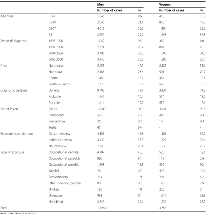
In the period 1993-2008, 15,322 incident cases of all-site MM were recorded. Most MM cases (93%) occurred in the pleural site. Standardised incidence rates for pleural mesothelioma in 2008 were 3.84 per 100,000 for men and 1.45 for women, respectively. Among the 15,845 mesothelioma cases, exposure to asbestos fibres was assessed for 12,065 individuals (76.1%), identifying 530 (4.4%) with familial exposure (they lived with an occupationally exposed cohabitant), 514 (4.3%) with environmental exposure to asbestos (they lived near sources of asbestos pollution and were never occupationally exposed) and 188 (1.6%) exposed through hobby-related or other leisure activities. (Tab. 2). Clusters of cases due to environmental exposure are mainly related to the presence of asbestos-cement industry plants (Casale Monferrato, Broni, Bari), to shipbuilding and repair activities (Monfalcone, Trieste, La Spezia, Genova) and soil contamination (Biancavilla, Sicily) (55). (Fig. 3). The mean age at diagnosis was 69.3 years with no significant difference by gender (70.3 for women and 68.9 for men). Less than 10% of cases occurred in subjects younger than 55 years.
15
Table 2. Incident cases of malignant mesothelioma recorded by the Italian national mesothelioma registry (ReNaM) selected for cluster analysis. (54).
uncorrected standardized incidence ratios and of distri-bution of posterior probability of the estimated RR exceeding 1 for all areas are reported in Additional files 1, 2, 3, 4, 5, 6, 7 and 8.
The distribution of cases in the clusters by gender and by geographic area, as well as the environmental fraction of cases (i.e. the percentage of cases attributed to envir-onmental exposure to asbestos based on expert evalu-ation of the informevalu-ation collected through interviews) and the size of unrecognized exposures among defined
cases are shown in Table 2. Table 3 reports detailed in-formation about asbestos exposure referring to the main economic activities involved, i.e. those causing more than 3% of defined MM cases in each cluster. For several large clusters less represented economic sectors (less than 3%) also need to be mentioned. A significant num-ber of MM cases was attributable in the Genoa cluster to military defense (N = 28), an oil refinery (N = 19) and an electric power plant (N = 13), and in the Trieste cluster to many wood furniture factories (N = 14), a steel industry
Table 1 Incident cases of malignant mesothelioma recorded by the Italian national mesothelioma registry (ReNaM) selected for the cluster analysis
Men Women
Number of cases % Number of cases %
Age class 0-54 1,046 9.6 450 10.3 55-64 2,646 24.1 856 19.7 65-74 4,015 36.6 1,404 32.2 75+ 3,257 29.7 1,648 37.8 Period of diagnosis 1993-1996 1,042 9.5 382 8.8 1997-2000 2,271 20.7 884 20.3 2001-2004 3,706 33.8 1,503 34.5 2005-2008 3,945 36.0 1,589 36.4 Area Northwest 5,158 47.1 2,423 55.6 Northeast 2,586 23.6 901 20.7 Centre 1,450 13.2 454 10.4
South & Islands 1,770 16.1 580 13.3
Diagnostic certainty Definite 8,708 79.4 3,234 74.2
Probable 1,142 10.4 574 13.2
Possible 1,114 10.2 550 12.6
Site of lesion Pleura 10,312 94.0 3,941 90.4
Peritoneum 579 5.3 403 9.3
Pericardium 26 0.2 14 0.3
Testis 47 0.4 -
-Exposure ascertainment Direct interview 4,585 41.8 1,401 32.2
Indirect interview 4,139 37.8 1,727 39.6
No interview 2,240 20.4 1,230 28.2
Type of exposure Occupational, definite 4,987 45.5 526 12.1
Occupational, probable 890 8.1 112 2.6 Occupational, possible 1,291 11.8 402 9.2 Familial 76 0.7 446 10.2 Environmental 214 1.9 294 6.7 Other non-occupational 80 0.7 108 2.5 Unlikely 193 1.8 223 5.1 Unknown 993 9.1 1,017 23.3 Undefined 2,240 20.4 1,230 28.2 Total 10,964 4,358 Italy, 1993–2008 (N = 15,322).
16
Figure 3. Crude incidence rates of malignant mesothelioma (MM) cases by Italian municipalities. Italian National Mesothelioma Register (ReNaM). Italy, men and women, 1993–2008. Diagnosis of MPM definite, probable or possible, all site of lesion (56).
17
MOLECULAR PATHOGENESIS OF MPM
MPM develops insidiously in patients who are usually diagnosed at advanced stages, because radiological diagnostic tools are not effective for early detection, while early serum biomarkers have not been established yet. The anatomical location and characteristics of the body cavities where MPM initially develops also cause malignant cells to easily spread and invade tissues.
Pathologically MPM is subdivided into three major subtypes, viz. epithelioid, sarcomatoid, and biphasic, the latter showing both epithelioid and sarcomatoid components (57).
MPM subtypes differ in their morphology, biological/clinical phenotypes, aggressiveness and response to treatment (58).
The epithelioid MPM consists of cuboidal or polygonal cells with abundant cytoplasm and uniform round nuclei forming a tubular and papillary pattern. The sarcomatoid MPM consists of sheets or fascicles of spindle cells of variable cellularity and pleomorphism. According to the World Health Organization, biphasic MPM must contain at least 10% epithelial and at least 10% sarcomatoid patterns (59). Epithelioid is the most common sub-type among MPM patients, being associated with a relatively better prognosis (60). (Fig. 4).
18
Figure 4. Histologic types of MPM. Microphotograph (H&E stain) of epithelioid mesothelioma (A), sarcomatoid mesothelioma (B) and biphasic mesothelioma (C) (61).
The antibodies chosen for sarcomatoid mesothelioma are very different from those used for epithelioid meso-thelioma. In the case of sarcomatoid mesothelioma, cytokeratin (AE1/AE3 or CAM5.2 as antibodies) exhibits a high specificity and is the most useful [24]. On the other hand, because the diagnosis for true sarcoma is based on the specific differentiation of tumor cells, mesothelioma is eliminated by making its differentiation clear. For example, the following antibodies are known to be useful: MyoD1, desmin and myoglobin for rhabdomyosarcoma; desmin, a-SMA and h-caldesmon for leiomyosarcoma; S-100p for malignant nerve sheath tumor; KP-1 for malignant fibrous histiocytoma [25].
The most difficult tumor to be differentiated from sar-comatoid mesothelioma of pleura is sarcomatoid carcinoma (spindle cell carcinoma, pleomorphic carci-noma) of the lung. When immunohistochemical stainings are used, both respond positively to cytokeratin [24]. In this
case, therefore, the gross finding or the clinical diagnosis by imaging is very important, as already mentioned.
Immunohistochemical stains may be useful in differen-tiating between fibrous pleuritis and desmoplastic mesothelioma. Desmin is positive for spindle cells of the fibrous pleuritis, while desmin is negative for tumor cells of the sarcomatoid mesothelioma [26]. The combination of EMA, desmin and p53 is useful for differentiating between reactive mesothelial hyperplasia and early-stage epithelioid mesothelioma. Reactive mesothelial cells are positive for desmin and negative for EMA and p53 [27].
Compensation or relief of patients
In the compensation system for occupational exposures to asbestos and in the new law for non-occupational exposure to asbestos, if the diagnosis of mesothelioma is certain, it
Fig. 4 Microphotograph of desmoplastic mesothelioma (H&E stain). The feature of granulation or fibrous pleuritis are dominant Fig. 1 Microphotograph of epithelioid mesothelioma (H&E stain).
Papillo-tubular structure is prominent
Fig. 2 Microphotograph of sarcomatoid mesothelioma (H&E stain). Proliferation of spindle cells mimies true sarcoma
Fig. 3 Microphotograph of biphasic mesothelioma (H&E stain). The features of epithelioid mesothelioma and that of sarcomatoid meso-thelioma are mixed within one tumor
62 Environ Health Prev Med (2008) 13:60–64
123
A
B
19
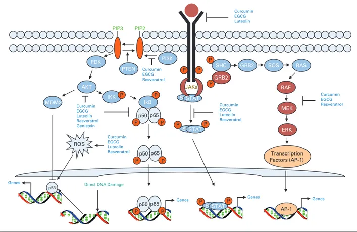
The exact mechanisms of mesothelioma pathogenesis are unknown. However, several recent MM reviews report comprehensive lists of genetic, epigenetic and signaling alterations (62). A main mechanism is thought to be triggered by asbestos fiber penetration within the lung tissue. When long and thin asbestos fibers are inhaled deeply into the lung and penetrate the pleural space, they start to interact with mesothelial cells and inflammatory cells. (Fig. 5). This is thought to trigger prolonged cycles of damage/repair in lung tissue, accompanied by prolonged local inflammation, allegedly leading to MPM carcinogenesis (63).
Figure 5. Development of malignant pleural mesothelioma (MPM) in response to asbestos exposure
Alternative routes of fiber translocation to the parietal pleura include lymphatic and haematogenous dissemination (17). The exact way in which asbestos fibers cause these changes is uncertain, but researchers believe such changes are responsible for cancer development. It also remains unclear why the initial affected site of MPM development by asbestos exposure is the parietal instead of the visceral pleura. Compared with other cell types, human mesothelial cells are very susceptible to asbestos cytotoxicity, which raises a paradoxical issue of how asbestos causes MPM if human mesothelial cells exposed to
20
asbestos die (64). It is however known that several features of asbestos fibers contribute to their carcinogenicity, including chemical composition, fibers length and form, and their biopersistence (65). It has been also shown that asbestos has pleiotropic effects on cell signaling pathways (66, 67). (Fig. 6). First, reactive oxygen species generated by asbestos fibers lead to DNA damage and strand breaks (68-70). Second, macrophages that phagocyte asbestos fibers are unable to digest them, resulting in further production of reactive oxygen species (70). These events activate MAP-kinase signaling pathways through the epithelial growth factor (EGF) receptor, which is frequently highly expressed in mesothelioma together with downstream transcription factors.
The long latency period of the disease (up to 40 years) suggest that multiple genetic alterations are important in the conversion from normal to malignant mesothelial cell. Comprehensive karyotypic analysis has revealed that malignant mesotheliomas display multiple clonal chromosomal abnormalities (more than 10 of them in most cases). This can be explained by considering that asbestos fibers engulfed by mesothelial cells can physically interfere with the mitotic process of the cell cycle, thereby damaging the mitotic spindle. This may result in chromosomal structural abnormalities and aneuploidy of mesothelial cells (71). The loss of one copy of chromosome 22 is the most typical single chromosomal change in patients with mesothelioma. Specific deletions of chromosomal sites involve the short arm (p) of chromosomes 1, 3 and 9, as well as the long arm (q) of chromosome 6. Other non-random cytogenetic alterations have been detected on other chromosomes (72). Mutations of the p53 gene (TP53) are occasionally observed in MPM (73). Loss and/or inactivation of tumor suppressor genes like TP52 may play a role in the development and progression of MPM. Asbestos fibers can absorb chemicals to their surface, with the consequent accumulation of hazardous molecules (including carcinogens). Fibers can also bind cellular proteins, possibly leading to detrimental deficiency for normal mesothelial cells (74). Finally, asbestos-exposed mesothelial cells and macrophages release a variety of cytokines and growth factors, including inflammation and tumor promotion agents like transforming growth factor beta (TGF-β), which might have a role in stimulating tumor growth; platelet-derived growth factor (PDGF), which may act as a regulatory factor in MM cell proliferation, insulin-like growth factor (IGF), which promotes tumor proliferation and cell migration (75); interleukins such as IL-6 and IL-8, which may promote tumor growth and the development of new capillaries (76); vascular endothelial growth factor (VEGF), which also promotes tumor angiogenesis (77), and hepatocyte growth factor (HGF), which stimulates mesothelioma cell migration and invasiveness (78).
21
Tumor necrosis factor-α (TNF-α ) has been shown to activate nuclear factor-κB (NF-κβ), which leads to mesothelial cell survival and inhibits asbestos-induced cytotoxicity (79, 80). High-mobility group box 1 (HMGB1) protein is also released from mesothelial cells, which are exposed by asbestos and then undergo necrotic cell death, promoting an inflammatory response (81).
Figure 6. Possible mechanisms of asbestos-induced carcinogenesis. HMGB1, high-mobility group box 1 protein; ROS, reactive oxygen species; TGF-β, transforming growth factor-β; VEGF, vascular endothelial growth factor (74).
22
CLINICAL FEATURES AND DIAGNOSIS
Clinical MPM symptoms are usually atypical and nonspecific. The most common one is dyspnea (90% of cases) that is typically caused by pleural effusion. Commonest initial symptoms are pain (35%) caused by irritation of intercostal nerves or by infiltration into the chest wall, distension of abdomen (31%), and less frequently night sweats. Patients also commonly develop cachexia, anorexia or ascites (77%) (47). Weight loss is a late sign of disease, along with generalized malaise and failure to thrive (82). Rarely, patients present with fever of unknown origin, intestinal obstruction or acute abdominal surgical emergency.
Symptomatic metastases are unusual. In addition, the majority of patients report late due to very elusive and atypical symptoms, leading to diagnostic delays of up to six months (83). Considering the elusive nature of the disease, the diagnosis of MPM should be considered in any patient with a unilateral pleural effusion or thickening, especially if chest pain is present. Whenever MPM is suspected, a detailed occupational history should be taken and the patient should be referred to an experienced center for pulmonary medicine.
An important help to clinicians in this phase can be provided by imaging diagnostic tools, which also play an important role in staging, treatment planning (especially in terms of resectability), response assessment, and follow up of MPM patients. Initially, non-invasive tests such as ultrasonography, computerized tomography (CT), and magnetic resonance imaging (MRI) can be used to obtain further support for the suspected diagnosis and assess the extent of disease. CT is useful to judge the extent of tumor and to detect lymphonode metastases. MRI is the best way to determine whether the tumor has invaded the diaphragm or the chest wall. Positron emission tomography (PET) is also coming into wide use; its main advantage is a greater sensitivity for the detection of distant metastases (84). The integration of PET functional data coupled to radio-pharmaceutical FDG, with anatomical data from CT (PET/CT) could be useful in the diagnosis and preoperative staging of patients. (85).
In order to render an accurate diagnosis, a biopsy sample must provide adequate diagnosable tissue. For the diagnosis of MPM, a biopsy sample typically is obtained form open procedures such as thoroscopy. Biomarkers would be helpful in managing three clinical aspectsof MPM: early diagnosis, prognosis, and outcome prediction. Researchers have actively sought MPM biomarkers for more than 20 years. However, at present, there are no biomarkers in clinical use for MPM. In fact, MPM remains a rare disease and the small number of patients and the difficult accessibility to a uniform tumor population renders the search for biomarker frustrating. For early diagnosis, optimal serum biomarker for mesothelioma would predict mesothelioma development in asbestos-exposed subjects, differentiate mesothelioma from
23
benign pleural disease or metastatic cancer, could be useful for pathologic subtypes, and correlate with disease extent in order to monitor treatment response and predict prognosis. (86).The biomarkers osteopontin (87), soluble mesothelin-related protein (SMRP) (88), and megakaryocyte potentiating factor (MPF) are currently most promising for diagnosis, but each of them has limitations (89, 90).
24 CURRENTLY ESTABLISHED THERAPY
Treatment options depend on multiple factors, including patient age, performance status, tumor histology, and disease stage at presentation.
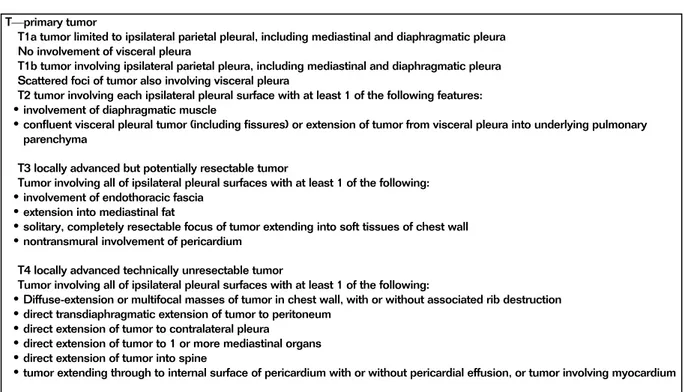
The staging system proposed by the International Mesothelioma Interest Group was accepted by the International Union Against Cancer and the American Joint Commission on Cancer (Tab. 3 and 4). This system describes the extent of tumor according to the tumor-node-metastasis (TNM) classification, i.e. local extent of the primary tumor (T descriptor), presence and location of lymphnodes (N descriptor), and presence of distant metastatic disease (M descriptor) (91).
Table 3.Staging Evaluation: TNM International Staging System for Diffuse MPM (92). mammary and anterior diaphragmatic lymph nodes. The
posterior diaphragmatic lymphatics drain into the para-aortic and posterior mediastinal lymph nodes. There is communi-cation between lymphatics on both sides of the diaphragm, including the retrocrural, inferior phrenic, gastrohepatic, and celiac regions. Extrathoracic metastatic disease has been seen at autopsy in 50%-80% of cases.15 Distant hematogenous metastases are common and can involve the lungs, liver, spleen, adrenal glands, lymph nodes, bones, and brain.
Clinical Presentation
Patients with MPM usually present with slow onset of chest pain, shortness of breath, and cough. Chest wall invasion may lead to intractable pain. Pleural effusion can be seen in up to 95% of cases. Tumor infiltration of the pleura may encase the lung. Mediastinal invasion may lead to dysphagia, phrenic
nerve paralysis, cardiac tamponade, and superior vena cava syndrome.
Differential Diagnosis
The radiologic manifestations of MPM include a unilateral pleural effusion, a pleural mass or diffuse pleural thickening. The differential diagnosis of a unilateral pleural effusion is extensive, including congestive heart failure, infection, pul-monary embolism, collagen vascular, and subdiaphragmatic diseases. The differential diagnosis of a pleural mass includes metastatic disease, sarcoma, lymphoma, and localized fibrous tumor of the pleura. Localized fibrous tumors of the pleura arise from mesenchymal cells, can be benign or malignant, are not related to asbestos exposure and have better prognosis than MPM. The differential diagnosis of diffuse nodular pleural thickening is limited and includes MPM, metastatic disease, and in cases with a mediastinal mass, thymic Table 1 Staging Evaluation: TNM International Staging System for Diffuse MPM
T—primary tumor
T1a tumor limited to ipsilateral parietal pleural, including mediastinal and diaphragmatic pleura No involvement of visceral pleura
T1b tumor involving ipsilateral parietal pleura, including mediastinal and diaphragmatic pleura Scattered foci of tumor also involving visceral pleura
T2 tumor involving each ipsilateral pleural surface with at least 1 of the following features:
!involvement of diaphragmatic muscle
!confluent visceral pleural tumor (including fissures) or extension of tumor from visceral pleura into underlying pulmonary parenchyma
T3 locally advanced but potentially resectable tumor
Tumor involving all of ipsilateral pleural surfaces with at least 1 of the following:
!involvement of endothoracic fascia
!extension into mediastinal fat
!solitary, completely resectable focus of tumor extending into soft tissues of chest wall
!nontransmural involvement of pericardium T4 locally advanced technically unresectable tumor
Tumor involving all of ipsilateral pleural surfaces with at least 1 of the following:
!Diffuse-extension or multifocal masses of tumor in chest wall, with or without associated rib destruction
!direct transdiaphragmatic extension of tumor to peritoneum
!direct extension of tumor to contralateral pleura
!direct extension of tumor to 1 or more mediastinal organs
!direct extension of tumor into spine
!tumor extending through to internal surface of pericardium with or without pericardial effusion, or tumor involving myocardium N—lymph nodes
NX regional lymph nodes not assessable N0 no regional lymph node metastases
N1 metastases in ipsilateral bronchopulmonary or hilar lymph nodes
N2 metastases in subcarinal or ipsilateral mediastinal lymph nodes, including ipsilateral internal mammary nodes
N3 metastases in contralateral mediastinal, contralateral internal mammary, and ipsilateral or contralateral supraclavicular lymph nodes
M—metastases
MX distant metastases not assessable M0 no distant metastases
M1 distant metastases present
25 Table 4. Classification of Stage by TNM Description (92).
The relatively low incidence of MPM poses a challenge to the evaluation of the effectiveness of treatment options because of difficulties in the accrual of patients in clinical trials, particularly large, randomized prospective trials. Given this limitation, the failure of single-modality therapy, i.e. chemotherapy, radiation therapy, or surgery alone, has led to the increasing use of multimodality regimens, by combining these treatment options (93).
Surgery
The role of surgery in MPM treatment is still uncertain. In general, patients with stage I disease can be considered candidates for radical surgery. The three most common surgical procedures are debulking surgery (also known as cytoreductive surgery or pleurectomy/decortication (P/D)), extra pleural pneumonectomy (EPP), and surgical pleurodesis via video assisted thoracoscopic surgery (VATS). P/D is a lung-sparing operation in which the pleura is mobilized off the chest wall, mediastinum, diaphragm, and pericardium, and stripped from the surface of the lung. Pleurectomy has not been shown to prolong survival but can reduce the recurrence of pleural effusion better than talc pleurodesis (94). In contrast, EPP is the radical en bloc resection of the lung, visceral and parietal pleura, ipsilateral diaphragm, and adjacent pericardium (95). It is a rather complex operation, which should be performed by skilled surgeons and in selected centres. VATS is a type of thoracic surgery performed using a small video camera that is introduced into the patient's chest via a scope. It is a minimally invasive surgical technique used to diagnose and treat problems in the chest. Operations that were traditionally carried out with thoracotomy or sternotomy and are currently performed with VATS include: biopsy for diagnosis of pulmonary, pleural or
malignancy with pleural metastases. CT features that aid in differentiating malignant from benign pleural disease include pleural thickening with a circumferential distribution encas-ing the lung (sensitivity 100%, specificity 41%), pleural thickening of greater than 1 cm in thickness (sensitivity 94%, specificity 36%), and nodular morphology (sensitivity 94%, specificity 51%).16
Staging
A clinically and pathologically accurate staging system is essential in the selection of homogeneous groups of patients with similar prognoses for entry into clinical trials to better assess new treatment options. The staging system proposed by the International Mesothelioma Interest Group was accepted by the International Union Against Cancer and the American Joint Commission on Cancer (Tables 1 and 2).17 This system describes the extent of tumor according to the tumor-node-metastasis (TNM) classification that is, the local extent of the primary tumor (T descriptor), the presence and location of lymph node involvement (N descriptor), and the presence of distant metastatic disease (M descriptor). Recently, the International Association for the Study of Lung Cancer Staging Committee developed a large international database of 3101 patients from 15 centers on 4 continents to examine outcomes in surgically managed MPM patients that suggest the need to revise T and N staging.18Initial analysis of this database showed that significant differences in overall survival were seen for: T4 vs T3 and T3 vs T2 but not T2 vs T1; N0 vs N1 and N2 but not N1 vs N2; stages III and IV vs stage I but not stage II vs stage I.
This analysis also confirmed the importance of distinguish-ing patients who would benefit from surgical resection from those needing palliative treatment. Limitations in staging using imaging modalities such as CT, MRI, and PET and the morbidity and mortality associated with surgical resection have resulted in the need for extended surgical staging in patients being considered for resection. Thus, cervical media-stinoscopy or endobronchial ultrasound-guided lymph node biopsy, laparoscopy and peritoneal lavage may be performed in MPM patients undergoing preoperative evaluation for aggressive multimodality therapy. Rice et al. reported that extended surgical staging precluded 15 of the 118 patients (12.7%) assessed by clinical staging alone to be candidates for extrapleural pneumonectomy (EPP).19
Imaging is limited in determining the true extent of MPM. When compared with surgical staging, CT has been shown to underestimate the extent of disease in patients with early chest wall involvement, small positive lymph nodes, transdiaph-ragmatic extension, peritoneal implants, and abdominal organ metastases less than 2 mm in size.20 Despite these limitations, CT is still the imaging modality of choice in the initial staging and surveillance of patients with MPM due to its easy accessibility and cost-effectiveness.
T Staging
Accurate T staging is important in determining resect-ability.17 In patients with locally advanced tumors, imaging is usually aimed at distinguishing T3 disease (a solitary focus of chest wall involvement, involvement of the endothoracic fascia, mediastinal fat extension, or nontransmural pericardial involvement) (Fig. 2) from unresectable (T4) disease (diffuse tumor extension or multiple chest wall foci, direct extension to the media-stinal organs, spine, internal pericardial surface or con-tralateral pleura, and transdiaphragmatic invasion). However, the pathologic descriptors for T staging are often difficult to determine on CT and MRI.
In locally advanced (T4) disease, the poor accuracy of CT and MRI in detecting transdiaphragmatic extension of MPM is due to their inability to identify microscopic disease. Thus, preoperative laparoscopy and peritoneal lavage are routinely performed in some institutions in patients being considered for EPP. In the study by Rice et al., laparoscopy identified 10 of 109 (9%) patients with transdiaphragmatic invasion or peritoneal metastases compared with 3 of 109 patients identified by cross-sectional imaging.19
N Staging
The N descriptor denotes the presence and location of lymph node involvement. Large retrospective studies have shown intrathoracic nodal metastases in up to 50% of MPM patients who undergo radical resection.21 CT is typically used to evaluate for hilar and mediastinal nodal disease and uses size of greater than 1 cm in short-axis dimension as an indicator of suspicious nodal disease. The specificity of CT for detecting nodal disease is poor because metastases can be seen in nodes less than 1 cm in size whereas enlarged lymph nodes greater than 1 cm in size can be hyperplastic. Furthermore, it can be difficult to differentiate hilar or mediastinal lymph nodes from tumor involving the mediastinal pleura (Fig. 3). Owing to the poor outcome of patients with extrapleural nodal involvement who undergo radical resection, inva-sive nodal sampling is important in patient selection.22 Schouwink et al. performed mediastinoscopy in 43 patients with MPM and compared the staging accuracy with that of CT. Sensitivity, specificity, and accuracy were 80%, 100%, and 93%, respectively, for mediastinoscopy compared with 60%, 71% and 67%, respectively, for CT.23 Table 2 Classification of Stage by TNM Description
Stage Description 1a T1aN0M0 1b T1bN0M0 II T2N0M0 III Any T3M0 Any N1M0 Any N2M0 IV Any T4 Any N3 Any M1 M.T. Truong et al 326
26
mediastinal pathology, decortication, pleurodesis for recurrent pleural effusions or spontaneous pneumothorax, surgical stapler-assisted wedge resection of lung masses, and resection of mediastinal (96). The goal of surgery is gross total resection of the tumor. As mesotheliomas tend to grow diffusely, they are usually not totally resectable; some residual tumor tissue (often microscopic) is generally leftbehind. Adjuvant chemotherapy is therefore frequently administered in order to achieve full elimination of remaining tumor cells (97).
Chemotherapy
The use of chemotherapy in malignant mesothelioma aims to lengthen survival, improve quality of life and provide symptomatic relief. At present, there is no single drug or combination therapy that could be considered as a standard treatment for mesothelioma. Chemotherapy is the mainstay of therapy because of the fact that most MPM patients present with advanced disease. In a meta-analysis of studies published between 1965 and 2001, cisplatin was found to be the most active single drug, while its use in combination chemotherapy has been associated with higher response rates, but not with longer survival (98). Several new cytotoxic agents with definite activity in mesothelioma have recently been evaluated, including gemcitabine, imatinib, and the antifolates pemetrexed and raltitrexed. The pemetrexed/cisplatin combination significantly improved response rates, time to progression, overall survival and quality of life compared to cisplatin alone (99).
Radiotherapy
The role of radiation therapy in MPM is as controversial as the role of surgery. Radiotherapy can be used to control local MPM growth and occasionally causes regression of disease, but there is no evidence that radiotherapy alone affects survival (100). Patients with mesothelioma are given prophylactic radiotherapy at puncture sites and after surgical interventions to prevent local recurrence and to relieve pain in palliative care. Radical radiotherapy of the entire tumor is not currently feasible, because these tumors tend to grow in a complex geometrical configuration, and the resulting high radiation load of treatment would be likely to cause collateral damage to heart and lungs (101).
27 Multimodal approaches
Specialized mesothelioma centers employ multi-modality approaches, including surgical resection, chemotherapy, and radiation, with survival in excess of 20 months depending on stage (102).
In a clinical trial, neo-adjuvant chemotherapy combined with pleuropneumectomy and followed by radiotherapy has led to a survival rate 3-year longer, on average, then those obtained with unimodal treatments (76 %) (103). In another study, multimodal approach has prolonged the median survival time up to 22 months for stage I disease (104).
28 NOVEL THERAPIES
Many novel strategies are being attempted to improve survival for patients with MPM.
Several classes of targeted therapies have emerged from preclinical work and are being evaluated. These treatments focus on the following broad mechanisms:
• Tyrosine kinase inhibitors • Antibody conjugated toxins • Immune checkpoint inhibitors • Gene therapy
• Tumor vaccines
Tyrosine kinase inhibitors
Molecular studies of mesothelioma specimens have shown marked overexpression of protein targets such as epidermal growth factor receptor (EGFR) and vascular endothelial growth factor (VEGF) (105).
The inhibition of EGFR-dependent signaling pathway in mesothelioma cell lines leads to decreased cell survival (106); based on these findings, several clinical trials have been conducted but, disappointingly, these studies did not show improved survival.
Higher levels of VEGF may be reflective of more-advanced disease and have been associated with shorter survival. In vitro studies have demonstrated that increased mesothelioma cell proliferation occurs upon treatment with VEGF and that significant inhibition of cell growth can be achieved by blocking this pathway (107). It is therefore thought that interference with this pathway could potentially lead to successful therapy. Numerous clinical trials have evaluated the effect of VEGF inhibitors alone and in combination with other drugs, but also the results of these trials have been disappointing.
Therapy targeting cell-surface receptors
Mesothelin is a 40-kDa, glycosylphosphatidylinositol-anchored cell-surface glycoprotein present on normal mesothelial cells and overexpressed on the surface of mesothelioma. Mesothelin overexpression, occurring more prominently on epithelioid tumors, may serve to alter cell adhesion and/or invasion. Preclinical studies in nude mice suggested that MORAb-009 (amatuximab), a monoclonal antibody with high affinity for human mesothelin combined with chemotherapy (gemcitabine or paclitaxel) was more effective than either chemotherapy alone (108). BAY 94-9343 (anetuman ravtansine) is a human antimesothelin antibody
29
coupled via a reducible disulfide bond to DM4, a microtubule-targeting toxophore that shows highly selective cytotoxicity against cells with high mesothelin expression. Preclinical studies have shown a dose-dependent and receptor-dependent, 94% reduction of tumor growth with BAY 94-9343 compared to 70% reduction with cisplatin and pemetrexed (109).
SS1P is an immunotoxin consisting of an antimesothelin antibody variable fragment linked to a cytotoxic fragment of Pseudomonas exotoxin A. Phase I trial including 16 mesothelioma patients has shown that SS1P was well tolerated up to 25 µg/kg/day × 10 days, with modest clinical activity and minor responses, and that two mesothelioma patients had symptomatic improvement (110).
High levels of interleukin-4 (IL-4) receptor expression have been shown on fresh human mesothelioma specimens and correlated with a worse outcome. In a human mesothelioma xenograph, nude mouse model, intratumoral injection of the recombinant toxin IL-4 (38–37)-PE38KDEL significantly reduced tumor volumes in a dose-dependent manner compared to the control and IL-4-treated mice (111).
Immune checkpoint inhibitors
Cytotoxic T lymphocyte antigen-4 (CTLA-4) is vital for maintaining host immune tolerance to established tumors. A Phase II trial evaluating anti-CTLA-4 antibody (tremelimumab) in 29 patients with chemotherapy-resistant advanced mesothelioma (28 pleural and 1 peritoneal) has been recently reported (112). Objective clinical responses have been observed in only 2 of 29 patients (6,9%) and disease stabilization has been noted in 9 patients (31%).
Programmed death receptor is found on the surface of cells and its stimulation leads to T-cell deactivation, thus allowing escape from the immune system surveillance in the presence of otherwise antigenic substrate. Activation of this receptor occurs by a programmed death ligand 1 (PD-L1), which is present in the tumor microenvironment and on the surface of tumor cells. The effect of PD-L1 blockade on different subpopulations of T-cells has pro-duced opposing effects on tumor progression, suggesting that tumor-derived immune suppression is mediated by specific subsets of T-cells (113). Several trials are currently evaluating the inhibition of this pathway using different agents (lambrolizumab and nivolumab) in cancers other than MPM.
Gene therapy
Multiple genetic abnormalities have been identified in MPM, and a variety of genetic manipulation strategies have been employed in preclinical studies.