Research Article
Combination of Coenzyme Q
10
Intake and Moderate Physical
Activity Counteracts Mitochondrial Dysfunctions in a SAMP8
Mouse Model
C. Andreani
,
1C. Bartolacci,
1M. Guescini
,
2M. Battistelli,
2V. Stocchi,
2F. Orlando,
3M. Provinciali,
3,4A. Amici,
1C. Marchini,
1L. Tiano
,
5P. Orlando
,
5and S. Silvestri
5,6 1University of Camerino, via Gentile III da Varano, 62032 Camerino, Italy2University of Urbino, via Aurelio Saffi, 61029 Urbino, Italy
3Experimental Animal Models for Aging Unit Scientific Technological Area, IRCCS INRCA, via del Fossatello, 60127 Ancona, Italy 4Advanced Technological Center for Aging Research Scientific Technological Area, IRCCS INRCA, via Birarelli 8, 60121 Ancona, Italy 5Polytechnic University of Marche, Department of Life and Environmental Sciences (DISVA), via Brecce Bianche, Ancona, Italy 6Biomedfood srl, Spinoff of Polytechnic University of Marche, via Brecce Bianche, 60131 Ancona, Italy
Correspondence should be addressed to P. Orlando; [email protected] Received 12 June 2018; Accepted 29 August 2018; Published 24 October 2018 Academic Editor: Carine Smith
Copyright © 2018 C. Andreani et al. This is an open access article distributed under the Creative Commons Attribution License, which permits unrestricted use, distribution, and reproduction in any medium, provided the original work is properly cited. Aging skeletal muscles are characterized by a progressive decline in muscle mass and muscular strength. Such muscular dysfunctions are usually associated with structural and functional alterations of skeletal muscle mitochondria. The senescence-accelerated mouse-prone 8 (SAMP8) model, characterized by premature aging and high degree of oxidative stress, was used to investigate whether a combined intervention with mild physical exercise and ubiquinol supplementation was able to improve mitochondrial function and preserve skeletal muscle health during aging. 5-month-old SAMP8 mice, in a presarcopenia phase, have been randomly divided into 4 groups (n = 10): untreated controls and mice treated for two months with either physical exercise (0.5 km/h, on a 5% inclination, for 30 min, 5/7 days per week), ubiquinol 10 (500 mg/ kg/day), or a combination of exercise and ubiquinol. Two months of physical exercise significantly increased mitochondrial damage in the muscles of exercised mice when compared to controls. On the contrary, ubiquinol and physical exercise combination significantly improved the overall status of the skeletal muscle, preserving mitochondrial ultrastructure and limiting mitochondrial depolarization induced by physical exercise alone. Accordingly, combination treatment while promoting mitochondrial biogenesis lowered autophagy and caspase 3-dependent apoptosis. In conclusion, the present study shows that ubiquinol supplementation counteracts the deleterious effects of physical exercise-derived ROS improving mitochondrial functionality in an oxidative stress model, such as SAMP8 in the presarcopenia phase.
1. Introduction
Aging is characterized by a progressive decline in skeletal muscle mass and muscular strength [1–3]. In healthy people, there is a 1% per year decline in muscle mass between 20 and 30 years of age. This decline is accelerated above 50 years of age [4, 5]. The progressive decline in muscle mass and strength with aging is known as sarcopenia [1, 6–8]. Sarcope-nia is defined as a geriatric syndrome characterized by age-related muscular loss and dysfunction that cause physical
disability, a poor quality of life, and death. The prevalence of this pathology in adults under the age of 70 is about 25% but increases up to 40% in 80-year-old or older people [9, 10]. This condition can lead to decreased physical activity increasing the risk of falls in aged individuals [11]. Under-standing the mechanisms underneath aging-induced skeletal muscle atrophy and promoting health and mobility in the elderly are crucially important goals in order to develop ther-apeutic strategies [12]. Several studies pointed towards a crit-ical role of mitochondria and their implication in age-related Volume 2018, Article ID 8936251, 15 pages
degenerative processes, and many therapeutic attempts have been focused on mitochondria [13]. Indeed, these organelles play a key role in cellular bioenergetics and represent a sensi-tive target in muscle cells [14, 15]. Moreover, metabolism of reactive oxygen species (ROS), Ca2+homeostasis, and apo-ptosis are controlled by mitochondria [16]. Aging of skeletal muscle determines the alteration of the structure and func-tion of these organelles leading to mitochondrial dysfunc-tion [17]. In this context, a growing body of evidence has highlighted a major role of oxidative stress and inflamma-tion in promoting aging of skeletal muscle [18]. Accordingly, it has been recently reported that excessive production of mitochondrial ROS in skeletal muscle is strongly associated with sarcopenia and the impairment of energy homeostasis [19]. In fact, the physiologic equilibrium between ROS pro-duction and antioxidant defense is disrupted in aging sub-jects, and the accumulation of ROS during mitochondrial respiration can cause mutations in mitochondrial DNA (mtDNA) [20] which in turn lead, through a vicious cycle, to further impaired mitochondrial functionality. Moreover, many studies have reported that a decline in mitochondria content may also account for the loss of skeletal muscle mass [18], further impairing oxidative phosphorylation and ATP production [21, 22]. Mitochondrial biogenesis is regulated by the expression of nuclear and mitochondrial genes, controlled by the transcriptional coactivator peroxisome pro-liferator gamma coactivator-1α (PGC-1α) [23]. Vainshtein et al. [24] suggested a role of this coactivator also in the regulation of autophagy and mitophagy in skeletal muscle. These two are distinct but interconnected degradation pro-cesses aimed at eliminating damaged cellular components in response to stress stimuli. Both mitophagy and autophagy are regulated by autophagy-related genes (Atgs) including Beclin-1 and LC3 [25].
During aging, skeletal musclefibers gradually lose the capability to remove dysfunctional mitochondria [13]. This condition could further impair mitochondrial respiration and enhance ROS production [26] contributing to the onset of sarcopenia. Previous studies suggested that an appropriate physical activity regimen can counterbalance age-associated muscular deficits by promoting mitochondrial biogenesis [27–29]. Exercise training has been reported to modulate skeletal muscle metabolism, regulating intracellular signaling pathways and thus mediating mitochondrial homeostasis [30, 31]. However, some authors raised doubts regarding the beneficial role of exercise in the elderly, claiming that physical activity-dependent ROS production could exacer-bate oxidative damage inside aged skeletal muscles [32–34]. In this scenario, association of physical activity with antioxi-dant therapies might be an effective strategy to prevent the adverse effects of exercise in the elderly. Coenzyme Q10 rep-resents a valuable candidate for oxidative stress prevention and for supporting muscle functionality [35–39]. Coenzyme Q (CoQ) consists of a quinone head which, in mammalian cells, is attached to a chain of 9 (CoQ9) or 10 isoprene units CoQ10 [40]. In human tissues, the most abundant form is coenzyme Q10, while in mice and rats it is coenzyme Q9, although CoQ10 represents a significant proportion of
total CoQ and its level is able to increase following oral
supplementation [41–46]. As part of the mitochondrial elec-tron transport chain (ETC), CoQ actively participates in oxi-dative phosphorylation and plays a key role in energy and redox state balance [47]. In addition, CoQ has been found in other subcellular localizations and in circulating plasma lipoproteins, where it acts as an endogenous lipophilic anti-oxidant in synergism with vitamin E [48]. Endogenous CoQ10synthesis, the principal source of CoQ [49], has been shown to significantly decrease during aging and in certain degenerative diseases [50, 51], thus triggering cellular dys-functionality. These evidences underlie the rationale for CoQ use in clinical practice and as a food supplement. CoQ exists in three states of oxidation: ubiquinone (CoQ), the fully oxidized form; ubisemiquinone (CoQH.), the partially reduced form; and ubiquinol (CoQH2), the fully reduced form. In particular, the CoQH2form has several advantages being more bioavailable and readily usable by the organism not requiring reductive steps [52]. This is of particular rele-vance in conditions when reductive systems might be less efficient such as during aging or following intense physical exercise. Here, we investigated the effect of a combined approach of mild physical exercise and ubiquinol supple-mentation on the senescence-accelerated mouse-prone 8 (SAMP8) model in a presarcopenia phase [53, 54]. SAMP strains derived from AKR/J series [55] show senescence acceleration and age-related pathological phenotypes, similar to aging disorders seen in humans. In particular, we focused on SAMP8 mice since they exhibit the most striking features among SAMP strains in terms of life span, fast aging progres-sion due to high oxidative stress status [56, 57], dramatic decrease in muscle mass and contractility [58, 59], and a huge reduction in type II muscle fiber size [60, 61]. The aim of this study is to develop prevention strategies able to preserve skeletal muscle health during aging by maintaining mito-chondrial function through regular physical exercise and antioxidant supplementation using a senescence-accelerated mouse-prone model (SAMP8).
2. Materials and Methods
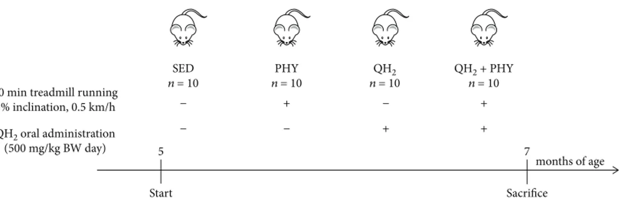
2.1. SAMP8 Housing and Treatment. Senescence-accelerated mice (SAMP8, Harlan) [58, 59, 62], aged 5 months, have been randomly divided into 4 groups (n = 10) as summarized hereafter: untreated controls (SED), trained (PHY), ubiqui-nol 10-administered (QH2), and both trained and ubiquinol 10-supplemented (QH2+ PHY). The PHY and QH2+ PHY groups underwent treadmill running at 0.5 km/h, on a 5% inclination, for 30 min, 5 days per week, for 2 months up to 7 months of age [63, 64] (Figure 1). The QH2 and QH2
+ PHY groups were supplemented with ubiquinol 10 (Kaneka) (500 mg/kg body weight/day in sunflower seed oil) via oral administration. Such QH2formulation was
pre-viously prepared and stored at−80°C in 500μL aliquots to avoid repeated freezing-thawing cycles. An aliquot was thawed daily in a water bath at 60°C in the dark just prior to administration. An equal amount of sunflower seed oil was given to SED and PHY mouse groups. The animals were bred and housed under controlled temperature (20°C) and a circadian cycle (12-hour light/12-hour dark). The animals
were fed on chow diet and water ad libitum. Male mice were used for all experiments. The animal procedures followed the 2010/63/EU directive on the protection of animals used for scientific purposes and were approved by the Ethic Com-mittee on Animal Use of the University of Camerino (proto-col number 14/2012).
2.2. Tissue Collection and Analysis. Mice were rapidly sacri-ficed by isoflurane inhalation followed by cervical disloca-tion, two days after the last exercise/administration session, to avoid possible metabolic effects of the last exercise/ administration bout. Gastrocnemius (GA), tibialis anterior (TA), soleus (SO) muscle, and cardiac muscle were carefully excised. GA samples were either immediately used forflow cytometry (FACS) analysis or preserved in liquid nitrogen for mtDNA quantification or mRNA extraction. TA and cardiac muscles were used for CoQ9 and CoQ10 (total and
oxidized form) quantification. TA muscles were used also for protein extraction and Western blot analysis, while SO samples werefixed in 3% glutaraldehyde for 4 hours, to be analyzed for fiber morphology, number, and ultrastructure of mitochondria by electron microscopy. Other tissues including liver, spleen, and kidneys were recovered for even-tual future applications and preserved at−80°C.
2.3. Coenzyme Q9and Q10Extraction and Quantification. TA
and cardiac muscles were mechanically homogenized (two bouts at 30 Hz for 5 min) in propanol (Sigma) using 7 mm steel beads (Qiagen) and TissueLyser II (Qiagen). After cen-trifugation (2 min at 20,000 g, 4°C), 40μL of the supernatant was injected into a high-performance liquid chromatography (HPLC) apparatus with an electrochemical detector (ECD), model 3016 by Shiseido Co. Ltd, to measure total coenzymes Q9and Q10and Q9oxidative status. The mobile phase was
50 mM sodium perchlorate in methanol/distilled water (95/ 5 v/v) with a flow rate of 0.2 mL/min. Using a column-switching system, coenzymes were eluted from the concen-trating column by mobile phase 2, 50 mM sodium perchlo-rate in methanol/isopropanol (70/30 v/v) with aflow rate of 0.08 mL/min. The column oven was set at 40°C. Pumps one and two of model 3001, autosampler model 3033, and switch valve model 3012; concentration column CQC (C8 DD; 10 mm× 4.0 mm ID); and separation column CQS (C18 AQ; 150 mm× 2.0 mm ID, particle size at 3 μm diameter) were used, all from Shiseido Co. Ltd. A peculiarity of the
system was the use of a postseparation reduction column (Shiseido CQR) capable of fully reducing the peak of ubiqui-none. CoQ9 and CoQ10standard solutions were previously prepared in ethanol and stored at −80°C. The oxidation potential for ECD was 650 mV. TA and cardiac muscle con-tents of CoQ9and CoQ10were expressed asμg/g muscle and
the oxidized form as percentage of total CoQ9.
2.4. Electron Microscope Analysis. Control and treated SO samples were washed and immediatelyfixed with 2.5% glu-taraldehyde in 0.1 M phosphate buffer for 1 hour, postfixed with 1% of osmium tetroxide (OsO4) in the same buffer for 2 hours, and embedded in araldite, as previously reported [65, 66]. The sections were collected on 400-mesh nickel grids, stained with uranyl acetate, lead citrate andfinally ana-lyzed with an electron microscope at 80 kV.
2.5. Flow Cytometry Analysis
2.5.1. Skeletal Muscle Dissociation. GA muscles were dissoci-ated into single-cell suspensions using a skeletal muscle disso-ciation kit (Miltenyi Biotec) according to the manufacturer’s instructions. Mechanical disaggregation was performed via gentleMACS Dissociator using the m_muscle_01 program (Miltenyi Biotec).
2.5.2. Cell Viability and Cell Count. Cell viability and cell count of the obtained single-cell suspensions were evalu-ated using Guava ViaCount® Reagent Kit (Millipore) that discriminates among viable, apoptotic, and dead cells. Briefly, the assay exploits a mixture of cell membrane-permeable (red) and cell membrane-impermeable (yellow) DNA-binding fluorescent probes, diluted 1 : 10 in PBS, and used to stain cells immediately before reading. Cells were incu-bated with reagent for 5 min in the dark, and the analysis of the distribution allows the discrimination of the percentage of cell debris (R−/Y−), live cells (R+/Y−), and dead cells (R+/Y+) with Guava ViaCount software using a Guava easy-Cyte™ flow cytometer (Millipore).
2.5.3. Mitochondrial Membrane Depolarization. Mitochon-drial membrane depolarization was measured by incubating 2.5× 10⁵viable muscle cells with MitoProbe™ DiIC1(5) (Life Technologies) (40 nMfinal concentration) at 37°C for 20 min in the dark. During the experimental setup, a suspension of control cells, before staining, was incubated with 1 μL
months of age
5 7
Start Sacrifice
30 min treadmill running 5% inclination, 0.5 km/h QH2 oral administration (500 mg/kg BW day) SED n = 10 PHY n = 10 QH2 n = 10 QH2 + PHY n = 10 − − + − − + + +
of carbonyl cyanide 3-chlorophenylhydrazone (CCCP) 50 mM for 5 min at 37°C in the dark. After washing with phosphate-buffered saline (PBS), cells were centrifuged at 300 g for 5 min at room temperature andfinally resuspended in PBS and analyzed using the Guava easyCyte™ flow cyt-ometer (Millipore), equipped with a red laser at 633 nm. Using the Guava InCyte software, a gate relative to cells con-taining depolarized mitochondria was arbitrarily set using as a reference CCCP-treated cells assuming that in this condi-tion 90% of the cells contained depolarized mitochondria. This gate was subsequently used for all further analyses. 2.6. Mitochondrial DNA (mtDNA) Quantification. To assess mtDNA content, DNA was extracted from GA muscle using QIAamp DNA Mini kit (Qiagen) and then used for quantita-tive real-time PCR (qRT-PCR) on the StepOne Plus system (Applied Biosystems). The 36B4 gene was used as a nuclear DNA (nDNA) marker while the COX1 gene was used for mtDNA. The primers used are summarized in Table 1. Briefly, 10 ng of DNA was amplified using 1x SYBR Select Master Mix (Applied Biosystems), using the following proto-col: 10 min denaturation at 95°C, followed by 45 cycles (95°C for 15 sec, 60°C for 15 sec, and 72°C for 30 sec) and melting curve (95°C for 15 sec, 60°C for 30 sec, and 95°C for 15 sec). Relative copy number quantification was carried out using theΔΔCt method.
2.7. Western Blot Assay. TA muscle samples were mechani-cally homogenized in RIPA buffer (0.1% SDS, 1% NP40, and 0.5% CHAPS) supplemented with protease inhibitors aprotinin, sodium orthovanadate, and phenylmethylsulfonyl fluoride (Sigma-Aldrich). Lysates were incubated on ice for 30 min and then centrifuged at 16.000 g, 4°C, for 20 min.
The supernatant was collected, quantified via Bradford method (Bio-Rad), and stored in aliquots at−80°C to avoid repeated freezing-thawing cycles. For Western blot analysis, an equal amount of protein lysates (20–40 μg depending on the protein assayed) were separated onto Criterion™ TGX™ precast gels (Bio-Rad) and transferred to a polyvinylidene difluoride (PVDF) membrane (Millipore) using Criterion™ Blotter (Bio-Rad). Membranes were blocked with 5% BSA-TBS-T and then overnight incubated with primary antibod-ies at 4°C. Secondary antibody binding was performed at RT for 1 hour. After TBS-T washing, immunoreactive bands were incubated with enhanced chemiluminescent reagent (EuroClone) and detected via ChemiDoc™ XRS+ System (Bio-Rad). Densitometry analysis was accomplished through ImageJ software using H2B (nuclear), VDAC1 (mitochon-drial), andβ-actin (total) as protein normalizers. The results are representative of at least three independent experiments. The antibodies used are listed in Table 2.
2.8. Gene Expression Analysis. Total RNA was extracted from GA muscles. RNA purification was performed using the E.Z.N.A.® Total RNA Kit I (Omega Bio-tek) according to the manufacturer’s instructions, and contaminant DNA was digested with DNase I enzyme (Ambion). cDNA was synthesized using the Maxima Reverse Transcriptase kit (Thermo Fisher Scientific). Real-time PCR amplifications were conducted using SensiFAST SYBR Green (Bioline) according to the manufacturer’s instructions, with 300 nM primers and twoμL of cDNA (20 μL final reaction volume). Specific primers used are listed in Table 3.
Thermocycling was conducted using LightCycler 480 (Roche) initiated by a 2 min incubation at 95°C, followed
Table 1: pRT-PCR primers for nDNA and mtDNA.
Target Primer sequence_forward Primer sequence_reverse
36B4 5′-CGACCTGGAAGTCCAACTAC-3′ 5′-ATCTGCTGCATCTGCTTG-3′
COX1 5′-TCTACTATTCGGAGCCTGAGC-3′ 5′-CAAAAGCATGGGCAGTTACG-3′
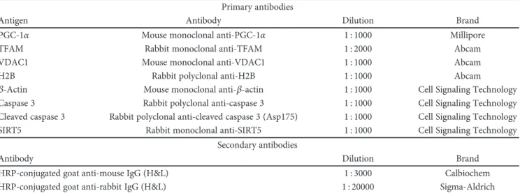
Table 2: Summary of used antibodies. Primary antibodies
Antigen Antibody Dilution Brand
PGC-1α Mouse monoclonal anti-PGC-1α 1 : 1000 Millipore
TFAM Rabbit monoclonal anti-TFAM 1 : 2000 Abcam
VDAC1 Mouse monoclonal anti-VDAC1 1 : 1000 Abcam
H2B Rabbit polyclonal anti-H2B 1 : 1000 Abcam
β-Actin Mouse monoclonal anti-β-actin 1 : 1000 Cell Signaling Technology
Caspase 3 Rabbit polyclonal anti-caspase 3 1 : 1000 Cell Signaling Technology Cleaved caspase 3 Rabbit polyclonal anti-cleaved caspase 3 (Asp175) 1 : 1000 Cell Signaling Technology SIRT5 Rabbit monoclonal anti-SIRT5 1 : 1000 Cell Signaling Technology
Secondary antibodies
Antibody Dilution Brand
HRP-conjugated goat anti-mouse IgG (H&L) 1 : 3000 Calbiochem HRP-conjugated goat anti-rabbit IgG (H&L) 1 : 20000 Sigma-Aldrich
by 40 cycles (95°C for 5 sec, 60°C for 5 sec, and 72°C for 10 sec) with a single fluorescent reading taken at the end of each cycle. Each reaction was conducted in triplicate and completed with a melting curve analysis to confirm the specificity of amplification and lack of primer dimers. Quantification was performed according to the ΔCq method, and the expression levels of GAPDH and S16 were used as a reference [67].
3. Statistical Analysis
Data are presented as mean ± SEM. All statistical analyses were performed using GraphPad Prism® 6.0 software. Unpaired two-tailedt-test was employed when 2 groups were compared and ANOVA for comparison between three or more groups. Two-way ANOVA with Bonferroni correction for multiple comparisons was used when 3 or more groups were compared over time. The GraphPad Prism routine for outlier identification was used to identify any out-of-range values to be excluded from the statistical analysis.
4. Results
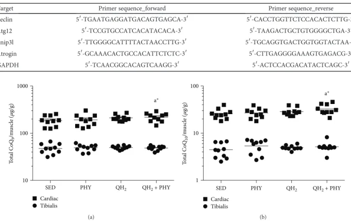
4.1. Combination of Physical Activity and Ubiquinol Supplementation Increases Q9 and Q10Content in Cardiac Muscles and Lowers the Oxidation of Endogenous Coenzyme Q9. Total coenzyme Q9 and Q10 (CoQ9 and CoQ10) levels
and oxidative status of coenzyme Q9were quantified by an HPLC-ECD instrument on skeletal and cardiac muscles. The results were normalized on muscle weight and expressed
as μg/g of muscle. Coenzyme Q levels were very different
between skeletal and cardiac muscles, the latter showing remarkably higher levels of both coenzymes (Figure 2). After ubiquinol and physical exercise (QH2+ PHY) treatment,
both coenzymes were significantly increased in the cardiac tissue, in particular +23% CoQ9 (p = 0 05, Figure 2(a))
and +27% CoQ10(p = 0 03, Figure 2(b)) with respect to the sedentary group.
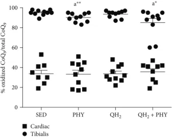
To evaluate the effect of exogenous CoQ supplementa-tion on the oxidative status of endogenous coenzyme Q9, its
oxidized form was measured as well. As shown in Figure 3, skeletal muscle is characterized by a higher extent of oxida-tion (on average 95% of Q9is oxidized) compared to cardiac
muscle (35% of oxidized Q9). Ubiquinol supplementation
alone was not able to lower the oxidation of endogenous muscular CoQ. On the contrary, a significant decrease of oxidized coenzyme Q9was observed in skeletal muscle after regular physical exercise alone or in combination with ubiquinol supplementation (Figure 3,−4.7%, p = 0 009, and −3.6%, p = 0 03, respectively).
4.2. Physical Exercise Alone or in Combination with Ubiquinol Administration Stimulates Muscle Hypertrophy in SAMP8 Mice. To evaluate the impact of the different
Table 3: Summary of used primers for Beclin, Atg12, Bnip3l, Atrogin, and GAPDH.
Target Primer sequence_forward Primer sequence_reverse
Beclin 5′-TGAATGAGGATGACAGTGAGCA-3′ 5′-CACCTGGTTCTCCACACTCTTG-3′
Atg12 5′-TCCGTGCCATCACATACACA-3′ 5′-TAAGACTGCTGTGGGGCTGA-3′
Bnip3l 5′-TTGGGGCATTTTACTAACCTTG-3′ 5′-TGCAGGTGACTGGTGGTACTAA-3′ Atrogin 5′-GCAAACACTGCCACATTCTCTC-3′ 5′-CTTGAGGGGAAAGTGAGACG-3′
GAPDH 5′-TCAACGGCACAGTCAAGG-3′ 5′-ACTCCACGACATACTCAGC-3′
To ta l C o Q9 /m us cl e ( 𝜇 g/g)
SED PHY QH2 QH2 + PHY 10 100 1000 Tibialis Cardiac a⁎ (a)
SED PHY QH2 QH2 + PHY
Tibialis Cardiac a⁎ To ta l C o Q10 /m us cl e ( 𝜇 g/g) 1 10 100 (b)
Figure 2: Total coenzyme Q9(a) and Q10(b) levels in cardiac and tibialis anterior muscles, expressed asμg coenzyme/g of muscle in sedentary
(SED), physical exercise (PHY), ubiquinol (QH2), and ubiquinol associated with physical exercise (QH2+ PHY) mouse groups (n = 10).
treatments on musclefiber atrophy/hypertrophy, fiber diam-eter was measured for each experimental condition. Morpho-metrical analyses of fiber diameter revealed a gradual increase in the ubiquinol (QH2), ubiquinol and exercise (QH2 + PHY), and exercise (PHY) groups, with the PHY
fibers having the largest average diameter (+33% compared to the SED group,p = 0 0009, Figures 4(a) and 4(b)). Ubiqui-nol treatment alone did not produce any significant variation infiber size, nor was it able to outweigh the effect of physical exercise alone (+23%; p = 0 02). These data suggest that physical exercise alone or in combination with ubiquinol is able to induce musclefiber hypertrophy.
4.3. Ubiquinol Supplementation Is Able to Improve Mitochondrial Structure and Morphology Counteracting Physical Activity-Induced Mitochondrial Depolarization. Mitochondrial ultrastructure was evaluated in skeletal muscle by transmission electron microscopy (TEM), and at functional level, mitochondrial membrane potential was evaluated in dissociated skeletal muscle cells by flow cytometry using a Nernstianfluorescent probe. As shown in Figure 5(a), in the SED experimental group, mitochondria appeared rounded or elongated, strongly damaged with rather dilated and disorga-nized cristae. Strikingly, muscle mitochondria of the PHY group appeared even more compromised presenting typical matrix swelling and poorly organized or absent cristae. On the contrary, mitochondria from mice supplemented with 500 mg/kg BW/day of ubiquinol alone (QH2) or in
association with physical exercise (QH2+ PHY) appear
slightly smaller but with well-preserved cristae. Mitochon-drial membrane potential analysis (Figures 5(b) and 5(c)) confirmed that the altered mitochondrial ultrastructure observed in the PHY group was associated with signi fi-cantly increased mitochondrial depolarization (+20% cells with depolarized mitochondria vs. SED group, p = 0 03, Figure 5(c)), suggesting that exercise might account for a
bioenergetics impairment in aged muscles of 7-month-old SAMP8 mice. Notably, this increase was significantly coun-teracted following ubiquinol supplementation in association with regular physical exercise (QH2+ PHY) (−12.7%, p =
0 01), while QH2 alone was not able to decrease the basal
level of depolarized cells which was similar to sedentary mice. These data suggest that ubiquinol supplementation in combi-nation with regular physical exercise prevents exercise-dependent mitochondrial dysfunctions.
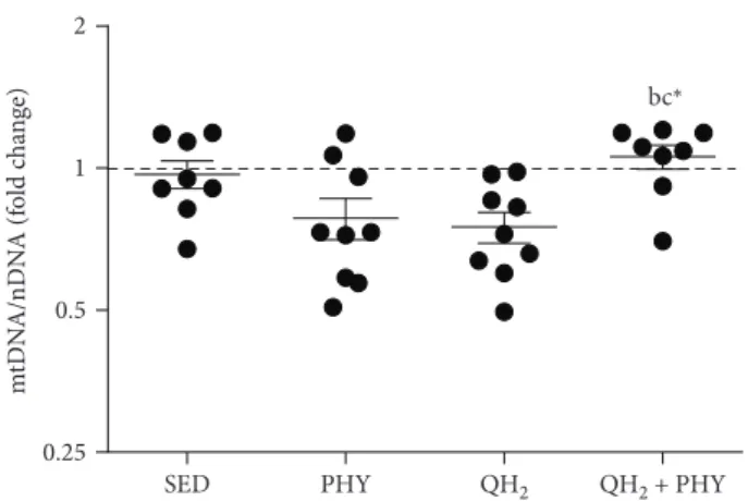
4.4. Combination of Physical Exercise and Ubiquinol Promotes Mitochondrial Biogenesis in the Muscles of the QH2+ PHY Group. Mitochondrial DNA content and
PGC-1α, Tfam, and SIRT5 protein levels of TA muscles were
ana-lyzed to evaluate the mitochondrial biogenesis. Notably, QH2+ PHY treatment was not only able to preserve mito-chondrial morphology and functionality (Figure 5) but also capable of modulating mitochondrial biogenesis. In particu-lar, a significantly higher mtDNA copy number was detected in the QH2+ PHY group compared to the two treatments
alone (QH2+ PHY vs. PHY, 1.35-fold change,p = 0 03, and
QH2+ PHY vs. QH2, 1.41-fold change, p = 0 01, Figure 6).
Moreover, the combined treatment promoted a highly signif-icant increase in the expression of proteins involved in mito-chondrial biogenesis, such as PGC-1α (+284.9%, p < 0 0001) and SIRT5 (+39.5%, p = 0 02), compared to sedentary mice (Figure 7). Regular physical exercise in association with ubi-quinol supplementation was also able to increase the expres-sion levels of TFAM although not in a significant manner. On the contrary, individual treatments, both physical exercise and ubiquinol supplementation, did not induce any changes in these markers, with the exception of a significant downreg-ulation of SIRT5 in the trained mice.
4.5. Combination of Ubiquinol and Physical Exercise Thwarts Activation of Autophagy/Mitophagy Signals and Lowers Caspase 3-Dependent Apoptosis in the Muscles of QH2+ PHY Mice. To assess whether the different treatments impacted muscular autophagy/mitophagy, we analyzed the mRNA expression (Figure 8) of Beclin-1 (a), Atrogin-1 (b), Atg12 (c), and Bnip3l (d) genes encoding key players involved in both these degradation processes. The associa-tion of ubiquinol supplementaassocia-tion and physical exercise pro-duced a significant decrease in the mRNA expression of Beclin-1 (−1.96-fold, p = 0 005), Atg12 (−3.34-fold, p = 0 003), and Bnip3l (−4.1-fold, p = 0 004) if compared to the PHY group. Atrogin-1 expression decreased significantly only compared to sedentary mice (p = 0 04). Ubiquinol sup-plementation alone was able to induce a significant decrease only for Bnip3l mRNA expression to a similar extent to the combined treatment of ubiquinol and physical exercise (−3.7-fold, p = 0 009). To determine whether apoptosis was also modulated, cleaved caspase 3 level was also exam-ined via Western blot assay (Figure 9). Notably, despite that all the treatments were able to significantly decrease cas-pase 3-dependent apoptosis with respect to SED controls, QH2+ PHY combination triggered the most pronounced antiapoptotic effect in PHY (−76.9%), QH2 (−82.6%), and
QH2+ PHY (−96.9%) mouse groups, respectively, compared
% o xidized C o Q9 /t o tal C o Q9 0 20 40 60 80 100 a *
SED PHY QH2 QH2 + PHY
Tibialis Cardiac
a⁎⁎ a⁎
Figure 3: Oxidized coenzyme Q9 level in cardiac and tibialis anterior muscles, expressed as percentage of oxidized of coenzyme Q9in sedentary (SED), physical exercise (PHY), ubiquinol (QH2),
and ubiquinol associated with physical exercise (QH2+ PHY) mouse groups (n = 10).∗p < 0 05 and∗∗p < 0 01; (A) = vs. SED.
SED QH2 + PHY QH2 PHY (a) Fib er size ( 𝜇 m)
SED PHY QH2 QH2 + PHY
20 30 40 50 60 a⁎⁎⁎ a⁎ (b)
Figure 4: (a) Representative microphotographs of fibers (SED, bar = 25 μm; PHY, bar = 65 μm; QH2,bar = 35 μm; and QH2+ PHY,ba
r = 55 μm). (b) Fiber size quantification of soleus muscle, expressed in μm, in sedentary (SED), physical exercise (PHY), ubiquinol (QH2), and ubiquinol associated with physical exercise (QH2+ PHY) mouse groups (n = 5). ∗p < 0 05 and ∗∗∗p < 0 001; (A) = vs. SED.
SED
QH2
QH2 + PHY
PHY
(a)
Red2 fluorescence (RED2-HLog) Plot P04, gatedon P01.R10.R5 100 101 102 103 104 0 20 40 60 80 Dep_cells C o un t (b)
SED PHY QH2 QH2 + PHY 0 20 40 60 80 100 a⁎ b⁎⁎ b⁎⁎ % dep o la rized cells (c)
Figure 5: (a) TEM analysis of mitochondrial ultrastructure of soleus muscle (SED, bar = 200 nm; inset SED, bar = 500 nm; PHY, bar = 1 μm; QH2,bar = 500 nm; and QH2+ PHY, inset, and QH2+ PHY,bar = 500 nm). (b) Mitochondrial membrane depolarization expressed as Red2 Fluorescent (RED2-HLog). (c) Percentage of depolarized cells of gastrocnemius muscle, respectively, in sedentary (black histogram; SED), physical exercise (light grey histogram; PHY), ubiquinol (dark grey histogram; QH2), and ubiquinol associated with physical exercise
(dark grey histogram; QH2+ PHY) mouse groups (n = 10). Dashed histogram (b) represents sample control treated with CCCP∗p < 0 05
to SED (p < 0 0001, Figures 9(a) and 9(b)). Overall, these data suggest that QH2+ PHY combination successfully lowers the
expression of autophagy/mitophagy-associated genes and prevents apoptotic cell death inside the aging muscles.
5. Discussion
In the present study, senescence-accelerated prone 8 (SAMP8) mice, characterized by premature aging and high degree of oxidative stress [68], were used to investigate if a combined approach of mild physical exercise and ubiquinol (CoQH2) supplementation was able to improve
mitochon-drial function and preserve skeletal muscle health during aging. In our experimental settings, SAMP8 mice were treated with ubiquinol, physical exercise, and a combination of both for two months starting in the presarcopenia phase (5 months) until sarcopenia onset (7 months) [53]. As expected, the skeletal muscle of 7-month-old SED mice (used as control) presented high oxidative stress, damaged mito-chondria, high extent of apoptosis, and mitophagy. While ubiquinol or physical exercise alone was able only to partially rescue these impairments, the combination of ubiquinol and physical exercise significantly improved the overall structural and functional status of the skeletal muscle. Skeletal muscle senescence is associated with decreased muscle mass and mitochondrial dysfunction, and the excessive production of mitochondrial ROS seems to strongly associate with the dis-ruption of mitochondrial energy metabolism [19]. In this context, physical exercise has been proposed as a strategy to stimulate mitochondrial respiration and biogenesis counter-acting muscle decline in older subjects [27, 28]. Nonetheless, some studies have shown that ROS production could indeed exacerbate the oxidative stress in senescent muscle, which is characterized by a severely impaired antioxidant response [32–34, 69]. For these reasons, the association of mild reg-ular physical activity and antioxidant therapies could be a powerful strategy to minimize the adverse effects of exercise during aging. In particular, coenzyme Q in its reduced and
active form (ubiquinol), being a key player both in the mitochondrial electron transport chain and in the antioxi-dant response in biological membranes [48], may represent an ideal candidate in improving oxidative status and func-tionality of the senescent muscle.
CoQ10level correlates to high rates of metabolism, and for this reason, it is highest in organs such as the heart, kid-ney, and liver (114, 66.5, and 54.9 g/g tissue, respectively) [70], probably due to the large amounts of mitochondria where it is acting as an energy transfer molecule. In fact, coenzyme Q wasfirst isolated from beef heart mitochondria, in 1957 [71].
Accordingly, at 7 months of age, skeletal muscle con-tent of endogenous CoQ9was significantly lower and more
oxidized in comparison to cardiac muscle. Oral ubiquinol supplementation (500 mg/kg body weight/day) alone was unable to increase skeletal and cardiac muscle CoQ content and only the association of ubiquinol supplementation and mild treadmill running significantly increased the amount of both coenzymes in the cardiac muscle but not in the skel-etal muscle. Increase of both coenzymes (endogenous CoQ9
and dietary absorbed CoQ10) in the heart suggests a higher
biosynthesis rate that could be related to different mitochon-drial requirements triggering both biosynthesis and incorpo-ration. In the skeletal muscle, these changes that could be required for efficient tissue incorporation seem to occur at a much lower extent or might be less evident due to a lower mitochondrial content. Accordingly, Ernster and Dallner have previously shown that feeding rats with a comparable dosage of oxidized CoQ10significantly increased its plasma
content, while tissue CoQ10accumulation was very moderate
and variable in different tissues/organs [47]. In particular, skeletal muscle seems to have a very low ability to incorpo-rate CoQ. However, Sohal and Forster showed that CoQ10
dietary supplementation in rodents was able to change the subcellular localization of CoQ, increasing the mitochondrial content of both coenzymes in various mitochondria-rich tis-sues, such as liver, heart, and skeletal muscle [72]. In another study, the same authors confirmed that skeletal muscle increase in CoQ10following supplementation was the lowest
in all analyzed tissues [73]. In the present study, we verified that the use of orally administered reduced CoQ10did not
provide any significant improvement in tissue uptake, show-ing results in line with previous reports where ubiquinone was used as active substance. This is a relevant observation since ubiquinol has been proposed as a more bioavailable form of Coenzyme Q10; nonetheless, in the proposed experi-mental condition, the oxidative state of ubiquinol does not seem to provide any significant improvement in terms of tissue uptake.
Taken together, these data suggest that ubiquinol dietary supplementation alone might not be enough to produce its cellular accumulation, but additional stimuli, such as physi-cal activity and mitochondrial biogenesis, could improve ubi-quinol incorporation [74]. Indeed, we reported that physical exercise could therefore act as a trigger for CoQ accumula-tion or rearrangement at the subcellular level. This effect was particularly evident in mitochondria-rich cardiac mus-cle, resulting in a significant increase in the overall cellular
SED PHY QH2 QH2 + PHY
0.25 0.5 1 2 m tD N A/nD N A (f o ld c h an ge) bc⁎
Figure 6: Fold change of copy number of mitochondrial DNA/ nuclear DNA (mtDNA/nDNA) measured on gastrocnemius muscle in sedentary (SED), physical exercise (PHY), ubiquinol (QH2), and ubiquinol associated with physical exercise (QH2+ PHY) mouse groups (n = 10). ∗p < 0 05; (b) = vs. PHY and c = vs. QH2.
PGC-1𝛼
H2B
TFAM
SIRT5
VDAC1
SED PHY QH2 QH2 + PHY
(a) Rela ti ve p ro tein le ve l (A U) PGC-1𝛼 TFAM SIRT5 0 1 2 3 4 SED PHY QH2 QH2 + PHY abc⁎⁎⁎⁎ ac⁎b⁎⁎ a⁎ (b)
Figure 7: Western blot analysis (a) and relative protein quantification (b) of PGC-1α, TFAM, and SIRT5 expressed in tibialis anterior (TA) muscle, in sedentary (SED), physical exercise (PHY), ubiquinol (QH2), and ubiquinol associated with physical exercise (QH2+ PHY) mouse groups (n = 5). PGC-1α protein levels were normalized to H2B levels. TFAM and SIRT5 were normalized to VDAC1 levels. AU: arbitrary units.∗p < 0 05,∗∗p < 0 01, and∗∗∗∗p < 0 0001; (a) = vs. SED, (b) = vs. PHY, and (c) = vs. QH2.
B ec lin-1 (f o ld c h an ge) b⁎⁎c⁎ 0.1 1 10
SED PHY QH2 QH2 + PHY
(a) A tr o gin-1 (f o ld c h an ge ) 0.1 1 10 a⁎
SED PHY QH2 QH2 + PHY
(b) A tg12 (f o ld c h an ge)
SED PHY QH2 QH2 + PHY b⁎⁎ 0.01 0.1 1 10 (c)
SED PHY QH2 QH2 + PHY
B ni p3I (f o ld c h an ge) 0.01 0.1 1 10 b⁎⁎ b⁎⁎ (d)
Figure 8: Gene expression (mRNA) expressed as fold change of genes Beclin (a), Atrogin (b), Atg12, (c) and Bnip3l (d) measured on gastrocnemius muscle in sedentary (SED), physical exercise (PHY), ubiquinol (QH2), and ubiquinol associated with physical exercise (QH2+ PHY) mouse groups (n = 5).∗p < 0 05 and∗∗p < 0 01; (a) = vs. SED, (b) = vs. PHY, and (c) = vs. QH2.
content. Moreover, even if we did not observe a significant CoQ accumulation in the skeletal muscle, we observed func-tional modifications at mitochondrial and cellular levels sug-gesting CoQ activity without accumulation in this tissue.
We focused our investigation on skeletal muscle con-sidering its primary involvement in physical exercise. In the SAMP8 model, physical exercise alone produces het-erogeneous responses at the cellular level. On the one hand, it had a clear ergogenic effect being able to promote an increase in skeletal musclefiber size and to improve the oxidative status of endogenous coenzyme Q9. Accordingly, observational and intervention studies have demonstrated that physical exercise has a positive effect on muscle mass, muscle strength, and physical function in the older popula-tion [1, 2, 75, 76]. However, physical exercise also induced mitochondrial disturbances in terms of membrane depolari-zation. This result could be due to a reduced antioxidant activity [68] and to an intrinsic impairment of the electron transport chain [77]. Both conditions characterizing the pre-senescent SAMP8 mice might be further exacerbated by physical exercise in our experimental settings.
Notably, the combined treatment was able to counteract the mitochondrial impairment induced by physical exercise alone and also increased the mitochondrial number assessed as mtDNA/nDNA ratio. These data are confirmed by the analysis of PGC-1α muscle protein level, a key protein involved in the control of mitochondrial biogenesis, oxida-tive metabolism, and autophagy [23, 78, 79] which was significantly increased after 2 months of regular physical exercise associated with ubiquinol supplementation. Several studies reported that induction of PGC-1α, NRF-1, and Tfam expression [80–82] during physical exercise is triggered by oxidative stimuli [83]. On the contrary, aging-derived oxida-tive stress does not produce similar effects altering PGC-1α expression through different mechanisms [63].
Moreover, in our experimental model, increased fiber diameter in trained animals was not associated with a par-allel increase in mtDNA copy number after 2 months of physical exercise, confirming that muscle hypertrophy was not linked with enhanced mitochondrial biogenesis.
Shrinkage of the mitochondrial pool is a feature typical of the senescence process, and it is characterized by decreased enzymatic activity and level of mitochondrial proteins [21, 84, 85] as well as low mtDNA content [63, 86]. In this regard, it is remarkable that PGC-1α upregulation was asso-ciated with a concomitant increase in mtDNA copy number only in skeletal muscle of QH2+ PHY mice and not in mice
subjected to single interventions.
Moreover, SIRT5, which has been recently found to pro-tect mitochondria from fragmentation and degradation, by supporting mitochondrial elongation [87], was significantly increased following 2 months of ubiquinol supplementation and regular physical exercise, further supporting a positive effect of the QH2+ PHY combined treatment that could
sug-gest improvement in mitochondria biogenesis.
Intriguingly, despite the improvements in the mito-chondrial pool and the functionality observed in the QH2
+ PHY group, an increase in musclefiber size was produced by physical exercise alone and to a lower extent by the combination treatment. Indeed, muscle mass depends on different factors other than mitochondrial biogenesis, such as the balance between protein synthesis and degradation. van Wessel et al. described how high oxidative fibers are small in size, despite their high capacity for protein synthe-sis if compared to low oxidative fibers [88]. The authors suggest that cellular energy status may be crucial in mediat-ing either a low oxidative phenotype and a large size or a high oxidative phenotype but a small size. They also suggest that oxidative fibers have a higher rate of muscle protein degradation in the presence of low cellular energy or high oxidative stress status, a condition similar to our experi-mental model.
During aging, senescent cells respond to a wide range of damaging stimuli produced by the accumulation of dys-functional proteins and organelles, among which mito-chondria play a pivotal role due to their bivalent role as source, target of ROS, and master regulator of programmed cell death processes. Throughout evolution, cells developed strategies like autophagy, mitophagy, and apoptosis to manage these constraints. These tightly regulated and
SED Caspase 3 −35 kDA −19 kDA 𝛽-Actin Cleaved Caspase 3 PHY QH2QH2 + PHY (a)
SED PHY QH2 QH2 + PHY 0.0 0.5 1.0 1.5 2.0 Cle av ed caspas e 3/ to ta l caspas e 3 a⁎⁎⁎⁎ a⁎⁎⁎⁎ a⁎⁎⁎⁎ (b)
Figure 9: Immunoblot image (a) and relative protein quantification normalized to β-actin (b) of caspase 3 and cleaved caspase 3, measured on tibialis anterior muscle in sedentary (SED), physical exercise (PHY), ubiquinol (QH2), and ubiquinol associated with physical exercise (QH2+ PHY) mouse groups (n = 5).∗∗∗∗p < 0 0001; (a) = vs. SED.
interconnected processes are of pivotal importance also in the maintenance muscle homeostasis, which is dysregulated during aging and sarcopenia [89, 90]. In this context, ROS originating from mitochondria have been reported to acti-vate both autophagy machinery [91] and caspase-dependent apoptosis [92].
In this regard, the combined treatment with ubiquinol and physical exercise not only ameliorated muscle oxidative stress and bioenergetics but also associated with a decreased expression of autophagy/mitophagy-associated genes, such as Bnip3l, Atg12, and Beclin-1 known to promote mito-chondrial fragmentation and mitophagy and autophago-some formation [93, 94].
Finally, the combined treatment was also able to prevent caspase 3-dependent apoptosis. Since caspase cleavage cas-cade is a readout of mitochondria-associated apoptotic cell death [95–97], these data strengthen the positive impact of QH2+ PHY combination on mitochondria. Furthermore, previous studies have shown that cytochrome c release from mitochondria activates caspase 3 that in turn cleaves respira-tory complex proteins exacerbating mitochondrial dysfunc-tion increasing ROS producdysfunc-tion [92]. The loss of muscle mass, caused by an imbalance between muscle protein syn-thesis (MPS) and muscle protein breakdown (MPB) [98] and consequently decline in strength, can also be due to increased activity of the ubiquitin proteasome pathway (UPP), which is also responsible for mitochondrial protein quality control. Our data, showing a decrease in Atrogin-1 expression and myogenin (MyoG) protein level (supplemen-tary material (available here)) and highlighting a potential effect of the combined treatment, suggest also a possible downregulation of UPP proteolytic pathway, which might concur to muscle health preservation [99].
Taking into consideration the limitations of this study, associated to the fact that we did not take into account muscle fiber type composition and we did not measure the actual intramuscular ROS levels, overall in this context, our data demonstrate that ubiquinol might directly pre-vent cell death by acting both as mitochondrial nutrient and as ROS scavenger.
6. Conclusion
In conclusion, the present study shows that ubiquinol supplementation and physical exercise synergize at improv-ing mitochondrial functionality, counteractimprov-ing the deleteri-ous effects of physical exercise-induced ROS in the muscles of a SAMP8 mouse model. These results suggest that ubiqui-nol could be a powerful dietary supplement in sports nutri-tion and in particular in the elderly. The use of antioxidants in sports practice is still a debated topic, because it was demonstrated that some of these molecules are able to turn off the hormetic signals generated by physical exercise. How-ever, antioxidant compounds represent a very heterogeneous family of molecules with different targets and cellular tropism so that evidences reported for some of them should not be simply extended to all molecules with sim-ilar activities. Moreover, data available in the scientific liter-ature referring to a quenching effect of antioxidants on
adaptive response commonly refer to trained healthy subjects. Our study shows that ubiquinol, while reducing harmful effects generated by physical exercise, improves exercise-induced hormetic response in a model character-ized by elevated oxidative stress and prone to premature aging such as SAMP8 mice.
Data Availability
The data used to support thefindings of this study are avail-able from the corresponding author upon request.
Conflicts of Interest
The authors declare that they have no conflicts of interest.
Authors
’ Contributions
Andreani C. and Bartolacci C. contributed equally to this work.
Acknowledgments
This study was supported by a grant fellowship 2017 from the Fondazione Umberto Veronesi (Silvestri S. was a recipient of grant“Post-doctoral Fellowship 2017”). The authors wish to thank Kaneka for kindly providing ubiquinol.
Supplementary Materials
Immunoblot image and relative protein quantification of myogenin (MyoG) normalized to H2B (Figure 10) were measured on tibialis anterior muscle of all mouse groups. The results showed how ubiquinol supplementation, in association with physical exercise (QH2+ PHY mouse
group), was significantly (∗p < 0 05) able to prevent the
increase in MyoG protein level induced after physical exercise alone (PHY mouse group). These data highlight a protective role of ubiquinol towards the deleterious effect of mild physical exercise in an old skeletal muscle of senescence-accelerated mice, suggesting also a possible downregulation of the UPP proteolytic pathway. Figure 10: immunoblot image and relative protein quantification of myogenin (MyoG) normalized to H2B measured on tibialis anterior muscle in sedentary (SED), physical exercise (PHY), ubiquinol (QH2), and ubiquinol associated
with physical exercise (QH2+ PHY) mouse groups (n = 5),
∗p < 0 05. (Supplementary Materials)
References
[1] H. Kim, T. Suzuki, K. Saito et al.,“Effects of exercise and tea catechins on muscle mass, strength and walking ability in community-dwelling elderly Japanese sarcopenic women: a randomized controlled trial,” Geriatrics & Gerontology Inter-national, vol. 13, no. 2, pp. 458–465, 2013.
[2] M. Leenders, L. B. Verdijk, L. van der Hoeven, J. van Kranenburg, R. Nilwik, and L. J. C. van Loon,“Elderly men and women benefit equally from prolonged resistance-type exercise training,” The Journals of Gerontology Series A:
Biological Sciences and Medical Sciences, vol. 68, no. 7, pp. 769– 779, 2013.
[3] C. . K. Liu, X. Leng, F. -C. Hsu et al.,“The impact of sarcopenia on a physical activity intervention: the Lifestyle Interventions and Independence for Elders Pilot Study (LIFE-P),” The Jour-nal of Nutrition, Health & Aging, vol. 18, no. 1, pp. 59–64, 2014.
[4] K. Keller and M. Engelhardt,“Strength and muscle mass loss with aging process. Age and strength loss,” Muscles , Ligaments and Tendons Journal, vol. 3, no. 4, p. 346, 2014.
[5] N. Montero-Fernández and J. A. Serra-Rexach,“Role of exer-cise on sarcopenia in the elderly,” European Journal of Physical and Rehabilitation Medicine, vol. 49, no. 1, pp. 131–143, 2013. [6] M. Brotto and E. L. Abreu, “Sarcopenia: pharmacology of today and tomorrow,” Journal of Pharmacology and Experi-mental Therapeutics, vol. 343, no. 3, pp. 540–546, 2012. [7] D. Scott, L. Blizzard, J. Fell, G. Giles, and G. Jones,
“Associa-tions between dietary nutrient intake and muscle mass and strength in community-dwelling older adults: the Tasmanian Older Adult Cohort Study,” Journal of the American Geriatrics Society, vol. 58, no. 11, pp. 2129–2134, 2010.
[8] A. J. Cruz-Jentoft and F. Landi,“Sarcopenia,” Clinical Medi-cine, vol. 14, no. 2, pp. 183–186, 2014.
[9] I. H. Rosenberg,“Sarcopenia: origins and clinical relevance,” Clinics in Geriatric Medicine, vol. 27, no. 3, pp. 337–339, 2011. [10] W. J. Evans,“Skeletal muscle loss: cachexia, sarcopenia, and inactivity,” The American Journal of Clinical Nutrition, vol. 91, no. 4, pp. 1123S–1127S, 2010.
[11] G. A. Power, B. H. Dalton, and C. L. Rice,“Human neuromus-cular structure and function in old age: a brief review,” Journal of Sport and Health Science, vol. 2, no. 4, pp. 215–226, 2013. [12] U. Granacher and T. Hortobágyi,“Exercise to improve
mobil-ity in healthy aging,” Sports Medicine, vol. 45, no. 12, pp. 1625-1626, 2015.
[13] H. N. Carter, C. C. W. Chen, and D. A. Hood,“Mitochondria, muscle health, and exercise with advancing age,” Physiology, vol. 30, no. 3, pp. 208–223, 2015.
[14] H. M. McBride, M. Neuspiel, and S. Wasiak,“Mitochondria: more than just a powerhouse,” Current Biology, vol. 16, no. 14, pp. R551–R560, 2006.
[15] L. D. Tryon, A. Vainshtein, J. M. Memme, M. J. Crilly, and D. A. Hood,“Recent advances in mitochondrial turnover dur-ing chronic muscle disuse,” Integrative Medicine Research, vol. 3, no. 4, pp. 161–171, 2014.
[16] M. P. Mattson,“Hormesis defined,” Ageing Research Reviews, vol. 7, no. 1, pp. 1–7, 2008.
[17] A. R. Konopka and K. Sreekumaran Nair,“Mitochondrial and skeletal muscle health with advancing age,” Molecular and Cel-lular Endocrinology, vol. 379, no. 1-2, pp. 19–29, 2013. [18] D. Y. Seo, S. R. Lee, N. Kim, K. S. Ko, B. D. Rhee, and J. Han,
“Age-related changes in skeletal muscle mitochondria: the role of exercise,” Integrative Medicine Research, vol. 5, no. 3, pp. 182–186, 2016.
[19] I. Sanchez-Roman, A. Gómez, I. Pérez et al.,“Effects of aging and methionine restriction applied at old age on ROS genera-tion and oxidative damage in rat liver mitochondria,” Bioger-ontology, vol. 13, no. 4, pp. 399–411, 2012.
[20] H. P. Indo, M. Davidson, H. C. Yen et al.,“Evidence of ROS generation by mitochondria in cells with impaired electron transport chain and mitochondrial DNA damage,” Mitochon-drion, vol. 7, no. 1-2, pp. 106–118, 2007.
[21] K. R. Short, M. L. Bigelow, J. Kahl et al., “Decline in skel-etal muscle mitochondrial function with aging in humans,” Proceedings of the National Academy of Sciences of the United States of America, vol. 102, no. 15, pp. 5618– 5623, 2005.
[22] O. E. Rooyackers, D. B. Adey, P. A. Ades, and K. S. Nair, “Effect of age on in vivo rates of mitochondrial protein synthe-sis in human skeletal muscle,” Proceedings of the National Academy of Sciences of the United States of America, vol. 93, no. 26, pp. 15364–15369, 1996.
[23] Z. Wu, P. Puigserver, U. Andersson et al., “Mechanisms controlling mitochondrial biogenesis and respiration through the thermogenic coactivator PGC-1,” Cell, vol. 98, no. 1, pp. 115–124, 1999.
[24] A. Vainshtein, E. M. A. Desjardins, A. Armani, M. Sandri, and D. A. Hood,“PGC-1α modulates denervation-induced mito-phagy in skeletal muscle,” Skeletal Muscle, vol. 5, no. 1, p. 9, 2015.
[25] N. Mizushima and B. Levine, “Autophagy in mamma-lian development and differentiation,” Nature Cell Biology, vol. 12, no. 9, pp. 823–830, 2010.
[26] J. J. Wu, C. Quijano, E. Chen et al.,“Mitochondrial dysfunc-tion and oxidative stress mediate the physiological impairment induced by the disruption of autophagy,” Aging, vol. 1, no. 4, pp. 425–437, 2009.
[27] L. DiPietro, “Physical activity in aging: changes in patterns and their relationship to health and function,” The Journals of Gerontology Series A: Biological Sciences and Medical Sci-ences, vol. 56, no. 2, pp. 13–22, 2001.
[28] J. Dziura, C. Mendes de Leon, S. Kasl, and L. DiPietro, “Can physical activity attenuate aging-related weight loss in older people? The Yale Health and Aging Study, 1982–1994,” American Journal of Epidemiology, vol. 159, no. 8, pp. 759– 767, 2004.
[29] R. N. Baumgartner, S. J. Wayne, D. L. Waters, I. Janssen, D. Gallagher, and J. E. Morley,“Sarcopenic obesity predicts instrumental activities of daily living disability in the elderly,” Obesity Research, vol. 12, no. 12, pp. 1995–2004, 2004. [30] A. P. Russell, V. C. Foletta, R. J. Snow, and G. D. Wadley,
“Skeletal muscle mitochondria: a major player in exercise, health and disease,” Biochimica et Biophysica Acta (BBA) -General Subjects, vol. 1840, no. 4, pp. 1276–1284, 2014. [31] S. Ketkar, A. Rathore, A. Kandhare et al.,“Alleviating
exercise-induced muscular stress using neat and processed bee pollen: oxidative markers, mitochondrial enzymes, and myostatin expression in rats,” Integrative Medicine Research, vol. 4, no. 3, pp. 147–160, 2015.
[32] P. V. Komi,“Stretch-shortening cycle: a powerful model to study normal and fatigued muscle,” Journal of Biomechanics, vol. 33, no. 10, pp. 1197–1206, 2000.
[33] G. S. Lynch, J. A. Faulkner, and S. V. Brooks,“Force deficits and breakage rates after single lengthening contractions of sin-gle fastfibers from unconditioned and conditioned muscles of young and old rats,” American Journal of Physiology-Cell Phys-iology, vol. 295, no. 1, pp. C249–C256, 2008.
[34] I. M. Conboy, M. J. Conboy, G. M. Smythe, and T. A. Rando, “Notch-mediated restoration of regenerative poten-tial to aged muscle,” Science, vol. 302, no. 5650, pp. 1575– 1577, 2003.
[35] Y. Shimomura, M. Suzuki, S. Sugiyama, Y. Hanaki, and T. Ozawa, “Protective effect of coenzyme Q10 on
exercise-induced muscular injury,” Biochemical and Biophysical Research Communications, vol. 176, no. 1, pp. 349–355, 1991. [36] M. Kon, F. Kimura, T. Akimoto et al.,“Effect of Coenzyme Q10 supplementation on exercise-induced muscular injury of rats,” Exerc Immunol Rev, vol. 13, pp. 76–88, 2007.
[37] M. Kon, K. Tanabe, T. Akimoto et al., “Reducing exercise-induced muscular injury in kendo athletes with supplementa-tion of coenzyme Q10,” British Journal of Nutrition, vol. 100, no. 4, pp. 903–909, 2008.
[38] A. Abadi, J. D. Crane, D. Ogborn et al.,“Supplementation with
α-lipoic acid, CoQ10, and vitamin E augments running
perfor-mance and mitochondrial function in female mice,” PLoS One, vol. 8, no. 4, article e60722, 2013.
[39] G. Tian, J. Sawashita, H. Kubo et al.,“Ubiquinol-10 supple-mentation activates mitochondria functions to decelerate senescence in senescence-accelerated mice,” Antioxidants & Redox Signaling, vol. 20, no. 16, pp. 2606–2620, 2014. [40] A. Lass, S. Agarwal, and R. S. Sohal,“Mitochondrial
ubiqui-none homologues, superoxide radical generation, and longev-ity in different mammalian species,” Journal of Biological Chemistry, vol. 272, no. 31, pp. 19199–19204, 1997.
[41] S. Kamzalov, N. Sumien, M. J. Forster, and R. S. Sohal, “Coen-zyme Q intake elevates the mitochondrial and tissue levels of Coenzyme Q and alpha-tocopherol in young mice,” The Jour-nal of Nutrition, vol. 133, no. 10, pp. 3175–3180, 2003. [42] C. Gómez-Díaz, M. I. Burón, F. J. Alcaín et al.,“Effect of
die-tary coenzyme Q and fatty acids on the antioxidant status of rat tissues,” Protoplasma, vol. 221, no. 1-2, pp. 11–17, 2003. [43] L. K. Kwong, S. Kamzalov, I. Rebrin et al.,“Effects of coenzyme
Q10administration on its tissue concentrations, mitochondrial oxidant generation and oxidative stress in the rat,” Free Radi-cal Biology & Medicine, vol. 33, no. 5, pp. 627–638, 2002. [44] H. Maruoka, K. Fujii, K. Inoue, and S. Kido,“Long-term effect
of ubiquinol on exercise capacity and the oxidative stress reg-ulation system in SAMP1 mice,” Journal of Physical Therapy Science, vol. 26, no. 3, pp. 367–371, 2014.
[45] C. Schmelzer, J. G. Okun, D. Haas et al.,“The reduced form of coenzyme Q10mediates distinct effects on cholesterol
metabo-lism at the transcriptional and metabolite level in SAMP1 mice,” IUBMB Life, vol. 62, no. 11, pp. 812–818, 2010. [46] M. Kitano, D. Watanabe, S. Oda et al., “Subchronic oral
toxicity of ubiquinol in rats and dogs,” International Journal of Toxicology, vol. 27, no. 2, pp. 189–215, 2008.
[47] L. Ernster and G. Dallner, “Biochemical, physiological and medical aspects of ubiquinone function,” Biochimica et Bio-physica Acta (BBA) - Molecular Basis of Disease, vol. 1271, no. 1, pp. 195–204, 1995.
[48] G. P. Littarru and L. Tiano,“Clinical aspects of coenzyme Q10: an update,” Nutrition, vol. 26, no. 3, pp. 250–254, 2010. [49] G. Dallner and P. J. Sindelar, “Regulation of ubiquinone
metabolism,” Free Radical Biology & Medicine, vol. 29, no. 3-4, pp. 285–294, 2000.
[50] G. P. Littarru, M. Battino, and K. Folkers,“Clinical aspects of coenzyme Q: improvement of cellular bioenergetics or antiox-idant perfection?,” in Handbook of Antioxidants, E. Cadenas and L. Packer, Eds., pp. 203–239, Marcel Decker, New York, NY, USA, 1996.
[51] A. Rötig, E. L. Appelkvist, V. Geromel et al., “Quinone-respon-sive multiple respiratory-chain dysfunction due to widespread coenzyme Q10deficiency,” Lancet, vol. 356, no. 9227, pp. 391– 395, 2000.
[52] D. Alf, M. E. Schmidt, and S. C. Siebrecht,“Ubiquinol supple-mentation enhances peak power production in trained athletes: a double-blind, placebo controlled study,” Journal of the International Society of Sports Nutrition, vol. 10, no. 1, p. 24, 2013.
[53] A. Y. Guo, K. S. Leung, P. M. Siu et al.,“Muscle mass, structural and functional investigations of senescence-accelerated mouse P8 (SAMP8),” Experimental Animals, vol. 64, no. 4, pp. 425– 433, 2015.
[54] T. Takeda,“Senescence-accelerated mouse (SAM): a biogeron-tological resource in aging research,” Neurobiology of Aging, vol. 20, no. 2, pp. 105–110, 1999.
[55] J. E. Morley, H. J. Armbrecht, S. A. Farr, and V. B. Kumar, “The senescence accelerated mouse (SAMP8) as a model for oxidative stress and Alzheimer’s disease,” Biochimica et Bio-physica Acta (BBA) - Molecular Basis of Disease, vol. 1822, no. 5, pp. 650–656, 2012.
[56] Y. Chiba, A. Shimada, N. Kumagai et al., “The senescence-accelerated mouse (SAM): a higher oxidative stress and age-dependent degenerative diseases model,” Neurochemical Research, vol. 34, no. 4, pp. 679–687, 2009.
[57] F. Derbré, A. Gratas-Delamarche, M. C. Gómez-Cabrera, and J. Viña,“Inactivity-induced oxidative stress: a central role in age-related sarcopenia?,” European Journal of Sport Science, vol. 14, Supplement 1, pp. S98–S108, 2014.
[58] W. Derave, B. O. Eijnde, M. Ramaekers, and P. Hespel,“Soleus muscles of SAMP8 mice provide an accelerated model of skel-etal muscle senescence,” Experimental Gerontology, vol. 40, no. 7, pp. 562–572, 2005.
[59] T. Nishikawa, J. A. Takahashi, T. Matsushita et al.,“Tubular aggregates in the skeletal muscle of the senescence-accelerated mouse SAM,” Mechanisms of Ageing and Develop-ment, vol. 114, no. 2, pp. 89–99, 2000.
[60] C. Moorwood, M. Liu, Z. Tian, and E. R. Barton,“Isometric and eccentric force generation assessment of skeletal muscles isolated from murine models of muscular dystrophies,” Journal of Visualized Experiments, vol. 8, no. 71, article e50036, 2013. [61] M. Romanick, L. D. V. Thompson, and H. M. Brown-Borg,
“Murine models of atrophy, cachexia, and sarcopenia in skele-tal muscle,” Biochimica et Biophysica Acta (BBA) - Molecular Basis of Disease, vol. 1832, no. 9, pp. 1410–1420, 2013. [62] T. Takeda, M. Hosokawa, S. Takeshita et al.,“A new murine
model of accelerated senescence,” Mechanisms of Ageing and Development, vol. 17, no. 2, pp. 183–194, 1981.
[63] C. Kang, E. Chung, G. Diffee, and L. L. Ji, “Exercise training attenuates aging-associated mitochondrial dysfunction in rat skeletal muscle: role of PGC-1α,” Experimental Gerontology, vol. 48, no. 11, pp. 1343–1350, 2013.
[64] N. Okudan, S. Revan, S. S. Balci, M. Belviranli, H. Pepe, and H. Gökbel,“Effects of CoQ10supplementation and swimming
training on exhaustive exercise-induced oxidative stress in rat heart,” Bratislava Medical Journal, vol. 113, no. 07, pp. 393– 399, 2012.
[65] S. Salucci, S. Burattini, F. Buontempo, A. M. Martelli, E. Falcieri, and M. Battistelli, “Protective effect of different antioxidant agents in UVB-irradiated keratinocytes,” Euro-pean Journal of Histochemistry, vol. 61, no. 3, p. 2784, 2017. [66] M. Battistelli, S. Salucci, E. Olivotto et al., “Cell death in
human articular chondrocyte: a morpho-functional study in micromass model,” Apoptosis, vol. 19, no. 10, pp. 1471– 1483, 2014.
[67] M. W. Pfaffl, “A new mathematical model for relative quanti-fication in real-time RT–PCR,” Nucleic Acids Research, vol. 29, no. 9, article e45, pp. 45e–445, 2001.
[68] S. Taniguchi, M. Hanafusa, H. Tsubone et al., “Age-depen-dency of the serum oxidative level in the senescence-accelerated mouse prone 8,” Journal of Veterinary Medical Sci-ence, vol. 78, no. 8, pp. 1369–1371, 2016.
[69] M. J. Jackson and A. McArdle,“Age-related changes in skeletal muscle reactive oxygen species generation and adaptive responses to reactive oxygen species,” The Journal of Physiol-ogy, vol. 589, no. 9, pp. 2139–2145, 2011.
[70] R. Saini,“Coenzyme Q10: the essential nutrient,” Journal of Pharmacy and Bioallied Sciences, vol. 3, no. 3, pp. 466-467, 2011.
[71] F. L. Crane, Y. Hatefi, R. L. Lester, and C. Widmer, “Isolation of a quinone from beef heart mitochondria,” Biochimica et Biophysica Acta, vol. 25, no. 1, pp. 220-221, 1957.
[72] R. S. Sohal and M. J. Forster,“Coenzyme Q, oxidative stress and aging,” Mitochondrion, vol. 7, pp. S103–S111, 2007. [73] R. S. Sohal, S. Kamzalov, N. Sumien et al.,“Effect of coenzyme
Q10intake on endogenous coenzyme Q content,
mitochon-drial electron transport chain, antioxidative defenses, and life span of mice,” Free Radical Biology & Medicine, vol. 40, no. 3, pp. 480–487, 2006.
[74] M. Guescini, L. Tiano, M. L. Genova et al.,“The combination of physical exercise with muscle-directed antioxidants to counteract sarcopenia: a biomedical rationale for pleiotropic treatment with creatine and coenzyme Q10,” Oxidative Med-icine and Cellular Longevity, vol. 2017, Article ID 7083049, 19 pages, 2017.
[75] J. D. Crane, L. G. MacNeil, and M. A. Tarnopolsky, “Long-term aerobic exercise is associated with greater muscle strength throughout the life span,” The Journals of Gerontology Series A: Biological Sciences and Medical Sciences, vol. 68, no. 6, pp. 631–638, 2013.
[76] S. Zampieri, L. Pietrangelo, S. Loefler et al., “Lifelong physical exercise delays age-associated skeletal muscle decline,” The Journals of Gerontology Series A: Biological Sciences and Medi-cal Sciences, vol. 70, no. 2, pp. 163–173, 2015.
[77] H. Nakahara, T. Kanno, Y. Inai et al.,“Mitochondrial dysfunc-tion in the senescence accelerated mouse (SAM),” Free Radical Biology & Medicine, vol. 24, no. 1, pp. 85–92, 1998.
[78] M. Sandri, J. Lin, C. Handschin et al.,“PGC-1α protects skele-tal muscle from atrophy by suppressing FoxO3 action and atrophy-specific gene transcription,” Proceedings of the National Academy of Sciences of the United States of America, vol. 103, no. 44, pp. 16260–16265, 2006.
[79] J. Lin, C. Handschin, and B. M. Spiegelman,“Metabolic con-trol through the PGC-1 family of transcription coactivators,” Cell Metabolism, vol. 1, no. 6, pp. 361–370, 2005.
[80] K. Baar, A. Wende, T. Jones et al., “Adaptations of skeletal muscle to exercise: rapid increase in the transcriptional coacti-vator PGC-1,” The FASEB Journal, vol. 16, no. 14, pp. 1879– 1886, 2002.
[81] J. W. Gordon, A. A. Rungi, H. Inagaki, and D. A. Hood, “Selected Contribution: Effects of contractile activity on mitochondrial transcription factor A expression in skeletal muscle,” Journal of Applied Physiology, vol. 90, no. 1, pp. 389–396, 2001.
[82] I. Irrcher, P. J. Adhihetty, T. Sheehan, A. M. Joseph, and D. A. Hood,“PPARγ coactivator-1α expression during thyroid
hormone- and contractile activity-induced mitochondrial adaptations,” American Journal of Physiology-Cell Physiology, vol. 284, no. 6, pp. C1669–C1677, 2003.
[83] C. Kang, K. M. O'Moore, J. R. Dickman, and L. L. Ji, “Exer-cise activation of muscle peroxisome proliferator-activated receptor-γ coactivator-1α signaling is redox sensitive,” Free Radical Biology & Medicine, vol. 47, no. 10, pp. 1394– 1400, 2009.
[84] B. Chabi, V. Ljubicic, K. J. Menzies, J. H. Huang, A. Saleem, and D. A. Hood,“Mitochondrial function and apoptotic sus-ceptibility in aging skeletal muscle,” Aging Cell, vol. 7, no. 1, pp. 2–12, 2008.
[85] V. Ljubicic, A. M. Joseph, P. J. Adhihetty et al., “Molecular basis for an attenuated mitochondrial adaptive plasticity in aged skeletal muscle,” Aging, vol. 1, no. 9, pp. 818–830, 2009. [86] S. Welle, K. Bhatt, B. Shah, N. Needler, J. M. Delehanty, and
C. A. Thornton,“Reduced amount of mitochondrial DNA in aged human muscle,” Journal of Applied Physiology, vol. 94, no. 4, pp. 1479–1484, 2003.
[87] H. Guedouari, T. Daigle, L. Scorrano, and E. Hebert-Chatelain, “Sirtuin 5 protects mitochondria from fragmentation and degradation during starvation,” Biochimica et Biophysica Acta (BBA) - Molecular Cell Research, vol. 1864, no. 1, pp. 169–176, 2017.
[88] T. van Wessel, A. de Haan, W. J. van der Laarse, and R. T. Jaspers,“The muscle fiber type-fiber size paradox: hypertro-phy or oxidative metabolism?,” European Journal of Applied Physiology, vol. 110, no. 4, pp. 665–694, 2010.
[89] A. Dirks and C. Leeuwenburgh,“Apoptosis in skeletal muscle with aging,” American Journal of Physiology-Regulatory, Inte-grative and Comparative Physiology, vol. 282, no. 2, pp. R519–R527, 2002.
[90] R. A. Gottlieb and R. S. Carreira,“Autophagy in health and disease. 5. Mitophagy as a way of life,” American Journal of Physiology-Cell Physiology, vol. 299, no. 2, pp. C203–C210, 2010.
[91] Y. Chen, M. B. Azad, and S. B. Gibson, “Superoxide is the major reactive oxygen species regulating autophagy,” Cell Death & Differentiation, vol. 16, no. 7, pp. 1040–1052, 2009. [92] J. E. Ricci, R. A. Gottlieb, and D. R. Green,“Caspase-mediated
loss of mitochondrial function and generation of reactive oxy-gen species during apoptosis,” The Journal of Cell Biology, vol. 160, no. 1, pp. 65–75, 2003.
[93] E. Masiero, L. Agatea, C. Mammucari et al., “Autophagy is required to maintain muscle mass,” Cell Metabolism, vol. 10, no. 6, pp. 507–515, 2009.
[94] E. Wirawan, S. Lippens, T. vanden Berghe et al.,“Beclin1: a role in membrane dynamics and beyond,” Autophagy, vol. 8, no. 1, pp. 6–17, 2012.
[95] M. Enari, H. Sakahira, H. Yokoyama, K. Okawa, A. Iwamatsu, and S. Nagata,“A caspase-activated DNase that degrades DNA during apoptosis, and its inhibitor ICAD,” Nature, vol. 391, no. 6662, pp. 43–50, 1998.
[96] S. Sahara, M. Aoto, Y. Eguchi, N. Imamoto, Y. Yoneda, and Y. Tsujimoto, “Acinus is a caspase-3-activated protein required for apoptotic chromatin condensation,” Nature, vol. 401, no. 6749, pp. 168–173, 1999.
[97] M. L. Coleman, E. A. Sahai, M. Yeo, M. Bosch, A. Dewar, and M. F. Olson, “Membrane blebbing during apoptosis results from caspase-mediated activation of ROCK I,” Nature Cell Biology, vol. 3, no. 4, pp. 339–345, 2001.
[98] R. J. Stefanetti, E. Zacharewicz, P. Della Gatta, A. Garnham, A. P. Russell, and S. Lamon,“Ageing has no effect on the reg-ulation of the ubiquitin proteasome-related genes and proteins following resistance exercise,” Frontiers in Physiology, vol. 5, no. 30, 2014.
[99] V. Moresi, A. H. Williams, E. Meadows et al., “Myogenin and class II HDACs control neurogenic muscle atrophy by inducing E3 ubiquitin ligases,” Cell, vol. 143, no. 1, pp. 35– 45, 2010.
Stem Cells
International
Hindawi www.hindawi.com Volume 2018 Hindawi www.hindawi.com Volume 2018 INFLAMMATIONEndocrinology
International Journal ofHindawi www.hindawi.com Volume 2018 Hindawi www.hindawi.com Volume 2018
Disease Markers
Hindawi www.hindawi.com Volume 2018 BioMed Research InternationalOncology
Journal of Hindawi www.hindawi.com Volume 2013 Hindawi www.hindawi.com Volume 2018Oxidative Medicine and Cellular Longevity
Hindawi
www.hindawi.com Volume 2018
PPAR Research
Hindawi Publishing Corporation
http://www.hindawi.com Volume 2013 Hindawi www.hindawi.com
The Scientific
World Journal
Volume 2018 Immunology Research Hindawi www.hindawi.com Volume 2018 Journal ofObesity
Journal of Hindawi www.hindawi.com Volume 2018 Hindawi www.hindawi.com Volume 2018 Computational and Mathematical Methods in Medicine Hindawi www.hindawi.com Volume 2018Behavioural
Neurology
Ophthalmology
Journal of Hindawi www.hindawi.com Volume 2018Diabetes Research
Journal ofHindawi
www.hindawi.com Volume 2018
Hindawi
www.hindawi.com Volume 2018
Research and Treatment
AIDS
Hindawi
www.hindawi.com Volume 2018 Gastroenterology Research and Practice
Hindawi www.hindawi.com Volume 2018