SCUOLA DI DOTTORATO DI RICERCA IN SCIENZE BIOMEDICHE
Direttore della scuola: Prof. Andrea PianaINDIRIZZO IN NEUROSCIENZE
Responsabile di Indirizzo: Prof.ssa Maria Speranza Desole – Dott.ssa Rossana Migheli
XXVII CICLO
Rational design and applications of a new
Cell-penetrating Peptide targeting Mitochondria
Direttore
Prof. Andrea Piana
Tutor
Dott.ssa Rossana Migheli
Co-tutor
Tesi di Dottorato di
Prof. Ülo Langel
Dott. Pirisinu Marco
ANNO ACCADEMICO 2013-2014
La presente tesi è stata sviluppata nell’ambito della scuola di dottorato in Scienze
SCUOLA DI DOTTORATO DI RICERCA IN SCIENZE BIOMEDICHE
Direttore della scuola: Prof. Andrea PianaINDIRIZZO IN NEUROSCIENZE
Responsabile di Indirizzo: Prof.ssa Maria Speranza Desole – Dott.ssa Rossana Migheli XXVII CICLO
Rational design and applications of a new Cell-penetrating
Peptide targeting Mitochondria
Direttore
Prof. Andrea Piana
Tutor
Dott.ssa Rossana Migheli
Co-tutor
Tesi di Dottorato di
University of Sassari University of Stockholm
My PhD was carried out during three years at the Department of Clinical and Experimental Medicine of the Medical School of the University of Sassari and for a period of ten months at the Department of Neurochemistry ant toxicology, University of Stockholm. The collaboration between these two groups contributed to developing of following thesis.
CONTENTS
ABBREVIATIONS ... 1
INTRODUCTION ... 5
Literature Overview ... 5
Mitochondria ... 9
Organization and functions ... 9
Mitochondrial electron transport chain ...11
Mitochondrial Membrane Potential ...14
Mitochondrial generation of ROS ...15
Mitochondrial Antioxidants ...16
Oxidative stress ...18
Cell-Penetrating Peptides ...20
Classification ...20
CPPs uptake mechanism ...21
CPPs in drug delivery strategy: Applications and organelle specific delivery ...25
Nuclear Localization Sequences ...26
Mitochondria-Target Antioxidants ...27
GOAL OF THE THESIS ...34
MATHERIAS AND METHODS ...35
Solid phase Peptide Synthesis (SPPS) ...35
Manual Synthesis of MitPep-peptide and SS31 peptide ...38
Purifiction of peptides by HPLC and mass evaluation by MALDI-TOFF ...39
Cell culture ...40
Cell viability assay...41
Mitochondrial membrane potential assay ...42
ROS production assay ...43
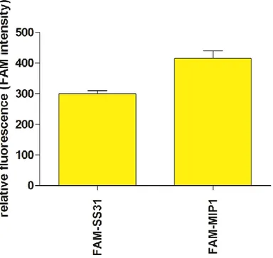
Measurement of mitochondrial uptake of (FAM) 5(6)carboxyfluorescein-MIP1 conjugated on isolated mitochondria ...45
Design of MIP1 peptide ...47
Effects of MIP1 peptide on the cell viability of HeLa 705 cells ...48
Effects of MIP1 peptide on mitochondrial membrane potential of HeLa 705 cells ...50
Effects of MIP1 peptide on ROS production in HeLa 705 cells ...52
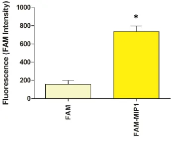
MIP1 cellular uptake and localization ...54
Mitochondrial uptake of MIP1 ...56
DISCUSSION AND CONCLUSION ...58
SUMMARY IN ITALIAN ...62
ABBREVIATIONS
ACN Acetonitril
ALS Amyotrophic lateral sclerosis
Arg Arginine
BBB Blood brain barrier
CME Clathrin- mediated endocytosis
CPPs Cell Penetrating Peptides
CvME Caveolae–mediated endocytosis
DA Dopamin
D-Arg D-arginine
DIC 1,3-diisopropylcarbodiimide
DIPEA N,N-Diisopropylethylamine
DMEM Dulbecco's Modified Eagle Medium
DMF Dimethylformamide DMSO Dimethylsulfoxide DTT Dithiolthreitol EDT 1, 2-Ethanedithiol EDTA Ethylenediaminetetraacetic FAM Carboxyfluorescein
FCCP Carbonyl cyanide 4-(trifluorometoxy)phenylhydrazone)
FDRA Friedreich’s Ataxia
GSH Glutathione
GSSG Glutathione disulfide
HOBt N-hydroxybenzotriazole
IMM Inner mitochondria membrane
IMS Inner membrane space
Lys Lysine
MALDI Matrix-assisted desorption ionization
MP Macropinocytosis
MPT Mitochondrial permeability transition
NAC N-Acetyl-L-Cysteine
NLS Nuclear Localization Sequences
NMP N-Methyl-2-pyrrolidone
NS Nervous system
OMM Outer mitochondrial membrane
PBS Phosphate buffered saline
PD Parkinson's disease
PMSF Phenylmethanesulfonyl fluoride
ROS Reactive oxygen species
RP-HPLC Reversed phase-High-performance liquid chromatography SNpc Substantia nigra pars compacta
SOD Superoxido dismutase
SPPS Sintesi peptidica in fase solida Tboc Di-tert-butyl dicarbonate
TFA Trifluoroacetic acid
TMRE Tetramethylrhodamine methyl ester
TOF Time of flight
TPP+ Triprenylphosphonium ion
INTRODUCTION
Literature Overview
Cellular oxidative stress is implicated in a wide array of cellular dysfunctions that give rise to onset of clinical disorders as ischemia-reperfusion injury (Takizawa et al.
2011); neurodegenerative disease (Aoun et al., 2013); diabetes; infiammatory
diseases; drug induced toxicity (Rivas, 2010). Over the years, different antioxidant approaches has been assessed but most of them did not show appreciable positive effects. The cellular membrane is a stumbling-block hard to overcome for most of natural and synthetic antioxidants, because of that their applicability window were strongly restricted. The efficacy of any drugs or gene therapy is related to two properties: ability of crossing cellular membranes and delivering a bioactive molecule on a specific cellular organelle. The cellular membrane of eukaryotic cell acts as a buckler that protects the cell from unregulated flow of bioactive molecules, ions and unwanted substances, in this way cell regulates the internal environment. Small molecules are able to cross the cellular membrane on their own instead of the larger drugs that because of their physicochemical characteristics are not capable to get into the cells and they need a special “help” as a delivery system. Delivery system must be efficient, safe and healthy. Mainly, there are two kind of delivery system: viral and non viral (Lajoie et al., 2015). This tesis is about one of the most novel non viral delivery system: cell penetrating peptide (CPPs). CPPs are short peptides sequences consisting up to 30 amino acids able to cross the cellular membrane and transport bioactive cargo into cells in an efficient and non toxic way. These short peptides have a positive charge, they are amphipathic and show both hydrophilic and lipophilic properties. A major breakthrough on CPPs date back in 1980s and early 1990s, when a series of short natural peptides sequences able efficiently cross the plasma membrane were identified (Green et al., 1988). Over
received a great deal of attention when their ability to cross cellular membrane and accumulate into cellular nucleus was demonstrated (Derossi et al., 1994). Those discoveries served like a cornerstone for a new subfield focused on the use of CPPs as molecular transporters: that was the beginning of molecular drug delivery strategy. Day by day, chemists and biochemists developed many variations of peptide structures in order to improve crossing activity, keeping low toxicity and immunogenic effects. As of today, hundreds of CPPs are available. They show different amino sequence, physicochemical properties and several mechanism of internalization. Some CPPs has been obtained from natural sequences (Vivès et al., 1997), while others from artificial constructs engineered (Pujals et al., 2008) to keep and exalt the important features of the molecules designed by nature. The identification of cellular targets for treatment of different disease states required the development of an efficient system able to delivery drugs into a target of interest. Different cargoes can be conjugated to CPPs as fluorophores, small molecular drugs, larger cargoes such as oligonucleotides, plasmids or proteins (Fawell et al., 1994). Uptake and efficacy of several therapeutic compounds are improved by CPPs conjugation, opening new opportunities to study biological process and making the treatment of several diseases more controlled and less toxic. The key of success in delivery strategy it depends on delivering drugs into specific target associated with the onset of particular disorder. Several studies demonstrated the mitochondrial involvement in the occurrence or worsening of the most disabling diseases. In view of all this mitochondria are interesting intracellular target for drug delivery. Nowadays, nucleus and mitochondria are great targeted. Cellular nucleus is targeted by Nuclear Localization Sequences (NLS), short cationic sequences, 10 amino acids in length (Goldfarb et al., 1986). These sequences are widely used to achieve nuclear delivery for a variety of DNA damaging agents or nucleic acids for gene therapy (Cartier et al., 2002). NLS find in cancer disease treatment the most promising field of application. Between NLS, simian virus 40 (SV40) showed high levels of cell permeability and low toxicity, for this reason has been applied in a number of studies to drive uptake of DNA for non-viral gene
therapy (Sing et al., 1998). This thesis is focused on mitochondrial targeting peptide. Mitochondrion is an organelle structurally discernable to the others cellular organelles being characterized by two membranes that underline pivotal cellular functions played by mitochondria. A lot of studies are focused on this organelle and it is of gaining attention in pharmaceutical and medical research since has been confirmed that it is involved in several diseases showing a great diversity of clinical appearance (Saraste, 1999). Literature shows several examples of CPPs targeting mitochondria, using artificial, rather than natural signal sequences (Mahon et al., 2007). Mitochondrion is an important target for drug therapy given its role in the pathology of cancer, neurodegenerative diseases, and others where reactive oxygen species are linked with pathological conditions (Dai et al., 2014). To date, the short SS peptide developed by Szeto and Schiller (Schiller et al., 2000) are the most promising mitochondria-targeted antioxidants. SS-compounds are tetrapeptides and have been designed alternating aromatic residues and basic amino acids (aromatic-cationic peptides). They are characterized by tyrosine (tyr) or dimethyltyrosine (Dmt) residues in order to increase antioxidant activity, the presence of D-amino-acid in either the first or second position minimizes aminopeptidase degradation and amidation of C-terminus to protect against hydrolysis (Szeto, 2006). The antioxidant action of SS peptides can be attributed to the Tyr or Dmt residues. The sequence position of the Tyr or Dmt residue is not important in scavenging ROS and reducing LDL oxidation (Zhao et al., 2004). All of SS peptides show 3+ net charge at physiologic pH and they can get into cells in an energy-independent non saturable manner. The uptake is really fast, several studies showed that SS-20 peptide is taken up into cells in less than 30 minutes (Zhao et al., 2004). The SS peptides target preferentially the inner mitochondrial membrane, indeed they concentrate in mitochondria 1,000-fold more compared with the cytosolic concentration (Zhao et al., 2004). The mechanism of uptake into
al., 2007). To date, SS peptides are the most important targeting mitochondria of all know CPPS, since they are potent in reducing intracellular ROS and as well as preventing cell death. The first part of this thesis will start from a short description of mitochondria, structure and underline hub role on cellular metabolism and functions, why is important to preserve the right functionality of this organelle and why it is a promising target for drug delivery. After that, CPPs and characteristics of most important CPPs targeting mitochondria will be discussed. The last part of the thesis will be focused on the synthesis and application of a new short antioxidant cell penetrating peptide targeting mitochondria, outlining its applicability on drug delivery strategy and on antioxidant treatments, obtained results and future aspects finalize this thesis.
Mitochondria
Organization and functions
Mitochondria are located in the cytoplasm of eukaryotic cells and play a pivotal role in cellular metabolism. Mitochondrion is fundamental in the generation of metabolic energy and it is responsible for most of the useful energy derived. Into mitochondria, the breakdown of carbohydrates and fatty acids is converted to ATP by the process of oxidative phosphorylation. Mitochondria are the main organelle for the synthesis of ATP under normal aerobic condition. The last oxidation step for fats and carbohydrates takes place in the mitochondria. The complicated structure of mitochondria is fundamental to perform these functions. During oxidation of fats and glycolysis, electrons are transferred from bioenergetic substrates to nicotinamide adenine dinucleotide (NAD) and flavin adenine dinucleotide (FAD). The high energy electron reduced forms, NADH and FADH2, is processed by a
complex carrier system called electron chain reaction (ETC) (Cooper, 2000). Briefly, two electrons and two protons combine with ½ O2 to produce H2O. In the same
time, protons are pumped from the mitochondrial matrix into inner membrane space (IMS) generating potential energy across the membrane, which is used to drive ATP synthesis. In short, all mitochondrial compartments are involved in ATP synthesis through a complex multistep process. In addition to this critical metabolic role mitochondria store calcium for cell signaling activities, generate heat, mediate cellular growth and death (Scheffler, 1999). Both number and size of mitochondria varies widely by organism and tissue type, but usually size range from 0.5 to 10μm and number range from a few hundreds to thousands each cell, it depending on both of the energy demands and cell type (Scheffler, 1999). Organization and function of mitochondria is widely studied. Mitochondria are rod-shaped,
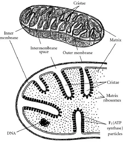
components plays distinct functional roles. IMM separates the mitochondrial matrix from the intermembrane space, it forms numerous folds (cristae) that extend into the interior (or matrix) of the organelle (Fig. 1). The cristae greatly increase the total surface area of the IMM. The IMM includes all the most important complexes that mitochondria need to fulfil their functions: all the complexes of the electron transport system, the ATP synthetase complex and transport proteins complex. The IMS is the smallest component of mitochondrion and it is located between the IMM and OMM. The IMS provides a redox active space, necessary environment to oxidize metabolic residues (Riemer, 2011). The IMS can exchange proteins, lipids, metal ions, and various metabolites with other cellular compartments, as OMM, allowing mitochondrial metabolism to adapt to cellular homeostasis. In particular, the biogenesis and activity of the respiratory chain is controlled by various proteins of the IMS (Vögtle, 2012). OMM is in direct contact with cellular cytoplasm. OMM is freely permeable to small molecules and contains special protein called porin that form channels allowing the free diffusion of molecules smaller than 6000 Daltons (Lin et al., 2014). OMM shows enzymes involved in the elongation of fatty acids, oxidation of epinephrine (adrenaline), and the degradation of tryptophan. Latest studies showed that apoptosis, longevity control are regulated by protein of OMM (Ran, 2014). The mitochondrial matrix is the space between cristae. The matrix contains the mitochondrial genetic system as well as the enzymes responsible for the central reactions of oxidative metabolism.
Fig. 1. Mitohondrial structure. Adapted from Freitas Jr., Basic Capabilities, Landes Bioscience, Austin, Tex, USA, 1999
Mitochondrial electron transport chain
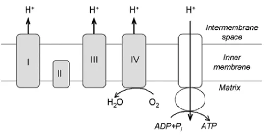
Mitochondrial electron transport chain is bound to inner mitochondrial membrane and mainly consists of five complexes called Complexes I through V (Fig. 2). Complex I, called also NADH-ubiquinone oxidoreductase, transports two electrons from NADH to the mitochondrial matrix and to coenzyme Q within the membrane. Complex I accepts electrons from NADH and it acts like a bridge between glycolysis, tricarboxylic acid cycle (TCA), fatty acid oxidation, and electron transport chain. Its
or boot, can be dissociated in two sub-complexes knows as sub-complexes 1α and sub-complexes 1β, containing 23 and 17 subunits respectively (Grigorieff, 1999). The ankle of “boot” is thought to protrude from the membrane so as to be predominant in the aqueous phase and contains the binding site for NAD(H) and the input electron transfer chain. The foot (the hydrophobic protein) is membrane-linked. Many inhibitors and several iron sulfur centers are localized on it as well as
a catalytic site where reduction of ubiquinone occurs. Many disease conditions are
associated to this complex, including leber hereditary optic neurophaty, melas syndrome, Altzheimer‘s disease and Parkinson’s disease (Meyers, 2013. Gaweda-Walerych, 2013).
Complex II, also known as succinate-coenzyme Q reductase or succinate dehydrogenase, is the only membrane-bound component of the Krebs cycle and in addition functions as a member of the electron transport chain in mitochondria and in many bacteria (Cecchini, 2003). Complex II acts as link between the TCA and electron transport chain. It is the only TCA cycle enzyme that is an integral membrane protein. Complex II oxidizes succinate to fumarate reducing FAD to FADH2. The structure of complex II is well investigated, has a mass of 124 kD and
composed of two hydrophilic subunits, a flavoprotein and an iron-sulfur protein, and two hydrophobic subunits linked to membrane.
Complex III, also named Cytochrome reductase, is a multisubunit transmembrane protein acceptor of electrons from reduced Coenzyme Q and uses them to reduce the second mobile electron carrier Cytochrome C. For each coenzyme Q fully oxidized, complex III moves four hydrogen ions outward from the matrix to the mitochondrial intermembrane space (Bolsover, 2011).
Complex IV, or Cytochrome C oxydase, is the terminal enzyme of the respiratory chain. It is a transmembrane protein and consists of 13 polypeptide subunits, 3 of which are encoded by mitochondrial DNA. The Complex IV moves an electron from each of four Cytochrome C and reduces one oxygen molecule to two molecule of water and moves four hydrogen ions from the matrix to the intermembrane space,
contributing to generate a differential transmembrane difference of proton that the ATP synthase will use to synthesize ATP.
ATP synthase, called also complex V, catalyzes ATP-Pi exchange, and ATP, GTP, and ITP hydrolysis. The synthesis of ATP from ADP and phosphate is driven across the membrane by a flux of protons gradient generated by IMS electron transfer (Galante et al., 1979). The ATP synthase catalyses a reversible reaction for this reason ATP hydrolysis generates a proton gradient by a reversal of this flux.
Fig. 2. Electron transport chain and ATP synthesis on the mitochondria inner membrane. Adapted from Szeto H.H. The AAPS Journal 2006; 8 (3) Article 62
Mitochondrial Membrane Potential
The optimum mitochondrial membrane potential (ΔΨm) is critical for preserving important cellular functions and mitochondrial processes as well keeping the physiological function of respiratory chain. The Δψm controls ATP synthesis, generation of ROS, mitochondrial calcium sequestration, import of proteins into the mitochondrion and mitochondrial membrane dynamics (Dai et al., 2014). Conversely, Δψm is controlled by ATP utilization, mitochondrial proton conductance, respiratory chain capacity and mitochondrial calcium. Depolarization might be found in oxidative stress conditions as consequence of mitochondrial calcium overload (Joshi et al., 2011). A significant loss of ΔΨm renders cells
depleted of energy with subsequent increased mitochondrial membrane
permeability. Hyperpolarization might be related to ATPase inhibition, inadequate supply of ADP, increased supply of NADH and apoptosis due to oxidative stress. The most used method for driving compound to mitochondria uses of the potential gradient across the mitochondrial inner membrane. As a result of moving proton
and electron through mitochondrial electron transport complexes, a negative
potential from 150 to 180 mV is generated across the IMM. Lipophilic cations may therefore accumulate 100-to 1000-fold in the mitochondrial matrix. A number of studies showed increased uptake into mitochondria in TPP+ and VitE -conjugated lipophilic sequence (Murphi et al., 2000. Jauslin et al., 2003). The uptake failed in depolarized mitochondrial membrane potential (Dhanasekaran et al., 2004), because of that the utility of TPP+-conjugated antioxidants is limited in model of neurodegenerative disease where mitochondrial membrane potential is impaired. To overcoming this problem in 2004 a new class of small cell permeable peptide antioxidants targeting mitochondria was described by Zhao and coworkers (Zhao et al., 2004). This class of CPPs is described in specific chapter of this thesis.
Mitochondrial generation of ROS
Mitochondria are small cellular organelle but consume around 85% of cellular oxygen to run the oxidative phosphorylation. As a consequence of mitochondrial metabolism, around 2% of oxygen is turned on superoxide anion (O2-) (Chance,
1979). The amount of O2- production is subordinated to mitochondrial metabolic
state and mitochondrial potential. Superoxide anion is not able to cross cellular membranes, but is converted to hydrogen peroxide (H2O2) by mitochondrial matrix
enzyme MnSOD or by CuZnSOD in the intermembrane space. H2O2 is more stable
than O2¯ and can diffuse out of the mitochondria into the cytosol (Szeto, 2006).
H2O2 can be readily converted on water by mitochondrial glutathione peroxidase or
catalase. Moreover, H2O2 can reacts with ferrous iron and resulting, through
Fentom reaction, in highly reactive hydroxyl radicals (OH•). As discussed above mitochondria are a major source of reactive oxygen species and superoxide is constantly generated during normal respiration by healthy mitochondria. Complex I, Complex II and Complex III are mainly involved in ROS production. Complex I transfers electrons from NADH to coenzyme Q, at the same time protons pass from the matrix to the intermembrane space and the anion superoxide is generated during movement of charge by complex I. Complex II reduces Coenzyme Q and is also responsible for production of low levels of superoxide anion. Complex III is responsible for increased ROS production in a state of decreased electrons transfer. In 1966, Jensen and colleagues understood the pivotal role mitochondria in ROS producing (Jensen, 1976), but to date, in spite of great knowledge of mitochondrial metabolism, a lot of pathway are still unclear. It is important to understand that ROS production is inevitable and useful process. Many studies showed the implication of ROS in important cellular pathway such as autophagy, signal transduction and immune function (Chen, 2007. Niess, 1999).
Mitochondrial Antioxidants
Excess of ROS can be highly dangerous and damage cellular components as protein, lipid and DNA, leading to death of cell. Mitochondria and cells in general posses different defense system to avoid excess of ROS. These antioxidant network system can be rated as enzymatic (superoxide dismutase, catalase) or non enzymatic systems (glutathione).
Superoxide Dismutase (SOD) is widely spread between living organism. All oxygen-metabolizing cells (Gregory, 1974), many anaerobic bacteria (Hewitt, 1975) also fungi (Rapp, 1973) own SOD able to run the dismutation of superoxide radical to H2O2. Superoxide is converted to hydrogen peroxide (H2O2) by two types of
intracellular superoxide dismutase (SOD) under physiological conditions: Cu/Zn-SOD in the cytosol and Mn-SOD in the mitochondrial matrix. Subsequently, the produced H2O2 is catalyzed into water and molecular oxygen by catalase or gluthatione
peroxidase (GPx) 2 O2¯+ 2H+ → O2+ H2O2
Catalase (CAT, oxidoreductase, EC1.11.1.6) is an enzyme found in all aerobic organism and some anaerobic organisms (Brioukhanov et al., 2006). Catalase is located in cellular and subcellular compartements (Roels, 1976) and in mitochondria matrix (Radi, 1991).The primary function of catalase enzymes is the rapid breakdown of hydrogen peroxide into water and safe oxygen.
2 H2O2 → 2 H2O + O2
Glutathione (L-y-glutamyl-L-cysteinyl-glycine; GSH,) is a tripeptide synthesized in cytosol. Cysteine and glycine are linked by peptide bond and the carboxyl group of the glutamate side-chain to the amine group of cysteine by gamma peptide linkage. Glutathione has been discovered in animals, plants and fungi (Penninckx, 2000) and one of the most important antioxidant in our body, it protects against free radicals,
H2O2 and reactive nitrogen species (RNS), it is involved in many cellular functions as
cell signaling, protein function, gene expression, cell differentiation/Proliferation, and its depletion is correlated with many disease as neurodegenerative disease. GSH is not required by diet but every cells of animal organism can synthesize it, so GSH is distributed overall the human body and levels vary according to organs and tissues and subcellular compartments (Wu, 2004). GSH is synthesized in the cytosol then delivered in different cellular compartments; 12% of total cellular GSH is located in mitochondria. The antioxidant action of molecule of GSH is related to thiol groups of cysteine by serving as an electron donor. In presence of H2O2, GSH is
oxidized by Glutathione peroxidase (GPx) to GSH disulfide (GSSG), which is then regenerated as GSH by the reaction with GSSG reductase (GSSG red) (Drigen, 2002).
Oxidative stress
Generation of ROS is a physiological consequence of cellular metabolism and useful tool to different biological pathways. Cells own different antioxidant systems in order to maintain right concentration of ROS. The physiological production of ROS is not dangerous, but a spatiotemporal imbalance between ROS production and ROS defense systems the starting point of cellular impairments. The reduction of antioxidant systems lets to oxidation of membrane phospholipids, proteins, and nucleic acids and this condition, named oxidative stress, can lead to necrotic or apoptosis cellular death (Zamzami et al., 1997). Oxidative stress has been associated to many diseases, including cancer, renal disease, neurodegenerative and cardiovascular disease (Hroudová 2014). Several studies showed increased levels of ROS in diabetes type 1 and type 2. At the moment, the relations between increased ROS and diabetes is not clear but seems that high level of reactive species of oxygen contribute to insulin resistance, the basis of diabetes (Rösen, 2001). Mitochondria are the major site of ROS, generated as byproducts of the electron transport chain. Moreover, mitochondria are continuously exposed to ROS and because of that particularly susceptible to oxidative damage. Mitochondrial DNA has been shown to undergo oxidative damage. In addition to lipid peroxidation, protein oxidation and nitration results in altered function of many metabolic enzymes in the mitochondrial matrix as well as in the electron transport chain. A particularly relevant protein that loses function upon oxidation is SOD, which would further compromise antioxidant capacity and lead to further oxidative stress (Szeto, 2006). In addition, the excess of ROS seems to be involved in cytochrome C release from mitochondria. Cytochrome C is normally bound to the inner mitochondrial membrane linked to cardiolipin. Cytocrom C participates supporting function of ATP synthesis. High levels of ROS lead to peroxidation of cardiolipin, then to the dissociation of Cytochrome C release through the OMM into the cytosol. Cytochrome C in the cytoplasm triggers the activation of caspase-9, which triggers the caspase cascade and ultimately leads to apoptosis (Liu, 1996). Mitochondria are
critical regulators of cell death and a key feature of neurodegeneration, and they play important role in cell processes, signaling pathways, calcium homeostasis, cell cycle regulation, apoptosis, ROS production, and thermogenesis rendering this organelle an important target for the delivery of radical scavengers. Achieving successful mitochondrial drug delivery could produce enhanced treatments for mitochondria-related disorders and also advance our knowledge of the roles that mitochondria play in cellular biology.
Cell-Penetrating Peptides
Classification
Over the last two decades, many different short peptide sequences able to transport diverse types of cargo molecules across cellular membrane have been identified. The continuous development of this field indicates that chemical space is rich in peptide sequences that exhibit high levels of cellular uptake. There are different ways to classify CPPs, on according to their origin, such as their charge, function, hydrophobicity or amphypathicity. One way to classify is to subdivide them into protein-derived, chimeric and synthetic or designed CPPs (Table. 1).
Protein–derived CPPs were the first type of CPPs discovered, Penetratin and Tat
belong to this category. The basic domain of HIV-Tat, which is sufficient for cell penetration, and the penetratin peptide, residues 43–58 derived from the third helix of Antennapedia protein homeodomain from Drosophila, are arguably the most studied CPPs. Both of them are still being used to this date and have in many cases been further modified to obtain new CPPs (Saleh et al., 2010).
Synthetic CPPs are entirely designed, this class ranges from simple polypeptides
such as poly–argynine (Futaki et al., 2007) to more complicated synthetic sequences including the model amphipatic peptide (Oehlke et al., 1998). Synthetic CPPs are still less common than protein derived or chimeric CPPs.
Chimeric CPPs are combinations of protein–derived and synthetic sequences.
Transportan is typical examples of chimeric CPPs. Transportan is a 27 amino acid-long peptide. The sequence of Transportan was designed using the natural amino terminus of the neuropeptide Galanin and the carboxyl terminus of Mastoparan by means of a lysine. (Pooga et al., 1998).
CPPs Origin Sequence Ref.
Tat HIV-1transactivator protein GRKKRRQRRRPPQ Vivès et al.1997 EB1 Chimeric LIRLWSHLIHIWFQNRRLWKKK Lundberg et al. 2007
SAP Designed VRLPPPVRLPPPVRLPPP Pujals et al. 2008
R9 Designed RRRRRRRRR Futaki et al.2001
TABLE 1. Example of common CPP by classification
CPPs uptake mechanism
A numbers of investigations have been conducted to elucidate how CPPs get into the cells (Duchardt et al., 2007). Most of naked peptides CPPs use endocytosis mechanism but a variety of uptake mechanisms appear to be operative in different systems, and in some cases, the mechanism is cell-type or cargo-specific (Mueller et al., 2008). For example, in 2003, Fittipaldi and coworkes showed that TAT uses a lipid raft mediated endocytosis when conjugated to a protein (Fittipaldi et al., 2003) and clathrin–dependent endocytosis if conjugated to a fluorophore (Richard et al., 2005). The uptake mechanism was initially considered direct, non–endocytic and receptor independent, but later studies showed CPPs can access the cell by two distinct routes: energy-dependent vesicular mechanisms, collectively referred to as endocytosis, or via a direct process involving translocation of the lipid bilayer, especially at high concentrations of peptide, also the same peptide can be taken via endocytic and direct pathways (Fig.3) (Duchardt et al., 2007).
Endocytosis
phospholipids. In addition, when linked to larger cargos, CPPs can utilize certain receptors in order to improve their uptake, such as class A scavenger receptors (Lindberg et al., 2013). Endocytosis is commonly divided in phagocytosis and pinocytosis. Phagocytosis has generally not been associated with CPPs and is reserved only for specialized cells, such as macrophages, monocytes, dendritic cells and neutrophiles; is a complex process used to engulf large particles (Aderem et al., 1999) such as bacteria or dead cells, it has been recently shown that a phagocytic process occurs in particles larger than 0.5 µm and is influenced by particle shape (Aderem et al., 1999).
Pinocytosis can be further classified in diverse pathways: clathrin-mediated endocytosis (CME), caveolae–mediated endocytosis (CvME), macropinocytosis (MP), clathrin and caveolin independent endocytosis (Hillaireau et al., 2009). Pinocytosis occurs in all cell types. The exact mechanisms of each of these pinocytic processes differ with regard to vesicle structure and the protein machinery utilized, they all share a common outcome: extracellular molecules are encapsulated into lipid vesicles, which are internalized after resealing of the plasma membrane.
Clathrin-mediated endocytosis
CME is the most well studied endocytic pathway. This mechanism starts with
formation of vesicles, size around 100-150 nm in diameter and coated with a complex of proteins mainly consisting of clathrin. Vesicles are formed in specialized regions of the plasma membrane called clathrin coated pills. The mechanism of their formation is characterized by several steps. Primarily, the enzyme GTPase dynamin drives the invagination of the plasma membrane, as the invagination gets deeper turns in vesicles (Takey et al., 2001). Afterwards, endocytosed vesicles move from the surface of plasma membrane to deeper region of cell, during moving, vesicles turn into endosomes, pH around 6, to lysosomes, pH around 5, where the cargo is enzymatically degraded (Luzio et al., 2009).
Caveolae–mediated endocytosis
CvME, also called as lipid raft-mediated endocytosis, is a cholesterol,
dynamin-dependent and receptor-mediated pathway (Nichols, 2003). It is characterized by endosomes formed from non-clathrin coated plasma membrane but consists of thecholesterol-binding protein caveolin and a cholesterol and glycolipid bi-layer. The fission of the caveolae from membranes is run by dynamin. Caveolae are approximately 50–80 nm in diameter. Their composition and function are highly cell-type dependent. Caveosomes are not degraded by acidic pH, therefore the cargo can be directly driven to the Golgi and/or endoplasmatic reticulum, avoiding normal lysosomal degradation (Bengali et al., 2007).
Macropinocytosis
MP is a mechanism of uptake able to take relatively large amounts of non-specific substances. MP usually occurs in macrophages and cancer cells, it is characterized by formation of actin–driven membrane protrusions which collapse into and fuse with the plasma membrane (Hillaireau et al., 2009). The size of vesicles, called macropinosomes, is around 200 nm-5 µm in diameter. The fate of macropinosomes is still unclear and seems to be cell type dependent. Futaki and colleagues reported arginine-rich CPPs are preferentially taken by the cells via macropinocitosis (Futaki et al., 2007).
Clathrin and caveolin independent endocytosis
This pathway is less studied than the others mechanism of uptake. Endocytosis occurs in cells depleted of both CME events and caveolin in cholesterol dependent manner, implying distinct endocytic pathways that require specific lipid compositions for internalization (Doherty et al., 2009). Clathrin and caveolin independent endocytosis can be further divided into dynamin-dependent and
Direct translocation
Latest studies suggest that the translocation of polycationic CPPs across biological membranes occurred via an energy-independent cellular process, controlled by cholesterol and membranous protein (Pae et al., 2014). Direct translocation is characterized by destabilization of cellular membrane in an energy and temperature-independent manner (Bechara et al., 2013). Different models have been proposed to explain this kind of mechanism: inverted micelle formation, adaptive translocation and pore formation. Inverted micelle formation starts with an intussusception of cellular membrane due an electrostatic interactions and subsequent interaction of hydrophobic residues with the membrane core. Micelle origin from reorganization of neighbouring lipids at cellular surface, CPP is encapsulated by micelle and will be release inside after disruption of micelle (Derossi et al., 2002). Adaptive translocation seems to be exclusive for arginine-rich CPPs. Guanidium groups of arginines form bidentate hydrogen bonds with the phospholipid headgroups on the cell membrane: in this way CPPs get inside the cell. The pore formation model allows the passive diffusion of CPPs across the plasma membrane. This mechanism of uptake is mainly used by arginine and lysine rich CPPs. The attraction between the side chain of amino acid and the phospholipid headgroups of the distal layer leads to the formation of a transient pore (Morciano et al., 2014)
CPPs in drug delivery strategy: Applications and organelle specific delivery
Identification of cellular targets for treatment of different diseases required the development of a successful system able to delivery drugs into a target of interest. Some therapeutics exhibited excellent properties on in vitro studies, on the other hand on in vivo model the utilization was limited by their physicochemical characteristics. Uptake and efficacy of several therapeutic compounds are improved by CPPs conjugation, creating new opportunity to study biological process and making the treatment of several diseases more controlled, less toxic (Veldhoen et al. 2006). Different cargoes such as small molecules, imaging agents (Rao et al. 2007), small molecular drugs and larger cargoes such as oligonucleotides (Meade et al. 2007), plasmids and protein (Morris et al. 2001) can be conjugated to CPPs
Nuclear Localization Sequences
Signal peptides are an effective strategy for organelle-specific targeting, used by cellular machinery to identify newly translated peptides and traffic them to the correct destination in the cell. As the storehouse of genomic DNA, the nucleus is a desirable target and the necessary destination for agents used in gene therapy (Cartier et al., 2002). Between Nuclear Localization Sequences (NLS) are cellular penetrating peptides targeting nucleus. These short cationic sequences, 10 amino acids in length, are widely used to achieve nuclear delivery for a variety of DNA damaging agents or nucleic acids for gene therapy (Cartier et al., 2002), between NLS, Simian virus 40 (SV40) showed high levels of cell permeability and low toxicity, because of that has been applied in a lot of studies to drive uptake of DNA in nonviral gene therapy (Singh et al., 1998). Other strategy adopted to delivery DNA inside the nucleus is encapsulating in polymer nanospheres or phage particles with NLS peptide displayed on the exterior (Akuta et al., 2002). L. Benimetskaya and coll. used NLS to delivery Antisense oligonucleotides and block translation of Bcl-2 and
Fig.4. Applications of cell-penetrating peptidies as molecular devilery machine. Modified from Stewart et al., 2008
PKC-a in prostate and bladder carcinoma cells (Benimetskaya et al., 2002). Gold nanoparticles, carboplatin-based anti-cancer therapeutics, and green fluorescent protein (GFP) can be driven and localized inside the nucleus by NLS (Tkachenko et al., 2003. Wagstaff et al., 2007). At today, many studies show an improvement in nuclear localization and demonstrate an improved transfection efficiency (Ludtke et al., 1999)
Mitochondria-Target Antioxidants
Literature shows several examples of CPPs targeting mitochondria, using artificial, rather than natural signal sequences (Mahon et al., 2007). Mitochondrion is an important target of drug therapy due to its role in the pathology of cancer, neurodegenerative diseases and other diseases dealing with reactive oxygen species. Oxidative stress is the beginning or a consequence of several pathological conditions. Mitochondria are often the organelles where oxidative stress starts or main target of oxidative stress, therefore the mitochondrion represents candidate of significant interest for organelle-specific exogenous molecules. Hindering oxidative stress with delivering of antioxidant has been found to be effective in many animal models of diseases associated with oxidative damage (Chao, 2014. Kim, 2014). Sometimes, administration of antioxidants have exacerbated the oxidative condition instead of leading to significant benefits, this conflicting action is named antioxidant paradox (Halliwell, 2000). Some antioxidants beside their antioxidant action can have a prooxidant action especially in the presence of metals such as iron (Fe) which starts Fenton reaction (Murakami et al., 2007). High hopes and expectations in vitro about promising antioxidants have turned into delusions when the same results were not obtained in vivo studies. Up to now, there are literally dozens of completed or ongoing clinical trials using such antioxidants as
positive results, since several showed a negative association between antioxidant supplementation and positive outcomes. A significant challenge to mitochondrial drug delivery is the impervious structure of the hydrophobic inner membrane. Some antioxidants do not penetrate cellular or mitochondrial membranes therefore they are not effective against intracellular ROS, others are very lipophilic and tend to be retained in cell membranes (Dixon et al. 2007). Many efforts have been done in order to develop an ideal antioxidant and there is still a long way to go and crucial questions remain to be answered. Does the “ideal” antioxidant really exist? Several research groups are looking for this answer. The ideal antioxidant should be cell-permeable and able to target mitochondria therefore it can protect against oxidative damage and prevent mitochondrial impairmet. Latest results showed that two properties are important for passage across both the plasma and mitochondrial membranes: positive charge takes advantage of the potential gradient to get into mitochondria, lipophilic character in order to allow partitioning of the molecule through the lipid bilayer (Rosania et al., 2003).
TPP+-conjugated
Several mitochondria-targeted antioxidants have been developed and are currently undergoing preclinical testing. In the past, the most common strategies used for delivering compounds into mitochondria have relied on the conjugation of known redox agents to triprenylphosphonium ion (TPP+) (Adlam et al., 2005). It is an example of a cationic hydrophobic molecule with delocalized charge that can cross the mitochondrial membrane without a transporter. Another strategy is aromatic-cationic tetrapeptides that selectively target the inner mitochondrial membrane without relying on mitochondrial potential. TPP+ has been conjugated to lipophilic antioxidants such as coenzyme Q (MitoQ) (Skulachev et al., 2009), plastoquirone (SkQ1) (Smith et al., 2012) and Vitamin E (MitoVitE) (Murphy et al.,2000. Sheu et al., 2006). They showed an increased uptake in comparison to the same molecule without carrier and improved action in reducing intracellular ROS, preserving
reduced thiols, and reducing oxidative cell death (Kelso et al., 2001). In particular, MitoVitE was reported to be 800-fold more potent than idebenone protecting against GSH depletion in cultured fibroblast from patients with Friedreich’s Ataxia (FDRA). Furthermore, MitoVitE is 350-fold more potent than trolox (water soluble Vitamin E analog) (Szeto, 2006). Intraperitoneal and intravenous administrations of TPP+ were used in mice and the times of uptake and distribution were controversial. Uptake was obtained in the liver 1 hour after intraperitoneal injection, but was not detected in the brain and in the heart after 20 hours. TPP+ conjugated to MitoVitE was detectable in the heart 4 days after administration while levels in the brain were still really low (Smith et al., 2003). These TPP+-conjugated were evaluated on ischemia-reperfusion injury but not yet on models of neurodegenerative diseases. The utility and applicability of TPP+ is also limited by their mechanism of uptake. They need a normal mitochondrial potential to get this organelle, but all the neurodegenerative diseases are associated with abnormal mitochondrial potential. Furthermore, study on isolate mitochondria demonstrated that concentrations of TPP+ greater than 20 µM lead to wasting of IMM potential (Smith et al., 1999).
Choline Esters of Glutathione and N-Acetyl-Cysteine
Glutathione is a tripeptide (L-γ-glutamyl-L-cysteinyl-glycine) with strong antioxidant activity, detoxifying ROS and preventing thiol oxidation. Mitochondria are not capable to synthesize glutathione. The synthesis of glutathione occurs in the cytoplasm and transported into mitochondria by dicarboxylate and 2-oxoglutarate carriers (Chen et al., 2008). N-Acetyl-Cysteine (NAC) arises from the amino acid L-cysteine, it exhibits antioxidant activity (Xue et al., 2011. Reliene et al., 2004) as well is also used to provide cysteine for glutathione synthesis. Both of them are really useful in protecting mitochondria against oxidative damage. Using the same
NAC into mitochondria (Sheu et al., 2005). These compounds make use the negative internal potential of mitochondria, which leads and regulates their concentration in mitochondria. Ester of Glutathione is a membrane/lipid permeable derivative of GSH that is used to restore the GSH pool within cells subjected to cysteine and/or GSH depletion. Preliminary in vitro studies demonstrated that they are able to avoid depolarization due to hydrogen peroxide in neonatal rat ventricular myocytes and striatal neurons, but to date but in vivo animal studies are not yet accessible (Szeto et al., 2006).
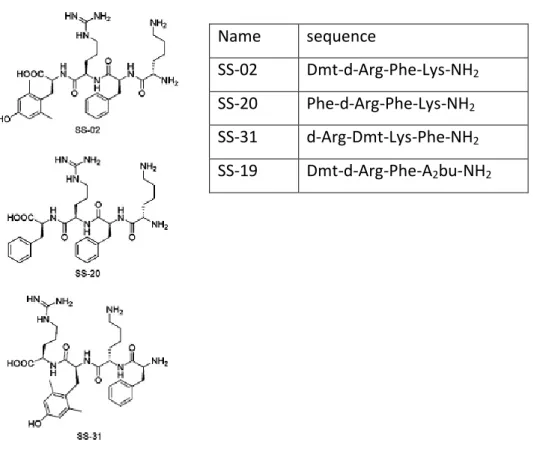
Szeto-Schiller Peptides
Date back in 2000, synthesis of the short Szeto-Schiller peptides (SS-peptides), the most promising mitochondria-targeted antioxidant peptide (Schiller et al., 2000). The Szeto-Schiller (SS) compounds are tetrapeptides (Fig. 5) designed for alternating aromatic residues and basic amino acids (aromatic-cationic peptides), they are characterized by tyrosine (tyr) or dimethyltyrosine (Dmt) residues in order to increase antioxidant activity and the presence of D-amino-acid in either the first or second position to minimize aminopeptidase degradation, amidation of C-terminus to reduce hydrolysis from C-C-terminus (Schiller, 2006). Scavenge activity of SS-peptides was proved first in vitro using luminol chemiluminescence, then antioxidant properties of SS-peptides were further established by inhibition of fatty acid peroxidation and low density lipoprotein (LDL) oxidation (Zhao et al., 2004). The antioxidant action of SS-peptides is dose dependent manner and can be attributed to Tyr or Dmt residues can scavenge H2O2, OH. and ONOO.. The sequence
position of the Tyr or Dmt residue is not important in scavenging ROS and inhibiting LDL oxidation but replacement of Dmt with phenylalanine (Phe) resulted in complete loss of antioxidant activity (Zhao, 2004). Tyr, or Dmt, can scavenge oxyradicals forming relatively unreactive tyrosyl radicals, which can be followed by radical-radical coupling to give dityrosine, or react with superoxide to form tyrosine
hydroperoxide (Hazel et al., 2006). Dmt is more effective than tyrosine in scavenging of ROS because bears much structural similarity to vitamin E, indeed both have the methylated phenol structure. Zhao and colleagues showed that upon induction of oxidative stress by tertbutylhydroperoxide (tBHP), cells treated with the SS-peptides decreased levels of mitochondrial reactive oxygen species and halted the progression of apoptosis (Zhao et al., 2004). All of SS peptides show 3+ net charges at physiologic pH and they are taken up into cells in an energy-independent non saturable manner. The uptake is really fast, several studies showed that SS-20 peptide is taken up into cells in less than 30 minutes and can freely pass through the plasma membrane in both directions (Zhao, 2004). The SS-peptides targeting preferentially the inner mitochondrial membrane, indeed SS-31 and SS-02 are taken 1000-fold and 10,000-fold respectively, in liver and brain mouse mitochondria (Zhaoet al., 2004. Zhao et al., 2005) and concentrate in mitochondria 1,000-fold more than the cytosolic concentration (Zhao, 2004). The mechanism of uptake into mitochondria is not self limiting but how it works is still unclear. During uptake of SS-peptides there is not a well defined formation of vesicles, typical of an endocytosis uptake. The uptake of these aromatic-cationic peptides is not dependent on mitochondrial membrane potential, a study showed that they are also concentrated in FCCP depolarized mitochondria (Doughan, 2007). Experience has taught us that talking about drug development to cross the blood brain barrier (BBB) is the maximum impediment for new drugs. Studies have shown ability of SS02 to get mouse brain in 5 minutes after intraventricular injection (Gifford A., 2004, unpublished data). Between SS-peptides, the short SS-31 peptide developed by Szeto is the most promising mitochondria-targeted antioxidants (Zhao, 2004) its antioxidant potentialities were confirmed against different types of adverse treatments in both in animal (Huang et al., 2013) and in vitro models (Zhao et al., 2013). A number of studies confirmed the potential applicability of SS-peptides on
administered to mice after acute cerebral ischemia (Cho et al., 2005). SS-31 was evaluated in animal models of neurodegenerative disease, PD (Yang et al., 2009) and Amyotrophic lateral sclerosis (ALS) (Petri et al., 2006). Nowadays, SS-peptides are the most promising approach with targeted delivery of antioxidants to mitochondrial organelle. Their extraordinarily potent in protecting against oxidative cell death is already proven by a number of publications. Moreover, they have an excellent pharmacokinetic profile and they are easily “druggable”, small, easy and fast to synthesize, readly soluble in water and resistant in human serum until to six months to (Schiller et al., 2000).
Name sequence
SS-02 Dmt-d-Arg-Phe-Lys-NH2
SS-20 Phe-d-Arg-Phe-Lys-NH2
SS-31 d-Arg-Dmt-Lys-Phe-NH2
SS-19 Dmt-d-Arg-Phe-A2bu-NH2
GOAL OF THE THESIS
Mitochondria are subcellular organelles involved in pivotal metabolic cellular pathways. Mitochondria developed a special membrane structure and network of antioxidant systems to help preserve its functions. A number of studies showed that the onset or the exacerbation of many diseases is caused by mitochondrial impairment (Szarka et al., 2014). Mitochondrion is an expecially interesting organelle for drug therapy given its role in the pathology of cancer, neurodegenerative diseases, and other diseases characterized by oxidative stress (Weissig et al., 2004). Drugs could be necessary for both inhibiting mitochondria in order to kill cancer cells as well as to protect the cells from oxidative damage and to repair dysfunctions. Over the years different strategies have been developed in order to get access to mitochondria, but its complex structure was often a tough hurdle to overcome so gaining access to this organelle could be difficult. Lately, the most useful strategy is based on cell penetrating peptide targeting mitochondria. Nowadays a novel class of small cell-permeable peptide antioxidants reported a great deal of attention. The structural motif of these peptides, named Szeto-Schiller peptides (SS-peptide), is characterised on alternating aromatic residue and basic amino acids (Szeto, 2006). This thesis was focused on the synthesis of a new short cell penetrating antioxidant peptide able to cross cellular membrane and target mitochondria. Gaining access to mitochondria means make easier treatments on it. The final purpose was to use antioxidant actions of this peptide to protect mitochondria against oxidative stress and due of its “druggable” properties use it as new tool on drug delivery strategy.
MATHERIAS AND METHODS
Solid phase Peptide Synthesis (SPPS)
A Peptide is a chemical compound consisting of amino acids condensed with each other through a peptide bond or amide bond between α-carboxyl group of residue and the α-amino group of next amino. Peptides have a maximum of 50 amino acids (Jones, 1991) and the term protein describes molecules with more than 50 amino acids. Peptides have a wide range of applications in medicine and biotechnology, for this reason solid phase peptide synthesis (SPPS) plays today a pivotal rule in the area of development of new therapeutic strategies, allowing the chemical synthesis of peptides and small proteins. SPPS has been used for the first time by Merrified (Merrefield, 1973), starting a revolutionary approach to the chemical synthesis of polypeptides. The basic principle of SPPS is the stepwise addition of protected amino acid to a growing peptide chain bounded by its C-terminal carboxylic acid by a covalent bond to a solid, stable and inert resin particle (Fig. 6). By-products and excess reagents may be removed easily by filtration and washing. Peptide Synthesis starts from Carboxyl-Terminus (COOH-Terminus, or C-terminal end) to Amino-Terminus (NH2-Terminus, or N-terminal end), adding one by one activated amino
acids. Activation of the carboxyl component of amino acids is based on formation of active esters. The activation makes more electrophilic carboxyl group, because of that α-carboxyl group of the amino acids is activeted to facilitate nucleophilic attack by the α-amino group of the previously coupled amino acid. Peptide bond is assisted by presence of coupling reagents, called also auxiliary nucleophiles. The newest coupling reagents belong to uranium and phosphonium salt. The use of these reagents has been reported to be more convenient and superior. Some of the
than amount of resin. Good outcome of reaction depends on availability of only a single nucleophile during acylation reaction, therefore is necessary to block those functional groups that must not participate in the peptide bond formation. In peptide synthesis the α-amino group are protected with temporary protecting groups, which are cleaved after each coupling reaction, and the functional groups of the amino acid side chains are protected with permanent protecting groups, which are cleaved after the synthesis is completed. In SPPS two main strategies are used: Boc /Benzyl-based strategy, Fmoc/tert-butyl-based strategy. Tert-butyloxycarbonyl (Boc) was first applied in 1950s; this group is stable towards most nucleophiles and bases, it is cleaved by acid e.g. Trifluoroacetic Acid (TFA) in Dichloromethane (DCM) or others strong acids e.g. HBr in TFA. This strategy is based upon the graduated acid lability of the side-chain protecting groups. In Boc/benzyl-based SPPS a number of side reactions may be caused by repetitive acid treatments during the synthesis and the use of a strong acid for the final deprotection. In order to avoid side reactions generated by Boc strategy, a new protocol based on orthogonal 9-fluorenylmethyl-oxycarbonyl (Fmoc) protecting group was developed by Carpino and Han. (Carpino, 1972). The Fmoc group is stable to acid, but is clave by base, e.g. piperidine in DMF, or tert-butyloxycarbonyl. Fmoc-based SPPS method is now the method of choice for the routine synthesis of peptides. Semi permanent protection groups for functional amino acid side chains are removed when synthesis ends. There are different protecting group strategies that allow in selecting amino acid being protected on the base of amino acids side chains. Cleavage and final deprotection are really important steps in peptide synthesis. The cleavage cocktails frequently used is a mixture of TFA and scavengers. The SPPS involves numerous repetitive steps. After loading the first amino acid, the desired peptide sequence is assembled in a linear fashion from the C-terminus to the N-terminus, alternanting deprotection and coupling of amino acids until the desired sequence is obtained. This technique has made the synthesis of peptides faster: averagely, each amino acid is added every hour, just the time required in order to complete the reaction.
In a final step, the peptide is released from the resin and the side-chain protecting groups concomitantly removed.
Fig.6. General scheme of solid phase peptide synthesis, SPPS. Adapted from Amblard et
Manual Synthesis of MitPep-peptide and SS31 peptide
All the peptides were synthesized manually by solid phase peptide synthesis using Fmoc-chemistry protected amino acids and 1,3-diisopropylcarbodiimide (DIC):1- (HOBt) as coupling agents (Soomets et al., 2005). SPPS can be performed in classical glass reaction vessels that can be made by glassblowers or purchased from manufacturers, alternatively syringes equipped with PTFE or glass frits may also be used. For the peptide synthesis Fmoc-Rink-Amide-MBHA-resin was usually used. Before starting the solid phase synthesis, the resin was swollen in an adequate solvent such as DCM or DMF. Couplings were performed with Fmoc-protected D or
L-amino acids and tree activators
1H-benzotriasole-1-yl)-1,1,3,3-tetramethyluronium tetrafluoroborate (TBTU), N-hydroxybenzotriazole (HOBt) and N,N-diisopropylethylamine (DIPEA) in N,N-dimethyl formamide (DMF) for 45’ each coupling (Table. 2), the Fmoc group was removed with piperidine (20% v/v) in DMF (20min). The success in each coupling and deprotection step were evaluated by qualitative Kaiser test. Typically compared to the resin, 2–10 times excess of activated amino acid is used. This excess allows a high concentration of reactants (typically 60–200 mM) to ensure appreciable amount of products (Muriel et al., 2006). The final cleavage from the resin/deprotection of the peptides was performed to minimize the by-products formation resulting from Rink amide resin at high concentrations of trifluoroacetic acid (TFA). The peptide was deprotected from permanent groups and detached from the resin and by adding trifluoroacetic for peptide lacking cysteine acid (TFA) 95% (v/v), water 2.5%(v/v), triisopropylsilane (TIS) 2,5% (v/v); for peptides containing cysteine or metionine TFA 94% (v/v), water 2,5% (v/v), 1,2-Ethanedithiol (EDT) 2,5%(v/v), TIS 1% (v/v), both mixture per 100 mg of resin. Cleavage and deprotection are one of the crucial steps in peptide synthesis, is not a simple reaction, but a series of competing reactions, it takes at least 3 hours at room temperature, under gentle stirring. To induce peptide precipitation, the cleavage mixture dropwise was added to cold (-20) ether in a 50-ml falcon tube,
about 40 ml ether for 3 ml cleavage cocktail, centrifuge at 7000 rpm for 5 minutes. Then slowly and carefully ether solution was decanted to a waste container avoiding shaking up the precipitate, was repeated two times to get rid of residual TFA and scavengers. The crude precipitate was left under fume hood until ether evaporated completely. The crude peptide was obtained in solid form through lyophilization of the acetic acid extract. A fluorescent analog containing 5,6-carboxyfluorescein (FAM) was prepared for mitochondrial and cellular uptake studies
Purifiction of peptides by HPLC and mass evaluation by MALDI-TOFF
The crude peptides were purified by reverse-phase high performance liquid chromatography (RP-HPLC) on a BioBasic C-8 column (Thermo Scientific, Sweden). The stationary phase was porous silica, covalently bound to a non–polar compound such as octadecyl silane (C18) or octysilane (C8). The convenient mobile phase for the separation of peptides was a gradient elution system where the composition of the solvent was continuously changed by a gradient programmer. All solvents used in HPLC systems must be of specific grade to keep save column and allow the use of
Compound MW Equivalents mmol
Peptide resin 1 0.065
Fmoc-amino acid 4 0.26
HCTU,0.5M in DMF 413.17 4 0.26
6-Cl-HOBt 0.5M in NMP 169.57 4 0.26
DIPEA, 5.84M neat 129.3 8 0.52
peptide was lyophilized, briefly, to dilute the correct peptide fraction in few millilitres of MQ water and freeze at -80°C for 24 h, then make holes into the cap of the tube with a syringe needle. Thereafter the tube of freezed peptide with solution was put into vacuum machine overnight. Each peptide was at least 98% pure as assessed by analytical reversed-phase HPLC. Molecular mass of peptides was determined by a matrix–assisted laser desorption/ionization time-of-flight mass-spectrometry (MALDI–TOFF) (Voyager-DE STR, Applied Biosystems), the values were compared with theoretical mass. According to the Applied Biosystems guidelines, saturated solution of α-cyano-4-hydroxycinnamic acid was chosen for the matrix (10 mg/ml in 50:50 ACN/water mix with 0.1% TFA content). MALDI is an ionization technique based. In this technique the sample is crystallized together with a matrix which is irradiated by a UV–laser beam. The matrix is ionized by the laser and some of its charges achieve the analyte, generating molecular ions of the analyte, in the same time protecting it from direct ionization by the laser. The time of flight detector evaluates the spent time for the analyte ion to reach a detector at known distance. This time depends on the mass-to-charge ratio of the particle and might be used to get the mass of the ion.
Cell culture
HeLa 705, cervical human cellular line was chosen to evaluate biological effects and uptake of MitPep. HeLa cells are important tool on biological science and medical research, they are the first immortal human cells ever grown in culture, many scientific landmarks since then have used HeLa cells, including cloning (Leid et al., 1992), gene mapping (He et al., 2014), in vitro fertilization (Ashizawa et al., 1992), drug delivery strategy (Bracht et al., 2014). HeLa 705 cells (human cervical cancer cell line) were cultured as subconfluent monolayers in 75cm2 cell culture flask. HeLa 705 in Dulbecco's Modified Eagle Medium (DMEM) (lifetechnologies) supplemented with 10% (v/v) fetal bovine serum (FBS), 1% non-essential amino acid (GIBCO) and
1% Penicillin-Streptomycin (life technologies), kept in a humidified incubator at 37o C, 5% CO2. Cells grown to subconfluence were enzymatically dissociated from the
surface with a solution of 0.05% trypsin/0.53mM EDTA
(ethylenediaminetetraacetic). To evaluate viability, mitochondrial membrane potential and ROS production, cells were plated at 1 X10⁴ cells/well in 96 well plates 24h prior to the experiment. To isolate mitochondria 7X10⁶ cells were plated in 275 cm2 cell culture flask 3 days prior to experiment. For cellular uptake experiments and microscopy studies cells were plated at 7,5X10⁴ cells were plated in glass bottom disches. For fluorescence studies was used a fluorescent analog FAM-MIP1 conjugated.
Cell viability assay
Cell viability was determined by conventional WST-1 assay (Roche Diagnostics Scandinavia AB, Sweden). WST-1 reagent is designed to be used for the quantification of cell proliferation using 96 well-plate format, it is a colorimetric assay based on reductases activity of mitochondrial succinato deidrogenase. A tetrazolium salt is reduced to formazan dye by active mitochondria, the amount of dye is correlated to the number of cell with active succinato deidrogenase. Briefly, HeLa 705 cells were plated at a density 1X10⁴ cells/well in 96-well plates and allowed to grow for 24 h before treatment with MIP1. The concentration of MIP1 stock solution was 1 mM in MilliQ water, different working solutions were prepared. Cells were treated at the final concentrations 0.5-5-50-100 μM in 100 μl of complete medium for 24 h and H2O2 200 μM for 24h was used as positive
control. Cell proliferation reagent WST-1 was added to each well at final dilution 1:10, in this case 10μl/100μl. After 4 h incubation the absorbance was measured at