R E S E A R C H A R T I C L E
Open Access
Acorn-related acquired pseudomelanosis in
Calabrian black pigs
Giovanni Lanteri
1*, Jessica Maria Abbate
1, Carmelo Iaria
2, Daniele Macrì
3, Vincenzo Ferrantelli
3and Fabio Marino
2Abstract
Background: Melanosis of lymph nodes in black pigs has generally been related to regression of congenital melanoma and, occasionally, to ingestion of acorns. The aim of this manuscript is to confirm the hypothesis of a possible acquired acorn-related pseudomelanosis in the Nero Calabrese pig, a swine breed belonging to the group of Italian native breeds and whose coverage area corresponds to the region of Calabria, southern Italy. This pig is characterized by slow-growing subjects, producing, however, high quality meat suitable for the production of sausages and fine hams. The study was carried out on 142 normally slaughtered pigs. All organs were examined. Lymph nodes and intestine (jejunum) were sampled. Histochemistry was performed on deparaffinized histological sections to identify the cell types involved and to characterize the pigment stored. To further confirm the pigmentation disorder, immunohistochemistry was carried out. Total phenolic substances were identified in acorns through the use of a biochemical reaction.
Results: Lymph node pigmentation appears directly related to acorn ingestion, with a higher incidence in the group which was 70% natural fed (acorn of Quercus virgiliana). Moreover, findings obtained revealed how different amounts of phenolic substrates present in Q. virgiliana and Q. ilex acorns can influence the incidence of such exogenous pigmentation.
Conclusion: The findings obtained in this study confirm the acquired nature of the melanin-like pigmentation detected in lymph nodes from acorn-fed swine. Acquired pigmentation must be differentiated from true melanosis as well as from melanosis related to tumor regression of congenital melanoma. This thesaurismosis can be proposed as a marker of wellbeing and quality, confirming that the pigs have been bred and fed in natural conditions.
Keywords: Acorn, Pseudomelanosis, Quercus, Swine, Thesaurismosis Background
Pathological color modifications in tissue include a vast number of different categories; pathological pig-mentations are identified as deposition of substances with a specific coloration in cells and tissues; exogen-ous pigments are introduced from the external envir-onment and are stored in cells and in tissues; endogenous pigmentations are products of metabol-ism; thus, their appearance is due to metabolic dis-turbance. Endogenous pigmentations are caused by colorant substances such as lipofuscin, melanin and the group of hem pigments, derived from blood
pigment hemoglobin [1]. The most important and
known endogenous pigmentation is melanosis, a grey-black pigmentation found in different organs due to melanin storage [2].
Melanin (in Greek melas = black), an autochthonous intracellular pigment, is characterized by a brown-black color.
It is produced by tyrosine oxidation, which repre-sents the structural basis of its molecule, tyrosinase (with Apo enzyme containing copper) through deoxy phenylalanine (DOPA) and indolquinone. Melanin is produced by specific cells, melanocytes, whose precur-sors - melanoblasts (non-pigmented cells) - derive from the neural crest [1, 3]; melanocytes can release pigment to other cells as opposed to melanophages
© The Author(s). 2019 Open Access This article is distributed under the terms of the Creative Commons Attribution 4.0 International License (http://creativecommons.org/licenses/by/4.0/), which permits unrestricted use, distribution, and reproduction in any medium, provided you give appropriate credit to the original author(s) and the source, provide a link to the Creative Commons license, and indicate if changes were made. The Creative Commons Public Domain Dedication waiver (http://creativecommons.org/publicdomain/zero/1.0/) applies to the data made available in this article, unless otherwise stated. * Correspondence:[email protected]
1Department of Veterinary Sciences, University of Messina, 98168 Messina, Italy
and keratinocytes, which are capable of storing but not of synthetizing it [3].
Melanin production may be influenced by neuroendo-crine, as well as exogenous, factors (X-rays, UV rays, ar-senic, phenolic substances, etc.). Phenols can influence polymerization of quinones, derived from phenol oxida-tion, which assume a dark discoloration [4].
Melanin contained in melanosomes is collected from pigmented cells of the epidermis, the hair matrix, intestine mucosa and pia mater of some spe-cific tracts of the brain (substantia nigra), and the choroid.
Several pathological modifications linked to melanin can occur: some tumors, e.g. melanoma, melanosis, acanthosis nigricans, skin anomalies in human beings, skin hyperpigmentation associated to hyperadrenocor-ticism, in the rare human hereditary disease known as Dubin-Johnson syndrome; in mutant Corredale sheep, in which an excretory defect of the liver produces an
accumulation of melanin pigment in hepatocytes [5].
Pathological changes related to melanin can be due to a perturbation of those processes related to produc-tion, transport and dispersion of pigments [4].
Melanosis of lymph nodes in some black pigs, such as Sinclair, Duroc-Jersey, Hormel, Vietnamese pot-bellied and their cross-breeds, has also been related to regres-sion of congenital melanoma [6–9].
In general, melanosis represents an unusual finding at the slaughterhouse [10] and to date, except in soli-peds, it is not considered a disease to notify, although
carcasses showing extensive melanosis are
con-demned. If the condition is localized, only the af-fected organ or part of the carcass needs to be
condemned according to FAO recommendations [11].
Recent investigations, performed at slaughterhouses on the autochthone swine breed Nero Siciliano pig reared free-range (in pleair), have shown an in-creased incidence of melanosis [4, 12].
The Nero Calabrese pig breed is characterized by slow growing subjects, producing high-quality meat suitable for the production of sausages and fine hams, with meat pH tending to decrease more quickly com-pared to the modern depigmented breeds. Monoinsa-ture and polyinsaMonoinsa-ture fatty acids of the omega-3 and omega-6 series are more present in the meat of the black pigs reared free range as compared to those reared intensively. Another advantage derives from the fact that animals, because of their rusticity, can be farmed free range all year and can adapt very well to any possible feed, including food derived from grazing. In such a kind of farming system, animals live in a large closed off woodland area, where they can feed on natural pabulum, generally composed of acorns, tubers, roots, etc. Commercial food is daily
provided only as an integrative supplement, thus re-ducing the environmental impact of these foodstuffs.
The aim of the present paper was to verify the possible acquired pathogenesis for a melanin-like pigmentation in Nero Calabrese pigs, by macroscopic, histological, immuno-histochemical and biochemical exams. Moover, considering the high frequency in the Calabrian re-gion of holm oak and quercus acorns being used as swine feed, it was possible to demonstrate how a differ-ent phenolic substrate available in differdiffer-ent types of acorns may influence the incidence of exogenous pigmentation.
Results
Results obtained by macroscopic and microscopic evaluation performed on pig specimens have been
in-cluded in Table 1. Briefly, subjects which had been
fed this food for a longer period of time/higher quan-tity showed the highest presence of phenolic substrate in quercus acorn and an increased incidence of the pigmentation. Moreover, groups fed with Q. virgiliana showed a greater melanosis prevalence than those fed with Q. ilex. On the other side, no significant differ-ent labelling was demonstrated by the histochemistry
and immunohistochemistry between the different
positive groups. In details: Group A (34 subjects, 70%
natural fed/present in grazing [quercus acorn
“Quer-cus virgiliana”, roots, tubers] + 30% commercial fed) showed pseudomelanosis of lymph nodes in 34/34 subjects (100% prevalence); group B (42 subjects, 40%
natural fed/present in grazing [quercus acorn
“Quer-cus virgiliana”, roots, tubers] + 60% commercial fed) showed a positivity of 36/42 subjects (85.71%); group C (35 subjects, 70% natural fed/present in grazing
[holm oak acorn “Quercus ilex”, roots, tubers] + 30%
commercial fed). showed a positivity of 28/35 subjects (80%); group D (31 subjects, 40% natural fed/present
in grazing [holm oak acorn “Quercus ilex”, roots,
tu-bers] + 60% commercial fed) showed a positivity of 23/31 (74.19); group E (29 subjects, 100% commercial fed) showed no pseudomelanosis (0%). Statistical ana-lysis highlighted a significant difference in pseudome-lanosis prevalence among groups. In particular, group A (total fed with Q. virgiliana) evoked a greater
pseudomelanosis prevalence than groups B (χ2
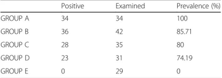
Table 1 Prevalence of pseudomelanosis in examined subjects
Positive Examined Prevalence (%)
GROUP A 34 34 100
GROUP B 36 42 85.71
GROUP C 28 35 80
GROUP D 23 31 74.19
189.50; P < 0.0001), C (χ2 7.57; P = 0.003), D (χ2 10.01; P = 0.0008) and E (χ2 63.00; P < 0.0001). Chi square test showed a significant higher pseudomela-nosis prevalence in groups B (χ2 50.42; P < 0.0001), C (χ2 41.20; P < 0.0001) and D (χ2 50.20; P < 0.0001) compared to group E.
Macroscopic exam carried out on lymph nodes did not show any modification in shape and volume; the only evident feature was the strong black-brownish dis-coloration of the tissue which, in all the positive cases observed, involved both cortical and medullar portions
(Fig. 1). None of the control animals showed any
pig-mentation in lymph nodes.
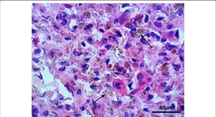
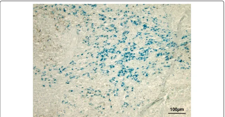
Histological exam performed on lymph node tissue sections stained with H&E permitted to show a macrophage infiltrate containing a granular brownish pigment from multiple observations (three independ-ent histopathologists) (Fig. 2). Nile Blue elective stain-ing showed a dark green pigment, differentiatstain-ing it from lipofuscin and thus suggesting a diagnosis of pseudomelanosis (Fig.3). Finally, Lillie’s method for ferric
and ferrous iron (Fig.4), a highly specific reaction, showed granulation of dark green color in macrophages, providing a further and final confirmation of the diagnosis. Both the two discolorating histochemical tests specific for melanin (Melanin Bleach and Hydrogen Peroxide) revealed the total disappearance of the pigment from the lymph node tissue (Fig.5).
Okun’s reaction, performed on cryostate intestine sec-tions (jejunum), showed formation of intracellular
pseudomelanin pigment in mastocytes detected in intes-tinal villi (Fig. 6), confirming the presence of tyrosinase enzymes at the level of the enteric wall in swine.
No skin melanomas were detected.
The immunohistochemical reaction with macrophage marker (Fig.7), performed on lymph node tissue sections, showed a strong cytoplasmic expression of macrophages containing variable amounts of pigment. The different concentrations checked for the anti-human melanoma antibody, as well as the immunoreaction against S-100, did not show any expression, thus excluding the hypoth-esis of melanoma metastasis in lymph nodes.
Results obtained from biochemical assay performed on acorn specimens showed the presence of a higher amount of phenolic substances (mg of gallic acid equiva-lents/mg of acorn tissue) in Quercus as compared to that of holm oak (Table2).
Discussion
Data here reported represent the first experimental evidence of a feeding related melanosis in Nero Calabrese pig. This conclusion is supported by the high incidence of melanosis in acorn fed groups the absence of neoplastic skin lesions, the absence of im-munoreaction for anti-human melanoma which cross reactivity has been demonstrated by Lanteri et al. [13], the absence of such pigmentation in control ani-mals, as well as from Okun’s reaction performed on the intestinal mucosa, confirming the acorn related enteric pathogenesis. Thus, the results obtained in
Fig. 2 Lymph nodes: Lymph node tissue sections shown a macrophage infiltrate containing a granular brownish pigment (Arrows) (H&E)
this study, considering the well-known process leading to melanin production, permit to hypothesize that the enzyme present at the level of the intestine in swine could start this catalytic process to form melanin, act-ing on the phenolic substrate of the acorn; moreover,
as already shown in Table 1, the higher presence of
such substrate in quercus acorn can explain the in-creased incidence of the pigmentation in those sub-jects which had been fed this food for a longer period of time/higher quantity. Moreover, groups fed
Fig. 4 Lymph nodes: Evidence of dark green granules in macrophages. (Lillie)
with Q. virgiliana showed a greater melanosis preva-lence than those fed with Q. ilex.
The findings obtained support the feed-related patho-genesis previously suggested also in Nero Siciliano Pig, demonstrating for the first time acorn ingestion as cause
of acquired melanosis or, rather, a pseudomelanic the-saurismosis in Nero Calabrese Pig [4,12].
Nevertheless, a similar pathogenic possibility was re-ported as a possible origin of melanosis in pigs fed with acorns [14] even if, considering the limited scientific and
Fig. 6 Lymph nodes: Evidence of intracellular pseudomelanin pigment in mastocytes (Arrows) detected in intestinal villi confirming the presence of tyrosinase enzymes at the level of the enteric wall in swine. (Okun’s reaction)
technical knowledge at that time, tannins were only sug-gested as a possible cause although no demonstration was provided.
The role of polyphenol-oxydase and/or peroxydase in pig intestine in activating the phenolic substrate present in acorn, determining the catalysis reaction of the melanin-like pigment, can be stated. Wilberts et
al. [15] reported the presence of a melanosis coli in
swine due to ceroid pigment storage with multifactor-ial etiology, studying the association among oxidative damage, protein nitrification and hypovitaminosis E [15].
In human beings, melanosis coli is characterized by a brown-black pigmentation of the colon mucosa and is commonly associated to chronic use of anthracen based laxative (cascara, senna, aloe and rhubarb, among others), containing antrachinone as the active principle. Histologically, melanosis coli is character-ized by macrophage carrying pigment in the lamina
propria of the colon-rectal mucosa [16]. Recent
la-boratory investigations have shown that the compos-ition of such pigment is not perfectly superimposable
with melanin pigment [17]; consequently, the term
pseudomelanosis was introduced, suggesting the pres-ence of a melanin-like pigment with exogenous origin. In our opinion, the pathogenesis of the lymph node pigmentations here reported resembles that of melan-osis coli, even if in our opinion the term pseudomela-nosis should be preferred.
In the field of comparative pathology, the chronic use of vegetable laxatives containing anthraquinone over a long time, in man, was reported to evoke the appearance of melanosis in the colon [17,18].
Conclusions
The typical characteristics of rusticity of the Nero Cala-brese Pig, as well as the Sicilian breed, generally farmed in plein-air, make this swine breed an interesting in vivo model to study some disorders which have disappeared or, at least are rare, in modern swine culture, and so it could be considered as a model for comparative study also for some human pathologies.
Methods
In this study, 142 pigs belonging to the“Nero Calabrese” breed, both males and females, of about 18–24 months in age, reared in plein-air in a forest area of Aspromonte
and fed with natural foods derived from the
environment, with a supplement of commercial food in different percentages depending on seasonal availability in the territory were analyzed. All the animals belong to
the “Strangio” farm and were selected on the basis of a
total absence of melanoma lesions, such as maculae, nodules or exophytic masses; moreover, because of con-tinuous meat production animals were randomly se-lected throughout the year, permitting to evaluate the effect of different acorns on lymph node discoloration. Pigs were subdivided into 5 groups and were regularly slaughtered at the public slaughterhouse of Siderno (RC), in the period 2015–16:
1) Group A: n° 34 subjects, 70% natural fed/present in grazing [quercus acorn“Quercus virgiliana” (crude protein 4.2%; etheric extract 2.9%; crude fiber 2.6%), roots, tubers] + 30% commercial fed (barley, field bean, maize, wheat feed, citrus fruit essential pulps, carob beans, sugar cane molasses, calcium
carbonate, bi-calcic phosphate, sodium chloride. Analytic Composition: crude protein 17%; etheric extract 3.4%; crude fiber 4.6%, ash 5.7%).
2) Group B: 42 subjects, 40% natural fed/present in grazing [quercus acorn“Quercus virgiliana” (crude protein 4.2%; etheric extract 2.9%; crude fiber 2.6%), roots, tubers] + 60% commercial fed (barley, field bean, maize, wheat feed, citrus fruit essential pulps, carob beans, sugar cane molasses, calcium
carbonate, bi-calcic phosphate, sodium chloride. Analytic composition: crude protein 17%; etheric extract 3.4%; crude fiber 4.6%, ash 5.7%). 3) Group C: 35 subjects, 70% natural fed/present in
grazing [holm oak acorn“Quercus ilex” (crude
protein 4.6%; etheric extract 2.8%; crude fiber 2.8%), roots, tubers] + 30% commercial fed (barley, field bean, maize, wheat feed, citrus fruit essential pulps, carob beans, sugar cane molasses, calcium
carbonate, bi-calcic phosphate, sodium chloride. Analytic composition: crude protein 17%; etheric extract 3.4%; crude fiber 4.6%, ash 5.7%).
4) Group D: 31 subjects, 40% natural fed / present in
grazing [holm oak acorn“Quercus ilex” (crude
protein 4.6%; etheric extract 2.8%; crude fiber 2.8%), roots, tubers] + 60% commercial fed (Composition: barley, field bean, maize, wheat feed, citrus fruit essential pulps, carob beans, sugar cane molasses, calcium carbonate, bi-calcic phosphate, sodium chloride. Analytic composition: crude protein 17%; etheric extract 3.4%; crude fiber 4.6%, ash 5.7%). 5) Group E: 29 subjects, 100% commercial fed
(barley, field bean, maize, wheat feed, citrus fruit essential pulps, carob beans, sugar cane molasses, calcium carbonate, bi-calcic phosphate, sodium chloride. Analytic composition: crude protein
Table 2 QV: Quercus virgiliana; QI: Quercus ilex
Acorn Phenolic Substances
QV 0.0137 ± 0.012 mg
17%; etheric extract 3.4%; crude fiber 4.6%, ash 5.7%).
Sample size was determined on the basis of the num-ber of pigs actually reared on the farm in a one-year period. Due to the homogeneity of the sample size in the different groups, all animals were considered for statis-tical evaluation.
At regular slaughtering, all organs were examined. Lymph nodes and intestine (jejunum), were sampled and photographed; parts of the tissue samples were fixed in 10% formalin solution and embedded in par-affin wax. Tissues coming from 10 specimens for each experimental group were used for histological,
histo-chemical and immunohistohisto-chemical analysis. 5μm
thick paraffin embedded sections were routinely
stained with haematoxilin-eosin (H&E).
Histochemical evaluation was performed on deparaf-finized histological sections to identify the cell types involved and to characterize the pigment stored, using the following staining and bleaching methods:
Histochemical staining reactions
Nile Blue (hydrogen sulfate): elective staining method based on hydrogen sulfate specific to differentiate neutral fat and cholesterol esters from free fat acids and from phospholipids; such reaction permits to
differentiate melanin pigment from lipofuscin [19].
Lillie’s method: this histochemical reaction, based on ferric sulfate and potassium iron cyanide, is highly se-lective for melanin, showing pigment granules stained
in dark green [20]. Okun’s method: elective
histo-chemistry for tyrosinase detection in intestine,
through catalysis of the L-tyrosine and DL-DOPA in melanin. This reaction, performed on cryostated
sec-tions, is severely damaging for tissue sections [21].
Bleaching histochemical methods. Hydrogen peroxide: histological sections are treated for 24–48 h with 10%
H2O2. After this treatment, melanin present in
tis-sues, both externally and in macrophages, is disco-lored. Melanin is the only pigment that bleaches with
this reaction [2]. Melanin bleach: this method is
based upon the same principle of bleaching using hydrogen peroxide but using potassium permanganate and oxalic acid [2].
Immunohistochemistry staining reactions
To further confirm the pigmentation disorder, elimin-ating every doubt on the possible pathogenetic
hy-pothesis of melanosis subsequent to regressive
congenital melanoma, the following
immunohisto-chemical stains were carried out on 10μm thick
histological sections, developed from an Avidin-biotin complex (BioSpa, 20,143, Milano, Italy) and revealed
by DAB (Diaminobenzidine, Vector Laboratories, Inc.
U.S. Headquarters, Burlingame, California, USA):
Macrophage marker (Novocastra, Newcastle Upon Tyne, UK, Product Code NCL-MAC387; dilution 1: 100), mouse monoclonal antibody with high specificity towards leucocyte antigens and L1 proteins, important for macrophage, monocyte and histiocyte labelling; to differentiate melanin pigmentation from immunohisto-chemistry labelling, sections were bleaching for 150 min by using 10% H2O2 in phosphate-buffered saline (PBS) at
60C° as proposed by Orchard [22]. Monoclonal mouse
anti-human melanoma (Dako, Denmark, Product Code M 7258; dilution 1:25, 1:50, 1:100, 1:200), specific against the Melan A antigen; this antibody, provided for use in im-munocytochemistry and immunohistochemistry, permits to show antigens at 45, 68, 75 KDa (positivity range known for pig) on the basis of concentration [23]. Exams were performed in duplicate with two different rates, on lymph node tissue sections. Polyclonal rabbit anti-S100 (Dako, Denmark, Product Code Z 0311; dilution 1:1000), antibody labelling normal melanocytes, Langerhans cells, histiocytes, condrocytes, adipocytes, cardiac and skeletal muscle, Schwann cells, epithelial and myoepithelial mam-mary cells, salivary and sweat glands, and glial cells. For each sample, negative controls were also performed by omission of primary antibody (Ab) and substitution of pri-mary antibody with an indifferent rabbit pripri-mary antibody (Additional files 2, 4). Moreover, melanoma tissue ob-tained from a Nero Siciliano Pig was used as positive con-trol [13] (Additional files1,3).
Biochemical reactions
Total phenolic substances were identified from acorns through the use of a methanolic extract with Folin-Ciocalteau reagents [24]; data obtained revealed the total amount of phenolic substances expressed as amount of gallic acid on mg of acorn tissue.
Statistical analysis
Chi square analysis was performed to evaluate
differ-ence in pseudomelanosis prevalence among the
Groups. P values < 0.05 were considered statistically significant.
Data were analyzed using statistical software Prism v. 4.00 (Graphpad Software Ldt., USA, 2003).
Additional files
Additional file 1:Melan A: positive control in melanoma from pig (bar 50μm). (TIF 4973 kb)
Additional file 2:Melan A: negative control from pig lymph node, obtained by omission of the primary antibody (AB) and substitution of primary antibody with an indifferent rabbit primary antibody (bar 50μm). (TIF 3738 kb)
Additional file 3:S100: positive control in melanoma from pig (bar 50μm). (TIF 3470 kb)
Additional file 4:S100: negative control from pig lymph node, obtained by omission of the primary antibody (AB) and substitution of primary antibody with an indifferent rabbit primary antibody (bar 100μm). (TIF 3500 kb)
Abbreviations
DAB:Diaminobenzidine; KDa: Kilodalton; DL-DOPA: D-form of Dihydroxyphenylalanine; FAO: Food and Agriculture Organization; H&E: Haematoxylin and eosin; RC: Reggio Calabria
Acknowledgments
We would like to thank the owner and Dr. Carmelo De Stefano, the veterinarian caring for the animals and Dr. Francesca Arfuso for support in the statistical analysis.
Authors’ contributions
GL and FM contributed to study design and preparing the manuscript. JA contributed to the acquisition and interpretation of data. CI, DM and VF contributed to the laboratory analysis, drafted and revised the manuscript. All authors have read and approved the manuscript.
Funding
The Authors declare that they have no received funding for this research. Availability of data and materials
The datasets used and analyzed during the current study are available from the corresponding author on reasonable request.
Ethics approval and consent to participate
There is no need of ethical approval as the study was carried out on swine reared in different groups in the farm and normally fed with different acorns available in different seasons. All the animals were evaluated at
slaughterhouse. Moreover, the Authors declare that have obtained written informed consent to use the animals in the study from the owner of the animals.
Consent for publication Not applicable. Competing interests
The authors declare that they have no competing interests. Author details
1Department of Veterinary Sciences, University of Messina, 98168 Messina, Italy.2Department of Chemical, Biological, Pharmaceutical and Environmental Sciences, University of Messina, 98166 Messina, Italy.3Istituto Zooprofilattico Sperimentale della Sicilia, Via Gino Marinuzzi 3, 90129 Palermo, Italy.
Received: 1 March 2019 Accepted: 27 May 2019 References
1. Dämmrich K, Loppnow H. Modificazioni regressive. In: In: Stünzi H., Weiss E. (1986): Patologia Generale Veterinaria. Edizione italiana a cura di F. Guarda, Editoriali Grasso; 1986. p. 244–5.
2. Sheehan DC, Hrapchak BB. Theory and practice of histotechnology. St Louis: CV Mosby Co; 1980. p. 48.
3. Dianzani MU, Dianzani I, Dianzani U. Istituzioni di patologia generale. UTET scienze mediche; 2004.
4. Lanteri G, Marino F, Liotta L, Stefano C, Macrì B. Experimentally induced melanin-like pigmentation (thesaurismosis) related to acorn ingestion in Nero Siciliano pigs. Acta Vet Hung. 2011;59:311–8.
5. Jones TC, Hunt RD, King NW. Veterinary pathology (6th edn) Williams and Wilkins. London Philadelphia. 1997:66–7.
6. Cerundolo R, Maiolino P, Roperto F, Lloyd DH, Visintin A, Maio M. Vitiligo following melanoma in Vietnamese pot-bellied pigs: gross, ultrastructural and immunological studies; 1998.
7. Hook RR Jr, Aultman MD, Adelstein EH, Oxenhandler RW, Millikan LE, Middleton CC. Influence of selective breeding on the incidence of melanomas in Sinclair miniature swine. Int J Cancer. 1979;24:668–72. 8. Oxenhandler RW, Berkelhammer J, Smith GD, Hook RR Jr. Growth and
regression of cutaneous melanomas in Sinclair miniature swine. Am J Pathol. 1982;109:259.
9. Ruth GR, Horstmann JP, Lanin DR, Flynn K, Wang N, Hordinski M, et al. Spontaneous regression of cutaneous melanotic tumors in Duroc pigs. In: Proceedings of annual meeting American Association of Veterinary Laboratory Diagnosticians; 1980.
10. Zaghini L, Della Salda L, Marcato PS. Melanosi e melanomi nei suini macellati. Obiettivi e Doc Vet. 1995;16:65.
11. Herenda D, Chambers PG, Ettriqui A, Seneviratna P, da Silva TJP. Manual on meat inspection for developing countries. Food and Agriculture Organization of the United Nations [FAO] Animal Production and Health Paper 119 [monograph online]. FAO; 1994. Specific diseases of pigs: Cysticercosis (Cysticercus cellulosae) infes. 2004.
12. Lanteri G, Marino F, Laganà G, Bellocco E, Barreca D, Liotta L, et al. Acquired melanosis caused by acorn ingestion in the Nero Siciliano pig. Vet Pathol. 2009;46:329–33.
13. Lanteri G, Marino F, Mazzullo G, Macrì B. Cutaneous melanoma and related melanosis of regional lymph nodes in Nero Siciliano pigs. Large Anim Rev. 2010;16:285–8.
14. Carta A. Sulla pseudomelanosi dei linfonodi di suini alimentati con ghiande. Atti Soc Ital Sci Vet. 1948;2:363–9.
15. Wilberts BL, Schwartz KJ, Gauger PC, Wang C, Burrough ER. Evidence of oxidative injury in pigs with melanosis coli. Vet Pathol. 2015;52:663–7. 16. Ghadially FN, Walley VM. Melanoses of the gastrointestinal tract.
Histopathology. 1994;25:197–207.
17. Freeman HJ.“Melanosis” in the small and large intestine. World J Gastroenterol WJG 2008;14:4296.
18. A. O. L’utilizzo della fitoterapia. Utifar. 2017.https://www.utifar.it/uploads/ model_5/l_utilizzo_della_fitoterapia.pdf.
19. Lillie RD. A Nile blue staining technic for the differentiation of melanin and lipofuscins. Stain Technol. 1956;31:151–3.
20. Botticelli AR, Villani M, Angiari P, Peserico L. Meningeal melanocytoma of Meckel’s cave associated with ipsilateral Ota’s nevus case report. Cancer. 1983;51:2304–10.
21. Okun MR, Edelstein LM, Hamada G, Donnellan B, Or N. The role of peroxidase vs. the role of tyrosinase in enzymatic conversion of tyrosine to melanin in melanocytes, mast cells and eosinophils. J Invest Dermatol. 1970; 55:1–12.
22. Orchard GE. Use of heat provides a fast and efficient way to undertake melanin bleaching with dilute hydrogen peroxide. Br J Biomed Sci. 2007;64:89–91.
23. Cui J, Chen D, Misfeldt ML, Swinfard RW, Bystryn J. Antimelanoma antibodies in swine with spontaneously regressing melanoma. Pigment Cell Res. 1995;8:60–3.
24. Velioglu YS, Mazza G, Gao L, Oomah BD. Antioxidant activity and total phenolics in selected fruits, vegetables, and grain products. J Agric Food Chem. 1998;46:4113–7.
Publisher’s Note
Springer Nature remains neutral with regard to jurisdictional claims in published maps and institutional affiliations.