Prodromal angina and risk of 2-year cardiac
mortality in patients with ST-segment elevation
myocardial infarction undergoing primary
percutaneous intervention
Gabriele Ghetti, MD, Maria Letizia Bacchi Reggiani, MSc, Claudia Rosetti, MD, Paola Battistini, MD,
Gianluca Lanati, MD, Maria Teresa Di Dio, MD, Anna Corsini, MD, Matteo Bruno, MD, Diego Della Riva, MD,
Antonio Giulio Bruno, MD, Miriam Compagnone, MD, Riccardo Narducci, MD, Francesco Saia, MD, PhD,
Claudio Rapezzi, MD, Nevio Taglieri, MD
∗Abstract
We sought to investigate the prognostic significance of prodromal angina (PA) in unselected patients with ST-segment elevation myocardial infarction (STEMI) undergoing primary percutaneous coronary intervention (PPCI) and its additive predictive value to the GRACE score.
We prospectively enrolled 3015 consecutive STEMI patients undergoing PPCI. Patients were divided in 2 groups according to the presence or absence of PA. Multivariable Cox regression was used to establish the relation to 2-year cardiac mortality of PA.
The mean age of the study population was 68 (±14) years; 2178 patients (72%) were male. During follow-up, 395 (13%) patients died with 278 of these (9.2%) suffering from cardiac mortality. Kaplan-Meier estimates showed a survival rate of 95% and 87% for patients with PA and no PA, respectively (log rank test<0.001). After multivariable analysis, patients with PA had still a lower risk of 2 years’ cardiac mortality compared with patients without PA (adjusted hazard ratio=0.50; 95% confidence interval [CI] 1.06–1.81, P=.001). Evaluation of net reclassification improvement showed that reclassification improved by 0.16% in case patients, whereas classification worsened in control patients by 1.08% leading to a net reclassification improvement of 0.93% (95% CI: 0.98, 0.88).
In patients with STEMI undergoing PPCI the presence of PA is independently associated with a lower risk of 2-year cardiac mortality. However, the incorporation of this variable to the GRACE score slightly worsened the classification of risk. Accordingly, it seems unlikely that the evaluation of PA may be useful in clinical practice.
Abbreviations: DES= drug-eluting stent, ECG = electrocardiogram, GP IIb/IIIa = glycoprotein IIb/IIIa, GRACE = Global Registry of Acute Coronary Events, LVEF= left ventricular ejection fraction, MI = myocardial infarction, NRI = net reclassification improvement, PA= prodromal angina, PPCI = primary percutaneous coronary intervention, SD = standard deviation, STEMI = ST elevation myocardial infarction, TIMI= thrombolysis in myocardial infarction.
Keywords:outcome, prodromal angina, STEMI
1. Introduction
In patients with ST-segment elevation myocardial infarction (STEMI), primary percutaneous coronary intervention (PPCI)
has emerged as the preferred reperfusion therapy to achieve early reperfusion, limit the infarct size, and ultimately reduce short and long-term fatality rates for MI.[1,2]
Despite obtaining a coronaryflow restoration in most of the cases, mechanisms that determine the extent of myocardial damage are multifactorial and poorly understood. They mainly comprise the area at risk, the total ischemic time, reperfusion injury, microembolization, and microvascular obstruction.[3] It
has been suggested that prodromal angina (PA) related to brief episode of myocardial ischemia before index MI (the so-called ischemic preconditioning) may contribute to limit thefinal infarct size.[4,5] Two recent studies[6,7] prospectively enrolling a small series of STEMI patients undergoing PPCI have shown that PA is independently associated with an increases myocardial savage andfinal infarct size as assessed with cardiac magnetic resonance. However, if these findings translate into an independent prognostic role of PA in the clinical setting of PPCI has not been established yet. Furthermore, whether the addition of PA to a current recommended GRACE risk score[8] improves the classification of risk has not been studied yet.
Therefore, the objectives of the present study were: to investigate the association between PA and 2-year cardiovascular mortality in an unselected cohort of STEMI patients undergoing
Editor: Yan Li.
This work was supported by Department of Experimental, Diagnostic and Specialty Medicine–DIMES, University of Bologna and by Fanti Melloni Foundation.
The authors report no conflicts of interest.
Polo Cardio-Toraco-Vascolare, Dipartimento di Medicina Specialistica, Diagnostica e Sperimentale, Alma Mater Studiorum Università di Bologna, Italy. ∗
Correspondence: Nevio Taglieri, Istituto di Cardiologia, Dipartimento di Medicina Specialistica, Diagnostica e Sperimentale, Alma Mater Studiorum Università di BolognaVia Massarenti 9, 40138 Bologna, Italy (e-mail: [email protected]). Copyright© 2018 the Author(s). Published by Wolters Kluwer Health, Inc. This is an open access article distributed under the terms of the Creative Commons Attribution-Non Commercial-No Derivatives License 4.0 (CCBY-NC-ND), where it is permissible to download and share the work provided it is properly cited. The work cannot be changed in any way or used commercially without permission from the journal. .
Medicine (2018) 97:37(e12332)
Received: 19 February 2018 / Accepted: 9 August 2018 http://dx.doi.org/10.1097/MD.0000000000012332
Observational Study
Medicine
PPCI and to evaluate its additive predictive value to the clinical risk model.
2. Material and methods
The present prospective cohort study (conceived in accordance with the principles of the most recent revision of the Declaration of Helsinki) was based on the database of one (S. Orsola-Malpighi Hospital) of the 2 centralized PPCI centers of the “Bologna STEMI hub and spoke network."[9,10]This prospective
database contains demographic information along with compre-hensive clinical, electrocardiogram (ECG), procedural, and outcome data from STEMI patients treated with PPCI at the center’s catheterization laboratory. The registry started in 2003 and was approved by the hospital’s Ethics Committee. For the purpose of the present study, we include consecutive out-of-hospital STEMI patients treated with PPCI from January 1, 2003 to December 31, 2015. We excluded patients with missing data and those with coma at presentation owing to the difficulty of ascertaining the presence or not of PA.
STEMI was defined as significant ST-elevation (in 2 adjacent leads and 0.1 mV in leads I–III, aVF, aVL, V4–V6, and 0.2mV in leads V1–V3) or in presence of STEMI-equivalent, namely new-onset (or presumably new) left bundle branch block, posterior STEMI and ST-T de Winter complex.[11]Stenosis≥50% in the lumen of left main artery or ≥70% in ≥1 of other major epicardial vessels or their main branches was considered significant. The culprit lesion was defined as the lesion with the highest degree of stenosis or in the presence of thrombolysis in myocardial infarction (TIMI) flow grade <3 or angiographic signs of endoluminal thrombi and/or plaque rupture. Coronary thrombus was defined as an intraluminal filling defect or an area of contrast staining within the coronary lesion. Plaque rupture was defined by the presence of a small crater or visible intimal flap. In case of TIMI flow <2, thrombotic coronary occlusion was defined on the basis of morphology and lack of collateral vessels. Percutaneous coronary intervention (PCI) was mainly performed within 12 hours of the self-reported onset of symptoms. Patients with symptom onset-to-STEMI diagnosis time interval between 12 and 24 hours were enrolled only in case of evident ongoing myocardial ischemia. Before PCI execution at the time of diagnosis, all patients received aspirin (250 mg i.v.) and heparin (5000 IU i.v). Vascular access, use of platelet glycoprotein IIb/IIIa agents, manual thrombectomy, and type of stent were at operator’s discretion. After PCI, P2Y12 inhibitors were adminis-tered according to current guidelines.
Cardiac arrest was defined as cardiac arrest occurring before revascularization and requiring resuscitation procedures, (e.g., ventilation, chest compression, defibrillation).
PA was defined as ≥1 episodes of angina (Canadian cardiovascular Sociaty class ≥1) up to 3 months before the index episode, based on previous results.[7,12]
Study endpoint was cardiac mortality at 2-year follow-up. Cardiac death was defined as death from cardiac cause, sudden death, or any death without another known cause. In-hospital re-MI was defined as the recurrence of typical clinical symptoms and new ECG changes with an increase of MB Creatin-Kinase (MB-CK) above≥ 50% of the previous levels. Out-of-hospital data concerning vital status of patients and cause of death were obtained by telephone interviews or independently from the Emilia-Romagna Regional Health Agency through the analysis of the Hospital Discharge Records and the Municipal Civil Registries, thus relying on treating physician’s diagnosis.
Categorical data are expressed as proportions and continuous variables reported as mean (±SD) or medians medians (25th–75th
percentiles). Patients were divided into 2 groups: those with no PA and patients with PA. For comparisons between groups, the x2test was used for categorical variables. Continuous variables
were tested for normal distribution and tested using Student t or Mann-Whitney tests as appropriate.
The Kaplan-Meyer method was used to analyze the occurrence of events during follow-up. Study groups were compared by means of the log-rank Cox-Mantel test. Patients were censored at 2 years or at the time of the last contact. The relation to 2-year cardiac mortality of PA was investigated with the use of multivariable Cox regression model adjusted for variable selected at univariable analysis (P <.1). Thus, the following variables were entered in a multivariable backward selection: age, sex, smoking status, hypercholesterolemia, diabetes, hypertension, family history of coronary artery disease, previous MI, previous stroke, peripheral artery disease, systolic blood pressure and heart rate on admission, anterior MI, Killip class, cardiac arrest, creatinine levels on admission, left ventricle ejection fraction (LVEF), radial access, multivessel disease, GP IIb/IIIa inhibitors, pre and post TIMIflow grade, pain to balloon interval, and PA. Proportional hazard assumption was checked by Shoenfeld tests. Model discrimina-tion was assessed using the Harrell C-index.[13]and confidence intervals were calculated using bootstraps estimation. Calibra-tion was evaluated by the Groennesby and Borgan test approach for survival data,[14] with a x2 ≥ 20 (P <.01)
indicating poor calibration.
The additional contribution of PA to the GRACE risk score for prediction of study endpoint was evaluated in terms of classification accuracy measuring the continuous net reclassifica-tion improvement (NRI) as previously described.[15]Confident
intervals were calculated using bootstraps estimation. AP value <.05 in the 2-tailed tests was considered significant. All analyses were performed with STATA 14.0 software (STATA Corpora-tion, College StaCorpora-tion, TX).
3. Results
During the study period, 3278 of hospital STEMI patients were treated with PPCI and enrolled in the prospective dataset of our Institution. For the purpose of the present study, 263 patients were excluded because of missing values and/or coma at admission (Fig. 1). Thus, the final study cohort comprises 3015 of hospital STEMI patients undergoing primary angioplasty.
Baseline characteristics are listed in Table 1. The mean age of the study population was 68 (± 14) years; 2178 patients (72%) were male. Among the study cohort, 685 (23%) had PA before the index MI. Patients with PA were generally younger than patients with no PA. They were more likely to be male and had a lower prevalence of hypertension, previous MI, and previous PCI. Patients with PA had a better clinical profile in terms of Killip class, LVEF, and pre-TIMIflow grade. They were more likely to receive radial access, drug-eluting stent (DES) implantation, and thrombus aspiration.
Overall, the rate of in-hospital mortality was 4.6% (Table 2). All deaths, but one (sepsis), were from cardiac origin. Patients with PA had a statistically significant lower rate of cardiac mortality than patients without PA (2.3% vs. 5.3%, respectively). There were no statistically significant differences in the rate of in hospital re-MI between the study groups (Table 2).
Table 1
Baseline characteristics.
Variable All patients No prodromal angina Prodromal angina
P
No. of patients n=3015 n=2330 n=685
Age, years, mean±SD 68±14 68±14 66±13 <.001
Male sex, no. (%) 2178 (72) 1657 (71) 521 (76) .011
Medical history
Previous MI, no. (%) 515 (17) 434 (18) 81 (12) <.001
Previous PCI, no. (%) 353 (12) 302 (13) 51 (8) <.001
Previous coronary bypass, no. (%) 69 (2.3) 55 (2.4) 14 (2.0) .626
Previous stroke/AIT, no. (%) 159 (5) 128 (6) 31 (5) .319
Peripheral artery disease, no. (%) 315 (10) 244 (11) 71 (10) .936
Risk factors
Diabetes, no. (%) 608 (20) 490 (21) 118 (17) .029
Hypercholesterolemia, no. (%) 192 (64) 1467 (63) 454 (66) .113
Hypertension, no. (%) 2099 (70) 1665 (71) 434 (63) <.001
Smokers, no. (%) 1966 (65) 1501 (64) 465 (68) .09
Family-history of CAD, no. (%) 971 (32) 744 (32) 227 (33) .552
Presenting characteristics
Anterior MI, no. (%) 1466 (49) 1114 (48) 352 (51) .1
Systolic BP, mmHg, mean±SD 128±30 128±30 129±29 .530
Heart rate, pulse/min, mean±SD 76±19 76±19 76±18 .8214
Cardiac arrest, no. (%) 115 (38) 94 (4.0) 21 (3.0) .245
Killip class>2, no. (%) 423 (14) 357 (15) 66 (10) <.001
Creatinine, mg/dL, mean±SD 1.13±0.7 1.14±0.68 1.12±0.78 .58
LVEF, mean±SD 49±11 49±12 51±12 .001
Procedure and medications
Radial access, no. (%) 1157 (39) 786 (34) 371 (54) <.001
Multivessel disease, no. (%) 1515 (50) 1146 (49) 369 (54) .031
Pre-TIMIflow grade <2, no. (%) 2374 (79) 1863 (80) 511 (75) .031
Post-TIMIflow grade <2, no. (%) 115 (38) 92 (3.9) 23 (3.4) .478
DES, no. (%) 476 (16) 336 (14) 140 (70) <.001
Thrombus aspiration, no. (%) 660 (22) 483 (24) 177 (26) .004
P2Y12 inhibitor, no. (%) 2879 (96) 2217 (95) 662 (97) .098
GP IIb/IIIa 2203 (73) 1710 (73) 493 (72) .402
Total ischemic time, min (25th–75th) 180 (120–295) 179 (121–297) 183 (120–290) .97
BP=blood pressure, DES =drug-eluting stent, LVEF=left ventricular ejection fraction, MI=myocardial infarction, PCI= percutaneous coronary intervention, TIMI=thrombolysis in myocardial infarction.
During a median follow-up of 2 years (only 34 [1.1%], patients had incomplete follow-up with a minimum value of 286 days), 395 patients died (13%). Of these, 278 patients (9.2%) suffered from cardiac mortality. The rates of both fatal and non-fatal re-MI were comparable (Table 2).
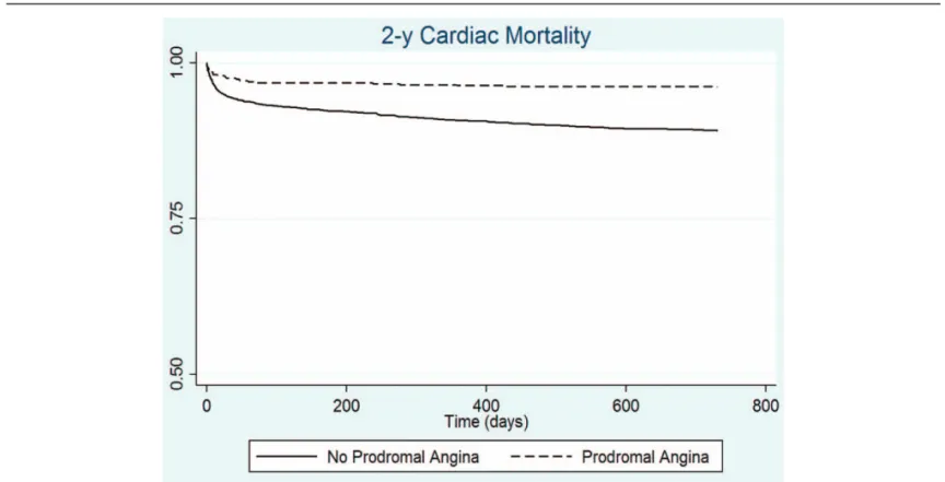
Kaplan-Meier estimates showed a survival rate of 95% and 87% for patients with PA and no PA, respectively (Fig. 2, log rank test<0.001).
After univariable and multivariable analysis, patients with PA had still a lower risk of 2 years cardiac mortality compared with patients without PA (adjusted hazard ratio [HR]=0.50; 95% confidence interval [CI] 1.06–1.81, P=.001) (Table 3). The model showed good discrimination power (Harrell C=0.876, 95% CI 0.858–0.897) and good calibration (Groennesby and Borganx2=8.065; P=.153).
Evaluation of NRI showed that reclassification improved by 0.16% in case patients, whereas classification worsened in control patients by 1.08% leading to a NRI of 0.93% (95% CI:
0.98, 0.88).
Table 2
Outcome.
Variable All patients No Prodromal angina Prodromal angina
P
No. of patients n=3015 n=2330 n=685
In-hospital outcome
All-cause mortality, no. (%) 140 (4.6) 124 (5.3) 16 (2.34) .001
Cardiac mortality, no. (%) 139 (4.6) 123 (5.3) 16 (2.34) .001
Re- myocardial infarction, no. (%) 55 (1.8) 37 (1.6) 18 (2.6) .074
Two-year outcome∗
All-cause mortality, no. (%) 395 (13) 350 (15) 45 (6.6) <.001
Cardiac mortality, no. (%) 278 (9.2) 251 (11) 27 (4) <.001
Fatal myocardial infarction 96 (35%) 86 (34.26) 10 (37.0) .77
Re-myocardial infarction 311 (10.3) 238 (10.2) 73 (10.6) .74
∗
Patients with complete follow-up=2981 (98.87%).
Table 3
Independent predictors of 2-year cardiac mortality. Multivariable Cox regression analysis.
Variable HR (95% CI) P Age, years, 1.05 (1.04–1.07) <.001 Diabetes 1.39 (1.06–1.81) .016 Systolic BP, mmHg 0.99 (0.99–1.00) .008 Cardiac arrest 1.68 (1.01–2.79) .045 Killip class>2 2.22 (1.65–2.97) <.001 Creatinine, mg/dL 1.16 (1.03–1.29) .012 LVEF 0.94 (0.93–0.95) <.001 Radial access 0.64 (0.46–0.89) .009 DES 0.41 (0.25–0.70) <.001 GP IIb/IIIa 0.71 (0.55–0.93) .009 Prodromal angina 0.50 (1.06–1.81) .001
BP=bloood pressure, DES =drug-eluting stent, HR=hazard ratio, LVEF=left ventricular ejection fraction.
Figure 2. Kaplan-Meier estimates for 2 year cardiac mortality in patients with PA and those without.
4. Discussion
The mainfindings of the present study enrolling 3015 unselected STEMI patients undergoing PPCI are as follows: patients with PA have a lower risk of 2-year cardiac mortality compared to those without; the incorporation of PA into the GRACE score does not improve classification of risk.
In acute MI with ST segment elevation the definite treatment is to rapidly restore coronary bloodflow and myocardial perfusion with the objective of reducing infarct size and improve outcome.[1,2] The size of the scar is directly associated with duration of myocardial ischemia. Previous studies have shown that PA leads to a more rapid reperfusion and smaller infarct size in patients with acute myocardial infarction treated with thrombolysis[16] with a mechanism not fully understood but likely involving ischemic preconditioning.[17,18]In the setting of
PPCI, 2 recent studies prospectively enrolling a small series of STEMI have shown that PA is independently associated with an increase myocardial savage and smaller final infarct size as assessed with cardiac magnetic resonance.[6,7]However, if these findings translate into a favorable effect of PA on long-term prognosis has not been established, yet.
In the present study, enrolling a large cohort of STEMI patients undergoing PPCI, we show that those experiencing episode of angina up to 3 months before the index episode have a lower risk of cardiac mortality. Patients with PA have a better risk profile in terms of some characteristics such as diabetes and DES utilization and this could be partly account for its prognostic role. However, the strength of thisfinding relies on a comprehensive multivari-able adjustment including also the LVEF, a main prognosticators of long-term prognosis after MI.[19] Of note, the rates of MI during follow-up between the study groups were comparable confirming that PA exercises its protective role reducing both the risk of life-threatening arrhythmias[20,21] and the risk of heart failure[22]in keeping with the theory that ischemic
precondition-ing reduces the infarct size and favors the post-ischemic recovery of global ventricular function.[23]In particular, the risk of
life-threatening arrhythmias remains high in post-MI survivors with a mortality rate of 25% at 2 years.[24] Along with prompt
revascularization, optimal medical therapy counteracting nega-tive cardiac remodeling and implantable cardioverter de fibrilla-tor are mainstay therapies to prevent death in patients with post-MI LV dysfunction.
Findings of our study contrast with those of a previous study that did not show a prognostic relevance of PA on the clinical course of STEMI patients undergoing PPCI.[25]However, there
are several differences between this study and ours that could explain the conflicting results.
First, we chose as primary endpoint the rate of long-term cardiac mortality, instead of in-hospital mortality, to better elucidate the cardiac consequences of PA in STEMI. Second, our sample size was 4-fold larger than the other one. Finally, patients of the present study were enrolled almost a decade later compared to those of the above mentioned study and had been treated in the context of current recommended strategies (clopidogrel and new P2Y12 inhibitors, DES, thrombus aspiration, radial access). Yet, to our knowledge, this is thefirst study that evaluates the additive contribution of PA to the GRACE score in a large unselected cohort of STEMI patients. We found that although PA independently stratifies risk of future cardiovascular death, there is no advantage in adding it to the GRACE score. On the contrary, we observed a slight worsening of risk classification.
The study is a prospective analysis of a single-center registry and it is not immune to sources of bias. Although we tried to control our results for known prognostic factors by means of multivariable analysis, the influence of other confounding factors cannot be ruled out. In particular, we did not collect information on the presence of collateralflow. However, it is known that in the context of acute coronary occlusion an efficient collateral flow is rarely observed. Besides, it has been recently shown that the presence of collateralflow has no protective role in terms of infarct size.[7] We did not collect information on LVEF during
follow-up. This is a crucial aspect as it is known that after coronary revascularization a LV function recovery may occur, usually within 1 month[26]. Thus, it would have been desirable to test the association between PA and left ventricle function recovery in order to provide information on the mechanisms linking PA and better outcome. In conclusion, in patients with STEMI undergoing PPCI, the presence of PA is independently associated with a lower risk of 2-year cardiac mortality. However, the incorporation of this variable to the GRACE score slightly worsens the classification of risk. Accordingly, its utility in clinical practice appears unlikely.
Author contributions
Conceptualization: Nevio Taglieri.
Data curation: Gabriele Ghetti, Claudia Rosetti, Paola Battistini, Gianluca Lanati, Maria Teresa Di Dio, Anna Corsini, Matteo Bruno, Diego Della Riva, Antonio Giulio Bruno, Miriam Compagnone, Riccardo Narducci.
Formal analysis: Maria Letizia Bacchi Reggiani, Nevio Taglieri. Investigation: Gabriele Ghetti, Claudia Rosetti, Paola Battistini, Gianluca Lanati, Maria Teresa Di Dio, Anna Corsini, Matteo Bruno, Diego Della Riva, Antonio Giulio Bruno, Miriam Compagnone, Riccardo Narducci, Nevio Taglieri. Methodology: Nevio Taglieri.
Supervision: Nevio Taglieri.
Writing– original draft: Gabriele Ghetti.
Writing– review & editing: Francesco Saia, Claudio Rapezzi, Nevio Taglieri.
References
[1] Ibanez B, James S, Agewall S, et al. 2017 ESC Guidelines for the management of acute myocardial infarction in patients presenting with ST-segment elevation: The Task Force for the management of acute myocardial infarction in patients presenting with ST-segment elevation of the European Society of Cardiology (ESC). Eur Heart J 39;133: 119–77.
[2] Levine GN, Bates ER, Blankenship JC, et al. 2015 ACC/AHA/SCAI Focused Update on Primary Percutaneous Coronary Intervention for Patients With ST-Elevation Myocardial Infarction: An Update of the 2011 ACCF/AHA/SCAI Guideline for Percutaneous Coronary Interven-tion and the 2013 ACCF/AHA Guideline for the Management of ST-Elevation Myocardial Infarction: A Report of the American College of Cardiology/American Heart Association Task Force on Clinical Practice Guidelines and the Society for Cardiovascular Angiography and Interventions. Circulation 2016;133:1135–47.
[3] Christian TF, Schwartz RS, Gibbons RJ. Determinants of infarct size in reperfusion therapy for acute myocardial infarction. Circulation 1992;86:81–90.
[4] Murry CE, Jennings RB, Reimer KA. Preconditioning with ischemia: a delay of lethal cell injury in ischemic myocardium. Circulation 1986; 74:1124–36.
[5] Eisen A, Fisman EZ, Rubenfire M, et al. Ischemic preconditioning: nearly two decades of research. A comprehensive review. Atherosclerosis 2004;172:201–10.
[6] Masci PG, Andreini D, Francone M, et al. Prodromal angina is associated with myocardial salvage in acute ST-segment elevation myocardial infarction. Eur Heart J Cardiovasc Imaging 2013;14:1041–8. [7] Lonborg J, Kelbaek H, Vejlstrup N, et al. Influence of pre-infarction
angina, collateralflow, and pre-procedural TIMI flow on myocardial salvage index by cardiac magnetic resonance in patients with ST-segment elevation myocardial infarction. Eur Heart J Cardiovasc Imaging 2012;13:433–43.
[8] Granger CB, Goldberg RJ, Dabbous O, et al. Predictors of hospital mortality in the global registry of acute coronary events. Arch Intern Med 2003;163:2345–53.
[9] Ortolani P, Marzocchi A, Marrozzini C, et al. Clinical impact of direct referral to primary percutaneous coronary intervention following pre-hospital diagnosis of ST-elevation myocardial infarction. Eur Heart J 2006;27:1550–7.
[10] Ortolani P, Marzocchi A, Marrozzini C, et al. A. Long-term effectiveness of early administration of glycoprotein IIb/IIIa agents to real-world patients undergoing primary percutaneous interventions: results of a registry study in an ST-elevation myocardial infarction network. Eur Heart J 2009;30:33–43.
[11] de Winter RJ, Verouden NJ, Wellens HJ, et al. A new ECG sign of proximal LAD occlusion. N Engl J Med 2008;359:2071–3.
[12] Solomon SD, Anavekar NS, Greaves S, et al. Angina pectoris prior to myocardial infarction protects against subsequent left ventricular remodeling. J Am Coll Cardiol 2004;43:1511–4.
[13] Harrell FEJr, Lee KL, Mark DB. Multivariable prognostic models: issues in developing models, evaluating assumptions and adequacy, and measuring and reducing errors. Stat Med 1996;15:361–87.
[14] Demler OV, Paynter NP, Cook NR. Tests of calibration and goodness-of-fit in the survival setting. Stat Med 2015;34:1659–80.
[15] Pencina MJ, D’Agostino RBSr, Steyerberg EW. Extensions of net reclassification improvement calculations to measure usefulness of new biomarkers. Stat Med 2011;30:11–21.
[16] Andreotti F, Pasceri V, Hackett DR, et al. Preinfarction angina as a predictor of more rapid coronary thrombolysis in patients with acute myocardial infarction. N Engl J Med 1996;334:7–12.
[17] Kloner RA, Jennings RB. Consequences of brief ischemia: stunning, preconditioning, and their clinical implications: part 2. Circulation 2001;104:3158–67.
[18] Kloner RA, Jennings RB. Consequences of brief ischemia: stunning, preconditioning, and their clinical implications: part 1. Circulation 2001;104:2981–9.
[19] Halkin A, Singh M, Nikolsky E, et al. Prediction of mortality after primary percutaneous coronary intervention for acute myocardial infarction: the CADILLAC risk score. J Am Coll Cardiol 2005;45: 1397–405.
[20] Shiki K, Hearse DJ. Preconditioning of ischemic myocardium: reperfu-sion-induced arrhythmias. Am J Physiol 1987;253:H1470–6. [21] Li YW, Whittaker P, Kloner RA. The transient nature of the effect of
ischemic preconditioning on myocardial infarct size and ventricular arrhythmia. Am Heart J 1992;123:346–53.
[22] Kloner RA, Shook T, Przyklenk K, et al. Previous angina alters in-hospital outcome in TIMI 4. A clinical correlate to preconditioning? Circulation 1995;91:37–45.
[23] Cave AC, Hearse DJ. Ischaemic preconditioning and contractile function: studies with normothermic and hypothermic global ischaemia. J Mol Cell Cardiol 1992;24:1113–23.
[24] Raviele A, Bonso A, Gasparini G. Vardas PE, et al. Prophylactic implantation of implantable cardioverter/defibrillator in post-myocardial infarction patients. Cardiac Arrhythmias, Pacing and Electrophysiology Kluger Academic Publishers, Dordrecht:1998; 305–10.
[25] Zahn R, Schiele R, Schneider S, et al. Effect of preinfarction angina pectoris on outcome in patients with acute myocardial infarction treated with primary angioplasty (results from the Myocardial Infarction Registry). Am J Cardiol 2001;87:1–6.
[26] Taglieri N, Saia F, Guiducci V, et al. Left ventricular function after ST-elevation myocardial infarction in patients treated with primary percutaneous coronary intervention and abciximab or tirofiban (from the Facilitated Angioplasty with Tirofiban or Abciximab [FATA] Trial). Am J Cardiol 2009;103:785–90.