A miniaturized silicon based device for nucleic acids
electrochemical detection
Salvatore Petralia
a,⁎
, Maria Eloisa Castagna
a, Emanuele Cappello
b, Fausto Puntoriero
c, Emanuela Trovato
c,
Antonio Gagliano
d, Sabrina Conoci
a,⁎
a
STMicroelectronics, Stradale Primosole 50, 95121 Catania, Italy
b
Distretto Tecnologico Sicilia Micro e Nano Sistemi, VIII strada Z.I., 5, 95121 Catania, Italy
cUniversity of Messina, Chemical Science Department, V.le Ferdinando Stagno d’Alcontres, 31, 98166 Messina Italy d
University of Catania, Department of Industrial Engineering, Viale Andrea Doria 6, 95125 Catania, Italy
a b s t r a c t
a r t i c l e i n f o
Article history: Received 31 July 2015
Received in revised form 9 September 2015 Accepted 17 September 2015
Keywords:
Electrochemical detection Real time PCR
Unspecific DNA intercalator
In this paper we describe a novel portable system for nucleic acids electrochemical detection. The core of the sys-tem is a miniaturized silicon chip composed by planar microelectrodes. The chip is embedded on PCB board for the electrical driving and reading. The counter, reference and work microelectrodes are manufactured using the VLSI technology, the material is gold for reference and counter electrodes and platinum for working electrode. The device contains also a resistor to control and measuring the temperature for PCR thermal cycling. The reac-tion chamber has a total volume of 20μL. It is made in hybrid silicon–plastic technology. Each device contains four independent electrochemical cells.
Results show HBV Hepatitis-B virus detection using an unspecific DNA intercalating redox probe based on metal– organic compounds. The recognition event is sensitively detected by square wave voltammetry monitoring the redox signals of the intercalator that strongly binds to the double-stranded DNA. Two approaches were here eval-uated: (a) intercalation of electrochemical unspecific probe on ds-DNA on homogeneous solution (homogeneous phase); (b) grafting of DNA probes on electrode surface (solid phase).
The system and the method here reported offer better advantages in term of analytical performances compared to the standard commercial optical-based real-time PCR systems, with the additional incomes of being potential-ly cheaper and easier to integrate in a miniaturized device.
© 2015 The Authors. Published by Elsevier B.V. This is an open access article under the CC BY-NC-ND license (http://creativecommons.org/licenses/by-nc-nd/4.0/).
1. Introduction
Nucleic acid analysis has been an attracting topic infields such as gene analysis, pathogen detection, environmental and forensic analysis
[1–3]. In this scenario polymerase chain reaction (PCR) has become a common method for nucleic acids detection. In thisfield, scientists had developed a series of technologies, such as multiplex PCR, isother-mal PCR, real time PCR and reverse transcriptase PCR. Recently the sci-entific community has focused its attention on the development of miniaturized microfluidic chips, made of silicon or plastic material, suit-able to perform PCR on a small sample volume (b25 μL)[4]. The main advantages of these chips include low the cost of analysis due to the low volume of reagent and sample, the low response time and the abil-ity to integrate upstream and downstream process such as sample prep-aration and detection directly on chip[5]. Several microfluidics chips are describes in the literature performing nucleic acids amplification.
However, only few examples that include nucleic acids detection by real time PCR are reported[6]and the most of these are based on optical detection methods.
The immobilization of oligonucleotides onto surfaces has been re-ported as one of the most successful strategy to enhance the sensitivity for biosensor systems[7–9]. In 2008, Hsing demonstrated, for thefirst time, the possibility to electrochemically monitor DNA during the am-plification process on a solid phase PCR, through the incorporation of redox-labeled base during the amplification[10,11]. Limoges et al. pro-posed a novel electrochemical detection method that indirectly detects DNA polymerization in homogeneous phase[12]. In particular, PCR pro-cess is detected in real time by monitoring the electrochemical signal of a intercalating redox probe, based on osmium complexes[13], that re-mains free in solution in presence of amplified DNA: the final result is an exponentially decreasing of the signal of the redox intercalation with the increasing of the amplified PCR sample by the PCR cycles.
Intercalating molecules such as metal complexes based on rutheni-um, osmirutheni-um, iridirutheni-um, platinrutheni-um, cobalt[14–17]or organic compounds
[18]may be a good method for electrochemical DNA probing because of their advantages such as reversibility of the redox reaction, chemical
⁎ Corresponding authors.
E-mail addresses:[email protected](S. Petralia),[email protected]
(S. Conoci).
http://dx.doi.org/10.1016/j.sbsr.2015.09.006
2214-1804/© 2015 The Authors. Published by Elsevier B.V. This is an open access article under the CC BY-NC-ND license (http://creativecommons.org/licenses/by-nc-nd/4.0/).
Contents lists available atScienceDirect
Sensing and Bio-Sensing Research
stability and simple functionalization. Recently researcher reported the use of bypyridine (bpy) and dipyridophenazine (DPPZ) osmium (II) as luminescence and electrochemical probes for real-time method. How-ever it is difficult to employ it as electrochemical probe since its high redox potential can destroy the species immobilized on the electrode (guanine and adenine oxidation).
In this paper we reported a novel silicon based device for nucleic acids detection based on the electrochemical monitoring of a unspecific DNA intercalating probe based on osmium complex. The miniaturized silicon chip integrates planar microelectrodes together with tempera-ture sensors and heaters manufactempera-tured by using the standard VLSI tech-nology. A PCR chamber is defined by a polycarbonate structure, so that a total reaction volume of 20μL is achieved. The chip is embedded on PCB board for the electrical driving and reading.
To demonstrate the ability to electrochemically detect DNA, experiments using HBV (Hepatitis B virus) clone as target and Os[(bpy)2DPPZ]2+as probe were performed. Two approaches were
here evaluated: (a) intercalation of electrochemical unspecific probe on ds-DNA on homogeneous solution (homogeneous phase); (b) grafting of DNA probes on electrode surface (solid phase). In the first approach the detection is proven by the decrease of redox signal due to the less easily electrochemically detectable probe intercalated into ds-DNA, while in the second one the DNA detection is confirmed by the increasing of redox signal with the increasing of PCR cycles. 2. Materials and methods
2.1. Chemicals
The osmium complex [Os(2,2 ′-bipyridine)(dipyrido[3,2-a:2′,3′-c]phenazine)]Cl2(Os(bpy)2(DPPZ)++) was synthesized according to
published procedures[17]. According to literature[12], the complex can reversibly exchange one-electron at a standard potential (E° ranging from 0.1 to 0.8 V vs SCE).
Hepatitis B virus (HBV) clone (ref. product CLO-05960116 HBV Com-plete Genome) an all the reagents for the HBV real time PCR were pur-chased from Clonit (kit ref. product CLO-FO2 HBV MMIX KIT 48) and used according to the Instruction for Use.
Human Genomic DNA (10 ng/μL) was purchased from Jena Bioscience.
Thiolated HBV capture probe 25-mer long was supplied form Clonit. 2.2. Amperometric device
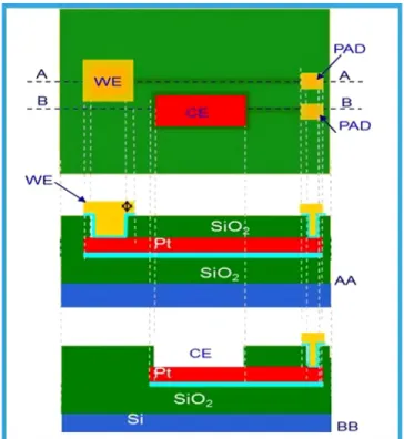
The silicon electrochemical device has been manufactured using the VLSI technology on a 6″ silicon substrate. To electrically isolate the elec-trodes from the substrate a silicon oxide layer has beenfirstly thermally grown (first passivation layer). Then a platinum film has been sputtered and lithographically defined in the electrodes areas and contact zones (PAD). A second passivation layer was then deposited (Silicon oxide by PECVD) to isolate thefirst metallization (Pt) form the second one (Au). A dry etch was performed to connect thefirst and the second met-allization (i.e., on interconnection tracks). The second metmet-allization (Au) was then sputtered and lithographically defined in complementa-ry electrodes regions and contacts areas.Fig. 1shows a schematic cross section of the electrochemical cell structure.
The chip is then assembled with a second silicon device containing integrated temperature sensors and heaters (Fig. 2a). Thefinal silicon device is mounted on a polycarbonate ring to create 4 reaction cham-bers of 20μL each that contains on their floor the electrochemical elec-trodes. The complete structure isfixed on a plastic holder for easily handling (Fig. 2c).
Each electrochemical cell is composed by three planar electrodes: a working electrode in platinum with size 1000 × 2000μm, a counter and a reference electrode made in gold with size 800 × 500 and
800 × 1250μm, respectively. The electrode distances are 100 μm and 200μm.Fig. 2b reports a scheme of the electrochemical cell layout. 2.3. Homogenous phase experiment (real time PCR)
The real time electrochemical PCR experiments were performed using a master mix solution of 20μL containing Clonit buffer (1×) and Hot start polymerase, 0.5μM of each forward and reverse primers, 2 μL of HBV-clone (105copies/μL). Different amount of Os(bpy)
2(DPPZ)++ranging
from 0.1 to 1μM were added. The PCR cycling was performed in a porta-ble thermalcycler (Q3-thermocycler developed by STMicroelectronic–
Fig. 2d) by using the following thermal program: preheating period of 10 min at 95 °C, followed by a maximum of 45 cycles of 95 °C for 15 s and 60 °C for 60 s.
The square wave voltammograms were recorded at the end of the PCR cycles.
Same real time PCR experiments (20μL of the above reported master mix) were executed on standard tube in Applied Biosystem 7500 real time PCR equipment.
2.4. Solid phase experiment (hybridization on immobilised capture probe) The working electrode of the electrochemical device has been func-tionalized by spotting of a solution containing 10μM of thiolated HBV capture probe (25-mer long). The chip was incubated for 4 h at 30 °C (90% RH) and washed by deionized water and dried by nitrogenflow.
The functionalized working electrode was hybridized (60 min at 55 °C) with solutions (20μL) of HBV PCR-cycled at 0, 20 and 40 PCR cy-cles, respectively. After hybridization, a solution (2μL) containing 0.1μM of redox probe was added and square wave curves recorded. 2.5. Electrochemical measurement
The square wave voltammetry measurements were recorded by a Parstat 2273 (Princenton Applied Research) equipment with the follow conditions: square-wave (SW), scan rate 10 mV/s, pulse high/pulse
width 0.025 V for 0.05 s, stop height 12 mV. All electrochemical mea-surement were carried out in potassium chloride 20 mM buffer.
For the solid state intercalation test a sodium phosphate 150 mM so-lution of oligonucleotide 5′-thiol terminated was grafted at working electrode surface overnight at room temperature, washed several time with water and dried by nitrogenflow.
3. Results and discussion
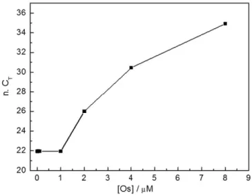
To evaluate possible PCR inhibition effect by osmium complex, real time PCR experiments in standard microtubes were performed. In par-ticular, different redox probe concentrations ranging from 0.1 to 8μM were tested. The results are reported inFig. 3. It can be noticed that no PCR inhibition occurs at osmium complex concentrations in the 0.1–1 μM range, while at upper concentration values the PCR was quite inhibited. The CTvalues measured at the osmium probe
concentra-tions of 0.1, 0.5 and 1μM were 21.35, 21.98 and 22.00 respectively. Ac-cording to no inhibition effect, these values are comparable with what obtained with a master mix without Os complex (CTequal to 21.94).
The compatibility of PCR with the electrochemical cells was investi-gated by performing real time PCR experiments into the electrochemi-cal device. For this purpose, 10μL of HBV mix (105copies/μL) were
cycled in the each of the four reaction chambers of the electrochemical device and read in real time by the Q3 reader (FAM labeled probe). The mean value of CTobtained in the 4 chambers was 22.9 ± 1.2, quite
com-parable with the number obtained in the microtubes (CT23.9– see
above). These data clearly indicate that the device structure and the ma-terials that compose the main modules of PCR chamber (gold and plat-inum for planar electrodes, silicon oxide and polycarbonate of chamber walls), did not inhibit the DNA amplification reaction.
In order to verify the capability of the system to detect redox probe signals upon unspecific intercalation, square wave measurements were recorded directly on the electrochemical cell by using different human genomic DNA amounts.Fig. 4reports the decrease of redox sig-nal for a solution 0.125μM of Os(bpy)2(DPPZ)++in potassium chloride
buffer 20 mM in absence and in presence of 50 and 100 ng of human DNA, respectively. The decrease of signal clearly indicates the effective-ness probe intercalation. It is completely intercalated with 100 ng of human DNA.
To evaluate the ability of the system to detect amplified product in homogeneous phase, square wave signals were recorded on solutions (20μL) containing a redox probe concentration of 0.1 μM and PCR prod-uct amplified at different cycles (0, 10, 20, 30 and 40 cycles). Data are showed inFig. 5. It can be noticed that the electrochemical response agrees with the intercalation process since the electrical signal de-creases with the increasing of the DNA amounts (insertedfigure).
The affinity binding of Os(bpy)2(DPPZ)++with PCR product was
measured by the Scatchard plot (data not showed)[19]. A Kbvalue of
1.5 × 10⁶M−1was found. This is in agreement with both the Kb=
4 × 10⁶M−1determined for calf thymus DNA and K
b= 5 × 10⁶M−1
de-termined for PCR product[12].
Tofinally demonstrate the efficiency to detect the PCR amplification in solid state, a thiolated HBV capture probe 25-mer long was grafted on working electrode surface. The layer was hybridized with HBV PCR-cycled product in presence of 0.1μM of redox probe.Fig. 6reports the square wave curves (SW) recorded (solid line). For comparison the same curve (PCR-cycled product containing 0.1μM of redox probe) ob-tained by measurements in solution (dashed line) is reported. The SW curve at solid phase shown a significantly shift of peak voltage at higher values (from 0.66 to 0.81 V) together with a huge increasing of sensitiv-ity. In addition, the peak current increases from 1.8 × 10⁻⁶A for the in-tercalation on homogeneous phase to 5.6 × 10⁻⁶A in case of the solid
state. This is in agreement with literature data for the redox process with monolayer immobilized on electrode surfaces.
4. Conclusion
In this paper we have described a novel portable system for nucleic acids electrochemical detection. Two different approaches were investigated: (a) intercalation of electrochemical unspecific probe on ds-DNA on homogeneous solution (homogeneous phase); (b) grafting of DNA probes on electrode surface (solid phase). The data here presented confirms the ability of electrochemical cells to detect the PCR amplified products with both the approaches. Real time PCR data, recorded in homogenous phase, prove the compati-bility of the PCR process towards both the presence of redox probe Os(bpy)2(DPPZ)++(concentration range 0.1–1 μM) and the
electro-chemical cell materials. However, the solid phase approach reveals a better potentiality for future applications. Actually, it is noteworthy that an increasing of sensitivity of about three times was found due
Fig. 4. SW curves for redox probe 0.1μM at different human DNA amount.
Fig. 5. Square wave curves for redox probe 0.1μM at different PCR cycles.
Fig. 6. Square wave curves of redox probe for solid state (solid line) and homogenous phase (dash line) PCR products intercalation. Inserted the redox signal increasing for 0, 20 and 40 PCR cycles.
to the intercalation on ds-DNA directly grafted to the electrode surface.
The miniaturized system here proposed could be a good candidate for point-of-care applications in the medicalfield.
Acknowledgments
This work has been funded by MIUR-PON (2007–2013) “Hyppocrates – Sviluppo di Micro e Nano-Tecnologie e Sistemi Avanzati per la Salute dell’uomo” (PON02 00355) and by PRIN (2010–2011) “Metodologie chimiche innovative per biomateriali intelligenti”. We also gratefully Roberta Giuffrida, Marco Branciforte, Giuseppe Tosto and Maria Grazia Amore from STMicroelectronics and Maria Letizia di Pietro e Scolatica Serroni from University of Messina for their support. References
[1] E. Palecek, M. Bartosik, Electrochemistry of nucleic acids, Chem. Rev. 112 (2012) 3427–3481.
[2] C.-H. Lu, B. Willner, I. Willner, DNA nanotechnology: from sensing and DNA ma-chines to drug delivery systems, ACS Nano 7 (2013) 8320–8332.
[3] A. Merkoci, A. De la Escosura-Munniz, M. Espinoza-castaneda, Nanoparticles/ nanochannels– based electrochemical biosensors, J. Nanosci. Nanotechnol. 96 (2015) 205–223.
[4] B. Foglieni, A. Brisci, F. San Biagio, P. Di Pietro, S. Petralia, S. Conoci, M. Ferrari, L. Cremonesi, Integrated PCR amplification and detection processes on a Lab-on-Chip platform: a new advanced solution for molecular diagnostics, Clin. Chem. Lab. Med.: CCLM 48 (3) (2010) 329–336.
[5] S. Petralia, R. Verardo, E. Klaric, S. Cavallaro, E. Alessi, C. Schneider, In-Check system: a highly integrated silicon Lab-on-Chip for sample preparation, PCR amplification and microarray detection of nucleic acids directly from biological samples, Sens. Ac-tuators B: Chem. 187 (2013) 99–105.
[6] M.S. Ibrahim, R.S. Lofts, P.B. Jahrling, E.A. Henchal, V.W. Weedn, M.A. Northrup, P. Belgrader, Real-time microchip PCR for detecting single-base differences in viral and human DNA, Anal. Chem. 70 (9) (1998) 2013–2017.
[7] A. Erdem, Nanomaterial based electrochemical DNA sensing strategies, Talanta 74 (2007) 318–325.
[8] G. Ventimiglia, S. Petralia, Recent advances in DNA microarray technology: an over-view on production strategies, detection methods and applications, Bionanoscience 3 (2013) 428–450.
[9] M.M. Rahman, X.B.B. Li, N.S. Lopa, S.J. Ahn, J.J. Lee, Electrochemical DNA hybridiza-tion sensors based on conducting polymers, Sensors 15 (2015) 3801–3829.
[10]Luo, T.M.H. Lee, I.M. Hsing, Immobilization-free sequence-specific electrochemical detection of DNA using ferrocene-labeled peptide nucleic acid, Anal. Chem. 80 (2008) 7341–7346.
[11] X. Luo, I.M. Hsing, Immobilization-free electrochemical detection of DNA mutation on a microchip, Proc.μTAS’09 (2009) 33–34.
[12]T. Defever, M. Druet, D Evrard, D. Marchal, B. Limoges, Real-time electrochemical PCR with a DNA intercalating redox probe, Anal. Chem. 83 (2011) 1815–1821.
[13] (a) H.A. Wagenknecht (Ed.), Charge Transfer in DNA: From Mechanism to Applica-tions, Wiley, New York, 2005;
(b)K.E. Augustyn, V.C. Pierre, J.K. Barton, Metallointercalators as Probes of DNA Rec-ognition and Reactions, Wiley, New York, 2008.
[14]F. Puntoriero, S. Campagna, M.L. Di Pietro, A. Giannetto, M. Cusumano, Lumines-cence of a Pt(II) complex in the presence of DNA. Dependence of luminesLumines-cence changes on the interaction binding mode, Photochem. Photobiol. Sci. 6 (2007) 357–360 (and Refs. therein).
[15] S. Campagna, M. Cavazzini, M. Cusumano, M.L. Di Pietro, A. Giannetto, F. Puntoriero, S. Quici, Luminescent Ir(III) complex exclusively made of polypyridine ligands capa-ble of intercalating into calf-thymus DNA, Inorg. Chem. 50 (2011) 10667–10672 (and Refs. therein).
[16] E. Trovato, M.L. Di Pietro, F. Puntoriero, Shining a new light on an old game– an OsII-based near-IR light switch, Eur. J. Inorg. Chem. (2012) 3984–3988 (and Refs. therein).
[17] E.M. Kober, J.V. Caspar, B.P. Sullivan, T. Meyer, Synthetic routes to new polypyridyl complexes of osmium(II), Inorg. Chem. 27 (1988) 4587–4598.
[18] S. Conoci, A. Mascali, F. Pappalardo, Synthesis, DNA binding properties and electro-chemistry to an electrode-bound DNA of a novel anthracene–viologen conjugate, RSC Adv. 4 (2014) 2845–2850.
[19]J.D. McGhee, P.H. von Hippel, Theoretical aspects of DNA–protein interactions: co-operative and non-co-co-operative binding of large ligands to a one-dimensional ho-mogeneous lattice, J. Mol. Biol. 86 (1974) 469.