1
UNIVERSITÀ POLITECNICA DELLE MARCHE
DIPARTIMENTO SCIENZE DELLA VITA E
DELL’AMBIENTE
Corso di dottorato ciclo XXIX in:
Biologia ed Ecologia Marina
Plastic and environmental safety: the effects of
EDCs on metabolism, reproduction and
epigenetic processes
Tesi di dottorato di: Tutor:
Stefania Santangeli Oliana Carnevali
Co-Tutor: Ferenc Mueller Gary Hardiman
2
3
Summary
1. INTRODUCTION 4
1.1. Plastic additives 4
1.2. Endocrine disruptor compounds 5
1.3. Non-monotonic dose-response curves 8
1.4. Epigenetic effects of EDCs 11
1.5. The different groups of EDCs studied in this project 12
1.5.1. Bisphenol A 12
1.5.2. Nonylphenol 14
1.5.3. 4-tert-octylphenol 14
1.5.4. Di-isononyl-phthalate 15
1.5.5. Diethylene glycol dibenzoate 15
1.6. Physiological processes studied in the present thesis work 16
1.6.1. Oogenesis 16
1.6.2. Lipid metabolism 18
2. OBJECTIVES 29
3. BPA- induced deregulation of epigenetic patterns: effects on female zebrafish reproduction. 31 4. Epigenetic changes in F1 zebrafish embryo from mother exposed to BPA. 57 5. Effects of diisononyl phthalate on Danio rerio reproduction. 76 6. Obesogenic effects of environmental pollutants: disruption of lipid metabolism in Danio rerio. 100
7. Xenobiotic-contaminated diets affect hepatic lipid metabolism: implications for liver in Sparus aurata
juveniles. 124
8. Effects of BPA on female reproductive function: the involvement of epigenetic mechanism. 150
4
1. INTRODUCTION
1.1. Plastic additives
Plastic are synthetic organic polymers, derived from the polymerization of monomers extracted from oil or gas (Derraik 2002; Rios et al. 2007). From the development of modern plastic, in 1907, a number of inexpensive manufacturing techniques have been optimized, resulting in the mass production, in a surplus of lightweight, durable, inert and corrosion-resistant plastics (Cole et al. 2011). These attributes have led to the extensive use of plastic in almost inexhaustible applications leading to the growth of plastics production from approximately 1.9 tons in 1950, to approximately 330 million tons in 2013 (PlasticsEurope 2015).
Whilst the social benefits of plastic are far-reaching (Andrady and Neal 2009), this valuable commodity has been the subject of increasing environmental concern. In first place the durability of plastic, that makes it such an attractive material to use, but also, makes it highly resistant to degradation (Barnes et al. 2009b; Sivan 2011). We must also consider the copious use of “throw-away” plastic users, the proportion of plastic that contributes to municipal waste constitutes 10% of waste generated worldwide (Barnes et al. 2009a). An estimate up to 10% of plastic produced end up in the oceans, where they may persist and accumulate (Cole et al. 2011). The impact that large plastic debris, known as “macroplastics” can have on the marine environment include: the injury and death of marine birds, mammals, fish and reptiles resulting from plastic entanglement and ingestion (Derraik 2002), the transport of non-native marine species to new habitats on floating plastic debris, and the smothering of the seabed preventing gas-exchange (Gregory 2009).
Macroplastics will gradually break down into ever-smaller-pieces due to sunlight exposure, oxidation, and the physical action of waves, currents, and grazing by fish and birds (Zettler et al. 2013) forming tiny plastic fragments called secondary microplastics with a diameter smaller than 1 or 5 mm. Degradation of larger pieces of plastic is not the only way microplastics end up in the ocean. For example microbeads used as scrubbing in personal care products can escape water treatment facilities and pass into environment (Fendall and Sewell 2009).
Although plastics are typically considered as biochemically inert (Roy et al. 2011), plastic additives, also called plasticizers may be incorporated into plastics during manufacture to change their properties or extend the life
5 of the plastic by providing resistance to heat, oxidative damage and microbial degradation (Browne et al. 2007). These additives are of environmental concern since they both extend the degradation times of plastic and may, in addition, leach out, introducing potentially hazardous chemicals to biota (Barnes et al. 2009b; Lithner et al. 2009) through the interference with biologically important processes, potentially resulting in endocrine disruption (Barnes et al. 2009a; Lithner et al. 2009, 2011).
1.2. Endocrine disruptor compounds
An endocrine disrupting compound (EDCs) is defined by the United States Environmental Protection Agency (EPA) as "an exogenous substance or mixture that alters function(s) of the endocrine system and consequently
causes adverse health effects in an intact organism, or its progeny, or (sub)populations".
The term endocrine-disrupting chemicals is used to define a structurally diverse class of synthetic and natural compounds that possess the ability to alter various components of the endocrine system and potentially induce adverse health effects in exposed individuals and populations. EDC, have been reported to interfere with the endocrine system and ultimately disturb the normal function of tissues and organs, producing adverse developmental, reproductive, neurological, cardiovascular, metabolic and immune effects (Schug et al. 2011). Given their physicochemical differences and distinct biological effects, it is not surprising that a variety of mechanisms are used by EDCs to influence the endocrine system (Henley and Korach 2006). The main evidence suggesting that exposure to environmental chemicals can lead to disruption of endocrine function comes from changes seen in a number of wildlife species. Effects related to endocrine disruption have been reported in molluscs, crustacea, fish, reptiles, birds and mammals in various parts of the world (http://ec.europa.eu/environment/chemicals/endocrine/index_en.htm).
The hormonal activity of some chemical compounds was well known some decades ago. For instance, in 1938 an unidentified hormonal activity for bisphenol A (BPA) was published (Dodds and Lawson 1938). After World War II, scientists located high levels of xenoestrogens (or EDCs related with human activity) in the environment because of their increased use in industrial and domestic areas. In 1990 only some pesticides, catalogued as dangerous for other negative effects, formed the EDCs list. The endocrine disruptor concept, the rules for specific studies of endocrine activity, and the awareness of human health and environmental exposure were established at the European Workshop on the Impact of Endocrine Disruptors on Human Health and
6 Wildlife (Weybridge, U.K.) in 1996. Alkylphenols, bisphenols, phthalates, polychlorinated biphenyls, polycyclic aromatic hydrocarbons, dioxins–furans (PCDD/F), some metals (Pb, Cd, and Hg), and organotin compounds have been finally included in the EDCs list.
Chemicals with hormonal activity, and thus potential endocrine disruptors, include:
Natural hormones from any animal, released into the environment, and chemicals produced by one species that exert hormonal actions on other animals. For example human hormones unintentionally reactivated during the processing of human waste in sewage effluent, may result in changes to fish.
Natural chemicals including toxins produced by components of plants (the so-called phytoestrogens, such as genistein or coumestrol) and certain fungi.
Synthetically produced pharmaceuticals intended to be highly hormonally active. For example the contraceptive pill and treatments for hormone-responsive cancers may also be detected in waste water. Man-made chemicals and by-products released into the environment. Laboratory experiments have
suggested that some man-made chemicals might be able to cause endocrine changes. These include some pesticides (e.g. DDT and other chlorinated compounds), chemicals in some consumer and medical products (e.g. plastic additives), and a number of industrial chemicals (e.g. polychlorinated biphenols (PCBs), dioxins). The hormonal activity of these chemicals, is many times weaker than the body's own naturally present hormones, e.g. nonyl-phenol (a breakdown product of alkylphenol ethoxylate surfactants), found as a low level contaminant in some rivers in Europe, has an estrogenic activity only about one-ten thousandth that of the natural hormone, estrogen.
Numerous toxic compounds have environmental concentrations below the thresholds risk, according to the regulations, but it must take into account that the human and environmental health problem regarding EDCs is related to their bioaccumulation, low level chronic exposures, and synergistic effect of them, according to scientists. Some EDCs frequently identified in the migration tests were BPA, phthalates and 4-NP (4-nonylphenol) or t-butylphenol. The main non-EDCs identified, were solvents, antioxidants, and rubber-like compounds (Romero et al. 2002). Some chemicals can act on the endocrine system to disturb the homeostatic mechanisms of the body or to initiate processes at abnormal times in the life cycle. They are usually either natural products or synthetic chemicals that mimic, enhance (an agonist), or inhibit (antagonist) the action of hormones (Soto et al. 1995).
7 The mechanisms through which these chemicals can exert their effects are different:
Due to structural similarity to the natural hormone, EDCs can mimic their actions in the body by binding its receptor or group of receptors on the cell surface, cytoplasm, or nucleus, acting as agonists and changing gene expression in a way characteristic for a given hormone (Roy et al. 2009);
Mimicry can also make EDCs act as partial agonists, or antagonists (they can bind the receptor but not activate it) of particular receptor (Diamanti-Kandarakis et al. 2009);
EDCs may alter receptor production;
Activation of receptors can be modulated by EDCs through receptor phosphorylation or other ways (Lohse et al. 1990);
EDCs may interfere with recruitment of transcriptional co-activators, in which receptor activity relies (especially nuclear receptors and aryl hydrocarbon receptor). (Swedenborg et al. 2009);
Some EDCs can impose the disruption without direct receptor interaction. They can affect the level of enzymes, influencing directly or indirectly the production of hormone (e.g. aromatase) (Swedenborg et al. 2009);
Concentration of the active hormone, reaching the receptor site with bloodstream, can also be altered by modulating hormone transport (they may bind to transport proteins in the blood, altering the amounts of natural hormones that are present in the circulation) or metabolism (Baker et al. 1998);
EDCs can alter signaling pathways in different and complex ways, including pathway crosstalk or may act by mechanisms yet unknown (Safe and Wormke 2003).
Endocrine disruption has been demonstrated to occur in wildlife, particularly in aquatic species or in species that are connected to aquatic food chains, like “fish-eating” birds (Tyler et al. 1998; Vos et al. 2000). The questions being addressed today, are not whether endocrine disruption occurs in wildlife but rather in which concentration it occurs, what mechanism are involved, and whether disruption of the endocrine system will lead to ecologically relevant effects (Guillette 2006). To answer these questions, zebrafish, together with fathed minnow, Pimephales promelas, and medaka, Oryzias latipes, are under evaluation as model species for regulatory test programs in the environmental risk assessment of EDCs (Ankley and Johnson 2004). Since EDCs can lead to chronic alterations of development and reproduction, the short life cycle of small laboratory
8 fish, such as zebrafish, represents an important advantage of these species as experimental animals (Bresch et al. 1990).
1.3. Non-monotonic dose-response curves
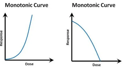
“The dose makes the poison” is the dogma of modern toxicology, derived from a quote from Paracelsus, a XV-century physician and alchemist considered to be a founder of toxicology as a discipline. This basic principle is not only popularly recognized, but also intuitive. It can be easily assumed, that the relation between chemical exposure dose and the observed biological effect is consequentially logical: the higher the dose, the higher the effect. This assumption underlies the modern system of toxicity testing for new compounds (and other effect-searching screening methods), so the belief in the principle is not only theoretical, but it has also very practical implications. The screening, usually employs a range of high doses of a particular compound, and obtained effects that are extrapolated to lower doses. Performed tests include assessing the LD50s (the dose lethal for 50% of the experimental animals), MTD (maximum tolerated dose), LOAEL (lowest levels observed adverse effect) and NOAEL (the no observed adverse effect level). The safe dose for humans and wildlife is subsequently calculated by an algorithm providing some safety coefficient and based on the NOAEL value. Assuming that the dose-response relationship had been linear, the “dose-safe” could have been deduced by simple dose response test. The relationship of dose and effect corresponding to this dogma can be visualized as a monotonic dose-response curve (Fig 1.3.1.), which may take slightly different forms, however, the effect curve always maintains a direction of change.
9 Even though accurate in many cases, the dogma does not hold true for some investigated compounds, and the number of them is rapidly increasing. It is possible for a chemical to elicit a more complex response pattern, than expected. While the number of possible response patterns shaped by complex molecular interactions is theoretically unlimited, it is common to observe a U-shaped or inverted-U-shaped dose-response curve characterized by the presence of a peak and subsequent effect decrease with a rising dose. That type of pattern is called a non-monotonic dose-response curve (NMDRC) (Fig. 1.3.2.), because the mathematical function defining this curve does not preserve the given order: the slope of the dose-response curve changes sign from positive to negative or conversely at some point of the range of examined doses, so the direction of biological effect change is not constant.
The consequence of the existence of non-monotonic dose-response curve phenomenon is that no toxicity testing results can be extrapolated to another range of doses, than those empirically tested. The lack of significant results observed during standard high dose chemical safety assessment does not entitle the researcher to assume safety of lower doses. This, however, undermines the paradigms of safety testing procedures, therefore questioning validity of conclusions reached from the majority of safety tests performed to this day and challenging the status quo. Even employing gold standard testing methods, it is necessary to expand extremely, the range of investigated compound concentrations in order to learn about its real potential to cause biologically relevant effects. The lack of such safety measures leads to an unrestricted use of
10 compounds, such as endocrine disruptor compounds, that later prove to be not safe, and often more toxic in low doses (which is recognized as a low-dose effect) (Vandenberg et al. 2012). It is important to underline that low doses are the kind of exposure experienced chronically by wildlife and humans in their living environment.
Numerous molecular mechanisms are proposed to explain the existence of non-monotonic dose-response curves and low-dose effects. These include such as:
Cofactors and receptors specific for a cell or tissue types: Some of the observed non-monotonic effects may be a result of overlapping monotonic effects, for example coming from a number of different cell subpopulations or tissues present in the sample or culture, that react to the hormone in a different way (Soto and Sonnenschein 2001) due to a different type and amount of receptors expressed (Morani et al. 2008) as well as the presence of specific cofactors capable of altering receptor selectivity and downstream signal processing (Jeyakumar et al. 2008), thus the response as well.
Receptor selectivity: Many of the discussed compounds can act via multiple receptors and pathways depending on the concentration. They are activated, almost exclusively, by one type of receptor at low dose, and become less specific at high dose, weakly binding other kinds of receptor too (Moriyama et al. 2002) and altering the biological endpoints.
Figure 1.3.2. Examples of monotonic dose-response curves; there is a theoretically unlimited potential for different shapes of
11 Desensitization of receptors: For a wide range of hormones (or agents mimicking them) the decreased response to ligands by receptor had been observed as a result of biochemical inactivation after prolonged exposure.
Down-regulation of receptor expression: Rising ligand levels often lead to an increase of inactivation and degradation of its receptors, at some point exceeding the capacity to produce new ones in the pace equal to the speed of degradation pathway (Ismail and Nawaz 2005). Also, receptor production can be regulated based on the level of circulating ligand, in a non-linear pattern. Additionally signaling from one receptor type can influence production process of another type (Kinyamu and Archer 2003).
Competition for receptor sites: If the artificial compound is competing with a natural hormone for the same receptor, they can both find an unoccupied site, bind and activate the responses; however at a high dose of the compound it outcompetes the natural hormone, altering the response. Mathematical models were used to determine high probability of NMDRC in all such cases (Kohn and Melnick 2002).
Negative endocrine feedback loops: Several animal hormones are regulated by positive or negative feedback loops: the circulating level of a given hormone or product of its action affects the production of this hormone based on activation of receptors related to it. Overstimulation of hormone receptor by other compounds decreases hormone production (Vandenberg et al. 2012).
Several studies demonstrates that EDCs do not follow the principle “the dose make the poison”, but rather exhibit a U-shaped and inverted U-shaped, non-monotonic, dose response curve, where the strongest responses may be elicited by the lowest and highest doses, or by intermediate concentrations, respectively, probably due to the receptor down regulation induced by higher hormone levels (Tibbetts et al. 1998; Vandenberg et al. 2009, 2012).
1.4. Epigenetic effects of EDCs
The term epigenetics was described for the first time in 1942 by Conrad Waddington as “…the interaction of genes with their environment which brings the phenotype into being” (Waddington 1942). Epigenetic modifications are now described as heritable and reversible chemical modifications of chromatin, resulting in adjustment of its activity without change in the underlying DNA sequence. Epigenetic plays an important role in cellular differentiation, growth, metabolism, and regulation of gene expression by silencing and activating
12 specific genes (Bernstein et al. 2007; Esteller 2007; Jirtle and Skinner 2007). In addition, it was shown that epigenetic mechanisms facilitate interaction between gene transcription and environment (Esteller 2007). The main types of epigenetic modifications include DNA methylation and histone modification (Tammen et al. 2013).
Recent studies have demonstrates the ability of EDCs to have epigenetic effects (Anway and Skinner 2006; Ho et al. 2006). 2,3,7,8-tetrachlorodibenzo-p-dioxin (TCDD), polychlorinated biphenyls (PCBs), and phthalate esters, have been found to affect the reproductive system or induce tumor development by altering DNA methylation, and steroid hormone metabolism and signaling (McLachlan et al. 2006; Wu et al. 2004). Plastics induce a delayed female pubertal onset in F1 rats but in F2 generation. Plastics, dioxin and jet fuel promote early onset of puberty in female rats (Manikkam et al. 2012). BPA shows trans generational effects on sperm fertility and other reproductive parameters in male rats (Wolstenholme et al. 2012) and was additionally found able to interfere with both DNA methylation and histone modification processes (Doherty et al. 2010; Dolinoy et al. 2007; Kundakovic et al. 2013). Plasticizer such as BPA, DEHP and DBP can produce inheritance effects in F3, such as testis, prostate, kidney and ovary effects, tumors and may induce obesity in subsequent generation (Manikkam et al. 2013).
1.5. The different groups of EDCs studied in this project
In this project, the different studies conducted, have together investigated the endocrine disrupting potential, of different compounds: Bisphenol A, nonylphenol, 4-tert-octylphenol, di-isononyl-phthalate and diethylene glycol dibenzoate. The current section will give a short general description of these four groups of endocrine disrupting compounds.
1.5.1. Bisphenol A
Bisphenol A (BPA) is a phenol formed by a hydroxyl residue directly bound to an aromatic ring (Flint et al. 2012) which was synthesized firstly in 1891 by the Russian chemist Alexander P. Dianin. Later in 1930, its estrogenic effects were discovered (Rubin 2011), while the use of this chemical in the plastics industry, started in 1940, since its ability to increase heat resistance and elasticity. Nowadays BPA is a key monomer in
13 production of epoxy resins and in the most common form of polycarbonate plastic and is one of the most produced chemicals in the world. According to recent estimates, a hundred ton of BPA per year can be found in the environment (Rubin 2011) through landfill or plastic degradation (Kang et al. 2006), realistically resulting in environmental waters concentrations ranging from 5 µg/L to 21 µg/L (Crain et al. 2007).
BPA is present in 95% of products containing epoxy resins and polycarbonates such as food-containers, bottles, toys, dental products, CD and DVD and water pipes. It is also used in the vinyl chloride and thermal paper production which are used for the manufacture of slips, books, booklets, ticket, kitchen rolls, toilet paper and food paperboard (Nam et al. 2010). For this reason, it is an important contaminant due to its ubiquitous presence and the increased exposure of humans and wild organisms via environment and food chain.
BPA is generally characterized as an oestrogen-like compound. Oestrogen classical action mode, is trough activation of the nuclear oestrogen receptors (ERs), ERα and ERβ. In addition to these nuclear receptors, estrogen-related receptor gamma (ERRγ) and the G-protein coupled oestrogen receptor (GPER) have been proposed as potential mediators of BPA-associated oestrogen disruption (Riu et al. 2011; Wang et al. 2012). Similar to other xenoestrogens, it is considered to be a selective oestrogen receptor modulators because its effects are pro-oestrogenic in some tissues and anti-oestrogenic in others (Kundakovic and Champagne 2011; Wetherill et al. 2007) while its capacity to bind also to membrane receptors make it harmful even at pico and nanomolar concentrations (Wetherill et al. 2007).
As an environmental pollutant, BPA is probably able, to alter epigenetic mechanisms through methylation of the CpG sites (Dolinoy et al. 2007). Even though we don’t have a good knowledge on the matter, BPA is supposed, to be able to alter histone modification and, hence, the structure of chromatin correlated with either activation or repression of transcription (Kundakovic and Champagne 2011).
Many studies revealed ontogenetic and endocrine disruptions by BPA on aquatic organisms at environmental relevant concentrations and there is an emerging concern that this pollutant may adversely impacts reproduction, lipid metabolism as well as brain development not only in animal but also in human (Lam et al. 2011; Saal et al. 2012; Sheng et al. 2013).
14
1.5.2. Nonylphenol
Nonylphenol (NP) is a xenobiotic compound consisting of a phenol ring and nine-carbon chain on the para-position. Nonylphenol ethoxylates are highly “cost effective” surfactants with exceptional performance and consequently used widely in industrial, institutional, commercial and household applications such as detergents, emulsifiers, wetting and dispersing agents, antistatic agents, demulsifiers and solubilisers. Nonylphenol ethoxylates reach sewage treatment works in substantial quantities where they biodegrade into several by-product including nonylphenol. Due to its physical-chemical characteristics, such as low solubility and high hydrophobicity, nonylphenol accumulates in environmental compartments that are characterized by high organic content, typically sewage sludge and river sediments, where it persists. The occurrence of nonylphenol in the environment is clearly correlated with anthropogenic activities such as wastewater treatment, landfilling and sewage sludge recycling (Soares et al. 2008). The first evidence that alkylpehnols could be oestrogenic was published in 1938 (Dodds and Lawson 1938). Nonylphenol was found to mimic the natural hormone 17β-oestradiol by competing for the binding site of the oestrogen receptor (White et al. 1994). Recently it has also been established that nonylphenol has antiandrogenic activity (Lee et al. 2003) and it has been proposed to induce lipid accumulation in hepatocytes (Hao et al. 2012).
1.5.3. 4-tert-octylphenol
Para-Alkylphenols are present in the aquatic environment due to their use in pesticide formulations, from leaching of plastics and from degradation of alkylphenol ethoxylates. 4-tert-octylphenol (t-OP) is the most estrogenic of the 4-alkylphenols and in vitro studies have demonstrated that it binds to the estrogen receptor, activates estrogen responsive genes, and stimulates mitogenesis of MCF breast cancer cells (Routledge and Sumpter 1997; White et al. 1994). In “vivo” studies, have demonstrated the ability of this pollutant to cause estrogenic-like effects, including, disruption of spermatogenesis, increased incidence of sperm deformities and lowered serum concentration of testosterone and gonadotropins (Boockfor and Blake 1997) while there are limited information on their effects on the metabolic pathway (Pedersen and Hill 2000).
15
1.5.4. Di-isononyl-phthalate
Di-isononyl-phthalate (DINP) is a compound belonging to the phthalate family.
Phthalates are chemicals with a broad worldwide distribution which are used as solvent in products for personal care but also as additives in plastics, specifically, in plasticized polyvinyl chloride (PVC) (David et al. 2001; Koch and Calafat 2009; Schettler 2006). The annual world production of phthalates is more than 4 Mt, of which about 1 Mt is produced in Europe (Peijnenburg and Struijs 2006). The most important compound of this category was bis(2-ethylhexyl)phthalate (DEHP), that, given its proven toxicity for reproduction (Carnevali et al. 2010), has recently been replaced by other compounds, such as di-isononyl phthalate (DINP) (Clara et al. 2010). DINP became then one of the main plasticizer used for PVC production with a consequent increase in its presence in the environment. Environmental concentration of DINP was found at concentration from 0.52 µg/L (1.2 X 10⁻⁸ M) and upwards (Quinn-Hosey et al. 2012).
The formation of DINP occurs through an esterification reaction between phthalic anhydride and two molecules of isononyl alcohol, with the production of a water molecule. This compound contains two alkyl chains of nine carbon atoms which confers a high molecular weight. DINP is consequently more permanent, it has a lower solubility rate and a low rate of migration compared to other phthalates (Vieira et al. 2011).
Because of its composition and chemical structure, DINP appears to be an endocrine disrupter, playing mostly an anti-androgen action in men by blocking the receptors of male sex hormones and inhibiting their biological effects. Very little is known about the effect of DINP in teleost since most of studies have focused primarily on mammals but several studies have shown its ability to act on the androgen responsive tissues, causing retention of nipples, testicular atrophy, epididymal agenesis and the decrease of testosterone levels, in both testes and plasma, of male rats (Borch et al. 2004; Gray et al. 2000).
1.5.5. Diethylene glycol dibenzoate
Diethylene glycol dibenzoate (DGB) was recently approved by European Chemical Agency as an alternative to phthalates in the processing of plastic (Kermanshahi et al. 2009). DGB is characterized as a high solvating plasticizer intended for the use in the manufacturing of PVC, vinyl flooring, adhesives, latex caulks, sealants and elastomers. The main mechanism of environmental removal for this chemical is microbial degradation.
16 Dibenzoate, has a higher biodegradation rates than phthalates, however studies have shown that interaction of Rhodotorula rubra with dibenzoate-based plasticizers, resulted to an incomplete microbial degradation leading to the accumulation of monobenzoates, which have higher toxicity than the original plasticizers. The biodegradatioin of this commercial plasticizer by the yeast R. rubra, results in the formation of substantial amounts of metabolites, such as diethylene glycol monobenzoate, which is toxic and resistant to further degradation (Kermanshahi et al. 2009).
Only a handful of research papers, concerning DGB are available, focused on understanding the pathways and results of DGB metabolism in a living organism. These publications suggest, that while DGB does not last long in the organism, the intermediates of its detoxification do, and the results do not support the use of DGB as an environmentally safer alternative to phthalates (Gartshore et al. 2003; Kermanshahi et al. 2009).
1.6. Physiological processes studied in the present thesis work
It is well known that EDCs can interfere with vertebrate reproductive system (Carnevali et al. 2010; Crain et al. 2008; Patisaul and Adewale 2009). In addition to reproductive effects, there is also a growing concern that metabolic disorders may be linked with EDCs (Casals-Casas and Desvergne 2011). Oogenesis and lipid metabolism process were thus investigated in this project.
1.6.1. Oogenesis
In teleost fish, oogenesis is under the control of hypothalamic-pituitary-gonadal (HPG) axis. The principal component of the axis includes the hypothalamus and hypophysis (pituitary gland) in the brain, the gonads and the liver. These tissues are structurally linked in one or both of the following mechanism: a relatively fast neuronal linkage and a slower vascular linkage. In teleost, pituitary is directly innervated by hypothalamic neurons that secrete directly into the intercellular space of the hypophysis. The main HPG axis neurohormone, secreted by hypothalamus is gonadotropin-releasing hormone (GnRH) which stimulates the production and the release of two types of glycoprotein gonadotropic hormones: the follicle-stimulating hormone (FSH), which primarily induces oogenesis, and luteinizing hormone (LH), whose primary activity causes final gametes maturation and induction of ovulation.
17 The ovaries of principal teleost are paired or bilobed organs and we will focus on those which are asynchronous spawners. The principal cell types of the ovary are the germinative cells, in various stage of development, starting with the primary oogonia, which develop into mature oocytes. At any particular time, the asynchronous ovary contains oocytes at all stage of development. Surrounding the developing oocyte, is a spherical layer of cells called the granulosa, which assists the translocation of the egg-yolk lipoprotein vitellogenin into developing oocyte. Finally, in the stromal layers of the ovary resides a cell type called thecal cells, which are involved in the synthesis of steroid hormone.
The primary effects in the ovary of FSH and LH are to stimulate the thecal and granulosa cells to produce the female’s steroid hormone 17β-estradiol. However, the synthetic pathway of this hormone includes the male hormone testosterone as an intermediate. Apparently, the thecal cells, under the stimulatory influence of LH, synthesize testosterone, which diffuses to the granulosa cells. FSH induces the enzyme aromatase in the granulosa cells, which converts testosterone to estrogen. The estrogen that is produced has several critical roles, including oocyte development and stimulation of the liver to produce and secrete vitellogenin into the bloodstream. Once in the ovary, the vitellogenin is translocated via the granulosa cells into the developing oocyte. Finally maturation is under control of LH which induces the production of progesterone and its receptors, accountable for inducing the first meiotic division, indicated as germinal vesicle break down. (Ankley and Johnson 2004; Nagahama 1994).
It is now evident that exposure to EDCs has the potential to adversely affect reproductive physiology in vertebrates. One of the most well studied is BPA which was found able to interfere with steroidogenic and apoptotic processes in the ovary (Peretz et al. 2011; Ziv-Gal et al. 2013) but also to delay or inhibit ovulation (Lahnsteiner et al. 2005). The critical question now is if wildlife and human health is at risk from chronic exposure to low doses of this compound, either alone or in mixture.
Furthermore the ability of these compounds to permanently affect the epigenome could be potentially catastrophic to the welfare of future generations and requires further attention by both toxicologists and endocrinologists.
18
1.6.2. Lipid metabolism
Recent studies have revealed the involvement of environmental chemicals in obesity. This chemicals are defined as obesogens, substances that increase weight gain acting directly on fat cells, or indirectly, by altering the regulators system of appetite and satiety, the metabolic rate and the energy balance and thus favoring calories storage (Alonso-Magdalena et al. 2011; Grün and Blumberg 2009; Janesick and Blumberg 2011). Many known obesogens are endocrine disruptor compounds (EDCs) often used for the manufacturing of plastic materials and then presents in the majority of the object of daily use (North and Halden 2013; Schug et al. 2011). Liver is a key metabolic organ which governs body energy metabolism. The maintenance of energy expenditure involves the dynamic integration of two metabolically opposed states, fasting and feeding. Fasting involves the induction of catabolic, or ATP generating pathways, whereas feeding engages anabolic or ATP-consuming pathways, that build carbon skeletons and store energy. The integration of these states is governed by endocrine signals, such as insulin and glucagon, nutrient signals such as long chain fatty acids and branched chain amino acids, adipose and gut-derived polypeptide hormones, such as leptin, and nutrient-sensing kinases.
In the feeding phase, food is digested in the gastrointestinal tract, and glucose, fatty acids, and amino acids are absorbed into the bloodstream and transported to the liver through the portal vein circulation system. Insulin is released from pancreatic β cells and suppresses hepatic glucose production; glucose is condensed into glycogen and/or converted into fatty acids or amino acids in the liver. Other nonesterified fatty acids (NEFAs) originate from chylomicrons, which are produced by enterocytes with dietary fat, and arrive to the liver by circulation where they are released through lipolysis process. In hepatocytes, free fatty acids are esterified with glycerol 3-phosphate to generate triacylglycerol (TAG) or with cholesterol to produce cholesterol esters. TAG and cholesterol esters are either stored in lipid droplets within hepatocytes or secreted into the circulation as very low density lipoprotein (VLDL) particles which deliver lipids to extra hepatic tissues through the circulation.
In the fasted state or during exercise, adipose tissue produces and releases NEFAs and glycerol via lipolysis, whereas muscle breaks down glycogen and proteins and releases lactate and alanine. Alanine, lactate, and glycerol are delivered in the liver and used as precursors to synthesize glucose through gluconeogenesis.
19 NEFAs are oxidized in hepatic mitochondria through fatty acid β oxidation and generate ketone bodies (ketogenesis). The liver also begins to produce glucose through glycogenolysis. These fuel substrates (glucose, Ketone bodies) are released from the liver into the circulation system and are essential for the extrahepatic tissues, including red blood cells and the brain.
The liver’s energy metabolism is tightly controlled. Transcriptional regulation of the genes involved in fatty acid metabolism is presently considered as the major long-term regulatory mechanism that controls the lipid homeostasis. It is executed by a variety of transcription factors among which the SREBPs, the C/EBPs, and members of the nuclear receptor family are particularly active agents. It is therefore important that different process such as fatty acid synthesis, storage, oxidation and mobilization are well balanced (Clarke 1993; Karanth et al. 2009; Lodhi and Semenkovich 2014; North and Halden 2013; Rogers et al. 2014).
20
References
Alonso-Magdalena P, Quesada I, Nadal A. 2011. Endocrine disruptors in the etiology of type 2 diabetes mellitus. Nat. Rev. 7: 346–35356.
Andrady AL, Neal M a. 2009. Applications and societal benefits of plastics. Philos. Trans. R. Soc. Lond. B. Biol. Sci. 364: 1977–1984.
Ankley GT, Johnson RD. 2004. Small fish models for identifying and assessing the effects of endocrine-disrupting chemicals. ILAR J. 45: 469–483.
Anway MD, Skinner MK. 2006. Epigenetic transgenerational actions of endocrine disruptors. Endocrinology 147:S43-9; doi:10.1210/en.2005-1058.
Baker ME, Medlock KL, Sheehan DM. 1998. Flavonoids Inhibit Estrogen Binding to Rat Alpha-Fetoprotein. Exp. Biol. Med. 217: 317–321.
Barnes DK a, Galgani F, Thompson RC, Barlaz M. 2009a. Accumulation and fragmentation of plastic debris in global environments. Philos. Trans. R. Soc. Lond. B. Biol. Sci. 364:1985–98;
doi:10.1098/rstb.2008.0205.
Barnes DKA, Galgani F, Thompson RC, Barlaz M. 2009b. Accumulation and fragmentation of plastic debris in global environments. Philos. Trans. R. Soc.
Bernstein BE, Meissner A, Lander ES. 2007. The Mammalian Epigenome. Cell 128: 669–681. Boockfor FR, Blake CA. 1997. Chronic administration of 4-tert-octylphenol to adult male rats causes
shrinkage of the testes and male accessory sex organs, disrupts spermatogenesis, and increases the incidence of sperm deformities. Biol. Reprod. 57: 267–77.
Borch J, Ladefoged O, Hass U, Vinggaard AM. 2004. Steroidogenesis in fetal male rats is reduced by DEHP and DINP, but endocrine effects of DEHP are not modulated by DEHA in fetal, prepubertal and adult male rats. Reprod. Toxicol. 18: 53–61.
Bresch H, Beck H, Ehlermann D, Schlaszus H, Urbanek M. 1990. A long-term toxicity test comprising reproduction and growth of zebrafish with 4-chloroaniline. Arch. Environ. Contam. Toxicol. 19: 419– 427.
Browne M a, Galloway T, Thompson R. 2007. Microplastic--an emerging contaminant of potential concern? Integr. Environ. Assess. Manag. 3: 559–561.
21 Carnevali O, Tosti L, Speciale C, Peng C, Zhu Y, Maradonna F. 2010. DEHP impairs zebrafish reproduction
by affecting critical factors in oogenesis. PLoS One 5: 1–7.
Casals-Casas C, Desvergne B. 2011. Endocrine disruptors: from endocrine to metabolic disruption. Annu. Rev. Physiol. 73:135–162; doi:10.1146/annurev-physiol-012110-142200.
Clara M, Windhofer G, Hartl W, Braun K, Simon M, Gans O, et al. 2010. Occurrence of phthalates in surface runoff, untreated and treated wastewater and fate during wastewater treatment. Chemosphere 78:1078–84; doi:10.1016/j.chemosphere.2009.12.052.
Clarke SD. 1993. Regulation of fatty acid synthase gene expression: an approach for reducing fat accumulation. J Anim Sci 71: 1957–1965.
Cole M, Lindeque P, Halsband C, Galloway TS. 2011. Microplastics as contaminants in the marine environment: a review. Mar. Pollut. Bull. 62:2588–97; doi:10.1016/j.marpolbul.2011.09.025.
Crain DA, Eriksen M, Iguchi T, Jobling S, Laufer H, Leblanc GA, et al. 2007. An ecological assessment of bisphenol-A : Evidence from comparative biology. Reprod. Toxicol. 24: 225–239.
Crain DA, Janssen SJ, Edwards TM, Heindel J, Ho SM, Hunt P, et al. 2008. Female reproductive disorders: the roles of endocrine-disrupting compounds and developmental timing. Fertil. Steril. 90: 911–940. David R, McKee R, Butala J, Barter R, Kayser M. 2001. Esters of Aromatic Mono-, Di-, and Tricarboxylic
Acids, Aromatic Diacids and Di-, Tri-, Or Polyalcohols. E. Bingham, B. Cohrssen, and C.H.
Powelleds. . John Wiley & Sons, Inc., Hoboken, NJ, USA.
Derraik JG. 2002. The pollution of the marine environment by plastic debris: a review. Mar. Pollut. Bull. 44: 842–852.
Diamanti-Kandarakis E, Bourguignon JP, Giudice LC, Hauser R, Prins GS, Soto a M, et al. 2009. Endocrine-Disrupting Chemicals: an Endocrine Society Scientific Statement. Endocr. Rev. 30: 293– 342.
Dodds EC, Lawson W. 1938. Molecular Structure in Relation to Oestrogenic Activity. Compounds without a Phenanthrene Nucleus. Proc. R. Soc. B Biol. Sci. 125:222–232; doi:10.1098/rspb.1938.0023.
Doherty LF, Bromer JG, Zhou Y, Aldad TS, Taylor HS. 2010. In utero exposure to diethylstilbestrol (DES) or bisphenol-A (BPA) increases EZH2 expression in the mammary gland: an epigenetic mechanism linking endocrine disruptors to breast cancer. Horm. Cancer 1: 146–55.
22 Dolinoy DC, Huang D, Jirtle RL. 2007. Maternal nutrient supplementation counteracts bisphenol A-induced
DNA hypomethylation in early development. Proc. Natl. Acad. Sci. U. S. A. 104: 13056–61.
Esteller M. 2007. Cancer epigenomics: DNA methylomes and histone-modification maps. Nat. Rev. Genet. 8: 286–98.
Fendall LS, Sewell MA. 2009. Contributing to marine pollution by washing your face: Microplastics in facial cleansers. Mar. Pollut. Bull. 58: 1225–1228.
Flint S, Markle T, Thompson S, Wallace E. 2012. Bisphenol A exposure, effects, and policy: A wildlife perspective. J. Environ. Manage. 104: 19–34.
Gartshore J, Cooper DG, Nicell JA. 2003. Biodegradation of plasticizers by Rhodotorula rubra. Environ. Toxicol. Chem. 22: 1244–1251.
Gray LE, Ostby J, Furr J, Price M, Veeramachaneni DN, Parks L. 2000. Perinatal exposure to the phthalates DEHP, BBP, and DINP, but not DEP, DMP, or DOTP, alters sexual differentiation of the male rat. Toxicol. Sci. 58: 350–65.
Gregory MR. 2009. Environmental implications of plastic debris in marine settings--entanglement, ingestion, smothering, hangers-on, hitch-hiking and alien invasions. Philos. Trans. R. Soc. Lond. B. Biol. Sci. 364: 2013–2025.
Grün F, Blumberg B. 2009. Endocrine disrupters as obesogens. Mol. Cell. Endocrinol. 304: 19–29.
Guillette LJ. 2006. Endocrine disrupting contaminants-beyond the dogma. Environ. Health Perspect. 114: 9– 12.
Hao C-J, Cheng X, Xia H, Ma X. 2012. The endocrine disruptor 4-nonylphenol promotes adipocyte differentiation and induces obesity in mice. Cell. Physiol. Biochem. 30.
Henley D V., Korach KS. 2006. Endocrine-disrupting chemicals use distinct mechanisms of action to modulate endocrine system function. Endocrinology; doi:10.1210/en.2005-1117.
Ho S-M, Tang W-Y, Belmonte de Frausto J, Prins GS. 2006. Developmental exposure to estradiol and bisphenol A increases susceptibility to prostate carcinogenesis and epigenetically regulates
phosphodiesterase type 4 variant 4. Cancer Res. 66:5624–32; doi:10.1158/0008-5472.CAN-06-0516. http://ec.europa.eu/environment/chemicals/endocrine/index_en.htm. Endocrine disruptors - Chemicals -
23 http://ec.europa.eu/environment/chemicals/endocrine/index_en.htm.
Ismail A, Nawaz Z. 2005. Nuclear hormone receptor degradation and gene transcription: an update. IUBMB Life 57:483–90; doi:10.1080/15216540500147163.
Janesick A, Blumberg B. 2011. Endocrine disrupting chemicals and the developmental programming of adipogenesis and obesity. Birth Defects Res. 93: 34–50.
Jeyakumar M, Webb P, Baxter JD, Scanlan TS, Katzenellenbogen JA. 2008. Quantification of ligand-regulated nuclear receptor corepressor and coactivator binding, key interactions determining ligand potency and efficacy for the thyroid hormone receptor. Biochemistry 47:7465–76;
doi:10.1021/bi800393u.
Jirtle RL, Skinner MK. 2007. Environmental epigenomics and disease susceptibility. Nat. Rev. Genet. 8: 253–262.
Kang J-H, Kondo F, Katayama Y. 2006. Human exposure to bisphenol A. Toxicology 226: 79–89. Karanth S, Lall SP, Denovan-Wright EM, Wright JM. 2009. Differential transcriptional modulation of
duplicated fatty acid-binding protein genes by dietary fatty acids in zebrafish (Danio rerio): evidence for subfunctionalization or neofunctionalization of duplicated genes. BMC Evol. Biol. 9: 219. Kermanshahi A, Cooper DG, Mamer OA, Maric M, Nicell JA. 2009. Mechanisms of biodegradation of
dibenzoate plasticizers. Chemosphere 77: 258–263.
Kinyamu HK, Archer TK. 2003. Estrogen receptor-dependent proteasomal degradation of the glucocorticoid receptor is coupled to an increase in mdm2 protein expression. Mol. Cell. Biol. 23: 5867–81.
Koch HM, Calafat AM. 2009. Human body burdens of chemicals used in plastic manufacture. Philos. Trans. R. Soc. 364: 2063–78.
Kohn MC, Melnick RL. 2002. Biochemical origins of the non-monotonic receptor-mediated dose-response. J. Mol. Endocrinol. 29: 113–23.
Kundakovic M, Champagne F a. 2011. Epigenetic perspective on the developmental effects of bisphenol A. Brain. Behav. Immun. 25: 1084–93.
Kundakovic M, Gudsnuk K, Franks B, Madrid J, Miller RL, Perera FP, et al. 2013. Sex-specific epigenetic disruption and behavioral changes following low-dose in utero bisphenol A exposure. Proc. Natl. Acad. Sci. U. S. A. 110: 9956–61.
24 Lahnsteiner F, Berger B, Kletzl M, Weismann T. 2005. Effect of bisphenol A on maturation and quality of
semen and eggs in the brown trout, Salmo trutta f. fario. Aquat. Toxicol. 75: 213–224.
Lam SH, Hlaing MM, Zhang X, Yan C, Duan Z, Zhu L, et al. 2011. Toxicogenomic and Phenotypic Analyses of Bisphenol-A Early-Life Exposure Toxicity in Zebrafish. PLoS One 6;
doi:10.1371/journal.pone.0028273.
Lee HJ, Chattopadhyay S, Gong EY, Ahn RS, Lee K. 2003. Antiandrogenic effects of bisphenol A and nonylphenol on the function of androgen receptor. Toxicol. Sci. 75: 40–46.
Lithner D, Damberg J, Dave G, Larsson Ak. 2009. Leachates from plastic consumer products--screening for toxicity with Daphnia magna. Chemosphere 74:1195–200; doi:10.1016/j.chemosphere.2008.11.022. Lithner D, Larsson A, Dave G. 2011. Environmental and health hazard ranking and assessment of plastic
polymers based on chemical composition. Sci. Total Environ. 409:3309–24; doi:10.1016/j.scitotenv.2011.04.038.
Lodhi IJ, Semenkovich CF. 2014. Peroxisomes: A Nexus for Lipid Metabolism and Cellular Signaling. Cell Metab. 19:380–392; doi:10.1016/j.cmet.2014.01.002.
Lohse MJ, Benovic JL, Caron MG, Lefkowitz RJ. 1990. Multiple Pathways of Rapid beta2-Adrenergic Receptor Desensitization. Biol. Chem. 265: 3202–3209.
Manikkam M, Guerrero-Bosagna C, Tracey R, Haque MM, Skinner MK. 2012. Transgenerational actions of environmental compounds on reproductive disease and identification of epigenetic biomarkers of ancestral exposures. PLoS One 7.
Manikkam M, Tracey R, Guerrero-Bosagna C, Skinner MK. 2013. Plastics Derived Endocrine Disruptors (BPA , DEHP and DBP) Induce Epigenetic Transgenerational Inheritance of Obesity , Reproductive Disease and Sperm Epimutations. PLoS One 8.
McLachlan JA, Simpson E, Martin M. 2006. Endocrine disrupters and female reproductive health. Best Pract. Res. Clin. Endocrinol. Metab. 20: 63–75.
Morani A, Warner M, Gustafsson J-A. 2008. Biological functions and clinical implications of oestrogen receptors alfa and beta in epithelial tissues. J. Intern. Med. 264:128–42; doi:10.1111/j.1365-2796.2008.01976.x.
25 disrupted by bisphenol A as an antagonist. J. Clin. Endocrinol. Metab. 87:5185–90;
doi:10.1210/jc.2002-020209.
Nagahama Y. 1994. Endocrine regulation of gametogenesis in fish. Int. J. Dev. Biol. 38: 217–229.
Nam S-H, Seo Y-M, Kim M-G. 2010. Bisphenol A migration from polycarbonate baby bottle with repeated use. Chemosphere 79:949–52; doi:10.1016/j.chemosphere.2010.02.049.
North EJ, Halden RU. 2013. Plastics and environmental health: The road ahead. Rev. Environ. Health 28: 1– 8.
Patisaul HB, Adewale HB. 2009. Long-term effects of environmental endocrine disruptors on reproductive physiology and behavior. Front. Behav. Neurosci. 3: 10.
Pedersen RT, Hill EM. 2000. Identification of novel metabolites of the xenoestrogen 4-tert-octylphenol in primary rat hepatocytes. Chem. Biol. Interact. 128: 189–209.
Peijnenburg WJGM, Struijs J. 2006. Occurrence of phthalate esters in the environment of The Netherlands. Ecotoxicol. Environ. Saf. 63: 204–15.
Peretz J, Gupta RK, Singh J, Hernàndez-Ochoa I, Flaws J a. 2011. Bisphenol A Impairs Follicle Growth, Inhibits Steroidogenesis, and Downregulates Rate-Limiting Enzymes in the Estradiol Biosynthesis Pathway. Toxicol. Sci. 119: 209–217.
PlasticsEurope. 2015. Plastics-The Facts 2013: An analysis of European latest plastics production, demand and waste data. http:// www.plasticseurope.org/Document/plastics-the-facts-2013.aspx 1–40.
Quinn-Hosey KM, Roche J, Fogarty A, Brougham C. 2012. A Toxicological Assessment of Endocrine Disrupting Chemicals Found in the BMW (Border, Midland and Western) Region of Ireland. J. Environ. Prot. (Irvine,. Calif). 3: 304–315.
Rios LM, Moore C, Jones PR. 2007. Persistent organic pollutants carried by synthetic polymers in the ocean environment. Mar. Pollut. Bull. 54: 1230–1237.
Riu A, Grimaldi M, le Maire A, Bey G, Phillips K, Boulahtouf A, et al. 2011. Peroxisome proliferator-activated receptor gamma is a target for halogenated analogs of bisphenol A. Environ. Health Perspect. 119: 1227–1232.
Rogers MA, Liu J, Song BL, Li BL, Chang CCY, Chang TY. 2014. Acyl-CoA:cholesterol acyltransferases (ACATs/SOATs): Enzymes with multiple sterols as substrates and as activators. J. Steroid Biochem.
26 Mol. Biol. 151: 102–107.
Romero J, Ventura F, Gomez M. 2002. Characterization of paint samples used in drinking water reservoirs: identification of endocrine disruptor compounds. J. Chromatogr. Sci. 40: 191–197.
Routledge EJ, Sumpter JP. 1997. Structural Features of Alkylphenolic Chemicals Associated with Estrogenic Activity. 272: 3280–3288.
Roy JR, Chakraborty S, Chakraborty TR. 2009. Estrogen-like endocrine disrupting chemicals affecting puberty in humans--a review. Med. Sci. Monit. Int. Med. J. Exp. Clin. Res. 15: RA137-45.
Roy PK, Hakkarainen M, Varma IK, Albertsson A-C. 2011. Degradable polyethylene: fantasy or reality. Environ. Sci. Technol. 45:4217–27; doi:10.1021/es104042f.
Rubin BS. 2011. Bisphenol A: an endocrine disruptor with widespread exposure and multiple effects. J. Steroid Biochem. Mol. Biol. 127: 27–34.
Saal F vom, Nagel S, Coe B, Angle B, Taylor J. 2012. The estrogenic endocrine disrupting chemical bisphenol A (BPA) and obesity. Mol. Cell. Endocrinol. 354:74–84;
doi:10.1016/j.mce.2012.01.001.THE.
Safe S, Wormke M. 2003. Inhibitory Aryl Hydrocarbon Receptor−Estrogen Receptor α Cross-Talk and Mechanisms of Action. Chem. Res. Toxicol. 16: 807–816.
Schettler T. 2006. Human exposure to phthalates via consumer products. Int. J. Androl. 29. Schug TT, Janesick A, Blumberg B, Heindel JJ. 2011. Endocrine disrupting chemicals and disease
susceptibility. J. Steroid Biochem. Mol. Biol. 127: 204–215.
Sheng L, Liu Y, Jiang L, Chen Z, Zhou Y, Cho KW, et al. 2013. Hepatic SH2B1 and SH2B2 regulate liver lipid metabolism and VLDL secretion in mice. PLoS One 8: 1–10.
Sivan A. 2011. New perspectives in plastic biodegradation. Curr. Opin. Biotechnol. 22: 422–426.
Soares A, Guieysse B, Jefferson B, Cartmell E, Lester JN. 2008. Nonylphenol in the environment: A critical review on occurrence, fate, toxicity and treatment in wastewaters. Environ. Int. 34: 1033–1049. Soto a. M, Sonnenschein C, Chung KL, Fernandez MF, Olea N, Olea Serrano F. 1995. The E-SCREEN
assay as a tool to identify estrogens: An update on estrogenic environmental pollutants. Environ. Health Perspect. 103:113–122; doi:10.1289/ehp.95103s7113.
27 and cell death. J. Natl. Cancer Inst. 93: 1673–5.
Swedenborg E, Ruegg J, Makela S, Pongratz I. 2009. Endocrine disruptive chemicals: Mechanisms of action and involvement in metabolic disorders. J. Mol. Endocrinol. 43: 1–10.
Tammen S a., Friso S, Choi SW. 2013. Epigenetics: The link between nature and nurture. Mol. Aspects Med. 34: 753–764.
Tibbetts TA, Mendoza-Meneses M, O’Malley BW, Conneely OM. 1998. Mutual and intercompartmental regulation of estrogen receptor and progesterone receptor expression in the mouse uterus. Biol. Reprod. 59: 1143–52.
Tyler CR, Jobling S, Sumpter JP. 1998. Endocrine disruption in wildlife: a critical review of the evidence. Crit. Rev. Toxicol. 28: 319–361.
Vandenberg LN, Colborn T, Hayes TB, Heindel JJ, Jacobs DR, Lee D-H, et al. 2012. Hormones and
Endocrine-Disrupting Chemicals: Low-Dose Effects and Nonmonotonic Dose Responses. Endocr. Rev. 33: 378–455.
Vandenberg LN, Maffini M V., Sonnenschein C, Rubin BS, Soto AM. 2009. Bisphenol-A and the Great Divide: A Review of Controversies in the Field of Endocrine Disruption. Endocr. Rev. 30:75–95; doi:10.1210/er.2008-0021.
Vieira MGA, da Silva MA, dos Santos LO, Beppu MM. 2011. Natural-based plasticizers and biopolymer films: A review. Eur. Polym. J. 47: 254–263.
Vos JG, Dybing E, Greim H a, Ladefoged O, Lambré C, Tarazona J V, et al. 2000. Health effects of
endocrine-disrupting chemicals on wildlife, with special reference to the European situation. Crit. Rev. Toxicol. 30: 71–133.
Waddington CH. 1942. The epigenotype. Endeavour 18–20.
Wang J, Sun B, Hou M, Pan X, Li X. 2012. The environmental obesogen bisphenol A promotes adipogenesis by increasing the amount of 11 b -hydroxysteroid dehydrogenase type 1 in the adipose tissue of
children. Intern. J. Obes. 1–7.
Wetherill YB, Akingbemi BT, Kanno J, McLachlan J a, Nadal A, Sonnenschein C, et al. 2007. In vitro molecular mechanisms of bisphenol A action. Reprod. Toxicol. 24: 178–98.
28 compounds are estrogenic. Endocrinology 135.
Wolstenholme JT, Edwards M, Shetty SRJ, Gatewood JD, Taylor JA, Rissman EF, et al. 2012. Gestational exposure to bisphenol a produces transgenerational changes in behaviors and gene expression. Endocrinology 153: 3828–3838.
Wu Q, Ohsako S, Ishimura R, Suzuki JS, Tohyama C. 2004. Exposure of Mouse Preimplantation Embryos to 2, 3, 7, 8-Tetrachlorodibenzo-p-dioxin (TCDD) Alters the Methylation Status of Imprinted Genes H19 and Igf2. Biol. Reprod. 1797: 1790–1797.
Zettler ER, Mincer TJ, Amaral-Zettler LA. 2013. Life in the “Plastisphere”: Microbial Communities on Plastic Marine Debris. Environ. Sci. Technol. 130619162220002.
Ziv-Gal A, Craig ZR, Wang W, Flaws J a. 2013. Bisphenol A inhibits cultured mouse ovarian follicle growth partially via the aryl hydrocarbon receptor signaling pathway. Reprod. Toxicol. 42: 58–67.
29
2. OBJECTIVES
The present thesis has the ambition of applying an interdisciplinary approach to gaining an understanding on the effect of EDCs on reproduction and lipid metabolism. Different groups of EDCs were investigated to address the following objectives:
1. Determine the epigenetic effects of BPA on female reproduction and embryo development. 2. Determine the EDCs effects in fish reproduction.
3. Determine the EDCs effects on fish metabolism.
To achieve these objectives this study has been divided into 5 chapters plus a review:
1. BPA-induced deregulation of epigenetic patterns: effects on female zebrafish reproduction. The aim of this work is to determine the effects of BPA on female reproductive physiology and investigate whether changes in the expression levels of genes related to reproduction are caused by histone modification.
2. Epigenetic changes in F1 zebrafish embryo from mother exposed to BPA.
This study had the aim to investigate the effects of BPA treated females crossed with control males on the subsequent generation embryos. General and locus specific methylation plus genes involved in embryo and gonadal development were especially investigated to answer this question.
3. Effects of diisononyl phthalate on Danio rerio reproduction.
The purpose of this study was to determine the effect of exposure to different concentration of DiNP, starting from low environmental concentration (420 ng/l) to higher levels on female zebrafish reproductive parameters, including number and size distribution of ovarian follicles, apoptosis and autophagy signals, spatially resolved biochemical information of vitellogenic oocytes using fourier transform infrared (FTIR) imaging, and transcript levels for genes involved in reproduction.
4. Obesogenic effects of environmental pollutants: disruption of lipid metabolism in Danio rerio.
The aim of the present study is to investigate the effects of two EDCs involved in plastic
manufacturing as the BPA, which nowadays is one of the main plasticizer used worldwide, and the DGB, approved by European Chemical Agency as an alternative to phthalates in the processing of plastic.
30
5. Xenobiotic-contaminated diets affect hepatic lipid metabolism: implications for liver in Sparus aurata juveniles.
The present study investigated the effects of feed contaminated with BPA, NP, or t-OP on the lipid metabolism of gilthead sea bream (Sparus aurata). Considering their broad diffusion and the high potential for human exposure, these EDCs may pose a concrete risk to human health.
6. Effects of BPA on female reproductive function: the involvement of epigenetic mechanism.
The purpose of this review is to summarize the effects of BPA on adult female reproduction with focus on epigenetic changes that can be heritable.
31
3. BPA-INDUCED DEREGULATION OF EPIGENETIC PATTERNS: EFFECTS ON FEMALE ZEBRAFISH REPRODUCTION
Stefania Santangeli1,4, Francesca Maradonna1, Giorgia Gioacchini1, Gilda Cobellis2, Chiara Carla
Piccinetti1, Luisa Dalla Valle3, Oliana Carnevali1,4*.
1Dipartimento Scienze della Vita e dell’Ambiente, Università Politecnica delle Marche, Via Brecce Bianche, 60131 Ancona, Italy;
2Dipartimento di Medicina Sperimentale, Seconda Università degli Studi di Napoli, Via S. Maria di Costantinopoli 16, 80138 Napoli, Italy.
3Dipartimento di Biologia, Università di Padova, Via Ugo Bassi 58/B, 35131 Padova, Italy. 4INBB Consorzio Interuniversitario di Biosistemi e Biostrutture, 00136 Roma, Italy.
*Corresponding author:
Oliana Carnevali,
Dipartimento Scienze della Vita e dell’Ambiente Università Politecnica delle Marche
Via Brecce Bianche, 60131, Ancona, Italy Phone: +39 071 220 4990
Fax: +39 071 220 4650
orcid.org/0000-0001-5994-0572
e-mail: [email protected]
Published in Scientific Reports 25 February 2016
32
Abstract
Bisphenol A (BPA) is one of the commonest Endocrine Disruptor Compounds worldwide. It interferes with vertebrate reproduction, possibly by inducing deregulation of epigenetic mechanisms. To determine its effects on female reproductive physiology and investigate whether changes in the expression levels of genes related to reproduction are caused by histone modifications, BPA doses consistent with environmental exposure were administered to zebrafish for three weeks. Effects on oocyte growth and maturation, autophagy and apoptosis processes, histone modifications, and DNA methylation were assessed by Real-Time PCR (qPCR), histology, and chromatin immunoprecipitation combined with qPCR analysis (ChIP-qPCR). The results showed that 5 µg/L BPA down-regulated oocyte maturation-promoting signals, likely through changes in the chromatin structure mediated by histone modifications, and promoted apoptosis in mature follicles. These data indicate that the negative effects of BPA on the female reproductive system may be due to its upstream ability to deregulate epigenetic mechanism.
1. Introduction
Endocrine Disruptor Compounds (EDCs) are chemicals that can interfere with vertebrate reproduction (Patisaul and Adewale, 2009). The vertebrate reproductive system is under the control of the hypothalamic-pituitary-gonadal axis, which operates through a hormonal cascade where production of gonadotropin-releasing hormone (GnRH) in the hypothalamus stimulates pituitary gonadotropin synthesis, eventually leading to production of steroid hormones by the gonads.
Bisphenol A (BPA) is commonly used in the manufacturing of polycarbonate plastics and epoxy resins, making it one of the most widespread EDCs worldwide. According to recent estimates, 100 ton of BPA per year may end up in the environment (Rubin, 2011) due to landfill or plastic degradation (Kang et al., 2006), resulting in concentrations of 5 µg/L to 21 µg/L in environmental water (Crain et al., 2007).
BPA alters gene expression by binding to nuclear estrogen receptors (ERs) ERα and ERβ. It is defined as a selective ER modulator because its effects are pro-estrogenic in some tissues and anti-estrogenic in others (Kundakovic and Champagne, 2011; Wetherill et al., 2007). Its ability to bind to membrane receptors makes it harmful even at pico- and nanomolar concentrations (Wetherill et al., 2007).
33
As an environmental pollutant, BPA is probably able to affect epigenetic mechanisms through methylation of
CpG sites (Dolinoy et al., 2007). It also seems to induce histone modification, altering the chromatin structure and through it transcription activation and repression (Kundakovic and Champagne, 2011).
The first goal of the present study was to determine the effects of BPA concentrations consistent with environmental exposure (5, 10, and 20 µg/L) on reproductive physiology in female vertebrates. To do this, the expression of several genes critical for reproduction was monitored in the ovary: ERs esr1 and esr2a,
steroidogenic acute regulatory protein (star), and a member of the cytochrome P450 family 11
(cyp11a1)(Arukwe, 2008; Cotter et al., 2013; King et al., 2008; Palter et al., 2001) for follicle development
and steroidogenesis; progesterone receptors pgrmc1 and pgrmc2 for oocyte growth and maturation; and bone morphogenetic protein 15 (bmp15) and growth differentiation factor 9 (gdf9) as local signals(Nagahama, 1994). Apoptosis, autophagy, and oocyte viability were also assessed.
The second goal of the study was to explore whether the changes in the expression levels of these genes were caused by histone modifications, as measured by lysine 4 and 27 trimethylation in the amino terminal of histone 3 (respectively H3K4me3 and H3K27me3). H3K4me3 is involved in the activation of transcription (Kouzarides, 2007), since it marks the Transcription Start Site (TSS) region, whereas H3K27me3 is chiefly associated with gene silencing (Lindeman et al., 2010). Based on the recent hypothesis of a possible interplay between histone modifications and DNA methylation patterns, where DNA methyltransferases (DNMTs) seem to play an important role (Cedar and Bergman, 2009), DNMT gene expression levels were also investigated. The experimental model selected for the study, Danio rerio, has a fully sequenced genome and shares with humans 70% gene orthology and body plan similarity (Howe et al., 2013); these features make it an ideal model to assess the effects of EDCs on human reproduction. The issue is both urgent and topical, since several studies have reported a decline in human fertility, especially in western countries (Brannian and Hansen, 2006; Nyboe Andersen and Erb, 2006). Even though the decrease may be related to a variety of factors, a role for environmental EDCs is strongly suspected (Patisaul and Adewale, 2009).
34
2. Methods
2.1. Animals and BPA administration
A total of 48 adult female zebrafish (D. rerio, AB wild-type strain) were placed in eight 10-L aquaria (6 fish/tank) with oxygenated water under controlled conditions (28.0 ± 0.5 ˚C) and maintained on a 14/10 h light/dark cycle. They were fed 4 times a day, twice commercial food (Vipagran; Sera, Loessnitz, Germany) and twice Artemia salina. There were a control and 3 exposed groups, which received 5, 10, or 20 µg/L BPA (98% analytical purity, Sigma-Aldrich, Milano, Italy) for 3 weeks. All tanks were maintained in duplicate. After 3 weeks, fish were lethally anesthetized with 500 mg/l MS-222 (3-aminobenzoic acid ethyl ester, Sigma Aldrich) buffered to pH 7.4. Five half ovaries from each experimental group, in duplicate, were removed and fixed in Bouin's solution; the remaining ovaries were used for Real Time semi-quantitative Polymerase Chain Reaction (qPCR) (5 half ovaries) and Chromatin ImmunoPrecipitation (ChIP) analysis (7 whole ovaries). All procedures complied with Italian animal experimentation laws and were approved by the Ethics Committee of Padua University (Prot. 112/2015-PR).
2.2. Egg collection and fertility
Starting on the 8th day of treatment, the 3 groups of BPA-exposed females and control females were crossed with untreated males, and fertility was determined during the following 15 days. Fertilized eggs were counted and the fertility rate was calculated as the mean ± standard deviation (SD) of fertilized egg number / female / day from the 8th to the 21th day of treatment.
2.3. RNA extraction and cDNA synthesis
Total RNA was isolated from the ovary with RNAzol solution (Sigma Aldrich) according to the manufacturer’s instructions. Its final concentration was determined using Nanophotometer TM P-Class (Implen GmbH, Mϋnich, Germany), whereas integrity was established by Gel Red staining of 28S and 18S ribosomal RNA fragments on 1% agarose gel.
35 Total RNA was treated with DNAse to remove genomic DNA (10 IU at 37 ˚C for 10 min; Fermentas MBI, Amherst, NY, USA). Then 1 µg total RNA was used for cDNAs synthesis with iScript cDNA Synthesis Kit (Bio-Rad, Milano, Italy), and kept at -20 °C until use.
2.4. Real-Time qPCR
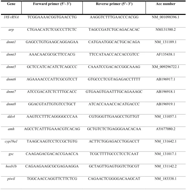
Relative quantification of gene expression was performed with the SYBR Green method in an iQ5 Multicolor Real-Time PCR Detection system (Bio Rad). All samples were analyzed in duplicate. Reactions contained 1 µl cDNA diluted 1/10, 5 µl 2X SYBR Green PCR Master Mix (Bio Rad) containing SYBR Green as a fluorescent intercalating agent, 0.1 µM of forward and reverse primers (ED Table 1), and 3.8 µl of milliQ water. The thermal profile was as follows: enzyme activation at 95 ˚C for 3 min; 45 cycles of denaturation (10 sec at 95 ˚C) followed by 20 sec annealing at 60 ˚C for star, dnmt6, esr2a, gdf9, ambra1a, beclin1, tp53,
caspase3, and cyp11a1, 59 ˚C for dnmt1, dnmt8, dnmt3, dnmt5, fshr, pgrmc1, pgrmc2, bmp15, and 58˚C for dnmt4, dnmt7, esr1, esr2b, and lhcgr, and 20 sec elongation at 72 ˚C. Fluorescence was monitored at the end
of each cycle. Dissociation curves for primer specificity and absence of primer-dimer formation check were performed and consistently showed a single peak.
Genes 18SrRNA (Tang et al., 2007) and arp (acid ribosomal protein) (Aursnes et al., 2011) were used as internal controls to enable result standardization by eliminating variations in mRNA and cDNA quantity and quality (Bustin et al., 2009). These genes were chosen because their mRNA levels did not vary either between experimental treatments or between follicular stages. No amplification product was observed in the negative control (absence of template). Data were analyzed using iQ5 Optical System version 2.1 (Bio-Rad). The quantification method was based on a ΔΔCt calculation implemented with the Pfaffl equation, to improve accuracy by accounting for varied reaction efficiencies depending on primers (Pfaffl, 2001; Vandesompele et al., 2002). All results are expressed with respect to control ovaries. Data were generated in duplicate from 5 biological replicates.
2.5. Histology
Five ovaries per fish group were fixed in Bouin's solution and prepared for histological examination using standard biological procedures. Gonads were embedded in paraffin and sectioned (7 μm) with a microtome.
36 Each ovary was fully sectioned, processed for hematoxylin-eosin staining, and observed at 200x final magnification under a light microscope (Leica Microsystems Inc., Milano, Italy). Atretic follicles were identified based on specific morphological markers of follicular atresia (i.e. granulosa cell hyperplasia, invagination and breakdowns of zona radiata, basal membrane disintegration, absorption of vitellus) as described by Üçüncü and Çakıcı (2009) (Üçüncü and Çakıcı, 2009).
Images (50x final magnification) were captured using a high-resolution digital camera (DC300F; Leica Microsystems) and atretic follicle number was counted and expressed as a percentage of the number of atretic/total follicles.
Student’s t-test and ANOVA followed by Duncan’s test for multi-group comparisons were performed, as appropriate, to assess the significance of differences. Data were expressed as percentages and reported as mean values ± SD.
2.6. Chromatin immunoprecipitation and antibodies
Chromatin was prepared from frozen ovaries following the instructions of the ChIP-IT High Sensitive Kit (Active Motif catalog no. 53040). Chromatin was fragmented by sonication with Active Motif's EpiShearSonicator (80% amplitude, pulse for 30 sec on and 30 sec off for a total sonication “on” time of 12 min –or 22 min of elapsed time) to produce fragments ranging from 200 to 600 bp. Approximately 200 µl of chromatin was used for each immunoprecipitation reaction. Then, 50 µl was removed from each sample and used as input control. ChIP was performed using antibodies specific for H3K4me3 (Abcam) and H27K4me3 (diagenode).
2.7. ChIP-qPCR
ChIP-qPCR was performed with real time PCR using the SYBR Green method. The reaction consisted in 5 µl SYBR Green Reaction Mix, 1 µl 0.1 µM primer pairs, 3 µl sterile water, and 1 µl DNA sample (ChIP or Input), for a total volume of 10 µl. Gene mapping and information on regions of interest were obtained using the UCSC Genome Browser. Primers were designed to target the TSS, the two flanking regions, and the gene body region using Pick Primers from the NCBI website, and were tested by in silico PCRs on the UCSC Genome Bioinformatics website. Primer sequences are reported in ED Table 2. For each pair, primer efficiency was