Annals of the Rheumatic Diseases, 1985; 44, 729-733
Amyloid
arthropathy in patients
undergoing
periodical haemodialysis
for
chronic
renal failure:
a
new
complication
J
MUNOZ-GOMEZ,l
E BERGADA-BARADO,2 R GOMEZ-PIREZ,E
LLOPART-BUISAN,l
E SUBfAS-SOBREVIA,2 J ROTES-QUEROL,AND M SOLI-ARQUES3
From the Departments of 'Rheumatology, 2Nephrology, and 3Pathology, Hospital Clinico y Provincial,
Barcelona, Spain
SUMMARY
Seven patients (five
male and twofemale)
with chronic renal failure(CRF) treated
by periodical haemodialysis presented
withswelling
and effusion of more thanthree
months'
duration in knees
(four bilateral),
shoulders(two,
oneof thembilateral),
elbow(one),
andankle
(one).
Four had acarpal
tunnelsyndrome
bothclinically
andelectromyographically (three
bilateral).
Allpatients
hadhyperparathyroidism secondary
to theirCRF,
whichwas notdue
toamyloidosis
in any of them. Thedialysis
durationperiod
varied from five to 14 years, with anaverage of8-6years.
Amyloid deposits (Congo
redpositive.areas
withgreenbirefringence
underpolarising microscopy)
wereshown in six of thesevensynovial biopsy specimens
oftheknee,
infive
of the sediments of thesynovial fluids,
and inspecimens
removedduring carpal tunnel
syndrome
surgery. Noamyloid
was found in thebiopsy specimen
of abdominal fat of six ofthepatients.
Thefinding
ofamyloid only
in thesynovial
membrane andfluid,
andcarpal tunnel,
its absence in abdominalfat,
and the lack of other manifestations ofgeneralised amyloidosis
(cardiomyopathy, malabsorption syndrome, macroglossia, etc.)
and of Bence Jonesmyeloma
(protein immunoelectrophoresis normal)
raises thepossibility
that this is aform ofamyloidosis
which is
peculiar
toCRF treated by periodical haemodialysis.
Key words: amyloidosis, synovial amyloidosis, synovial fluid, carpal tunnel syndrome.
Various musculoskeletal disorders have been
de-slribed
in patients with chronic renal failure(CRF) treated by periodicalhaemodialysis.'
Among these are synovitis and bursitis due to sodium urate, calcium pyrophosphate,hydroxyapatite,
and cal-cium oxalate microcrystals,2 bone and joint infec-tions, osteonecrosis, carpaltunnelsyndrome,35
soft tissue and vascular calcifications, and metabolic bone disease(hyperparathyroidism,
osteomalacia, osteoporosis,osteosclerosis).
Other
alterations have been described6: micro-traumatic olecranon bursitis caused by the armposition during haemodialysis,unexplained
'dialysis
cysts' (small cystic lesions in the hand and wrist
Accepted for publication 18 February 1985.
CorrespondencetoDr JMufioz-G6mez,Servicio de
Reumatolo-gia.HospitalClinico yProvincial, C/Villarroel 170,08036
Barce-lona, Spain.
bones), digital clubbing in one or more fingers induced by anoxia distal to thefistula, and aneurysm atthe site of the shunt, with orwithout calcification. Our relationship with the Nephrology Depart-mentinthe follow up of CRF patients in aperiodical haemodialysis programme enabled us to identify several patients with swelling and effusions that could not be explained by the above-mentioned causes.
The clinical picture in the first patient studied (case No 1) was a chronic 'swelling with
large
effusion in the knees and shoulders (similarto the 'shoulderpadsign'),which suggested the possibility of amyloid
arthropathy.7
Materials and methods
CRF patients undergoing
periodical
haemodialysis, with persistent swelling and effusion in the knee 729730 Mufioz-Gomez, Bergada-Barado,
Gomez-Perez,
joint of more than three months' duration, were
sought.
Among 312 patients undergoinga programme of
haemodialysis in the NephrologyDepartment seven
such patients were found, where the articular
problem couldnotbe explainedbyanyof the
above-mentioned causes.
Haemodialysis was performed by means of an
artificial kidney with automatic supply, continuous flow, and without capillary filterreuse.Thepatients were given ascorbic acid (1 g) and vitamin B complex after each haemodialysis session in addition
to continued treatment with aluminium hydroxide and 25-hydroxycholecalciferol.
Afull clinical and radiological examination of all patientswascompleted, in which the synovial fluid wasanalysedonvariousoccasions(leucocytecount,
search for crystals by regular and compensated polarised light microscopy both in fresh synovial fluid and after alizarin red staining) and a knee
synovial biopsy specimenwasobtained with Polley's
needle.
Synovial samples fixed in 10% formol and
syno-vial fluid sediments (after centrifugation) were
mounted in paraffin and the sections stained with Congo red and studied under polarising microscopy in search of green birefringence typical of
8
amyloidosis.
Some of the samplesweretreated with potassium
permanganateby Wright'stechnique,9and in sixout
of sevenpatients aneedle aspiration ofabdominal
fatt) was performed.
Results
The group studied included five men and two
women aged from 33 to 62 years (mean age 50)
(Table 1). The aetiology for their chronic renal failure was: glomerulonephritis (two), backflow
hydronephrosis (one), nephroangiosclerosis (one), and unknown (three). No patient had renal
amy-loidosis.
Thedialysis durationperiod varied from fiveto14
years, with an average duration of 8-6 years.
Clinicalexamination of the patients showed swell-ing and effusion in the major joints: knees (seven patients, four of them bilateral), shoulders (two patients,oneofthem bilateral), ankle (onepatient), andelbow(one patient). The swellingswere
accom-panied by minor pain, their onset being therefore difficult to ascertain; during our observations their
duration varied between six months andtwoyears.
Twopatientspresented withanacutecrisisinknees,
shoulder, and elbow, withvery'inflammatory' fluids
during the crisis, which caused the patients to be admitted andaculture of the fluidtobeundertaken
Table 1 Clinical features of the patients studied
Patient Sex Age Approx. CRF Dialysis Jointsigns and duration Carpaltunnel syndrome Radiology
No age (years)
1 M 59 49 9 Persistent swelling Yes. Bilateral. Geodes in shoulders.
knees/shoulders more Operated both sides Hyperparathyroidism
than one year. Acute crises shoulders, knee, elbow
2 M 48 ? 10 Persistent swelling No Geodes in shoulders,
knees and ankle, knees, and pubis.
two years Hyperparathyroidism 3 M 47 37 5 Persistent swelling Yes. Bilateral. Microgeodes in hand~.
knees and right Operated both sides Hyperparathyroidism
shoulder, six months
4 M 54 ? 8 Persistent swelling Yes. Bilateral. Geodes inknees, right
left knee, one year. Operated both sides femur,andrightshoulder.
Rightshoulder joint Hyperparathyroidism
pain
5 F 62 50 9 Persistent swelling Yes. Right operated Patella subluxation bilateral. both knees, oneyear. Vertebralosteodystrophia
Shoulderjoint pains
6 M 50 ? 5 Persistent swelling both No Arthrosis rightknee
knees, one year. (meniscectomy)
Acute crises both knees
7 F 33 19 14 Persistent swelling left No Hyperparathyroidism
knee, six months.
Joint pains inshoulders, ankles, right knee
Amyloid arthropathy in patients with chronic renal failure 731 Table 2 Synovial fluid characteristics and amyloid deposits localisation ofthe patients studied
Patienit No
SsYovial
flulidAmnYloid
deposits edinean oene Fat SYnovialmnemnbrante Ssnovial fluidI Non-inflammatory. Positive. Annular ligament (1) Negative Positivc. Mesothelial Positive. Shoulder(s)
Inflammatory in criscs svnovium (1) hvperplasia knee(s) 2 Non-inflammatory Negatise Positivc. Mesothelial Positive. Knee(s)
hvperplasia
3 Non-inflammatory Positive. Penneural region (2) Ncgativc Positise. Fibrous Positivc. Knee(s) connectivc tissue
4 Non-inflammatorv Positive. Annular ligament (2) Negative Positivc. Mesothelial Positive. Knee hyperplasia
5 Non-inflammatory Positive. Penneural region Negative Positive. Non-spccific Positivc. Knee(s) chronicsynovitis
6 Slight inflammatory. Negatise Negative. Mesothelial Not done Severc in cnses hyperplasia
7 Non-inflammatory Not donc Positive. Normal Ncgative.Knee. Small amount
tocheck thepossibility ofaninfectionin thejoints, but the results were negative. Between the crises there was persistence of the effusions with minor pain and low cellularity. Four patients complained of painsinshoulders. withnovisibleswellingatany time.
Four patients presented with a carpal tunnel syndrome both clinically andelectromyographically, bilateral in three of them.
Radiological signs of hyperparathyroidism or osteodystrophy, or both were seen in all the cases and were intense in three of them.
Three patients showed moderate or severecystic lesions in shoulders (three), knees
(two),
and the superior branch of thepubic symphysis (one). One patient showed microgeodes, and another patient developed a bilateral external subluxation of the patella.The blood biochemistry profileshowed CRF and theclassical alterations of secondary
hyperparathy-roidism in
all patients. Serumprotein
electrophoresis was normal; serum immunoglobulins levels (IgA, IgG, IgM) were normalinallpatients except forone with an IgG of 1760 mg/100 ml(17.6
g/W)
(normalth)
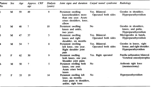
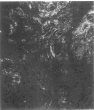
Fig. 1 Synovial tissue: (a) amorphous materialdeposition Congo red positive; (b) showing intense greenbirefringence
732 Mufioz-G6mez,
values600-1500 mg/100 ml(6-15g/l)). Immunoelec-trophoresis showed no qualitative abnormality of immunoglobulins nor the presence of light chains. These results excluded the presence of a Bence Jones myeloma in our patients.
The synovial fluid was 'non-inflammatory' in six patients (<2000 leucocytes/mm3 (2x10⁹/l)), and the remaining fluid showed moderate inflammatory characteristics (around 5000 cells/mm3 (5x10⁹/l)). Two patients who suffered an occasional acute crisis showed very cellular fluids during these crises, fluctuating between 8000 and 103 000 cells/mm3
(8-103
x109/l)
withan averageof 30 000leucocytes/ mm3 (30x109/l).No crystals were observed in any of the fluids, eitherin theunstainedexamination or afteralizarin red staining.
The histological study (Table 2) of the synovial biopsy specimens showed amyloid deposits (Figs la and lb) in six patients (Congo red positive areas with green birefringence); the other findings varied greatly: slight mesothelial hyperplasia (four patients, one of them amyloidnegative), non-specific chronic synovitis (one), fibrous connective tissue (one), and normal (one).
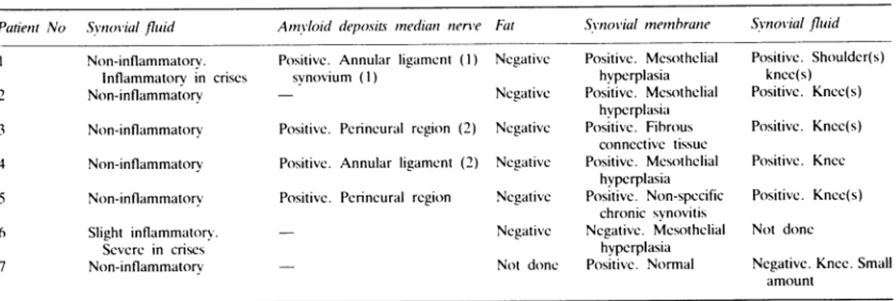
The presence of amyloid in the synovial fluid sediment (Fig. 2) was proved in five of the six
Fig.2 Synovialfluid centrifugate. Multiple deposits of Congoredpositivematerial with greenbirefringence. (Polarising microscopy, Congored, x100.)
patients studied (fluid from the knees in six occa-sions,from the shoulder in one); the negative fluid showed amyloid in the synovium. No fluid was studied in the seventh patient (No 6) with an amyloid negative synovial biopsy specimen.
This patient had a clinical picture similar to that of the others, with swelling and effusion in both knees for a little over one year, treated with aspiration and injections of a depot corticosteroid every three to four months. The patient had been referred from another hospital, and subsequent checks for the presence of amyloid in the effusion were not possible.
Surgery was performed on four of the patients with a carpal tunnel syndrome (two of them bilat-eral),showing amyloid deposits in all of them (in the annular ligament in two, in the perineural region in two, and in the synovium in another).
Abiopsy specimen of the abdominal fat was taken from six patients and proved amyloid negative in all of them.
Potassium permanganate staining was positive in all articular fluid sediments and in the perineural region for the only patient who had an operation for carpal tunnel syndrome in which it was studied. Discussion
Six of thepatientshadsynovial amyloidosis,four of them with a carpal tunnel syndrome, a frequent location of the so-called primary amyloidosis, but without any other manifestations of generalised amyloidosis (cardiomyopathy, malabsorption, mac-roglossia, etc.).
In patient No 6 the synovial biopsy specimen probablydidnotinclude anyaffectedsynovium, or the aetiology for thiscasemighthave been different. The positive results from the treatment of the samples by Wright'stechnique
pointed
towardsthis classification,but the lack ofamyloid
inthebiopsy
specimen of the abdominal fat,
negative
in the six patients in whichitwassoughtand which isusually positive in primary andgeneralised amyloidosis,
did notallow us toclassify thesepatients ashavingthis type of amyloidosis.The protein
immunoelectrophoresis
normal values without light chains confirmed that ourpatientsdidnotsuffer fromaBence Jones
myeloma.
All thepatientshad
hyperparathyroidism secondary
totheir chronic renal
failure,
and thiswas notduetoamyloidosis in any of them.
Theliterature todate hasdescribed
patients
with idiopathicgeneralisedamyloidosis
associated withaBence Jones myeloma. In our
patients
the clinical picturewas oneofchronicsynovitis
affecting mainly
Amyloid arthropathy in patients with chronic renalfailure 733 synovial membrane and in the carpal tunnel, but
without deposition in the abdominal fat or other manifestations of generalised
amyloidosis;
forthese reasons we think that we are probably dealing witha form of amyloidosis which is peculiar to CRF treated with periodical haemodialysis.Wethank Dr AStJDixon, RoyalNationalHospitalforRheumatic Diseases, Bath, England, for kindly reviewing the manuscript. References
I Massry S G, Bluestone R, Klinemberg J R, Coburn J W.
Abnormalities of the musculoskeletal system in hemodialysis
patients. SeminArthritis Rheum 1975; 4: 321-49.
2 HoffmanG S,Schumacher HR,PaulH, CimerianV.Calcium oxalate microcrystalline-associated arthritisinend-stagc renal
disease. Ann Intern Med 1982; 97: 36-42.
3 Clanet M, MausatM, Durroux R, etal. Syndromeducanal carpien, tenosynovite amyloideethemodialyseperiodique.Rev
Neurol (Paris) 1981; 137: 613-24.
4 AllienY,AsensioG.MailheD, BaldetP,Mion C. Syndrome
ducanalcarpien chezI'hemodialyse chronique. Approche
etio-pathogenique. A proposde 31 casoperes. Rev ChirOrthop
1983;69: 233-8.
5 Assenat H. Calemard E, CharraB, Laurent G.Terrat J C.
Vanel T.Hemodialyse.Syndromedu canal carpienetsubstance amyloide. Nouv Presse Med 1980; 24: 1715.
6 Resnick D, Niwayama G. Parathyroid disorders and renal
osteodystrophy.In: ResnickD, Niwayama G, eds. Diagnosisof bone and joint disorders. Philadelphia: Saunders, 1981:
1803-59.
7 IgnazakF T.Amyloidosis.In:KelleyWN, HarrisED.Ruddy
S, Sledge CB,eds. Textbookof rheumatology. Philadelphia:
Saunders, 1981: 1511-30.
8 Gordon D A, Pruzanski W, Ogryzlo M A. Synovial fluid examination for the diagnosis of amyloidosis. AnnRheum Dis
1973; 32: 428-30.
9 WrightJR, CalkinsE, Humphrey R. Potassium permanganate
reaction in amyloidosis. Lab Invest 1977; 36: 274-81.
10 WestermarkP,Stenkvist B. Diagnosis ofsecondaryainyloidosis
byfine needlebiopsyof the skin. Acta Med Scand 1971;190: 453-4.
Book
review
A Practical Handbook of Joint Fluid Analysis. By R A Gatter. Pp. 105. US$ 24-75. Lea andFebiger: Philadelphia. 1984.
Laboratory analysis of synovial fluid provides a firm
diagnosisinonlytwotypesofarthritis: microbial infections
andcrystalsynovitis. It canalsoidentifya haemarthrosis
andgivesomeideaof the acutenessof synovial
inflamma-tion; but here the laboratory does not addvery much to
what is apparent from naked eye inspection of the fluid.
The minimumrequirements for the clinician dealing with
rheumatic diseasesare,therefore, facilities for the reliable
identification of synovial crystals and infections.
Identification of synovial infections has so much in common with the requirements of other specialties that routine microbiology laboratories generally handle this
adequately. Crystal identification, however, requires
ex-perience and hardware that is not so often available in routine laboratories. For this reason, many
rheuma-tologists have found it necessary to equip themselves to carry outthisinvestigation, and a generation of traineesis
coming to regard this as an essential part of their experience.
Anexcellent account ofall aspects of the examination of synovial fluids is included in the recent 10th edition of
'Hollander', this section being written by the editor
himself-Dr Dan McCarty. Do we, therefore, need this new book on the same subject? I believe we do. This
handy,slim volume of about 100pages is wellsetoutand
includes excellent illustrations. Its place is beside the
polarising light microscope inthe outpatient department,
and on the shelf of the clinical pathology laboratory. It
needs to be within easy reach of both the beginner who cannot remember the optical properties of common crystals and also the more experienced, who will find
usefulinformation about suchmatters astheidentification
ofinjected corticosteroid material and other uncommon contaminants. Thereisafund ofotherusefulinformation
about all aspects of synovial analysis.
I was surprised at twopiecesofadvicegiven. Thefirstis
to use an anticoagulant when taking fluid for crystal examination;I find thatflecks ofclotareusefultofocuson
and, by trapping crystals, provide an enriched area in
which to search.Secondly. the authorrecommendsusinga high power oil immersion lens when studying 'wet'
preparationsforcrystals. This mayincreasethesensitivity
of the test, but it must also make it morecomplicatedand
time consuming.
However, in general theadvice given isclear,
compre-hensive, and essentially practical (the author's advice on
arthrocentesis includes keeping a small artery forceps
handy for removing needles when switching syringes or extracting the distal fragment of a broken needle!).
I recommend it.