Unusual Sites of Metastatic Malignancy
CASE 1.
Cardiac Metastasis in
Hepatocellular Carcinoma
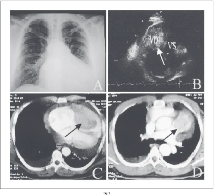
A 43-year-old white man was admitted to our hospital for severe dyspnea, orthopnea, cough, and peripheral ede-mas. Three years earlier, he underwent a partial left liver resection for a trabecular hepatocellular carcinoma (pT2, pN0, M0; stage II, International Union Against Cancer). Objective examination showed hepatosplenomegaly,
en-gorged jugular veins, right pleural effusion, edema of the legs, hypotension, and tachycardia. Chest x-ray (Fig 1A) docu-mented a moderately enlarged heart and a right pleural effu-sion. The two-dimensional echocardiogram (Fig 1B) demonstrated a right ventricular (arrow) mass associated with moderate pericardial effusion. Total-body computed tomog-raphy scan highlighted a right intraventricular mass (arrow; Fig 1C) protruding into the pulmonary conus (arrow, Fig 1D), pericardiac effusion, bilateral subpleural metastases, and a liver
Fig 1.
J
OURNAL OFC
LINICALO
NCOLOGY D I A G N O S I S I N O N C O L O G YV O L U M E 2 2 䡠 NUMBER 24 䡠 DECEMBER 15 2004
5012 Journal of Clinical Oncology, Vol 22, No 24 (December 15), 2004: pp 5012-5016
Downloaded from jco.ascopubs.org on December 31, 2011. For personal use only. No other uses without permission. Copyright © 2004 American Society of Clinical Oncology. All rights reserved.
recurrence in the eighth segment, without evidence of neoplas-tic thrombus into the suprahepaneoplas-tic and inferior cava veins. The patient had cardiac catheterization with biopsy that confirmed a hepatocellular carcinoma with intraventricular metastasis. Serum alpha-fetoprotein levels were normal. Biochemistry documented abnormalities of hepatic and renal functions, increased levels of D-dimers (20 ng/mL; normal, 0.0 to 0.5 ng/mL), low value of antithrombin III (57 ng/mL; normal, 80 to 120 ng/mL), and thrombocytopenia. The patient died after 20 days from acute congestive heart failure. Autopsy revealed liver recurrence of a well-differentiated trabecular hepatocellular carcinoma, multiple metastatic lung emboli, and a large right ventricular neoplastic mass infiltrating the cardiac wall and the pulmonary conus (arrow, Fig 2). Micro-scopic examination confirmed the presence of hepatocellular cancer in liver, lung, and myocardium (Fig 3; hematoxylin and eosin, 20⫻).
Several reports have described cardiac tumor metasta-ses, and their incidence appears to be approximately 10% in patients with hepatocellular carcinoma.1,2Such metastases, however, usually invade the heart through the vascular sys-tem or by infiltrating from neighboring organs. They mainly implant in the epicardium and myocardium, and rarely in the cardiac cavities.3 Intracardiac neoplastic thrombus may flow up to the left and right cardiac cavities through the pulmonary and the cava veins, respectively. This latter event mainly occurs in hepatocellular carcinoma, testicular teratoma, smooth muscle sarcoma, and renal car-cinoma.4,5 The incidence of hepatocarcinoma tumor thrombus in the right atrium is rare (range of incidence, 0.67% to 3%).6Hepatocellular carcinoma mostly produces direct invasion of the inferior vena cava and possible exten-sion to the right atrium. Right cardiac metastases without continuity from the primitive liver tumor are described in rare cases.7-9Patients who present with right cardiac cavities
tumor invasion are often misdiagnosed; however, even with correct diagnosis, effective treatment has not been well es-tablished. Palliative resection may be necessary owing to hemodynamic compromise, but the prognosis remains very poor.10In our case, a trabecular hepatocellular carcinoma surgically removed 3 years earlier produced a right massive intraventricular metastasis protruding into the pulmonary conus with extensive infiltration of the myocardial wall. This lesion was not due to a direct tumor extension to the right cardiac cavities, but to hematogeneous spread, as well documented by the massive endocardium involvement.
Raffaele Longo, David Mocini, Massimo Santini, Paride Giannantoni, Guido Carillio, Francesco Torino, Antonio Auriti, Roberto Marcello, Giovanna Lanzi, Francesco Cortese, and Giampietro Gasparini
“San Filippo Neri” Hospital, Rome, Italy
© 2004 by American Society of Clinical Oncology
Authors’ Disclosures of Potential Conflicts of Interest
The authors indicated no potential conflicts of interest. REFERENCES
1. Kato Y, Tanaka N, Kobayashi K, et al: Growth of hepatocellular
carcinoma into the right atrium. Ann Intern Med 99:472-474, 1983
2. Hanfling SM: Metastatic cancer to the heart: Review of the literature
and report of 127 cases. Circulation 22:474-483, 1960
3. Goudie RB: Secondary tumors of the heart and pericardium. Br
Heart J 17:183-188, 1955
4. Chua SO, Chiang CW, Lee YS, et al: Moving right atrial mass
associated with hepatoma: Two cases detected by echocardiography. Chest 89:148-150, 1986
5. Van Camp G, Abdulsater J, Cosyns B, et al: Transesophageal
echo-cardiography of right atrial metastasis of a hepatocellular carcinoma. Chest 105:945-947, 1994
6. Baba HA, Engers R, Heintzen MP: Right atrial metastasis as primary
clinical manifestation of hepatocellular carcinoma. Int J Cardiol 47:281-284, 1995
7. Vlasseros I, Tapanlis E, Katsaros A: Metastatic hepatocellular
carci-noma into the right atrium and ventricle: Echocardiographic diagnosis and follow-up. Echocardiography 20:387-388, 2003
Fig 2.
Fig 3. Diagnosis in Oncology
www.jco.org 5013
Downloaded from jco.ascopubs.org on December 31, 2011. For personal use only. No other uses without permission. Copyright © 2004 American Society of Clinical Oncology. All rights reserved.
8. Atkins KA: Metastatic hepatocellular carcinoma to the heart. Diagn
Cytopathol 23:406-408, 2000
9. Lei MH, Ko YL, Kuan P, et al: Metastasis of hepatocellular carcinoma
to the heart: Unusual patterns in three cases with antemortem diagnosis. J Formos Med Assoc 91:457-461, 1992
10. Chu MW, Aboguddah A, Kraus PA, et al: Urgent heart surgery for an
atrial mass: Metastatic hepatocellular carcinoma. Ann Thorac Surg 72:931-933, 2001
DOI: 10.1200/JCO.2004.10.198
■ ■ ■
CASE 2.
Burkitt’s Lymphoma Involving
the Gallbladder
A 51-year-old man with a history of gallstones presented with 2 to 3 weeks of intermittent fever, nausea, vomiting, anorexia, weight loss, jaundice, and progressive postprandial abdominal discomfort. Initial physical examination was nota-ble only for fever to 101°F, jaundice, and right upper quadrant abdominal tenderness to palpation. Laboratory evaluation re-vealed leukocyte count of 7.3⫻ 109cells/L with normal differ-ential and smear. Liver function tests were abnormal, with a total bilirubin of 10.0 mg/dL, alkaline phosphatase of 921 U/L, AST 261 U/L, ALT 187 U/L, amylase 560 U/L, and lipase 7,257 U/L. Routine radiologic evaluation with computed tomogra-phy scan showed a significantly thickened gallbladder wall at 2.25 cm and a solitary hepatic lesion that was consistent with an abscess. (Fig 1). The patient underwent a percutaneous fine-needle aspiration of the liver lesion to evaluate for infec-tion and was treated empirically with intravenous ampicillin/ sulbactam and metronidazole for presumed complicated cholecystitis. Before planned cholecystectomy, the biopsy of the liver lesion demonstrated non-Hodgkin’s lymphoma instead of an infection. Another percutaneous core biopsy, this time of the gallbladder wall itself, revealed an infiltra-tion of monotonous small noncleaved cells (Fig 2). Flow cytometry and immunohistochemical stains were positive for CD20, CD10, HLA-DR, Ki-67 (99%), and kappa light chain restriction, and negative for CD3 and CD23. Bone
marrow and CSF were uninvolved. A gastroenterologist per-formed esophagastroduodenoscopy showing diffuse lympho-matous studding of the upper gastrointestinal tract. (Fig 3)
Fig 1.
Fig 2.
Fig 3. Repine, DeArmond, and Lopez
5014 JOURNAL OFCLINICALONCOLOGY
Downloaded from jco.ascopubs.org on December 31, 2011. For personal use only. No other uses without permission. Copyright © 2004 American Society of Clinical Oncology. All rights reserved.