ALMA MATER STUDIORUM
UNIVERSITA' DI BOLOGNA
SCUOLA DI SCIENZE
Corso di laurea magistrale in Biologia Marina
Effects on photoperiod and feeding regimes on Neuropeptide Y (NPY)
and Agouti-related Protein (AgRP) expression in central areas
of Senegalese sole, Solea senegalensis
Tesi di laurea in Adattamenti degli animali all’ambiente marino
Relatore Presentata da
Prof.ssa Elena Fabbri Costanza Guidi
Correlatore
Prof. José Antonio Muñoz-Cueto
Correlatore
Dott. Águeda Jimena Martín Robles
III sessione
1 Introduction 1
1.1 Oveview of feeding in vertebrates 1
1.2 Neuroendocrine regulation of energy homeostasis 2
1.2.1 Orexigenic hormones 5
1.2.1.1 Neuropeptide Y (NPY) 5
1.2.1.2 Agouti-related Protein (AgRP) 8
1.3 Influence of intrinsic factors on feeding behaviour 9 1.3.1 Metabolic signals, energy reserve and feeding status 9
1.3.2 Ontogeny 11
1.3.3 Gender and reproductive status 11
1.3.4 Genetic influence 12
1.4 Influence of extrinsic factors on feeding behaviour 13 1.4.1 Temperature, salinity, hypoxia, pollutant and health status 13 1.4.2 Photoperiod, seasonal and circadian rhythms 14 1.4.2.1 Properties and parameters of biological rhythms 15 1.4.2.2 Circadian system: structure and molecular base 16 1.4.2.3 Light entrainment and food entrainment 17
1.5 The Senegalese sole: biology and development 18
1.5.1 Interest in acquaculture 21
2 Objectives 25
3 Materials and Methods 27
3.1 Animals and rearing system 27
3.2 Experimental design 27
3.3 Sampling 28
3.4 Selection and design of specific primers 28
3.5 RNA extraction 29
3.6 cDNA synthesis 30
3.7 Real Time quantitative PCR (RI-qPCR) expression analysis 30
3.8 Data analysis 31
3.8.1 Statistical analysis 31
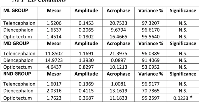
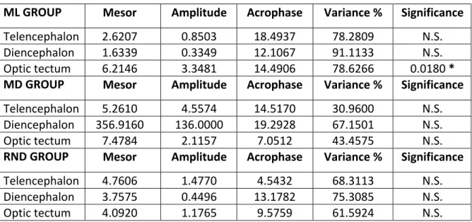
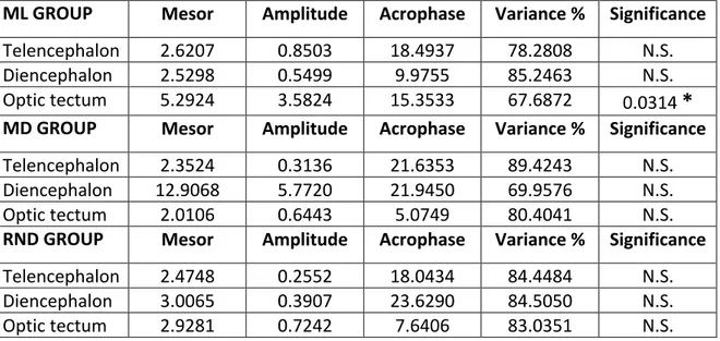
3.8.2 Cosinor analysis 31
4 Results 33
4.1 Daily expression of NPY, AgRP1 and AgRP2 in Senegalese sole reared under light-dark (LD)
conditions and different feeding schedules. 33
4.1.1 Daily expression of NPY mRNA under LD conditions 33
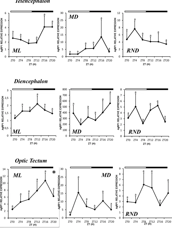
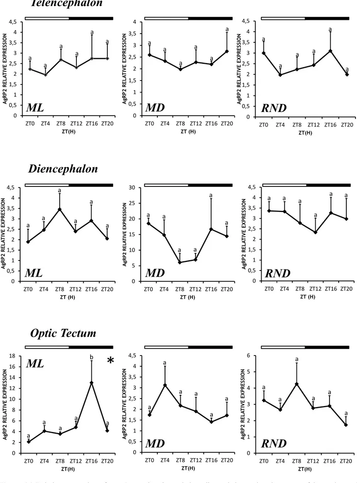
4.1.3 Daily expression of AgRP2 mRNA under LD conditions 37 4.2 Daily expression of NPY, AgRP1 and AgRP2 in Senegalese sole reared under constant dark (DD)
conditions and different feeding schedules. 39
4.2.1 Daily expression of NPY mRNA under DD conditions 39 4.2.2 Daily expression of AgRP1 mRNA under DD conditions 41 4.2.3 Daily expression of AgRP2 mRNA under DD conditions 43
5 Discussion 47
6 Conclusions 53
1 Introduction
1.1 Oveview of feeding in vertebrates
Feeding is a complex phenomenon, regulated by numerous central and peripheral endocrine factors, whose actions are modulated by both external and internal variables. Environment, season, time of day, but also circulating levels of nutrients, hormones and ontogeny affect food intake which, under optimum conditions, is adequate for basal metabolic needs, growth, development, reproduction and deposition of energy store.
In all vertebrates, feeding is regulated by orexigenic (appetite stimulating) and anorexigenic (appetite inhibiting) factors, which act on feeding centers in the brain to mediate the regulation of short-term and long-term dietary intakes (Volkoff et al., 2009a). The central nervous system (CNS), particularly the hypothalamus, has a central role in the regulation of energy homeostasis, but also peripheral organs, such as gastrointestinal tract (GI), pancreas, liver and adipose tissue, are involved in the regulation of feeding (Valassi et al., 2008). Peripheral hormones, in part released in response to the presence of food in the digestive tract, carry information to central feeding centers via the vagus nerve or by crossing the blood-brain barrier acting directly through central receptors and are integrated with other neuronal signals that reflect an animal’s energy status (Brightman & Broadwell, 1976; Holmgren & Olsson, 2009).
During the late 1980s and the 1990s, many brain neuropeptides related to feeding regulation were identified and characterized in mammals and then, some of these hormones or their fish homologs were examined to determine their regulatory effects on feeding in fishes (Volkoff et al., 2005; Gorissen et al., 2006). These peptides displayed similar appetite-regulating effects, showing that the regulation of food intake has been conserved along the vertebrate lineage, although differences exist: the presence of important anatomical and physiological differences between mammals (homeotermic) and fishes (ectothermic) (e.g. brain and gut morphology, caudal secretory organ in fishes) indicates that the endocrine control of feeding requires molecules and mechanism that may be specific to certain species of groups (Volkoff et al., 2009b).
Furthermore, fish represent a vast phylogenetic group, that include agnathans (jawless fishes), chondrichthyans (cartilaginous fishes), sarcopterygians (lobe-finned fishes), and actinopterygians (ray-finned fishes, which are 97% composed of teleosts). Because of this extent it shows a noteworthy level of diversity regarding morphology, ecology, behaviour and genomes (Volff, 2004; Volkoff et al., 2009a). For example it displays differences in gastrointestinal morphology and gut hormone profiles which are related to different feeding habits (Holmgren & Olsson, 2009), and furthermore variation of brain distribution of neuropeptides has been demonstrated (Cerdá-Reverter
& Canosa, 2009). Fishes also undergo several environmental challenges, that may involve long term fasting, trade-off in resource distribution between reproduction and growth (Heino & Kaitala, 1999) and this, added to indeterminate growth, has led to a number of feeding adaptations that suggests that the endocrine control of feeding in fish might also be diverse and involves species-specific molecules and mechanisms (Volkoff et al., 2009a).
Feeding might also display species-specific daily or circannual (seasonal) rhythms in fish, and can be affected by intrinsic factors (e.g., amount of energy stores and reproductive status) and by extrinsic factors (changes in environmental cues or holding conditions).
1.2 Neuroendocrine regulation of energy homeostasis
In mammals, stability of body weight and body composition over long periods of time requires that energy intake matches energy expenditure, that means that dedicated mechanisms are required to avoid changes in body weight, which in mature mammals mainly concerns adipose tissue mass, since protein and carbohydrate store slightly vary (Jéquier and Tappy, 1999; Lin et al., 2000). A renowned theory, the “Lipostatic model” (Kennedy, 1953), explains the well-regulated control of body weight an food intake: hormones produced from fat cells are the key signal to the brain centers that control eating behaviour and activity to regulate feeding an body-fat deposition (Inui, 1999; Lin et al., 2000). So, when body weight exceeds a certain value, eating behaviour is inhibited and energy consumption is increased.
Leptin is thought to be the lipostatic factor that governs energy balance through a negative-feedback loop that originates in adipose tissue and acts on hypothalamic centers in the brain. (Inui, 1999). Leptin is a proteinic hormone encoded by the obese gene (ob) and its effects are feeding inhibition, decrease in body weight and increase in thermogenesis and locomotor activity, as showed in studies involving rodents (Campfield et al., 1995; Halaas et al., 1995; Pelleymounter et al., 1995). Leptin and its receptor system provide an afferent negative feedback signaling system reflecting the amount of adipose energy stores to the brain hypothalamic centers (Schwartz and Seeley, 1997). A feedback regulatory loop with three distinct steps has been hypothesized, that comprise a sensor that monitors the level of energy, hypothalamic centers that receive and integrate through specific leptin receptors the intensity of the signal, and effector systems that influence energy intake and energy expenditure (Jéquier and Tappy, 1999). Leptin targets in the hypothalamus include neuropeptides, such as neuropeptide Y (NPY), agouti-related peptides (AGRP), α-melanophore-stimulating hormone (α-MSH), corticotropin-releasing factor (CRF),
energy expenditure. Also insulin has a similar effect on hypothalamus in the control of food intake (Inui, 1999; Schwarz et al.1992).
As mentioned before, hypothalamic sites, such as the arcuate nucleus (ARC), the lateral hypothalamus (LH), the ventromedial nucleus (VMN), the dorsomedial nucleus (DMN) and the paraventricular nucleus (PVN) have always been considered the centers involved in the control of feeding behaviour. In particular, the LH and the VMN were thought to be respectively the “feeding center” and the “satiety center”, according to the “Dual Center Model” for regulation of feeding (Anand and Brobeck, 1951; Lin et al., 2000). Over the years, new studies, which didn’t employ only discrete lesions or surgical transections, have permitted to redefine the hypothalamic pathways involved in the regulation of feeding and body weight, discovering localization and co-production of more than one signalling molecules in neurons belonging in the same neural area and a deep interconnection between neurons belonging to different areas (Lin, 2000).
In LH, two orexigenic hormones are produced, MCH and orexins (Bittencourt et al., 1992; de Lecea et al., 1998; Sakurai et al., 1998), but also anorexigenic molecules are expressed in LH, i.e. CART (Cocaine and amphetamine regulated transcript) and CRF, which decrease food intake when is centrally administered (Koylu et al., 1997; Kristensen et al., 1998; Kelly and Watts, 1998). Also NPY terminals are abundant in the LH, in contact with orexin and MCH cells. (Williams, 2001).
ARC is situated around the base of the third ventricle and it is indicated as the site which integrates circulating metabolic signals, such as adrenal and gonadal steroids, leptin and insulin (Kalra, 1999). In ARC neurons both orexigenic (e.g. NPY, opioids, galanin, AGRP) and anorexigenic peptides (CART, α-MSH, GABA and glutamate) are produced (Kalra, 1999), which extend into various hypothalamic sites, such as VMN, DMN, PVN, preoptic area (POA) and perifornical hypothalamus. This communication between hypothalamic pathways and the caudal brainstem, responding to meal-related satiety signals, is essential for the long-term regulation of energy homeostasis (Hillebrand, 2002). When ARC is reached by adiposity signals, anorexigenic peptides are released and thus a catabolic circuit is activated. On the contrary, the activation of anabolic pathway leads to the release of orexigenic peptides and this occurs when adiposity signal concentrations in the brain are low, thus indicating the urgency to eat There is an intense innervation of both MCH and orexin neurons in the LH by axons containing NPY, AGRP and α-MSH probably from the ARC (Broberger et al., 1998; Elias et al., 1998). The ARC also has extensively reciprocal connections with other hypothalamic regions, including the paraventicular nucleus (PVN), dorsomedial hypothalamic nucleus (DMH), ventromedial hypothalamic nucleus (VMH).
VMN is suggested to be a receptive field that transfers information to DMN-PVN for the releasing of orexigenic signal (Kalra, 1999), rather than a productive area.
DMN is suggested as site for NPY production and for NPY and leptin interaction, it is also supposed to be involved in the attenuation or inhibition of feeding by leptin (Yokosuka et at., 1998). Along with leptin receptors (Ob-Rb), the DMN contains insulin receptors. Some ARC-NPY/AGRP neurones also terminate in the DMN.).
PVN is the area responsible for the integration and interpretation of the different peripheral and central signals that inform the hypothalamus of surrounding conditions (De Gortari, 2006). PVN receives connections from different hypothalamic areas such as the lateral, median preoptic, arcuate and dorsomedial nuclei and expresses receptors for orexigenic and anorexic peptide signals, such as neuropeptide Y (NPY), agouti related peptide (AGRP), cocaine and amphetamine regulated-transcript (CART), α-MSH, CRH, orexins (De Gortari, 2006; Kalra et al, 1991, Dube et al.)
In fish, as in mammals, several brain region are involved in the regulation of food intake. In the past, from the 1960s to the early 1980s, studies which used electrical stimulation and lesioning showed that also olfactory tracts (Peter, 1979; Demski, 1983), inferior lobes of the hypothalamus (Demski & Knigge, 1971; Demski, 1973 and 1977; Savage & Roberts, 1975; Roberts & Savage, 1978), telencephalon (Grimm, 1960, Demski & Knigge, 1971; Stacey & Kyle, 1983) and optic tectum (Peter, 1979) are all substrates involved in feeding regulation.
In fish, the main neuroendocrine regions producing appetite-regulating hormones are the telencephalon, preoptic area and hypothalamus (Cerda Reverter &. Canosa, 2009) but the relative distribution of the peptides/mRNAs varies with the hormone considered and the species examined. For example, orexin immunoreactive (OX-ir) cells are found in the anterior hypothalamus and along the third ventricle within the preoptic area (POA) in zebrafish (Appelbaum et al., 2009) and medaka (Amiya et al., 2007), whereas in the Australian lungfish, OX-ir cells are also found in the POA, the infundibular hypothalamus and within the telencephalon (Lopez et al., 2009; Hoskins et al., 2012).
There is considerable variation in brain morphology and in the relative size of regions across species, which is often related to life history, ecology and behaviour. For example, it has been suggested that in cichlid fish, algal scrapers have small optic lobes and large telencephala whereas planktivores have enlarged optic lobes (Sylvester et al., 2010).
As mentioned before, the neuropeptides produced by hypothalamic nuclei and involved in feeding intake are divided in two main groups, regarding their action towards feeding: orexigenic, if they stimulate feeding intake, and anorexigenic, if they inhibit it. From the early 1990s, several
peptide or by cloning of their cDNA sequences. Table 1.1 summarizes them and their effects. So far, studies on the potential regulatory effects of these neuropeptides on food intake are very limited. Generally, these studies indicate that the brain hypothalamic area is involved in the regulation of food intake by these neuropeptides, but little work has been carried out on the precise mapping of these neuropeptides in relation to their feeding effects (Volkoff et al., 2009a).
1.2.1 Orexigenic hormones
1.2.1.1 Neuropeptide Y (NPY)
NPY is a 36-amino-acid peptide that belongs to the NPY-family peptides, which also includes peptide YY (PYY), pancreatic polypeptide (PP, found only in the pancreas of tetrapods), and peptide Y (Hoyle, 1999). The most important characteristic for all NPY-family peptides is that they exhibit certain amino acid residues necessary to adopt a specific three-dimensional structure, named the pancreatic polypeptide fold (PP-fold) (Blundell et al. 1981).
NPY was discovered in 1982, when Takemoto isolated it from pig brain homogenates, using a chemical assay that permitted identification of amidated peptides. The first member of this family to be discovered was the pancreatic polypeptide, but as Larhammar et al. (1993) showed, NPY had remained much more conserved throughout evolution than PP, and it seemed to be evolutionarily older than PP. Therefore, the family would be more appropriately called the NPY family than PP family, as it was at the beginning. (Cerda reverter and Larhammar, 2000).
In mammals, NPY is considered one of the most potent orexigenic agents (Halford et al 2004; Kalra et al 1999) and this action has been demonstrated also in fish, which also produce PYY whereas only some teleosts produce PY (Cerdá-Reverter et al., 2000).
These peptides bind to a family of G-protein-coupled receptors that compose the Y family, which has seven cloned members, namely Y1, Y2, Y4, Y5, and Y6 (Larhammar et al., 2001).To date, seven NPY receptor subtypes that bind both NPY and PYY have been identified in fishes (Y1, Y2, Y4–Y8) (Salaneck et al., 2008). Y1-like receptors have been identified in several fish species (Larhammar et al., 2001), whereas Y2-like receptors have only been characterized in zebrafish Danio rerio and rainbow trout, Oncochynchus mykiss (Fredriksson et al., 2004). Fish NPY receptors are expressed in brain but also in peripheral tissues such as eye and intestine (Fredriksson et al., 2004; Lundell et al., 1997).
In mammals, NPY is abundant in the CNS, particularly in the hypothalamic nuclei involved in the regulation of feeding, such as arcuate nucleus (ARC) and paraventricular nucleus (PVN) (Halford et al. 2004).
Central “Orexigenic” agents Ingestive effects Agouti-related protein (AgRP)
(Cerdá-Reverter and Peter, 2003) Increases food intake in fish (Schjolden et al., 2009), birds (Strader et al., 2003), and mammals (Rossi et al., 1998; Stark, 1998), and food hoarding in hamsters (Day and
Bartness, 2004)
Galanin
(Anglade et al., 1994; Unniappan et al., 2002; Wang and Conlon, 1994)
Increases food intake in fish (De Pedro et al., 1995a; Lin et al., 2000; Nelson and Sheridan, 2006; Volkoff et al., 2005) and rats (Kyrkouli et al., 1990) Neuropeptide Y (NPY) (Blomqvist et al., 1992; Cerdá- Reverter et al., 2000a) Increases food intake in fish (de Pedro et al., 2000; Lopez-Patino et al., 1999), frogs (Crespi et al., 2004), snakes (Morris and Crews, 1990), birds (Strader and Buntin, 2001), rats (Stanley and Leibowitz, 1984) and food hoarding in hamsters (Dailey and Bartness, 2009)
Orexin / Hypocretin
(Alvarez and Sutcliffe, 2002; Kaslin et al., 2004)
Increases food intake in fish (Lin et al., 2000; Volkoff et al., 1999; Volkoff et al., 2005), and rats (Sakurai et al., 1998), but not in birds (da Silva et al., 2008) Central “Orexigenic” agents Ingestive effects Cocaine- and Amphetamine- regulated transcript (CART) (Volkoff and Peter, 2000), Decreases food intake in fish (Volkoff et al., 2005), birds (Tachibana et al., 2003), rats (Kristensen et al., 1998) Cholecystokinin, CCK (Peyon et al., 1998),
Decreases food intake in fish (Himick and Peter, 1994; Volkoff et al., 2005), birds (Tachibana et al., 2012), rats (Gibbs et al., 1973) and food hoarding in Siberian hamsters (Bailey and Dela-Fera, 1995; Figlewicz et al., 1989; Teubner and Bartness, 2010) Corticotropin-releasing factor (CRF) (Ando et al., 1999; Bernier et al., 1999; Okawara et al., 1988) Decreases food intake in fish (De Pedro et al., 1993; Matsuda et al., 2008), amphibians (Crespi et al., 2004), birds (Denbow et al., 1999; Furuse et al., 1997), rats (Heinrichs and Richard, 1999; Levine et al., 1983; Morley and Levine, 1982; Negri et al., 1985) and food hoarding in rats (Cabanac and Richard, 1995) reviewed by (Carr, 2002) Melanin concentrating hormone (MCH) (Baker et al., 1995) Decreases food intake in fish (Shimakura et al., 2008), but increases food intake in rats (Presse et al., 1996) Peripheral Orexigenic Hormones Ingestive effects Grelin (gut) (Unniappan et al., 2002) Increases food intake in fish (goldfish and tilapia), but decreases food intake in rainbow trout (Jonsson, 2013; Jonsson et al., 2010), decreases food intake in birds (Kaiya et al., 2009), increases food intake in rats and mice (Tschop et al., 2000; Wren et al., 2000) and food hoarding in Siberian hamsters (Keen-Rhinehart and Bartness, 2005). Peripheral Anorexigenic Hormones Ingestive effects Leptin(adipocytes, liver)
Decreases body weight, adiposity, and food intake in fish (Crespi and Denver, 2006; Murashita et al., 2008), and mice (Campfield et al., 1995; Halaas et al., 1995; Pelleymounter et al., 1995) and food hoarding in Syrian hamsters (Buckley and Schneider, 2003) while increasing energy expenditure
Table 1.1: a partial list of chemical messengers with effects on ingestive behaviour in fish and other vertebrate taxa.
NPY sequences have been determined for several fish species and show strong evolutionary conservation among vertebrate species as the goldfish NPY has only five residues different from rat NPY (Blomqvist et al., 1992). NPY neurons are widely distributed in fish CNS of dipnoans (Trabucchi et al., 2000), elasmobranchs (Chiba, 2000) and teleosts (Cerdá-Reverter et al., 2000a; Doyon et al., 2003; Leonard et al., 2001; Peng et al., 1994; Silverstein et al., 1998). The presence of NPY in fish was first demonstrated in goldfish (Carassius auratus) by immunological and chromatographic studies (Kah et al., 1989). NPY immunoreactive neurons are present in the ventromedial-posterior hypothalamus and hypothalamic inferior lobes (Pontet et al., 1989). Further studies have shown that NPY mRNA is expressed in ventral telencephalon, POA, olfactory bulbs and also in optic tectum, locus coeruleus and other thalamic regions (Peng et al., 1994). A similar brain distribution of NPY mRNA has been described in coho salmon (Oncorhyncus kisutch )(Silverstein et al., 1998). NPY-immunoreactive fibers have also been identified in fish pituitary, pancreas and gastrointestinal tract, and nerve fibers surrounding blood vessels (Cerdá-Reverter and Larhammar, 2000; Chiba et al., 1996; Danger et al., 1991)
In Senegalese sole, Solea senegalensis, it has been demonstrated that NPY-like matter is widely distributed in the brain, with the highest density found in the forebrain, especially in hypothalamus and the ventral telencephalon, whose NPY-containing neurons constitute the major component of the NPY- system in the Senegalese sole (Rodriguez-Gomez et al., 2001).
Several studies have demonstrated that NPY is involved in the regulation of food intake in teleosts, as central injections of mammalian or fish NPY cause a dose-dependent increase in food intake in goldfish (De Pedro et al., 2000; Lopez-Patino et al., 1999; Narnaware et al., 2000), salmon, (Salmo salar) and catfish, (Ictalurus punctatus) (Silverstein and Plisetskaya, 2000). From studies on goldfish it emerged that NPY may act centrally through Y1 and Y5 receptors, which act independently to stimulate food intake in goldfish, but not throught Y2 (Narnaware and Peter, 2001b).
A further demonstration that NPY is involved in feeding is the fact that brain NPY mRNA levels increase following fasting in goldfish (Narnaware and Peter, 2001), winter skate, Raja ocellata (MacDonald and Volkoff, 2009), and chinook and coho salmon (Silverstein et al., 1999a) and undergo periprandial variations in goldfish. An increase in NPY mRNA levels in the telencephalon–preoptic area and hypothalamus shortly before feeding was observed, followed by a decrease in brain NPY mRNA levels after feeding (Narnaware and Peter, 2001a). Similar variations were observed also in Atlantic cod, Gadus morhua (Kehoe and Volkoff, 2007) and tilapia, (Oreochromis mossambicus) (Peddu et al., 2009). Besides in goldfish, NPY gene expression in
brain seems to be influenced by macronutrient intake (Narnaware and Peter, 2002) and by the peripheral metabolic status (Lin et al. 2000).
In fishes as in mammals, the actions of NPY on feeding occur in part by the modulation of other appetite regulators, e.g., corticotrophin-releasing factor (CRF) and cortisol (Bernier et al., 2004), cocaine- and amphetamine-regulated transcript (CART) (Volkoff and Peter, 2000), leptin (Volkoff et al., 2003), melanin-concentrating hormone (MCH) (Matsuda et al., 2008, orexins (OXs) and galanin (GAL) (Volkoff and Peter, 2001), growth hormone (GH) (Mazumdar et al., 2006) and ghrelin (Miura et al., 2006), and, in goldfish by estradiol a and testosterone (Peng et al., 1994).
1.2.1.2 Agouti-related Protein (AgRP)
Agrp1 and Agrp2 belong to the agouti family peptides, which also include the agouti signaling proteins (ASIP) and have potent and diverse functional roles in feeding, pigmentation and background adaptation mechanisms (Västermark et al., 2012).
In mammals, AgRP is one of the most potent orexigenic peptides and is co-expressed together with NPY in the ARC. It functions by increasing appetite and decreasing metabolism and energy expenditure (Hagan et al., 2000; Sainsbury and Zhang, 2010).
AgRP has been also identified in fish (Cerdá- Reverter and Peter, 2003; Klovins and Schiöth, 2005; Kurokawa et al., 2006; Song et al., 2003b; Stütz et al., 2005). Phylogenetic analyses of the deduced proteins showed that tetrapods have two agouti family members, i.e. ASIP and AgRP, while some teleosts have four: ASIP1, ASIP2, AgRP1, and AgRP2 (Braasch and Postlethwait, 2011).
All the agouti family peptides are characterized by a C-terminal polycysteine domain (Klovins and Schiöth, 2005). Orthologs of ASIP, AgRP, melanocortin receptors MC1R and MC4R have been identified in mammalian, teleost fish and avian genomes, but not in invertebrate genomes. This may suggest that the agouti-melanocortin system evolved by gene duplication in the last 500 million years.
Studies on rats showed that stimulate hyperphagia when administered intracerebroventricularly, furthermore, it displays long-lasting effects on food intake, with animals displaying hyperphagia even 7 days following a single intracerebroventricular (i.c.v.) injection (Hagan et al., 2000a).
The orexigenic function of AgRP is primarily performed through the antagonism of central melanocortin receptors (MCRs), specifically of melanocortin receptor 3 (MC3R) and melanocortin receptor 4 (MC4R), which are directly related to metabolism and body weight control (Oyama et al., 2010; Ollmann et al., 1997; Robinson et al., 2000). Studies on the sea bass, Dicentrarchus labrax,
However, the exact mechanism by which AgRP inhibits melanocortin-receptor signalling is not completely clear.
An mRNA encoding AgRP has been identified in goldfish (Cerdá-Reverter and Peter, 2003), zebrafish (Song et al., 2003), and pufferfish (Klovins et al., 2004; Kurokawa et al., 2006). The goldfish AgRP gene encodes a 128 amino acid precursor and is expressed in a variety of tissues including brain and peripheral tissues. Moreover, AgRP expression was identified only in hypothalamus of goldfish, while in Takifugu rubripes was detected throughout a wide region of brain in which suggests a species-specific expression of AgRP (Cerdá-Reverter and Peter, 2003; Ollman et al., 1997; Wei et al. 2013).
Several studies have revealed the involvement of AgRP in the control of food intake in fish: transgenic zebrafish overexpressing AgRP exhibited obesity, increased linear growth, and adipocyte hypertrophy, strongly suggesting that AgRP plays an important role in feeding control in teleost fish (Wan et al., 2012). Furthermore, AgRP levels are upregulated by fasting in different species of vertebrates, such as sheep (Adam et al., 2002), Japanese quail (Boswell et al., 2002), goldfish (Cerdá-Reverter and Peter, 2003) and zebrafish (Song et al., 2003), underlining a conserved role for AgRP in energy homeostasis. In goldfish, brain AgRP mRNA increases after 3 days of fasting, and it may exert its effect through MCRs (Cerdá-Reverter and Peter, 2003; Cerdá-Reverter et al., 2003). Fasting up-regulated hypothalamus AgRP mRNA levels in Danio rerio (Song et al., 2003. In Cyprinus carpio, fasting induces an initial reduction of expression and after the initiation of re-feeding, there was a significant induction of AgRP mRNA expression (Wan et al., 2012) All together, the identification of AgRP in several teleosts and the effect of fasting on AgRP expression in goldfish, suggest that it plays a part in appetite regulation, possibly through MCRs.
1.3 Influence of intrinsic factors on feeding behaviour 1.3.1 Metabolic signals, energy reserve and feeding status
Feeding behaviour and the expression of appetite-related peptides have been shown to be influenced by circulating metabolite levels, the ingestion of food and food deprivation (Volkoff et al., 2010).
Variations in circulating metabolite levels alter food intake in fish. A decrease in food intake has been observed in trout fed with high-carbohydrate enriched diets (Banos et al., 1998) and in carp injected intraperitoneally with essential amino acids (Kuz’mina, 2005). Also, protein and lipids can modify feeding in fish. Intraperitoneal administration of amino acids decreases food intake in carp (Kuz’mina, 2005) and also a lipostatic control of food intake has been suggested after noticing that,
in chinook salmon and channel catfish, fat fish eat less than thin fish (Shearer et al., 1997; Silverstein and Plisetskaya, 2000).
Gene expression of appetite regulators can be altered by meal consumption. Usually, the expression levels of orexigenic factors (NPY and Orexin) increase before or during a meal (Narnaware and Peter, 2001; Xu and Volkoff, 2007) while the expression levels of anorexigenic factors (CCK and CART) decrease after feeding (Peyon et al., 1999; Volkoff and Peter, 2001). Fasting generally induces an up-regulation of the expression of orexigenic factors, such as brain NPY, orexins and stomach ghrelin (Narnaware and Peter, 2001; Silverstein et al., 1999; Novak et al., 2005; Xu and Volkoff, 2007; Amole and Unniappan, 2008; Terova et al., 2008) and a down-regulation of the expression of anorexigenic hormones, such as brain CART and gut CCK (Kehoe and Volkoff, 2007; Kobayashi et al., 2008; Volkoff and Peter, 2001; MacDonald and Volkoff, 2009; Murashita et al., 2006). However, contradictory results have been reported. Fasting increases NPY mRNA expression after 3 days in goldfish hypothalamus (Narnaware et al., 2000), after 2 and 4 weeks in winter flounder hypothalamus (MacDonald and Volkoff, 2009), after 2-3 weeks in Chinook and coho salmon hypothalamus (Silverstein et al., 1999) and after 2 weeks in Brazilian flounder whole brain (Campos et al., 2010), whereas one-week fasting does not affect NPY mRNA expression in either cod forebrain (Kehoe and Volkoff, 2007) or tilapia whole brain (Riley et al., 2008). The duration of food deprivation might influence the results since, for example, in goldfish a fast of 10 days, but not of 3, decreases the hypothalamic mRNA expression of CART1 (Abbott and Volkoff, 2011). Besides, variations could be due to species-specific differences in response to fasting, as fishes might have different sets or patterns of appetite-related peptides and might respond differently to nutritional challenges or might be due to different expression analysis in different parts of the brain, as different regions of the brain might respond differently to fasting. For example, although both hypothalamus and telencephalon are brain regions that have been implicated in the regulation of feeding in fish, in skate (Raja ocellata), a 2-week fasting period increases NPY mRNA expression in the telencephalon but not in the hypothalamus (MacDonald and Volkoff, 2009). Moreover, different experimental conditions might influence the results (Hoskins & Volkoff, 2012).
Fish can also adapt their food intake to their energy needs and have been reported to respond with increased food intake when they are fed with low energy diets (Boujard and Medale, 1994; Geurden et al., 2006; Paspatis and Boujard, 1996; Yamamoto et al., 2000). The fact that brain NPY expression levels are influenced by macro-nutrient intake in goldfish suggests that these dietary adjustments in feeding might be mediated by variations in appetite-related hormones (Narnaware
1.3.2 Ontogeny
Appetite-regulating factors might play a role in embryogenesis (pre-hatch) as well as nutrient absorption/acquisition (pre- and post-hatch) in fish larvae, as they appear early in development. A later appearance of appetite-regulating factors, especially when associated with metamorphic events or mixed feeding phases (e.g., beginning of exogenous feeding), may reflect ontogenic shifts in feeding. Gastro instestinal CCK immunoreactivity has been detected upon first hatch in herring, Clupea harengus (Kamisaka et al., 2005), and ghrelin has been detected 48h post fertilization in zebrafish (Pauls et al., 2007). A number of other signals involved in the regulation of feeding in fishes appear to be present prior to hatching, such as somatostatin (Xing et al., 2005; Xu and Volkoff, 2008), NPY (Volkoff et al., 2009), OX (Xu and Volkoff, 2007), α-MSH and AgRP (Forlano and Cone, 2007).
1.3.3 Gender and reproductive status
In fish, there is a close relationship between feeding, gender and reproductive parameters.
Gender-specific differences in feeding behaviour have been noticed. During the spawning season, territorial male cunners (Tautogolabrus adspersus) feed less often and have different diets than females (Green et al., 1984).Sexual dimorphism in growth rates and size at maturity is common in fish and could be due in part to differences in feeding activity between sexes (Davis et al., 2008; Rennie et al., 2008; Shearer et al., 2006; Toguyeni et al., 1997). Differences in levels of appetite regulating hormones have also been noted in a few species. For example, in tilapia, gastric ghrelin mRNA levels are higher in females compared to males (Parhar et al., 2003), and in salmon the number of ghrelin cells per unit area in the stomach is higher in females than in males (Sakata et al., 2004). These observations would suggest that gender, or more specifically sex steroids, might influence feeding. It seems likely that these influences would be more pronounced when fish are either breeding or preparing to breed (e.g., migrating) as a decline in feeding is often seen during spawning migrations or other reproductive behaviors (e.g. courtship, spawning, guarding, territoriality) (van Ginneken and Maes, 2005). This happens in both Atlantic cod (Fordham and Trippel, 1999) and winter flounder (MacDonald and Volkoff, 2009), as feeding is suppressed in both sexes during the spawning period and it increases after spawning. Also European eels (van Ginneken et al., 2005), Atlantic salmon (Miller et al., 2005) eat very little during spawning and spawning migration, and male domino damselfish reduce time spent feeding during courtship and nest guarding (Mann et al., 2007).
al., 2002.) Seasonal changes correlated to gonadal cycles have been shown for NPY in ayu (Chiba et al., 1996) and catfish (Mazumdar et al., 2007) and CCK (MacDonald and Volkoff, 2009), suggesting that appetite-related hormones mediate in part these seasonal changes.
However, the exact roles of sex steroids on food intake are unclear as sex steroid treatment decreases food intake in some species, as seabass and eurasiatic perch (Leal et al., 2009; Mandiki et al., 2005), but increases feeding in others, as red sea bream (Woo et al., 1993). In goldfish, intracerebroventricular treatment with gonadotrophin-releasing hormone (GnRH) induces a decrease in food intake (Hoskins et al., 2008; Matsuda et al., 2008), which is in part due to down-regulation of brain OX mRNA expression (Hoskins et al., 2008). Conversely, intracerebroventricular OX-A injections induce a decrease in spawning behaviour and a decrease in GnRH mRNA expression levels in the brain (Hoskins et al., 2008). Also testosterone is thought to have an anorexigenic role, as testosterone treatment decreases food intake in male perch (Mandiki et al., 2005) and elevates MCH mRNA expression in the hypothalamus of both male and female goldfish (Cerdà-Reverter et al., 2006). Besides, castration reduces the density of NPY-immunoreactive fibers in the forebrain of tilapia (Sakharkar et al., 2005).
1.3.4 Genetic influence
In captive fish, food intake has shown genetic-based variations, such as in rainbow trout, in which different strains display variations in feeding activities and growth (Mambrini et al., 2004), feeding patterns (Boujard et al., 2007), nutrient utilization (Quillet et al., 2007) and body composition (Kause et al., 2002).
These variations have been found also in cod (Case et al., 2006) and Atlantic salmon (Glover et al., 2009) regarding variations in feeding activities and growth, while in striped bass (Wang et al., 2007) differences in body composition have been observed. However, since domestication selection has been achieved mostly empirically, very little is known about the genetic variations related to endocrine factors in these selected animals. Within the context of aquaculture, a better knowledge of these variations might lead to genetic improvement by selection for specific traits, such as better food efficiency and faster growth.
1.4 Influence of extrinsic factors on feeding behaviour
In fish, variations in several extrinsic factors have been shown to induce changes in swimming activity, feeding rhythms and growth (Boujard, 2001). Temperature and photoperiod are probably the major abiotic factors affecting feeding.
1.4.1 Temperature, salinity, hypoxia, pollutant and health status
Fish tend to increase both their food consumption and growth rates with rising temperatures within a “tolerable” range (Bendiksen et al., 2002; Kehoe and Volkoff, 2008;) and the optimal temperatures for growth usually decrease with fish size (Bjornsson et al., 2001). It has been noticed that they tend to decrease their food intake when placed in extreme temperatures (Volkoff et al. 2009). Little is known about the endocrine mechanisms regulating these temperature-induced changes in feeding. In Atlantic cod, brain NPY mRNA expression does not seem to be affected by temperature, but brain CART mRNA expression levels are higher in fish held at 2° C than in fish held at either 11° C or 15° C. This suggests that CART, but not NPY, may contribute to temperature-induced changes in appetite in this species (Kehoe and Volkoff, 2008).
Also salinity affects feeding in several fish species (De Boeck et al., 2000) but little is known about the appetite-regulating factors that may be involved. It is presumed that the somatotropic axis might play a role in salinity-related alteration on feeding, since it is a regulator of growth and salt water tolerance in salmonids (Boeuf and Payan, 2001). In conditions of acute osmoregulatory disturbance, fishes often momentarily reduce food intake, which has been shown to be concomitant with increases in brain corticotropin-releasing factor (CRF) in trout (Craig et al., 2005).
Hypoxia has distinct appetite suppressive effects (Buentello et al., 2000; Ripley and Foran, 2007). In rainbow trout, exposure to low oxygen levels increases forebrain CRF as well as plasma cortisol, suggesting that CRF-related peptides play a physiological role in mediating at least a portion of the reduction in food intake (Bernier and Craig, 2005).
Contaminants and disease agents also have impact on feeding but the responses vary depending on the species considered as well as on the agent and the method of exposure. For example, rainbow trout exposed to elevated waterborne concentrations of metals eats less (Todd et al., 2007), on the contrary coho salmon displays increased feeding rates, if fed with a high zinc diet (Bowen et al., 2006).
1.4.2 Photoperiod, seasonal and circadian rhythms
Photoperiod can affect feeding activity (Imsland et al.,2007; Noble et al., 2005; Sunuma et al., 2007; Tucker et al., 2006) as well as the expression of appetite-regulating hormones. In fish, natural daily feeding rhythms vary among species: some species are diurnal feeders, e.g. Atlantic salmon (Paspatis and Boujard, 1996), whereas others are nocturnal feeders, e.g. catfish (Boujard, 1995). In addition, many fish species have been shown to display preferred times of feeding throughout the day or night and it’s been demonstrated that alteration of normal daily feeding rhythms can induce poor performance and eventually diseases and death (Lopez-Olmeda et al., 2011)
Natural behavioural rhythms are often classified by their period, which is the interval needed to complete one cycle. Different rhythms have been studied in fish: tidal and lunar rhythms, circannual or seasonal rhythms (Bolliet et al., 2007; Boujard and Leatherland, 1992; Lall and Tibbetts, 2009).
One of the most evident expressions of adaptation to the environment, and a major characteristic of living organisms, are the Circadian rhythms. As the etymology of the name stresses (from the Latin circa, meaning "around" and dies, "day", meaning "approximately a day”, name created by Halberg) these rhythms develop on periods close to 24 h (Menaker et al., 1997) and persist in constant conditions. The circadian rhythm can be divided into routine cycles during the 24-hour day: diurnal, which describes organisms active during daytime; nocturnal, which describes organisms active at nighttime; crepuscular, which describes animals primarily active during the dawn and dusk hours.
Besides, rhythms that last longer than a day day (>28-hour) are defined infradian rhythms (annual migration or reproductive rhythms found in certain animals or the human menstrual cycle), while shorter ones, that last less than 20 hours, are called ultradian rhythms (the 90-minute REM cycle, or the 3-hour cycle of growth hormone production (Butler et al., 2005).
External stimuli are able to entrain circadian rhythms, in particular photoperiod: the 24 hours light-dark cycle is the most consistent environmental signal, because of its stability in period and phase. In fact most of the circadian rhythms of species studied until now are are synchronized in a daily manner by this cue. However, periodical fluctuations exhibited by biochemical, physiological and behavioural parameters and rhythms persist in constant conditions, thus demonstrating that they arise within organism itself and are not imposed by the environment (Kulczykowska et al., 2010)
The endocrine mechanisms regulating these rhythms are poorly known. Little is known about the fluctuations in circadian-related proteins or appetite-regulating peptides in fish.
1.4.2.1 Properties and parameters of biological rhythms
Circadian rhythms are present in all eukaryotic and some prokaryotic organisms and their wide diffusion show evolutionary conservation. Circadian rhythms are temperature-compensated, genetically determined and thus generated by a self-sustained endogenous pacemaker.
In vertebrates, a circadian timing system generates and regulates circadian rhythms; it consists in pacemakers and oscillators, entrainment pathways and pacemaker outputs connected to effector systems that express evident rhythms under circadian control (Guido et al., 2002).
Circadian rhythms “free-run”, displaying a period of approximately 24 hours, when organisms are in constant conditions and without environmental cues. The periods of the endogenous rhythms actually differ from those in nature, but thanks to exogenous synchronizing cues, commonly called
zeitgeber (“timegiver” in German), biological rhythms are adjusted in such a way that organisms
remain synchronised with their environment (Kulczykowska et al., 2010), as zeitgebers help to reset the biological clock to a 24-hour day.
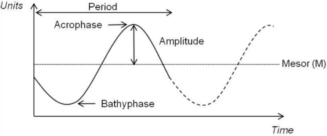
Circadian rhythms can be graphically represented in cronograms, where a sine wave can be fitted (Figure 1.1. Diagram of an oscillatory proess characterized by its 4 parameters.). Within each cycle, the phase is a particular value of the rhythm in the cycle and the time period at which the cycle peaks is called the acrophase. When the process is less active, the cycle is in its bathyphase or trough phase. The highest value of the rhythmic biological variable is the peak or maximum and the lowest value is the nadir. The difference between the peak and the mean value of a wave is measured by the amplitude and the period of the rhythm is the time between two points in the same phase (close to 24 h in the case of circadian rhythms).
1.4.2.2 Circadian system: structure and molecular base
All circadian systems are composed of three basic elements:
• an oscillator or clock that generates and sustains the endogenous rhythmic oscillations, • an input that allows the synchronization of the oscillator,
• and an output by which the pacemaker regulates many physiological and behavioural processes (Guido et al., 2002).
In vertebrates, three neural structures have been reported as oscillators or clocks: the retina, the pineal complex and the mammalian suprachiasmatic nucleus (SCN) of the hypothalamus or its analogous areas in other groups (Menaker et al., 2997). Nevertheless, independent or semi-independent, light-entrainable circadian clocks appear to exist at all levels of organization from cells, through tissues to organs.
Usually in animals, a master or central clock is present and it is located in the brain. Clocks located in other parts of the body are defined peripheral oscillators, in order to distinguish them from the central clock.
In mammals, entrainment of circadian rhythms depends upon photic information provided exclusively by the lateral eyes through the retina (Bertolucci et al., 2004). This photic information reaches the central oscillator localized in the suprachiasmatic nucleus (SCN) via the retinohypothalamic tract, synchronizing SCN daily rhythmic neuronal activity (Doyle et al., 2007; Maywood et al., 2007). In turn, signals from the SCN regulate the activity of many other targets, including melatonin synthesis in the pineal gland (Korf et al., 2003), which is considered to be a peripheral oscillator (Vatine et al., 2011). In this system, oscillation signals produced by the SCN are relayed to the pineal gland via the multisynaptic efferent pathway (Falcon, 1999; Takahashi, 1994).
In non-mammalian vertebrates, the pineal complex contains all elements required for photic entrainment and circadian rhythm generation, as it is photoreceptive and contains an intrinsic circadian oscillator (Korf et al., 1998; Falcon et al., 2003). Fish pineal cells are classical photoreceptor cells with structural and functional similarities to retinal photoreceptors. Pineal and retinal photoreceptor cells share a similar set of genes, or, in certain cases, paralogues (Falcon et al., 2003). In fish the pineal gland is considered to serve as central pacemaker, as transduce environmental light information into a neural and a neuroendocrine signal. It contains an intrinsic circadian clock that drives rhythmic synthesis of the hormone Melatonin, an indoleamine with well-known effect on internal biological rhythms, whose levels are high at night and low during the day as a result of regulated transcription and stability of serotonin-N-acetyl-transferase (AANAT).
Activity of this enzyme is dictated by the circadian clock and also shows a rapid suppression in response to illumination during the night (Ziv et al., 2007; Applebaum et al., 2006).
Internal biological clocks allow organisms to adapt their behavioural and physiological functions to daily and seasonal variation of environmental factors (Pevét, 2001). The basic clock mechanism is a feedback loop in which oscillating products of specific clock genes regulate their own expression. Each complete turn of the loop takes about 24 hours to accomplish, resulting in circadian oscillations of RNA and protein levels (Nanako et al., 2012).
In vertebrates this regulatory loop consists of positive elements (clock and bmal) that drive the expression of negative elements (period or per and cryptochrome or cry) that, in turn, feedback to down-regulate their own expression and allow the start of a new cycle of the feedback loop. Furthermore, the existence of an additional feedback loop that directs the rhythmic expression of bmal tends to confer robustness and stability on the core loop (Emery et al., 2004).
In fish, this clock machinery has been studied in goldfish (Carassius auratus), rainbow trout (Oncorhynchus mykiss), Atlantic salmon (Salmo salar), European sea bass (Dicentrarchus labrax), medaka (Oryzias latipes), the gilthead sea bream (Sparus aurata), the Senegalese sole (Solea senegalensis) and some reef fishes (Siganus guttatus and Halichoeres trimaculatus) (Davie et al., 2009, 2011; Del Pozo et al., 2012; Hur et al., 2012; Martín-Robles et al., 2011, 2012b; Park et al., 2007; Patiño et al., 2011; Sánchez et al., 2010; Velarde et al., 2009; Vera et al., 2013). Such studies has allowed identifying genes involved in many aspects of the circadian clock system, but much more work is needed in order to understand all aspects of temporal organization within each organism.
1.4.2.3 Light entrainment and food entrainment
Although light is the most noticeable zeitgeber, several non-photic stimuli, such as food, have been also shown to entrain circadian rhythms. Indeed, feeding time can act not only on the SCN and peripheral tissues, but also on an oscillator, called Food-Entrainable Oscillator (FEO), which is independent from the Light-Entrainable Oscillator (LEO) (Meijer and Rietveld, 1989) but not well-defined yet, as its anatomical location is still unknown (Stephan et al., 1979; Stephan, 2002; Davidson, 2006). In fish, datas on properties of feeding entrainment support the hypothesis of the existence of a FEO, even if it is still uncertain whether fish FEO and LEO are independent (Sánchez-Vázquez et al., 1997; Aranda et al., 2001). On the contrary in mammals studies suggested the independence of FEO from LEO (Stephan, 2002): lesions on SCN didn’t abolish feeding rhythms such as FAA (Food Anticipatory Activity), which is a phenomenon that displays an increase of activity in anticipation of an imminent meal, which is maintained for at least 30 minutes
(Mistlberger, 1994) and it’s clearly connected with FEO. One of the main characteristics of FAA is its gradual development, with several feeding cycles being required for its significance appearance. Moreover, after a shift of the feeding time, the time that fish require to resynchronize their FAA is directly related to the quantity of hours that the mealtime has been shifted. Locomotor activity, that includes behavioural and physiological variables such as feeding, reproduction and territoriality, exhibits daily rhythms that can be entrained by a feeding cycle. These behavioural patterns in many species have been fixed genetically due to the pressure generated by stable selective forces such as avoidance of predators, the availability of preys or the optimization of food (Daan, 1981) and ) and it is often conditioned by the existence of special sensory requirements such as the dependence on the vision for the capture of preys (Madrid et al., 2001). Anticipation of time meals has been reported for a wide variety of animals, from bees to higher vertebrates such as monkeys (Stephan, 2002) and it brings several advantages. For instance, when food availability is predictable, the animal can use this information to anticipate it and maximize food intake and nutrient utilization (Sánchez-Vázquez and Madrid, 2001).
In mammals behavioural/physiological parameters appeared to be controlled by the FEO, such as rinking behaviour can be synchronized to the periodic food access, increasing a few hours before mealtime or being maintained during food deprivation (Boulos et al. 1980; Clarke et al. 1986). Furthermore, along with an increase of locomotor activity, a rise of body temperature can also be measured prior to feeding time, which can be viewed as an adaptive mechanism that prepares an organism to ingest food efficiently
1.5 The Senegalese sole: biology and development
The Senegalese sole (Solea senegalensis, Kaup, 1858) is a marine teleost fish belonging to the Class Actinopterygii, Order Pleuronectiformes, Family Soleidae (Figure 1.2
The Soleidae family, which consists of 22 genera and 89 species inhabiting brackish, marine and fresh waters, is characterized by flat bodies and unusual asymmetrical external appearance.
The larval fish display a perfect symmetry, which is lost through a metamorphosis that takes place from a few weeks/months after hatching. This determines the migration of the left eye, that will take place on the right side of the skull/head, near the the other eye.
Thus the adult fish is characterized by an oval-shaped and flattened body, strongly compressed, with an ocular side which is slightly rounded and pigmented and a blind side which is white and totally flat, facing the bottom.
As benthic fishes, they normally lie down in the bottom/seafloor, usually covered by sand or mud, and they mimic their background by assuming a similar coloration. Their habit of burrowing in the sand represents an innate instinct of soles to avoid a possible aggression and it is kept also in individuals held in captivity. Soles can protrude the small eyes above the surface of the body, in such a way that the animal can see even if buried in the substrate.
The Senegalese sole inhabits mobile sand or muddy bottoms, around 100 m of depth, mainly in
coastal areas, but they can also be found in salt or brackish lagoons connected to the sea, rivers and estuaries. They are located in subtropical climates, between 14°N-47°N and 1°W-19°W. The coasts of Senegal are the southern boundary in the Atlantic, the Canarian Islands represent the western limit and the shores of Brittany constitute the limit in the north. Its geographical distribution in the Mediterranean is fairly broad, covering the south and east of the Iberian Peninsula, the north of Africa and Middle East until the coast of Turkey (Errore. L'origine riferimento non è stata
trovata.).
Sole is an euryhaline and eurythermal species, adapting perfectly to changes in temperature and salinity (Rueda-Jasso et al., 2004). Sole can tolerate low levels of dissolved oxygen and grow optimally in under saturated environments (Salas-Leiton et al., 2008). Senegalese sole is a predator principally of benthic invertebrates and it feeds preferentially polychaetes (i. e. Hediste diversicolor, Capitella capitata), crustaceans (orders Tanaidacea, Amphipoda and Decapoda) and bivalve molluscs (i. e. Scrobularia plana).
From a reproductive point of view, the Senegalese sole is a gonochoric species with separate sex and without apparent sexual dimorphism. The first sexual maturity is reached between the second and third year of life in males (first spermiation), and between the second and fourth year of life in females (first oviposition), when the size reaches 30 cm (Dinis et al., 1999). They have an asynchronic ovarian development, showing oocytes in different stages of development
(García-Figure 1.3: Computer generated distribution maps for Solea senegalensis (Senegalese sole), with modelled year 2100
native range map based on IPCC A2 emissions scenario. www.aquamaps.org. Note: Distribution range colors indicate degree of suitability of habitat which can be interpreted as probabilities of occurrence.
occurs in spring and autumn, with peaks in May and, to a lesser extent in October (Anguis and Cañavate., 2005; Oliveira et al., 2009). Spawning has an endogenous rhythmicity that synchronizes with dusk periods. It starts after dusk and it peaks about 4 hours later. It also shows synchronization with lunar phases, peaking at the new moon (Oliveira et al., 2009b). Spawning takes place between 13°C and 23°C, with higher fecundities between 15°C and 21°C (Anguís & Cañavate, 2005).
Nocturnal habits are shown not only for spawning (Oliveira et al., 2009a), but also for locomotor activity (Bayarri et al., 2004), feeding (Boluda Navarro et al., 2009) and Solea senegalensis displayed even a higher metabolic rate during the dark phase (Castanheira et al., 2011). Senegalese sole exhibites clearly nocturnal self-feeding patterns under laboratory and farming conditions, with 77% to 85% of feed demands occurring at night. Therefore, feeding during the photophase could be incompatible with the natural feeding rhythm of sole (Boluda Navarro et al., 2009). Nutritional requirements for sole larvae are still poorly known, and big efforts are being made to maximize survival and growth. Some experiments demonstrated the ability of sole to self- feed successfully and showed an accurate compensatory feeding behaviour by modifying their feeding activity accordingly to the reward level. (Boluda Navarro et al., 2009).
1.5.1 Interest in acquaculture
The Senegalese sole (Solea senegalensis) is a flatfish of high commercial importance that is almost indistinguishable by consumers from common sole (Solea solea, Linnaeus, 1758).
Southern Europe countries have been more focused in Solea senegalensis aquaculture due to the lower spawning temperature requirements of Solea solea (Howell, 1997), and the high abundance of Solea senegalensis in Mediterranean and Southern Atlantic waters (Dinis et al., 1999), which makes of Solea senegalensis the only sole species reared in Spain or Portugal nowadays
Ongrowing of sole is carried out under two major strategies: a traditional one and a more intensive one. Traditionally in Spain and in southern Portugal, soles were reared in earthen ponds in old salt marshes or other deltaic or estuarine environments. Fingerlings were either passively captured in these ponds. Feeding consisted in the occurrence of natural prey in the bottom of the ponds, with few or null effort from the aquaculturists, or supplementing or replacing these feed items with inert feed, hence taking a semi- intensive approach. On the other hand, the intensive approach is nowadays the leading trend although salt marshes are still used for semi-intensive and intensive sole aquaculture. Soles are stocked in fiberglass or concrete tanks, often in shallow raceways (Imsland et al., 2003), for the whole production cycle, and they are fed inert feeds in highly controlled environments.
Although interest in farming Senegalese sole intensively in southern Europe dates back to the early 1980s, it has failed to reach successful commercial development (Flos et al., 2001; Imsland et al., 2003; FAO Fisheries and Aquaculture Information and Statistics Service, 2011), mainly because of juvenile scarcity for stocking purposes, caused by the lack of full control over spawning, poor fry quality and high mortality rates during the weaning stage (Cañavate & Fernández-Díaz, 1999; Anguís & Cañavate, 2005). Furthermore a high incidence of skeletal malformations and pigmentation abnormalities in post-larvae and juveniles (Gavaia et al., 2002; Soares et al., 2002; Villalta et al., 2005a) is exhibited, along with disease outbursts affecting all ontogenetic stages caused by multiple pathogenic agents (Zorrilla et al., 2003).
Reproduction of Senegalese sole is one of the main difficulties that its domestication is still facing nowadays. There are several problems to obtain fertile spawning from second generation individuals, and this is a huge drawback, as broodstocks have to be collected from the wild as they loose reproductive performance or die (Cabrita et al., 2006; Guzmán et al., 2008, 2009a, 2011; Oliveira et al., 2011). Many approaches have been taken to investigate this problem. It has been seen that F1 females show normal vitelogenin steroid profiles and spontaneous spawning (Guzmán et al., 2008), with egg quality parameters within normal ranges (Guzmán et al., 2009a) but normally these eggs show no fertilization in the communal reproduction tanks.
Good broodstock management practices are required so to maintain genetic variation within farmed strains at comparable levels to those of wild source populations, e.g. the use of adequate numbers of effective parents, broodstock from different locations, or even more local ones if significant differences are present in the wild (Exadactylos et al., 2007). There are only a few studies focusing on assessing the optimal ongrowing photoperiod for sole, although it has been observed that sole is strongly influenced by it, as it shows a evident nocturnal activity pattern, with locomotor activity peaking in the first part of the dark period, and progressively decreasing during the night. Recent studies show that keeping a constant photoperiod after winter solstice deters the increase in melatonin production, characteristic of shorter days and longer nights, and also increases the production of steroid hormones and vitellogenin in the prespawning phase, also advancing spawning (Oliveira et al., 2011). These findings point out to the possible practicability of advancing/controlling spawning season of Senegalese sole in fish farms. A crucial step in the rearing of an aquaculture species is the ability to control maturation and, ultimately, spawning. Spawning of Senegalese sole females have been successfully induced through hormonal manipulation, although results have not been conclusive in suggesting a dosage, administration method or administration timing. The steroid of choice is an analogue of the gonadotropin-releasing
hormone (GnRHa) administered either via repetitive injections or through sustained-release implants (Agulleiro et al., 2006).
The Senegalese sole is acquiring an important relevance in chronobiological studies as the number of published works focused on the sole circadian system has increased in the last few years especially focused on rhythms of locomotor activity, feeding, melatonin, sex steroids in relation to temperature and photoperiod (Anguis and Cañavate, 2005; Bayarri et al., 2004; Boluda-Navarro et al., 2009; García-López et al., 2007; Guzmán et al., 2008; Oliveira et al., 2009). The molecular mechanisms underlying sole circadian rhythms have also been explored recently, both in adults and developing sole (Martín-Robles et al., 2011, 2012a, 2012b).
2 Objectives
The general objective of this study was to investigate how different photoperiods and feeding regimes modulate daily mRNA expression of three specific orexigenic hormones, i.e. the Neuropeptide Y (NPY) and the paralogues Agouti-related Protein 1 (AgRP1) and Agouti-related Protein 2, in adult specimens of Solea senegalensis, a flatfish species with a growing commercial interest in Southern Europe for marine aquaculture.
It is important to increase knowledge on the endocrine mechanisms that regulate feeding and growth in cultured fish, so to improve fish holding conditions and feeding strategies in aquaculture practices. In order to achieve this and to support the development of new techniques that could enhance feeding, food conversion efficiency and growth in cultured fish, two main experiments were developed:
• In a first experiment, adult fish, maintained under LD (12 h light:12 h dark) photoperiod, were divided in three different tanks, each one kept at a different feeding regime (feeding during daytime, feeding during night-time and feeding at random times).
• In a second experiment, adult fish were maintained under a DD (0 h light: 24 h dark) photoperiod and they were divided in two tanks, each one kept at a different feeding regime (feeding during the subjective daytime and feeding at random times).
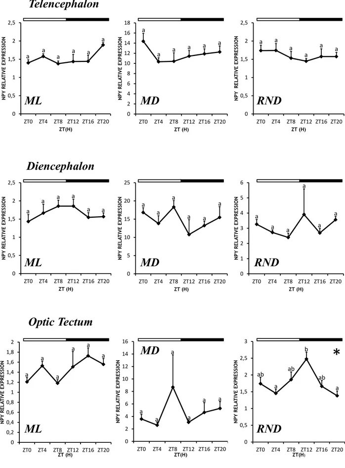
For both experiments, daily mRNA expression of selected orexigenic hormones (NPY, AgRP1 and AgRP2) has been studied in different central areas, i.e. telencephalon, diencephalon and optic tectum, which previous studied suggested to be the main areas involved in feeding in fish and rich in neurons related to orexigenic hormones.
This main objective also allowed to attain the following specific achievements:
• To gain skills on molecular biology techniques such as RNA extraction, cDNA synthesis and real time quantitative PCR.
• To gain skills on several bioinformatic tools and specific software such as the chronobiological software El Temps and the Bio-Rad CFX Manager v 3.1.
3 Materials and Methods
3.1 Animals and rearing system
This study was carried out on a total number of 128 Senegalese sole adult specimens (Solea senegalensis) which were collected from the “Laboratorio de Cultivos Marinos" of the University of Cádiz (Puerto Real, Spain). The animals had length between 20 and 25 cm in length and their weight spanned from 101 to 172 g. They were kept in five 250L tanks and maintained at constant temperature (19±1°C) and salinity (39 ppt), with continuous seawater renovation and gentle aeration. Each tank was equipped with an automatic feeder (EHEIM GmbH & co. KG, Germany) connected to a digital programmable timer (Data micro, Orbis, Spain), so to provide food at scheduled times. Commercial 3 mm dry pellets (Skretting S.A., Burgos, España) were supplied at a daily ration of 0,3% body weight. The tanks were also equipped with a lid containing two fluorescent lamps (Sylvania Gro-Lux, Germany) connected to an individual automatic photoperiod control system which allowed a light intensity of 400 lux at water surface level during the illumination period.
The experimental procedures were approved by the Animal Experimentation and Ethics Committee of the University of Cádiz (Spain) and performed according to international ethical standards.
3.2 Experimental design
Two experimental groups with different photoperiod conditions were designed. The photoperiod for the “LD group” was set at 12 h light:12 h dark (12L:12D) with lights on at 08:00 h local time, (zeitgeber time 0 or ZT0), while the animals of the “DD group” were reared in constant dark conditions, i.e. 0 h light: 24 h dark (0L:24D).
The animals reared in light-dark conditions, were subdivided in 3 groups, each one experiencing a distinct feeding regime:
• ML group: this group received food at a fixed time during the day, at 14:00h (ZT6), i.e in the middle of the light phase (ML as mid-light);
• MD group: this group comprised the animals that were fed at fixed time during the night, at 02:00h (ZT18), i.e., in the middle of the dark fase (MD as mid-dark);
• RND group, whose feeding was set at a random times, with an interval between 12 and 36
The animals kept in constant darkness, i.e., “DD group", were divided in 2 groups with different feeding time:
• sML group: this group received food at fixed time during the mid-light point of the subjective day, at 14:00h (circadian time 6 or CT6).
• RND group: this group was fed at random times, with an interval between 12 and 36 hours,
as described previously for the LD group.
3.3 Sampling
Sampling was conducted after four weeks under these different feeding regimes. during a 24 h daily cycle. The soles were anesthetized in MS-222 (Sigma, St. Louis, MO; 100-200 mg/l of water) and sacrificed by decapitation.
Animals held under LD conditions were sampled every 4 hours, starting at 8:00 h local time, which was considered ZT0 (zeitgeber time 0), at six different zeitgeber time points (ZT0, ZT4, ZT8, ZT12, ZT16, ZT20), where ZT0 corresponded to the light onset and ZT12 to the light offset.
The soles under DD conditions were sampled every 4 hours as well, at six different circadian time points: CT0, CT4, CT8, CT12, CT16, CT20.
The selected neural tissues, i.e. telencephalon, diencephalon and optic tectum, were dissected from every specimen, rapidly frozen in liquid nitrogen and stored at -80° C until used.
3.4 Selection and design of specific primers
Several specific primer pairs for Senegalese sole npy, agrp1 and agrp2 genes analysed by real-time quantitative PCR (RT-qPCR) were designed from the partial sequences available in the SoleaDB
(http://www.juntadeandalucia.es/agriculturaypesca/ifapa/soleadb_ifapa/sessions/new?locale=es) server using the Primer3 Plus software (http://www.bioinformatics.nl/cgi-bin/primer3plus/primer3plus.cgi).
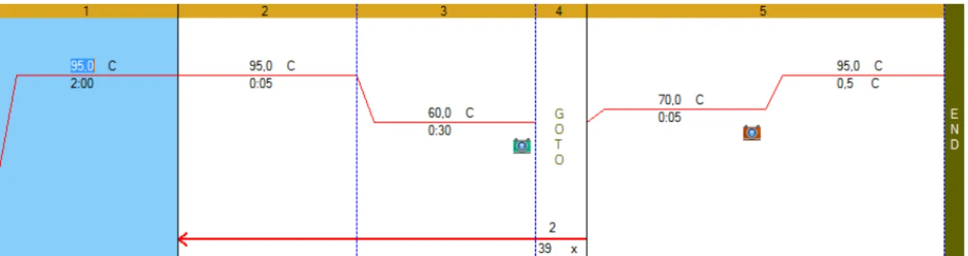
A gradient temperature RT-qPCR was run for each primer pair, in order to optimize PCR conditions, with the following temperatures: 58°C, 60,8°C, 62,5°C and 64,6°C. The resulting amplicons were analysed through agarose gel electrophoresis and DNA was extracted by using QIAquick Gel Extraction Kit (QIAGEN Group Inc, USA) following manufacturer instructions. Briefly, DNA fragments were cut out from the agarose gel with a clean, sharp scalpel, then put in a tube with 3 volumes of buffer QG available with the kit per 1 volume gel and incubated at 50°C for 10 min. After complete dissolution of the gel slices, the mixture was put in a collection tube and