A rapid diagnostic workflow for
cefotaxime-resistant Escherichia coli and Klebsiella
pneumoniae detection from blood cultures by
MALDI-TOF mass spectrometry
Elena De Carolis1☯, Silvia Paoletti1☯, Domenico Nagel1, Antonietta Vella1, Enrica Mello1, Ivana Palucci1, Giulia De Angelis1, Tiziana D’Inzeo1, Maurizio Sanguinetti1*,
Brunella Posteraro2‡, Teresa Spanu1‡
1 Institute of Microbiology, Università Cattolica del Sacro Cuore, Fondazione Policlinico Universitario Agostino Gemelli, Rome, Italy, 2 Institute of Public Health, Section of Hygiene, Università Cattolica del Sacro Cuore, Fondazione Policlinico Universitario Agostino Gemelli, Rome, Italy
☯ These authors contributed equally to this work. ‡ These authors also contributed equally to this work. *[email protected]
Abstract
Background
Nowadays, the global spread of resistance to oxyimino-cephalosporins in
Enterobacteria-ceae implies the need for novel diagnostics that can rapidly target resistant organisms from
these bacterial species.
Methods
In this study, we developed and evaluated a Direct Mass Spectrometry assay for Beta-Lac-tamase (D-MSBL) that allows direct identification of (oxyimino)cephalosporin-resistant
Escherichia coli or Klebsiella pneumoniae from positive blood cultures (BCs), by using the
matrix-assisted laser desorption ionization–time of flight mass spectrometry (MALDI-TOF MS) technology.
Results
The D-MSBL assay was performed on 93 E. coli or K. pneumoniae growing BC samples that were shortly co-incubated with cefotaxime (CTX) as the indicator cephalosporin. Sus-ceptibility and resistance defining peaks from the samples’ mass spectra were analyzed by a novel algorithm for bacterial organism classification. The D-MSBL assay allowed discrimi-nation between E. coli and K. pneumoniae that were resistant or susceptible to CTX with a sensitivity of 86.8% and a specificity of 98.2%.
a1111111111 a1111111111 a1111111111 a1111111111 a1111111111 OPEN ACCESS
Citation: De Carolis E, Paoletti S, Nagel D, Vella A, Mello E, Palucci I, et al. (2017) A rapid diagnostic workflow for cefotaxime-resistant Escherichia coli and Klebsiella pneumoniae detection from blood cultures by MALDI-TOF mass spectrometry. PLoS ONE 12(10): e0185935.https://doi.org/10.1371/ journal.pone.0185935
Editor: Massimiliano Galdiero, Seconda Universita degli Studi di Napoli, ITALY
Received: May 9, 2017 Accepted: September 21, 2017 Published: October 5, 2017
Copyright: © 2017 De Carolis et al. This is an open access article distributed under the terms of the Creative Commons Attribution License, which permits unrestricted use, distribution, and reproduction in any medium, provided the original author and source are credited.
Data Availability Statement: All relevant data are within the paper and its Supporting Information files.
Funding: The authors received no specific funding for this work.
Competing interests: The authors have declared that no competing interests exist.
Conclusion
The proposed algorithm-based D-MSBL assay, if integrated in the routine laboratory diag-nostic workflow, may be useful to enhance the establishment of appropriate antibiotic ther-apy and to control the threat of oxyimino-cephalosporin resistance in hospital.
Introduction
In the last decades, bloodstream infections caused by theEnterobacteriaceae family-members Escherichia coli and Klebsiella pneumoniae showing resistance to third-generation oxyimino-cephalosporin antibiotics [1,2], mainly to cefotaxime (CTX) [3], have increased. This has a negative impact on the infection-related mortality rates and hospital costs [4], and it is associ-ated with a delay in the administration of appropriate antibiotic therapy [5]. According to the European Antimicrobial Resistance Surveillance Network data, the percentages of third-gener-ation cephalosporin-resistantE. coli and K. pneumoniae isolates ranged from 19.8% and 45.9% in 2011 to 30.1% and 55.9% in 2015, respectively, in Italy alone; conversely, all over Europe the majority of countries reported resistance percentages of 25% or higher [6]. Resistant isolates produce extended-spectrum β-lactamases (ESBLs), the plasmid-mediated enzymes that can confer non-susceptibility to cefotaxime and other oxyimino-cephalosporins (e.g., ceftazidime) and to monobactams [7]. ESBLs are the result of mutations that occur in TEM, SHV, and, more prominently, CTX-M family β-lactamase-encoding genes [3]. CTX-M ESBLs hydrolyze cefotaxime better than ceftazidime, although several CTX-M variants with enhanced ceftazidi-mase activity have been detected [8]. The worldwide spread of ESBL-producing Enterobacter-iaceae organisms has led to increased use of carbapenems, resulting in the emergence of plasmid-mediated resistance to carbapenems [9].
In clinical laboratory practice, antimicrobial susceptibility testing is currently performed by using growth-based manual or automated methods, such as agar diffusion assays and broth microdilution systems [10]. However, these phenotypic methods are limited by turnaround times which do not satisfy the demand for timely information about the antimicrobial suscep-tibility of patient’s isolate. In contrast, molecular genetic methods are timely but able to only provide information about the absence or presence of an ESBL gene, which not always corre-lates to the phenotype [11]. Reasonably, matrix-assisted laser desorption ionization–time of flight mass spectrometry (MALDI-TOF MS), that is used for successful microbe identification [12], also direct from positive blood cultures (BCs) [13], in many clinical laboratories, has been explored as a tool for rapid antimicrobial resistance detection [14]. In one of these newly generated assays [15], enzymatic cleavage of the β-lactam ring (in the β-lactam antibiotic) leads to specific mass shifts that can be easily monitored by MALDI-TOF MS. In another assay [15], comparison of the growth rates derived from cultivations of the same organism in the presence or absence of an antibiotic (virtually from all classes) enables organism’s susceptibil-ity/resistance status to be assessed.
The aim of the present study was to evaluate the potentiality of a MALDI-TOF MS based assay, reported here as Direct Mass Spectrometry assay for Beta-Lactamase (D-MSBL), for the identification of cefotaxime-resistantE. coli and K. pneumoniae directly from positive BCs of patients treated at a tertiary-care Italian hospital.
Materials and methods
Study design and samples
The study was approved by the Ethics Committee of our Institution (protocol no. 0022585), but written informed consent was waived since D-MSBL analysis was performed only on a residual portion of clinical sample and patient-identifying information was not included. The study was conducted prospectively between April and December 2015 at the Università Cattolica del Sacro Cuore, Fondazione Policlinico Universitario Agostino Gemelli, which is a large tertiary-care teaching hospital in Rome, Italy. Blood samples were cultured in the BD Bactec FX BC system (Becton Dickinson Instrument Systems, Sparks, MD, USA), using sets of BD Bactec Plus Aerobic and Anaerobic bottles (Becton Dickinson). Gram staining was performed for all positive BCs. If Gram-negative rods were found, BC aliquots were directly used for MALDI-TOF MS identification and D-MSBL analysis (only BC positive forE. coli orK. pneumoniae were tested). Conventional cultures of the same sets of BC samples were performed for the isolates’ identification confirmation and reference antimicrobial suscepti-bility testing. Additionally, contrived samples were spiked at known concentrations of the twoEnterobacteriaceae species, using bacterial strains with molecularly characterized resis-tance mechanisms.
Laboratory methods
Direct MALDI-TOF MS analysis. For each sample included in the study, 8 mL of positive
BC fluid were inoculated in a VACUETTE Z Serum Sep Clot Activator tube (Greiner Bio-One International GmbH, Frickenhausen, Germany), centrifuged at 3500 rpm for 15 min, and the resulting pellet was transferred in an Eppendorf tube with 1 mL of HPLC-grade water to reach a turbidity of 2 McFarland. After centrifugation at 12000 rpm for 2 min, the supernatant was discarded and the bacterial pellet was used in parallel for direct MALDI-TOF MS identifica-tion on a ground-steel target plate with 1μL of formic acid ( 98%, Sigma-Aldrich, St. Louis, MO, USA) solution and for D-MSBL assay (i.e., β-lactam ring hydrolysis assay) with cefotax-ime (CTX, Sigma) (see below). MALDI-TOF MS analyses were performed using a Microflex LT mass spectrometer (Bruker Daltonics, Bremen, Germany). Spectra were analyzed with the Bruker’s Biotyper software (version 3.1), and the Bruker Biotyper library V.4.0.0.1 (5627 entries) was used to achieve bacterial identification [13]. BC samples that were found to con-tainE. coli (n = 58) or K. pneumoniae (n = 35) organisms were subjected to the D-MSBL assay on the same day. To ensure the assay reproducibility, the bacteria (pellets contained 108CFUs) were resuspended, in triplicate, in 30μL of CTX solution (0.5 mg/mL). The suspensions were vortexed and incubated at 37˚C with agitation (900 rpm) for 1 h. After centrifugation at 13000 rpm for 2 min, 1μL of supernatant was spotted in duplicate on a polished-steel target plate and covered with 1μL of HCCA (α-cyano-4-hydroxycinnamic acid) matrix solution (10 mg/mL in 50% acetonitrile). Prior to analysis, the Bruker antibiotic calibration standard, consisting of bradykinin 1–7 (M + H+: 757.39 Da), bradykinin 1–5 (M + H+: 573.31 Da),
Lys-Lys-Lys (M + H+: 403.30 Da), and Ser-His (M + H+: 243.10 Da), was resuspended in 25μL of HPLC-grade water and was used for the instrument calibration in the mass range from 100 to 1000 Da. Spectra were acquired automatically in the positive linear mode at a laser frequency of 60 Hz with an acquisition range from 100 to 1000 Da. The peaks corresponding to either non-hydrolyzed or hydrolyzed CTX forms were analyzed using the software ClinProTools (Bruker Daltonics) version 3.0 with the parameters chosen in a mass range between 300 and 600 Da. The reference strainsE. coli ATCC 25922 and K. pneumoniae ATCC 25955 (β-lacta-mase non-producers) andE. coli ATCC 35218 and K. pneumoniae ATCC 700603 (β-lactamase
producers) were used as negative or positive controls, respectively, and were included in every run. For quality control of the CTX solution and for detecting spontaneous hydrolysis, a con-trol without bacteria (CTX only) was also included. All the concon-trol samples were processed in the same way as the BC samples. Statistical analysis was performed on the mass peaks corre-sponding to the spectral profiles acquired from samples consisting of the CTX solution alone, the CTX solution plus test BC fluid, the CTX solution plus positive control (E. coli or K. pneu-moniae reference strain), and the CTX solution plus negative control (E. coli or K. pneupneu-moniae reference strain). Thus, four samples were included in each run of the MALDI-TOF MS analy-sis. For each sample, a total of 20 mass peaks showing highest intensity were automatically selected, in order to calculate the intensity average (Ave) with standard deviation (StdDev) of the peaks from all samples by means of the ClinProTools software peak statistic tool. Thus, a test sample was classified as CTX-resistant by an algorithm based on the following equations: Avex–StdDevx > Aves + StdDevs, calculated from CTX-resistance defining mass peaks (370.5 and 414.5 Da), andAvex + StdDevx < Aves–StdDevs, calculated from CTX-susceptibility defin-ing mass peaks (396.5 and 456.5 Da), where “x” and “s” were the test (unknown) sample and negative (susceptible) sample, respectively; if the equations were not satisfied, the test sample was classified as CTX-susceptible. In some cases, the analysis was conducted on three mass peaks, as the fourth could not be included among the 20 peaks mentioned above (see data in
S1 File).
Preliminarily, to validate the D-MSBL assay, we tested 24 bacterial reference strains that were selected to represent the third-generation cephalosporin-resistant strains most com-monly isolated in our clinical setting. BD Bactec Plus Aerobic bottles were spiked with 100μL of a 2-McFarland bacterial suspension obtained with each of 24 ESBL-producingK. pneumo-niae (KPC-3/SHV-11/ TEM-1, n = 4; CTX-M-15/KPC-2, n = 4; CTX-M-15/SHV-28/TEM-1, n = 4) andE. coli (CTX-M-1, n = 4; CTX-M-15, n = 4; CTX-M-27, n = 4) strains. As above described, BC bottles were incubated in the BD Bactec FX BC system until a positive growth was signaled by the instrument. The BC broths were then processed for the MALDI-TOF MS-based CTX hydrolysis analysis, and the peaks corresponding to non-hydrolyzed or hydrolyzed CTX forms were analyzed, as above described. Thus, each strain was classified according to the above-mentioned algorithm.
Conventional culture and susceptibility testing. Aliquots from each positive BC bottle
included in the study were subjected to routine Gram stain microscopy, and were subcultured in parallel on a set of selective and nonselective routine agar plates and incubated under appro-priate atmospheric conditions for 24 h or re-incubated for 48 h as necessary. Bacterial isolates were identified by the colony-smear method using MALDI-TOF MS, as previously described [13]. For allE. coli and K. pneumoniae isolates (n = 93), antimicrobial susceptibility testing (AST) was performed using Vitek 2 AST cards N201 (bioMe´rieux, Marcy l’E´toile, France). The minimum inhibitory concentration (MIC) results were interpreted according to the EUCAST breakpoints document version 6.0 2016 [10]. The CTX MICs were also determined using the Etest (bioMe´rieux) to confirm the EUCAST MIC results. Genotypic characterization in allE. coli and K. pneumoniae isolates was done by PCR for detection of ESBL or plasmidic AmpC-type enzyme genes (blaCTX-M,blaNDM,blaOXA-48,blaKPC,blaSHV,blaTEM,blaVIM,blaMOX-1,
blaCMY-2,blaLAT,blaDHA-1,blaACC,blaACT-1, andblaFOX-1), using the total DNA extracted from
each isolate. PCR amplification was performed with the primers and conditions described by Yeet al. [16], and therein references, and the PCR fragments, after purification, were sequenced by a 3100 genetic analyzer instrument (Applied Biosystems, Foster City, CA). Sequences were analyzed by ChromasPro version 2.4.1 (Technelsium Pty Ltd) and database searches were done by BLASTn program of the National Center for Biotechnology Informa-tion (NCBI,http://www.ncbi.nlm.nih.gov).
Results and discussion
All the 24 reference strains ofE. coli and K. pneumoniae used in the D-MSBL assay validation phase of the study were correctly classified by the MALDI-TOF MS-based algorithm here pro-posed for detection of CTX resistance. Thus, D-MSBL assay was directly applied on 93 BCs that were detected as positive forE. coli (58 isolates) and K. pneumoniae (35 isolates), as docu-mented by the MALDI-TOF MS analysis performed the same day the D-MSBL assay was done, and later confirmed on the isolates grown from subcultures. The D-MSBL assay results were compared with those obtained by the phenotypic or genotypic testing performed on the 93E. coli and K. pneumoniae isolates. The overall results are shown separately for E. coli and K. pneumoniae. As it can see for E. coli (Table 1), 17 of 19 CTX-resistant isolates and 38 of 39 CTX-susceptible isolates were correctly classified as resistant or susceptible, respectively, by the D-MSBL assay.
As it can see forK. pneumoniae (Table 2), 16 of 19 resistant isolates and all of 16 CTX-susceptible isolates were correctly classified as resistant or CTX-susceptible, respectively, by the D-MSBL assay.
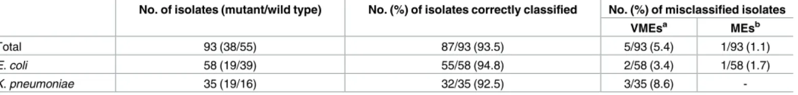
Overall, as shown inTable 3, there were 5 very major errors (2E. coli and 3 K. pneumoniae) and 1 major error (1E. coli), that corresponded to those isolates that were incorrectly classified as susceptible (3.4% and 8.6%) or resistant (1.7%), respectively, by the D-MSBL assay.
As exemplified inFig 1, we used a simple algorithm for MALDI-TOF MS analysis to cor-rectly identify CTX-resistant organisms in positive BCs of bacteremic patients, which takes into account the simultaneous reduction of non-hydrolysis peaks (456.5 and 396.5 Da) and increase of hydrolysis peaks (414.5 and 370.5 Da) associated with CTX and its forms [17]. Conversely, the inversion of these peak profiles indicated the presence of a CTX-susceptible organism.
Consistent with previous studies [18,19], our findings correlated well with the CTX resis-tance levels shown by subcultured organisms via conventional phenotypic testing (MICs from 4 to 64 mg/L). Notably, all the CTX-resistant organisms correctly detected by D-MSBL assay were ESBL producers, mainly due to a CTX-M β-lactamase that was possessed by all of 19E. coli and 6 of 19 K. pneumoniae (Tables1and2). The latter organisms were also possessors of a SHV type β-lactamase and/or KPC carbapenemase—specific β-lactamase with the ability to hydrolyze carbapenems [20]. ThreeK. pneumoniae isolates with CTX MICs of 16 to 64 mg/L were detected as CTX-susceptible organisms by the D-MSBL assay, although these isolates had a resistance mechanism (isolates 3K and 19K, KPC-3/SHV-11/TEM-1; and isolate 34K, CTX-M-15/SHV-28/TEM-1) compatible with the phenotype tested conventionally. Similarly, twoE. coli isolates (MIC, 64 mg/L) had a D-MSBL assay result (CTX-susceptible) that was in apparent disagreement with their resistance mechanism (isolate 7E, CTX-M-15/SHV-11/ TEM-1; and isolate 10E, CTX-M-1). In one study, Oviaño et al. [19] used a second cephalospo-rin (i.e., ceftazidime) to try to improve the detection of β-lactam resistance in those ESBL cases that were negative with the MALDI-TOF MS assay when CTX was used alone. Thus, it is con-ceivable that the use of ceftazidime would have enabled detection of our isolates carrying ESBL with a lower rate of CTX hydrolysis and/or higher catalytic efficiency for ceftazidime. How-ever, in 3 of 5 our isolates the ESBL phenotype was totally or partially represented by CTX-M type enzymes.
In summary, we found that the agreement between the D-MSBL assay and the genotypic method results was 94.8% with respect to theE. coli organisms and 92.5% with respect to the K. pneumoniae organisms. Data revealed that the assay has an overall sensitivity of 86.8% and specificity of 98.2% in predicting CTX resistance in clinical isolates ofE. coli and K. pneumo-niae. The positive predictive value and negative predictive value were 97.0% and 91.5%, respectively.
Table 1. Comparison of the cefotaxime resistance mechanism, MIC value, and D-MSBL assay results for 58 E. coli derived from the BCs tested.
Isolate Mechanism(s) of resistance Phenotype D-MSBL assay
MIC (mg/L) Categorya Category
1E CTX-M-1 8 R R 2E CTX-M-15 64 R R 3E CTX-M-15 64 R R 4E - 1 S S 5E - 1 S S 6E - 1 S S 7E CTX-M-15, SHV-11, TEM-1 64 R S 8E - 1 S S 9E - 1 S S 10E CTX-M-1 64 R S 11E - 1 S S 12E CTX-M-27 64 R R 13E CTX-M-15 64 R R 14E CTX-M-15 64 R R 15E CTX-M-27 64 R R 16E - 1 S R 17E - 1 S S 18E CTX-M-15 64 R R 19E - 1 S S 20E - 1 S S 21E CTX-M-15 64 R R 22E CTX-M-15 64 R R 23E - 1 S S 24E - 1 S S 25E CTX-M-15 64 R R 26E CTX-M-15 4 R R 27E CTX-M-15, TEM-1 64 R R 28E - 1 S S 29E - 1 S S 30E CTX-M-15 64 R R 31E - 1 S S 32E - 1 S S 33E CTX-M-15 64 R R 34E CTX-M-27 64 R R 35E CTX-M-15 64 R R 36E - 1 S S 37E - 1 S S 38E - 1 S S 39E - 1 S S 40E - 1 S S 41E - 1 S S 42E - 1 S S 43E - 1 S S 44E - 1 S S 45E - 1 S S 46E - 1 S S (Continued )
Table 1. (Continued)
Isolate Mechanism(s) of resistance Phenotype D-MSBL assay
MIC (mg/L) Categorya Category
47E - 1 S S 48E - 1 S S 49E - 1 S S 50E - 1 S S 51E - 1 S S 52E - 1 S S 53E - 1 S S 54E - 1 S S 55E - 1 S S 56E - 1 S S 57E - 1 S S 58E - 1 S S a
The MIC-based category was assessed according to the EUCAST guidelines version 6.0 2016 (S, 1 mg/L; R, >2 mg/L). https://doi.org/10.1371/journal.pone.0185935.t001
Table 2. Comparison of the CTX resistance mechanism, MIC value, and D-MSBL assay results for 35 K. pneumoniae derived from the BCs tested.
Isolate Mechanism(s) of resistance Phenotype D-MSBL assay
MIC (μg/mL) Categorya Category
1K KPC-3, SHV-11, TEM-1 64 R R 2K - 1 S S 3K KPC-3, SHV-11, TEM-1 64 R S 4K KPC-3, SHV-11, TEM-1 64 R R 5K KPC-3, SHV-11, TEM-1 64 R R 6K - 1 S S 7K KPC-3, SHV-12, TEM-1 64 R R 8K - 1 S S 9K - 1 S S 10K - 1 S S 11K KPC-3, SHV-11, TEM-1 8 R R 12K - 1 S S 13K CTX-M-15, KPC-2 64 R R 14K KPC-3, SHV-11, TEM-1 64 R R 15K CTX-M-15, SHV-11 16 R R 16K - 1 S S 17K KPC-3, SHV-11, TEM-1 8 R R 18K KPC-3, SHV-11, TEM-1 64 R R 19K KPC-3, SHV-11, TEM-1 16 R S 20K KPC-3, SHV-11, TEM-1 64 R R 21K KPC-3, SHV-11, TEM-1 64 R R 22K - 1 S S 23K - 1 S S 24K - 1 S S 25K - 1 S S 26K CTX-M-15, SHV-38 64 R R (Continued )
The emergence of multidrug-resistant Gram-negative organisms has a dramatic impact on the patient outcome, and it represents an urgent health-care problem either from a manage-ment or an economic standpoint. New and cost-effective diagnostic technologies offer the pos-sibility of overcoming the improper use of antibiotics and, in the meanwhile, controlling the spread of antibiotic-resistant bacteria. Our MALDI-TOF MS-based assay that couples direct identification and detection of CTX-resistantE. coli and K. pneumoniae organisms in positive BCs provides rapid results to clinicians who search for the most appropriate antibiotic therapy, without the need for expensive techniques such those based on PCR [11]. The overall expected time is <2 h, taking into account the identification step (30 min) and the D-MSBL assay step (60 min of hydrolysis assay plus 30 min of spectrum acquisition and analysis). Furthermore, our assay has the advantage that a given organism can be classified in the most objective way possible, without the need for presetting species-specific cutoff values to distinguish between susceptible and resistant strains, as recently reported by Junget al. [18]. Therefore, as our study shows, the proposed algorithm-based D-MSBL assay could be integrated into a diagnos-tic laboratory workflow to detect resistance against cephalosporin antibiodiagnos-tics with a high sensi-tivity 24 to 48 h earlier than conventional methods (Fig 2).
One limitation of the D-MSBL assay is that it determines susceptibility/resistance to CTX only, thus it does not indicates whether ESBL alone is likely. If this is the case, therapy should be switched to a carbapenem, or if already given, should be continued. Therefore, the assay applies better in epidemiological contexts where the prevalence of infections with carbapene-mase-producing bacteria is believed to be low. In our study, none ofE. coli isolates were
Table 2. (Continued)
Isolate Mechanism(s) of resistance Phenotype D-MSBL assay
MIC (μg/mL) Categorya Category
27K - 1 S S 28K - 1 S S 29K - 1 S S 30K - 1 S S 31K - 1 S S 32K CTX-M-15, KPC-3, SHV-11, TEM-1 64 R R 33K KPC-3, SHV-11, TEM-1 64 R R 34K CTX-M-15, SHV-28, TEM-1 64 R S 35K CTX-M-15, KPC-3, SHV-28, TEM-1 64 R R a
The MIC-based category was assessed according to the EUCAST guidelines version 6.0 2016 (S, 1 mg/L; R, >2 mg/L). https://doi.org/10.1371/journal.pone.0185935.t002
Table 3. Performance of the D-MSBL assay for 93 clinical isolates according to the presence or absence of cefotaxime resistance-associated β-lactamase mutant genes.
No. of isolates (mutant/wild type) No. (%) of isolates correctly classified No. (%) of misclassified isolates
VMEsa MEsb
Total 93 (38/55) 87/93 (93.5) 5/93 (5.4) 1/93 (1.1)
E. coli 58 (19/39) 55/58 (94.8) 2/58 (3.4) 1/58 (1.7)
K. pneumoniae 35 (19/16) 32/35 (92.5) 3/35 (8.6)
-a
Very major errors (VMEs) correspond to resistant isolates that were classified as susceptible by the D-MSBL assay. bMajor errors (MEs) correspond to susceptible isolates that were classified as resistant by the D-MSBL assay.
carbapenem-resistant, in contrast to ~40% ofK. pneumoniae isolates that were found to be resistant to carbapenems. Thus, in our clinical practice, D-MSBL assay may be a valuable tool for early therapeutic guidance in patients withE. coli bacteremia, whereas only a half of the patients withK. pneumoniae bacteremia can indeed benefit from this assay. Although this assay holds promise for the future, studies are yet needed to confirm its integration in a labora-tory workflow that will allow clinicians to diagnose bacteremia in real time and initiate appro-priate antibiotic therapy as early as possible.
Fig 1. Representative MALDI-TOF mass spectra of two K. pneumoniae organisms detected as susceptible (A) and CTX-resistant (B), respectively, by the D-MSBL assay in patients’ BC samples. As detailed on the bottom, classification results were determined by the analysis of both resistance-defining mass peaks (415.5 and 370.5) and susceptibility-defining peaks (456.5 and 396.5) in the test sample compared to those of a control negative (CTX-susceptible) sample (i.e., K. pneumoniae ATCC 25955 β-lactamase non-producer).
Supporting information
S1 File. Supplementary information that includes supplementary tables containing raw data relative to the MALDI-TOF MS analysis performed with the Bruker ClinProTools software.
(XLSX)
Author Contributions
Conceptualization: Elena De Carolis.
Data curation: Silvia Paoletti, Domenico Nagel, Antonietta Vella, Enrica Mello, Ivana Palucci. Formal analysis: Elena De Carolis, Silvia Paoletti, Domenico Nagel, Antonietta Vella. Funding acquisition: Maurizio Sanguinetti.
Supervision: Giulia De Angelis, Tiziana D’Inzeo, Maurizio Sanguinetti, Teresa Spanu. Writing – original draft: Elena De Carolis, Brunella Posteraro.
Writing – review & editing: Elena De Carolis, Maurizio Sanguinetti, Brunella Posteraro.
Fig 2. Rapid laboratory flowchart integrating the D-MSBL assay for direct detection of CTX-resistance in E. coli and K.
pneumoniae from positive blood cultures. The D-MSBL assay results were confirmed by the antimicrobial susceptibility testing that was
performed, on the next day, with the bacterial isolates grown from subcultures. https://doi.org/10.1371/journal.pone.0185935.g002
References
1. Kronenberg A, Hilty M, Endimiani A, Muhlemann K. Temporal trends of extended-spectrum cephalo-sporin-resistant Escherichia coli and Klebsiella pneumoniae isolates in in- and outpatients in Switzer-land, 2004 to 2011. Euro Surveill. 2013; 18. pii: 20484.
2. van der Steen M, Leenstra T, Kluytmans JA, van der Bij AK, ISIS-AR study group. Trends in expanded-spectrum cephalosporin-resistant Escherichia coli and Klebsiella pneumoniae among Dutch clinical iso-lates, from 2008 to 2012. PLoS One. 2015; 10: e0138088.https://doi.org/10.1371/journal.pone. 0138088PMID:26381746
3. Bevan ER, Jones AM, Hawkey PM. Global epidemiology of CTX-M β-lactamases: temporal and geo-graphical shifts in genotype. J Antimicrob Chemother 2017; 72: 2145–2155.https://doi.org/10.1093/jac/ dkx146PMID:28541467
4. Stewardson AJ, Allignol A, Beyersmann J, Graves N, Schumacher M, Meyer R, et al The health and economic burden of bloodstream infections caused by antimicrobial-susceptible and non-susceptible
Enterobacteriaceae and Staphylococcus aureus in European hospitals, 2010 and 2011: a multicentre
retrospective cohort study. Euro Surveill. 2016; 21.https://doi.org/10.2807/1560-7917.ES.2016.21.33. 30319PMID:27562950
5. Perez KK, Olsen RJ, Musick WL, Cernoch PL, Davis JR, Land GA, et al. Integrating rapid pathogen identification and antimicrobial stewardship significantly decreases hospital costs. Arch Pathol Lab Med. 2013; 137: 1247–1254.https://doi.org/10.5858/arpa.2012-0651-OAPMID:23216247 6. European Centre for Disease Prevention and Control (ECDC). Tables from Antimicrobial resistance
surveillance in Europe 2015.http://ecdc.europa.eu(30 January 2017, date last accessed).
7. Livermore DM. Defining an extended-spectrum beta-lactamase. Clin Microbiol Infect. 2008; 14 Suppl 1: 3–10.
8. Rossolini GM, D’Andrea MM, Mugnaioli C. The spread of CTX-M-type extended-spectrum beta-lacta-mases. Clin Microbiol Infect. 2008; 14 Suppl 1: 33–41.
9. Hawkey PM. Multidrug-resistant Gram-negative bacteria: a product of globalization. J Hosp Infect. 2015; 89: 241–247.https://doi.org/10.1016/j.jhin.2015.01.008PMID:25737092
10. European Committee on Antimicrobial Susceptibility Testing (EUCAST). Breakpoint tables for interpre-tation of MICs and zone diameters. Version 6.0, until 2016-12-31.http://www.eucast.org/antimicrobial_ susceptibility_testing/previous_versions_of_tables. (Assessed: 2017.02.01)
11. Tuite N, Reddington K, Barry T, Zumla A, Enne V. Rapid nucleic acid diagnostics for the detection of antimicrobial resistance in Gram-negative bacteria: is it time for a paradigm shift? J Antimicrob Che-mother. 2014; 69: 1729–1733.https://doi.org/10.1093/jac/dku083PMID:24677160
12. De Carolis E, Vella A, Vaccaro L, Torelli R, Spanu T, Fiori B, et al. Application of MALDI-TOF mass spectrometry in clinical diagnostic microbiology. J Infect Dev Ctries. 2014; 8: 1081–1088.https://doi. org/10.3855/jidc.3623PMID:25212071
13. Fiori B, D’Inzeo T, Giaquinto A, et al. Optimized use of the MALDI BioTyper system and the FilmArray BCID panel for direct identification of microbial pathogens from positive blood cultures. J Clin Microbiol. 2016; 54: 576–584.https://doi.org/10.1128/JCM.02590-15PMID:26677254
14. Sanguinetti M, Posteraro B. Mass spectrometry applications in microbiology beyond microbe identifica-tion: progress and potential. Expert Rev Proteomics. 2016; 13: 965–977.https://doi.org/10.1080/ 14789450.2016.1231578PMID:27598407
15. Sparbier K, Schubert S, Kostrzewa M. MBT-ASTRA: A suitable tool for fast antibiotic susceptibility test-ing? Methods 2016; 104: 48–54.https://doi.org/10.1016/j.ymeth.2016.01.008PMID:26804565 16. Ye Q, Wu Q, Zhang S, Zhang J, Yang G, Wang H, et al. Antibiotic-resistant extended spectrum
ß-lacta-mase- and plasmid-mediated ampC-producing Enterobacteriaceae isolated from retail food products and the pearl river in Guangzhou, China. Front Microbiol. 2017; 8: 96.https://doi.org/10.3389/fmicb. 2017.00096eCollection 2017. PMID:28217112
17. Sparbier K, Schubert S, Weller U, Boogen C and Kostrzewa M. Matrix-assisted laser desorption ioniza-tion–time of flight mass spectrometry-based functional assay for rapid detection of resistance against β-lactam antibiotics. J Clin Microbiol. 2012; 50: 927–937.https://doi.org/10.1128/JCM.05737-11PMID: 22205812
18. Jung JS, Popp C, Sparbier K, Lange C, Kostrzewa M, Schubert S. Evaluation of matrix-assisted laser desorption ionization-time of flight mass spectrometry for rapid detection of β-lactam resistance in
Enterobacteriaceae derived from blood cultures. J Clin Microbiol. 2014; 52: 924–930.https://doi.org/10. 1128/JCM.02691-13PMID:24403301
19. Oviaño M, Ferna´ndez B, Ferna´ndez A, Barba MJ, Mouriño C, Bou G. Rapid detection of enterobacteria-ceae producing extended spectrum beta-lactamases directly from positive blood cultures by
matrix-assisted laser desorption ionization-time of flight mass spectrometry. Clin Microbiol Infect. 2014; 20: 1146–1157.https://doi.org/10.1111/1469-0691.12729PMID:24942177
20. Papp-Wallace KM, Endimiani A, Taracila MA, Bonomo RA. Carbapenems: past, present, and future. Antimicrob Agents Chemother. 2011; 55: 4943–4960.https://doi.org/10.1128/AAC.00296-11PMID: 21859938