MALDI-TOF MASS SPECTROMETRY APPLICATION
TO THE STUDY OF VIRUS-HOST INTERACTIONS
Federica Fratini
Dottorato di Ricerca in Biologia
Cellulare e Molecolare
Ai miei genitori per tutte le possibilità che mi hanno offerto,
per essere stati delle guide e aver sempre incoraggiato
la mia ricerca... GRAZIE!
RINGRAZIAMENTI
Desidero ringraziare il Professor Mauro Piacentini per avermi dato la possibilità di svolgere il dottorato nei laboratori dell'Istituto Nazionale per le Malattie Infettive "L. Spallanzani" e la possibilità di introdurmi nell'affascinante mondo della Proteomica.
Desidero ringraziare il Dr. Fimia per l'importante e fondamentale collaborazione durante questi anni e tutti gli amici del laboratorio, in particolare Marta, Marco, Carmine, Valentina, Alessandra Rom, Fabiola, Cesca e Tony per il supporto scientifico e umano che mi hanno sempre dimostrato.
Desidero ringraziare gli amici della TUM e del Max Planck Institut di Monaco, in particolare Irena, per aver sempre condiviso e stimolato la mia passione per la ricerca scientifica e per avermi fatto sentire a casa ogni volta che sono tornata.
Desidero ringraziare Marco Inglessis, tutti i miei pari, e tutta la mia struttura, in paricolare Mara, Alessandra, Francesca, Stefano, Rossana, Sintu e Viviana, per il progetto e la crescita comune che stiamo condividendo e per l'inesauribile fonte di Energia e Forza che rappresentano.
Un grazie a Claudio e Francesca M. per avermi sopportato e supportato durante il periodo di scrittura della tesi.
Da cuore a cuore grazie a tutti voi e a tutti coloro che sicuramente ho dimenticato di menzionare!
INDEX pag. 1
1.INTRODUCTION 3
1.1 Definitions of Proteome and Proteomics 3
1.2 Mass Spectrometry in Biological Research 5
1.3 Virus-Infection Biology 7
2.MASS SPECTROMETRY PRINCIPLES and
INSTRUMENTATIONS 13
2.1 Ion Sources 13
2.2 Mass Analyzers 18
3.TWO- DIMENSIONAL ELECTROPHORESIS 28
4.HEPATITIS C VIRUS (HCV) 30
4.1 Epidemiology and history 30
4.2 Natural Course of Disease 31
4.3 HCV Genome and Polyprotein Processing 32
4.4 HCV Life Cycle 35
4.5 Cell-based model systems for HCV replication investigation 38
5.HCV and CRYOGLOBULNS 41
6.SEVERE ACUTE RESPIRATORY SYNDROME -
CORONAVIRUS (SARS-CoV) 47
6.1 Epidemiology and History 47
6.2 Natural history of SARS 48
6.3 SARS-CoV genome and expression 49
6.4 SARS-CoV Life Cycle 52
6.5 What is known about SARS-CoV? 54
7.AIMS OF WORK 57
8.MATERIALS AND METHODS 58
8.1 Samples preparation 58
8.2 2D-PAGE 60
8.3 Image Analysis 62
8.4 In gel-digestion 62
9.RESULTS and DISCUSSION 64 9.1 HuH7 proteome alteration induced by the presence
of HCV-replicon 64
9.2 HCV and Cryoglobulins 68
9.3 Vero E6 cells proteome alteration induced by
the presence of SARS-CoV 78
10.CONCLUSIONS 82
1. INTRODUCTION
1.1 Definitions of Proteome and Proteomics
The words “proteome” and “proteomics,” related to genome and genomics, were coined in 1995 by a group in Australia (Wasinger VC et al.,1995). "Proteome" derives from the words "PROTein" and "genOME": as the set of genes of an organism is its genome, the set of proteins expressed in a cell is its proteome. Proteomics, then, is the science concerned with the study of proteomes.
Genome and Proteome mirror the functional difference between DNA and proteins in the cell: while DNA is the central memory of the cell, coding for the whole repertoire of proteins and RNAs, proteins are the molecular machines of the cell and provide most of the functions the cell needs to live its life.
In this respect, the major difference between genomes and proteomes is immediately apparent. While the genome can be well-defined for an organism: is relatively static and remains unchanged in time (unless you spend, nowadays, too much time in the sun!) between the cell types of an organism and throughout its life span; the proteome is a highly dynamic entity, tissue and cell specific, which varies between cell types and within a cell type depending on the conditions the cell is facing. For example, our liver cells and our lung cells have the same genome, but they perform very different functions as a result of their different proteomes. A cell's proteome expression continually changes in response to environmental and intracellular stimuli.
The human genome contains the complete set of genes required to build up a functional human being. However, the genome is only a source of information. In order to function, it must be expressed (Fig.1.1). The transcription of genes is the first stage of gene expression and is followed by the translation of messenger RNA to produce proteins. The proteome is the complete set of proteins produced by the genome at any one time, much more complex than genome due to different patterns of gene expression and different patterns of post-translational protein modifications. Proteins show a tremendous chemical heterogeneity in virtually all parameters that can be measured (weight, solubility, hydrophilicity, Isoelectric point) and are present in extremely divergent concentrations in cells.
Fig.1.1 From genes to proteins.
Taking in mind the complexity and dynamicity of the proteome of an organism; all scientists attending the symposium entitled “Defining the Mandate of Proteomics in the Post-Genomics Era” (National Academy of Sciences, February 25, 2002; Molecular & Cellular Proteomics 1:763–780,
2002) agreed that the most useful definition of proteomics is likely to be the
broadest: "Proteomics represents the effort to establish the identities, quantities, structures, and biochemical and cellular functions of all proteins in an organism, organ, or organelle, and how these properties vary in space, time, and physiological state." Protein analysis is more complicated than figuring out the linear sequence of DNA genes, proteomics includes not only the identifying and quantifying of proteins, but also determining their localization, modifications, interactions, activities, and, ultimately, defining their function. Proteomics is thus a huge, long-term task, much more involved than sequencing the genome: a proteome being estimated an order of magnitude more complex than the genome (Fields, 2001)
1.2 Mass Spectrometry in Biological Research
The biological applications of MS currently encompass such diverse areas that a recent search on PubMed for the phrase “mass spectrometry” resulted in over 67183 total hits, with over 10660 of these articles published just in the last two years. The current importance of MS to biological research has been highlighted by the 2002 Nobel Prize in Chemistry, which was awarded to John Fenn and Koichi Tanaka “for their development of soft desorption ionization methods for mass spectrometric analysis of biological macromolecules”.
Ionization of larger molecules such as proteins was not possible until 1981 (see Tab.1 for brief history of MS), when the fast atom bombardment ionization method was introduced (Barber et al, 1981). The ability to ionize larger molecules was further improved with the advent of Electrospray Ionization (ESI) by Fenn and co-workers in 1988 (Fenn, 1989). The electrospray ion source was easily connected to on-line liquid chromatography (LC), which made possible the analysis of complex mixtures.
Time-of-flight (TOF) mass analysis had been developed and commercialized by 1956, but relatively poor mass resolution was a problem until improvements were made in the early 1970s (Thogersen et al., 1974). An ionization technique for biological molecules that could be used with TOF analysis was introduced in 1991. This ion source, Matrix-Assisted Laser Desorption/Ionization (MALDI), was the result of work in Germany by Hillenkamp, Karas, and coworkers (Karas et al, 1988) and in Japan by Tanaka and coworkers (Tanaka et al. 1988). Like ESI, the MALDI ion source was capable of ionizing and vaporizing large molecules, up to m/z 100,000, such as proteins.
This methods solved the problem of generating ions from large, non-volatile analytes, such as proteins or peptides, without significant analyte fragmentation. The ability to do that conferred to these two methods the attribute of "soft" ionization methods. Michael Gross, director of the Washington University Center for Biomedical and Bioorganic Mass Spectrometry, observes that the development of MALDI and ESI was "evolutionary. But their impact, he says, "is perhaps revolutionary". He notes, for example, that before there was MALDI, mass spectrometrists had fast atom bombardment ionization, a matrix-based technique that uses atomic collisions to ionize the sample, instead of a laser. Similarly, the concept of laser desorption has been known for over 20 years. The union of
these two techniques in MALDI, however, vastly enhanced the ability of researchers to analyze biological samples. (Perkel, 2001).
Tab. 1 A Brief History of the Development of Mass Spectrometry
A Brief History of the Development of Mass Spectrometry 1897 – J.J. Thompson discovers the electron and determines its m/z ratio
(Nobel Prize in 1906)
1912 – J.J. Thompson constructs first mass spectrometer and obtains mass spectra for O2, N2, CO, CO2, and COCl2
1918 – A.J. Dempster develops electron ionization source and magnetic sector with directional focusing
1948 – A.E. Cameron and D.F. Eggers publish design and mass spectra for a linear Time-of-Flight (TOF) mass spectrometer
1949 – H. Sommer, H.A. Thomas and J.A. Hipple describe the first application in mass spectrometry of Ion Cyclotron Resonance (ICR) 1953 – W. Paul and H.S. Steinwedel describe the quadrupole analyzer and
ion trap in a patent (Paul received Nobel Prize in 1989)
1956 – First coupling of gas chromatography with mass spectrometers by F.W. McLafferty and R.S. Gohlke
1966 – M.S.B Munson and F.H. Field discover chemical ionization
1974 – First coupling of liquid chromatography with mass spectrometry by A.J. Arpino, M.A.Baldwin and F.W. McLafferty
1974 – M.D. Comisarov and A.G. Marshall develop Fourier Transform ICR (FTICR)
1978 – R.A. Yost and C.G. Enke build first triple quadrupole masS spectrometer, one of the most common tandem mass spectrometers today
1987 – M. Karas, D. Bachmann, U. Bahr and F. Hillenkamp discover Matrix Assisted Laser Desorption Ionization (MALDI)
1988 – J. Fenn develops electrospray for Electrospray Ionization (ESI); First spectra for proteins larger than 20,000 Daltons (Nobel Prize in 2003)
1.3 Virus-Infection Biology
Viruses are obligatory intracellular parasite and use cellular biosynthetic machinery for replication. To replicate and cause disease, viruses must overcome cellular and humoral immune responses, defeat innate cellular defence systems, arrogate cellular factors, and reprogram the normal biology of the cell. During the course of evolution, viruses devised various strategies to exploit host cell transcription machinery; to regulate cellular signal transduction pathways; to take over the cellular RNA processing and transport machinery; to redirect translational machinery for optimization of viral protein synthesis and to circumvent host defences. They often utilise the most sophisticated methods in gene regulations in order to cope with and/or to modulate their host gene-expression systems.
Some of the cytopathic effects of virus infection on a host cell are caused by specific alterations in host-cell metabolism or structure that allow viral replication events. The effects on the host cell may be mediated by different mechanisms, such as the addition or substitution of a virus-specific macromolecule into a cellular complex or structure; a covalent or non-covalent modification of a host-cell molecule; a disassembly or rearrangement of a host-cell complex or structure, the assembly of a new specific complex or structure in the infected cell (for review see Gale et al., 2000). These effects are usually not simply cytopathic and/or toxic effects of virus infection. Interactions of viruses with host cells may involve subtle changes in the host cell, and an understanding of the nature of the interactions between viral gene products and the host-cell molecules often provides insight into the metabolic processes and critical regulatory events of the host cell.
The innate immune system is the first response to all types of pathogens prior to the appearance of the adaptive or specific response. It involves natural killer (NK) cells, the complement, cytokines and apoptosis. The NK cells are cytolytic cells that use an antigen-independent mechanism. They are activated by low level of autologous major histocompatibility complex class I (MHC-I) molecules on the surface of infected cells.
The complement is a component of the innate system as well as the specific immune system. It is composed of soluble molecules (C1q, C3b, etc.) that can interact directly with viruses, or with virus-antibody complexes, or with receptors on cells of the immune system. The complement-binding activates a cascade of proteases that leads to lysis or the activation of cells of immune response.
INFS and other cytokines are a large family of multifunctional secreted proteins involved in antiviral defensive responses and many other physiological process including cell proliferation and apoptosis. They play an important role in both innate and specific immune response. IFNs were originally discovered as antiviral proteins that inhibit viral replication. Upon virus infection, IFNs are induced in the host cell and thus mediate cellular defensive responses. The IFNs has been classified into two types. Type I IFNs are produced in most cell types and are typically induced by double-stranded RNA, which is either synthesized in the course of many viral infections, or by other cytokines and growth factor, such as interleukins 1 and 2 (IL-1 and IL-2) and tumour necrosis factor (TNF). Type I IFNs consist of the products of IFN-α multigene family, which are predominantly synthesized by leukocyte, and the product of IFN-β gene, which synthesized by most cell types but particularly by fibroblasts. Type II IFN (IFN-g) is synthesized mainly by T lymphocytes and is involved in the antigen- specific immune response. Type II IFNs consist of the product of the IFN-γ and is synthesized in response to the recognition of infected celles by activated T lymphocytes and natural killer cells (Vilcek & Sen, 1996). Both types of IFNs stimulate an antiviral state in target cells, whereby the virus replication is blocked or impaired due to the synthesis of a number of enzymes that interfere with cellular and viral process. The IFN signaling pathway has been extensively studied (for reviews see Samuel, 2001; Sen, 2001; Goodbourn et
al., 2000). Upon binding of IFNs to their cognate receptors, the JAK-STAT
signal transduction pathway is triggered, culminating in the transcription of IFN-stimulated genes (ISGs) that mediate IFN function. The proteins encoded by ISGs include many antiviral effectors such as the double-stranded RNA-activated protein kinase PKR (which inhibits viral protein synthesis by phosphorylation of the translational initiation factor eIF2α); the 2'-5' oligoadenylate synthetase (2'-5' OAS, which activates RNase L to degrade viral RNA); and the Mx GTPases (which block viral transport inside the cell).
The apoptotic process also appears to be a host innate defence mechanism against viral infections and tumourigenesis, as well as a component of the specific immune response, involving the cytotoxic activity. Programmed cell death is required to destroy cells that represent a threat to the integrity of the organism. In cells infected with viruses apoptotic cell death can be induced through the function of cytotoxic T lymphocytes (CTLs) and natural killer cells. The apoptotic process has been considered to be a frequent pathway of
interruption of viral replication and elimination of virus-infected cells by the host (Hasnain, 2003).
Besides the cellular effectors, the specific immune response makes use of humoral effectors comprised of antibodies secreted by activated B lymphocytes. CTL response is responsable for the elimination of infected cells and antibodies can bind free virus and mediate lysis of infected cells as well. Helper T cell (Th) secretes cytokines that are important for optimal responses of B cells and antibody production or CTL. Two types of Th are distinguished according to the type of cytokines they secrete. Th1, important for CTL activation, secretes IFN-γ, TNF-β and IL-2. Th2, important for B cell activation and secretion of antibodies, secretes 4, 5, 6 and IL-13. Several viruses impair the function of helper T lymphocytes by causing a shift from a Th1 to a Th2 cytokine profile. This induce an inappropriate and less-effective immune response which can results in virus escape and chronic infection (Goodbourn et al., 2000).
In most instances, host derived innate and acquired antiviral and immune responses are effective in suppressing virus replication and dissemination. On the contrary, many virus genomes encode proteins, which repress the apoptotic process so as to escape from the host immune attack. Viruses have devised fascinating tricks to inhibit host apoptosis and many such mechanisms are vital for viruses to establish their propagation and persistent infetion. These 'anti-death' mechanisms include the inhibition of IFNs pathway (Katze et al., 2002); the modulation of the anti-apoptotic members of the Bcl-2 family, resulting in inhibition of formation of ‘apoptosome’; the inactivation of the tumour suppressor p53, and caspase inhibition (Hay & Kannourakis, 2002).
In the case of persistent virus infections the host response is either inadequate and/or the virus employs strategies to thwart or avoid host responses. Continued disruption of host cell responses is also mechanistically involved in the various pathologies arising from chronic infection.
Hepatitis C virus (HCV) is a prime example of a virus which frequently (>75% of the time) progresses to chronicity because the virus modulates the host antiviral and immune responses. It is unclear which viral strategies contribute to virus persistence, pathogenesis and clinical manifestations (cryoglobulinemia, fibrosis, cirrhosis, hepatocarcinoma, etc.).
HCV has developed several strategies to counteract the immune system of the host. Combination of these different strategies probably allows HCV to establish persistence at a high frequency. The fact that HCV exists as an
evolving quasispecies (see pp. 30-31) plays an important role in the selection of escape mutants that are not recognized by the immune system. Furthermore, viral proteins can interact with effectors of various signalling pathways involved in cell proliferation, apoptosis or transformation. Intracellular interactions have been demonstrated between the viral NS5A, E2 or Core proteins with PKR (Gale et al., 1997; Pavio et al., 2000; Delhem
et al., 2001); between Core and several members of the TNFR family (Zhu et al., 1998; 2001) and between NS3 and PKC (Borowski et al., 1999).
These interactions may result in the modulation of apoptosis pathways or interference with the IFN-α pathway (Kountouras et al., 2003; Fimia et al., 2004). Modulation of cellular functions can also involve interaction of circulating virus particles with cell surface receptors, such as interaction between E2 and CD81 on NK cells (Tseng & Klimpel, 2002) or T cells (Wack et al., 2001) and sensitize these cells to apoptotic stimuli. These strategies impair both innate and specific immune responses. In addition HCV does not only infect hepatocytes but infect B and T cells as well and affect their normal functions.
However, depending on the experimental model used, a large but often contradictory literature exist concerning the impact of expression of HCV proteins on cellular processes.
Owing to its antiviral and immune-response properties, IFNs have been successfully applied as therapeutics against several types of cancers and infectious diseases including multiple sclerosis, hepatitis and genital warts (Sen, 2001). One of the most prominent clinical applications of IFNs is treatment of patients infected by hepatitis C virus (HCV). In the absence of a protective vaccine, the only useful therapeutic regimen is the interferon-alpha (IFN-α) together with ribavirin, a broad spectrum antiviral nucleoside. However, more than 50% of HCV-infected patients showed low rates of response to this therapy, in particular patients infected by genotype-1 HCV which is a more infectious sub-genotype among Americans and Europeans. Much of the search for the molecular basis of HCV resistance to IFN has centred on the viral non-structural 5A (NS5A) protein. This focus began with evidence that the amino acid sequence of a region of NS5A, termed the IFN sensitivity determining region (ISDR), correlates with therapeutic outcome (Enomoto et al., 1996). Subsequent in vitro studies demonstrated that NS5A from an IFN-resistant isolate inhibits the IFN-induced protein kinase PKR (Gale et al.,1997; 1999), suggesting a mechanism for IFN resistance. However, the link between NS5A and resistance to IFN still remain ambiguous because of the high divergence of results (Tan & Katze,
2001). A number of factors, including the level of NS5A expression, cells specific factors, or the presence of other HCV proteins may contribute to these disparate results (Geiss et al., 2003).
Precise knowledge of the pathogenic mechanisms by which viruses replicate in specific tissues, spread, cause persistent infection and disease must come, at least in part, from studies of the intracellular replication of the virus. A better understanding of the immunopathogenesis of viral infection may, in fact, facilitate the development of immunotherapeutic strategies and, much more, definition of antiviral strategies requires knowledge of the replicative mechanisms of viruses and the identification of replicative steps that involve virus-specific processes and not host-cell processes. Each virus may have a few specific steps of replication that may be used as targets for highly selective, carefully aimed chemotherapeutic agents. Therefore, proper use of such drugs requires a thorough knowledge of the suitable targets, based on a precise understanding of the replicative mechanisms of the offending virus. Until recently, investigation of HCV replication mechanisms and the cellular consequences of HCV infection have been hampered significantly by the absence of a robust in vitro infection-replication system, as well as the lack of a suitable small animal model. However, recent progress has led to the establishment of autonomously replicating subgenomic HCV RNAs, expressing only NS proteins (Lohmann et al., 1999; Blight et al., 2000), termed 'replicons'. These RNAs replicate efficiently and stably under selective pressure in Huh7 cells, a human hepatoma-derived cell line.
The work I will present in this thesis had the aim to investigate the mechanisms of HCV replication and the impact of infection into the host cell in such a way to get the most representative system of in vivo infection. We used this 'replicon system' and a proteomic approach looking for differences in the proteome expressed by Huh7 cells in the presence or absence of HCV replicon.
Looking to virus-host interaction, namely the host immune reaction to the presence of a virus, I have also apply the proteomic analysis to investigate Mixed Crioglobulinemia, the most common extracellular manifestation of HCV infection. Moreover, to better test this kind of technical approach in the investigation of virus-host interaction, I applied the same analysis to another RNA virus recently discovered that has determined a world-wide outbreak in 2003, the causative agent of the Severe Acute Respiratory Syndrome (SARS) coronavirus (CoV). The molecular basis of SARS-CoV induced pathology is still largely unknown. Anyway, SARS infection course has a completely different characteristics in respect to HCV, included the ability to
infect and replicate in different tissues (included liver) and cell lines, a latent period of only 5 hours post infection (h.p.i.) and, mainly, a fast growth (faster than other known human coronaviruses) that induce dramatic cytopathic effects within the first 24 h.p.i. and apoptosis within 48 h.p.i. (Ng
et al., 2003) SARS induced hepatitis has also been reported (Chau et al.,
2. MASS SPECTROMETRY PRINCIPLES and
INSTRUMENTATIONS
Mass spectrometric measurement are carried out in the gas phase on ionized samples. Mass spectrometers generally couple three devices (Fig.2.1):
¾ an ion source
¾ a mass analyzer which measure the mass-to-charge ratio (m/z) of the ionized analytes
Fig.2.1 Basic components of a mass spectrometer.
All mass spectrometers contain at least three major components: ion source, mass analyzer and a ion collection/detection system. The instrument must also be connected to a computer system to process and record data and a vacuum pump to control the pressure into the instrument : all internal components are maintained at very low pressure (10-6-10-8 torr) to limit the number of ions
collisions.
Mass
Analyzer DetectorIon Compute rSystem Ion Source
Vaccum System
¾ a detector that registers the number of ions at each m/z value.
2.1 Ion Sources
The most common ionization techniques used in biology to volatilize and ionize proteins and peptides are MALDI and ESI. ESI ionizes the analytes out of a solution and it is therefore readily coupled to a liquid-based separation tools, as liquid chromatography (Niessen & Tinke, 1995) or
capillary electrophoresis
(
Krylov & Dovichi, 2000), which may be used to concentrate and purify individual peptides prior to mass analysis. In an ESIsource (Fenn et al. 1989 e 1990, Fig.2.2) the sample is ionized when large
charged droplets are produced forcing the analyte solution through a needle, at the end of which is applied a potential sufficiently high to disperse the
emerging solution into a very fine spray of charged droplets all at the same polarity (Fig.2.3). The solvent evaporates away, shrinking the droplet size and increasing the charge concentration at the droplet's surface. When Coulombic repulsion overcomes the droplet's surface tension, the droplet explodes. This 'Coulombic explosion' forms a series of smaller, lower charged droplets. The process of shrinking followed by explosion is repeated until individually charged 'naked' analyte ions are formed and realised as gas-phase ions (Mann, 1990). The completely desolvated ions are formed at atmospheric pressure and then guided into the high-vacuum region of the mass spectrometer through a capillary or by a series of differentially pumped skimmers. Electrospray results in a continuous production of gas-phase ions, the charges are statistically distributed amongst the analyte's available charge sites, leading to the formation of multiply charged ions. In the case of proteins and peptides, on average, one positive charge per kDa (Mann, 1995), at the basically charged sites – the amino terminus, arginyl, histidyl and lysyl residues. Multiple charged ions [(M + nH)n+] are mathematically transformed into a simple mass spectrum that reveals the molecular weights of the fragments (Mann 1989; Reinhold 1992).
Increasing the rate of solvent evaporation, by introducing a drying gas flow counter current to the sprayed ions or a heated capillary (Fig.2.2), increases the extent of multiple-charging. Decreasing the capillary diameter and lowering the analyte solution flow rate i.e. in nanospray ionization, will create ions with higher m/z ratios (i.e. it is a softer ionization technique) which are of much more use in the field of bioanalysis
(Wilm & Mann,
1996).
Before a MALDI-MS can be acquired, a sample containing peptides/proteins has to undergo a sample preparation (Kussmann et al.,1997). Firstly, the sample has to be purified from any contaminants (buffers, salts, detergents or denaturants), secondly it has to be crystallize with the opportune matrix.
MALDI ionization involves a protein suspended or dissolved in a
crystalline structure (the matrix) of small, organic, UV-absorbing molecules. The matrix is normally an organic aromatic weak acid, which strongly absorb energy at the wavelength of the laser, most commonly N2-lasers (337
nm) and Nd-YAG (355 nm) have been successfully applied. The analyte is mixed with matrix material in solution and the mixture is allowed to dry as a crystalline coating on a metal target plate. (Fig.2.4).
Fig. 2.2. Schematic diagram of an electrospray source.
Fig.2.3 Electronspray ion source. The liquid sample exits a capillary on which a voltage
is applied. This process ionizes the sample and causes the exiting liquid to form a spray of small droplets.
The MALDI-matrix plays an important role in several ways: (i) it absorbs energy and protects the analyte from excessive energy i.e. analyte decomposition, (ii) it enhances the ion formation of the analyte by photoexcitation or photoionization of matrix molecules followed by proton transfer to the analyte molecule, (iii) the sample dilution into the matrix prevents association of analyte molecules .
The resulting crystals are irradiated with laser pulses at the wavelength at which the matrix has high spectral absorption. This process desorbs the mixture and photoexcites the matrix. The excited matrix then ionizes the analyte thought proton transfer causing the subsequent passage of matrix and analyte ions into the gas phase (Fig.6).
OH COOH HO CH=CHCOOH OCH3 OH H3C OH HC=C(CN)COOH Gentisic Acid 3,5-dihydroxybenzoic acid (DHBA) α-cyano-4-hydroxycinnamic acid (CHCA) Sinapinic Acid 3,5-dimethoxy-4-hydroxycinnamic acid
Fig.2.4 Commonly used matrix for MALDI-MS of peptides and proteins.
The principal ion detected using threshold laser intensity for MALDI is (M + H+), although signals for multiply charged ions and oligomeric forms of the analyte may be seen, especially for large proteins. At the time that the laser is pulsed a voltage is applied to the target plate, the ionized sample is then accelerated by the electrostatic field and expelled into a flight tube (Fig.2.6).
Fig.2.5 Matrix Assisted Laser Desorption Ionization (MALDI).
Fig.2.6 Acceleration of ionized sample through the extraction grid into the flight tube.
hν 1. Laser flash produces matrix neutrals (X), matrix ions (XH)+, (X-H)-, and sample
neutrals (M). 2. Sample molecules are
ionized by proton transfer from matrix ions:
XH++ M Æ X + MH+.
3. High voltage is applied to the sample plate, accelerating ions out of the Ion Source into the Flight Tube
MH+
+20 kV
Ground Grid
2.2 Mass Analyzers
Once a sample has been ionized, it must be mass analyzed. The beam of ions is then focused and directed into a mass analyzer which separates the ions in respect to their m/z value. In the context of proteomics, the key parameters of a mass analyzer are sensitivity, resolution and mass accuracy (Russel DH & Edmondson RD, 1997). There are four most commonly used mass analyzers for protein biochemistry applications: time-of-flight (TOF), quadrupole, ion trap and Fourier transform ion cynclotron (FT-MS). They are very different in design and performance, each with its own strength and weakness; often they are used in tandem to take advantage of differences
(Deutzmann R
., 2004).Quadrupole mass analyzers consists of four parallel rods or electrodes
arranged in pairs in an angles of 180° thereby able to form a "tube" of electric fields. (Fig.2.7). As the ions travel through the quadrupole they are filtered according to their m/z value so that only a single m/z value ion can strike the detector. The m/z value transmitted by the quadrupole is determined by the Radio Frequency (RF) and Direct Current (DC) voltages applied to the electrodes. If the pairs of rods have opposite DC voltages and an RF field phase shift of 180°, the quadrupole will produce an oscillating electric field that functions as a filter.
As the electric field is imposed, the gas-phase ions entering the quadrupole will oscillate according to their m/z value and, depending on the RF voltage, only ions of particular m/z value will have the correct oscillation path that enables it to pass the filter and reach the detector. The RF and DC fields are scanned (either by potential or frequency) to collect a complete mass spectrum.
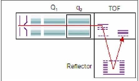
One common setup of the quadrupole mass analyzer is used in the
triple-quadrupole mass spectrometer (Yang L, 2002; Fig.2.8), in which three
quadrupoles are aligned with an ESI source. Ions of a specific m/z value are selected in the first quadrupole (Q1) and fragmented in the second, which is filled with a collision gas (es. Argon) and works as a collision chamber (MS/MS or CID experiments, Hunt DF. et al., 1986). The fragments are then analysed in the third quadrupole.
Fig.2.8 Triple-quadrupole mass analyzer
Ion trap analyzer can be aligned with both ESI and MALDI source
(Schwartz JC et al., 1996).It composed of a ring electrode and two end-cap electrodes (Fig.2.9). The pulses of an electrostatic ion gate inject ions into the ion trap. A combination of RF and DC voltages is applied to the electrodes to create a quadrupole electric field similar to the electric field for the quadrupole mass analyzer. Initially, the field within the trap is such that all ions that enter the analyzer begin a stable oscillation and are "trapped" in a potential energy well at the centre of the analyzer. Trapped ions are focused into the centre of the trap through the use of an oscillating potential
called the fundamental rf, applied to the ring electrode. An ion will be stably trapped depending upon its m/z value. The time during which ions are allowed into the trap, the "ionization period", is set to maximize signal while minimizing space-charge effects. Space-charge results from too many ions in the trap that cause a distortion of the electrical fields leading to an overall reduction in performance. The ion trap is typically filled with helium to a pressure of about 1 mtorr. Collisions with helium dampens the kinetic energy of the ions and serve to quickly contract trajectories toward the center of the ion trap, enabling trapping of injected ions. The mass spectrum is acquired by scanning the RF and DC fields to destabilize low mass to charge ions. These destabilized ions are ejected through a hole in one endcap electrode and strike a detector. The mass spectrum is generated by scanning the fields so that ions of increasing m/z value are ejected from the cell and detected.
Fig.2.9 Basic components of a typical ion trap mass analyzer.
Ion traps are especially suited to perform MS/MSn experiments It is possible to selectively isolate a particular m/z in the trap by ejecting all the other ions from the trap. Fragmentation of this isolated precursor ion can then be induced by CID experiments. The isolation and fragmentation steps can be repeated a number or times and is only limited by the trapping efficiency of the instrument.
The linear ion trap is based on the ion path of a triple quadrupole mass spectrometer in which Q3 is a linear ion trap with axial ion ejection (Fig.2.10). This arrangement allows de-coupling of precursor ion isolation and fragmentation from the ion trap itself. The result is a high sensitivity tandem mass spectrometer with triple quadrupole fragmentation patterns and no low mass cut-off, multiple-reaction monitoring (MRM),
as well as
precursor and constant neutral loss scanning (Hager JW et al., 2003;
Hopfgartner G. et al., 2004).
Fig.2.10 Linear Ion Trap mass analyzer
Fourier-transform ion cyclotron resonance mass spectrometry (FT-ICR-MS or just FT-(FT-ICR-MS) is probably the most complex method, has the highest
mass resolution (> 107) and is also useful for tandem mass spectrometry
experiments, of course it also very expensive (Zhang LK. et al., 2004). The standard arrangement for the analyser region, of the FT-ICR instrument, is an ion-trap located within a spatially uniform static magnetic field of strength B, that causes ions travel in a circular path (Fig.2.11). The cyclotron
frequency (ωc) of the ion's circular motion is determined by the magnetic
field strength (B) and the m/z value of the ion (ωc = Bz/2πm), this means that
mass. When the ions enter the ion trap pressure is in the range of 10-10 to 10-11 mBar with temperature close to absolute zero to minimize ion-molecule
reactions and ion-neutral collisions that damp the coherent ion motion. The signal is measured by placing electrodes on each side of the ions circular orbit.
Fig.2.11 FT-ICR-MS scheme.
A fast RF pulse is applied to the transmitter electrodes. Each individual excitation frequency will couple with the natural motion of resonant ions and excite them to a higher orbit where they induce an alternating current between the detector plates. The frequency of this current is the same as the cyclotron frequency of each m/z ion present and the intensity is proportional to the number of ions. The various frequencies and their relative abundances can be extracted mathematically by using a Fourier transform which converts a time signal (the image currents) to a frequency spectrum which is then converted to the mass spectrum (m/z = B/2πωc).
In tandem MS experiments, precursor ions are selected by applying an RF waveform to eject all other ions from the ion trap, so that only the precursors remain. These are accelerated to a higher kinetic energy by applying another RF pulse that increases the precursor ion's radius (kinetic energy). The accelerated ions are trapped for a period of several milliseconds or longer to allow them to collide with the inert gas. The collision gas is either introduced continuously or added with a pulsed valve. The product ions are
then excited into coherent motion with another RF pulse and the ion image currents are detected and transformed.
Time of Fligh (TOF) analyzer is conceptually the simplest spectrometer.
Applying a fixed voltage (V) at the source (ca. 20kV) the ionised gas-phase sample is accelerated to a fixed kinetic energy (Ek) and then guided into a
high-vacuum field-free flight tube, through which they reach the detector (Linear mode, Fig.13). The separation of ion species in the TOF occurs in agreement with the fundamental physical low of kinetic energy:
Ek = mv2 /2 = zV Ek= kinetic energy m = ion mass v = ion velocity z = number of charges V = source potential m/z = 2Vt2/L2 L = length of TOF t = time of flight
Because the flight tube is a field-free region, all the Ek of the ions results
from their initial acceleration, therefore a group of ions accelerated with the same constant voltage, at a fixed point and initial time, and allowed to drift in a field-free region, will traverse this region and separate in a time which depends upon their m/z ratios. Measuring that time-of-flight at the detector it is possible to determine the m/z value.
Earlier MALDI-TOF spectra were characterised by poor mass resolution due to the distribution of kinetic energy among ions that vary in initial desorption velocity (Beavis & Chait, 1991; Zhou et al.,1992) and in energy deficits from collisions within the plume of desorbed matrix during ion acceleration. In continuous ion extraction linear mode, there was no compensation for ions with the same m/z value but different initial velocity. To overcome this problem two improvements have been implemented in present TOF instruments configurations. Firstly, the events of desorption/ionization and acceleration has been separated by applying the acceleration field with a slight delay, up to hundreds of nanoseconds, relative to the laser pulse enabling the ions to be focused (Brown & Lennon, 1995; Vestal et al., 1995).
In the DE-mode, in fact, a variable voltage grid in front of the source (Fig.13) allows MALDI-generated ion cloud to expand in a transient
field-free region (grid has the same voltage of source). Then a fast high-voltage pulse (grid has slightly lower voltage than source) creates a potential gradient in the ionization region that accelerates slow ions more then fast ones. This pulse voltage and time delay corrects the initial velocity differences in such a way that identical m/z ions arrive simultaneously at the plane of the detector, enhancing resolution of the mass spectrum.
The second improvement was the introduction of a Reflectron (Mamyrin et
al.,1973) at the axial-end of the field-free drift region. Even applying
DE-mode, after ions are accelerated and velocity-focused (ions of the same mass align in time), they can defocus flying down the tube causing broadening of signal detector. The reflectron is an electrical mirror, with a voltage potential applied across the sides, which creates a retarding field at a voltage slightly higher than the accelerating one. The ions are sequentially slowed down through the reflector until they stop, turn around and re-accelerate back in a second drift region to a second detector (Reflector Detector, Fig.2.12). Ions species with a kinetic energy lower than the accelerating voltage will penetrate less in the reflector and will turn back sooner (as ions species with higher kinetic energy will penetrate deeper and turn back later) allowing ions of the same m/z value and slightly difference in flight times to be packed and focused in space and time to the detector.
Initial velocity Number
of ions
Velocity distribution of laser-desorbed ions
Sample plate Ground
grid Variable-voltage grid slow fast fast slow
Ions must line up at the beginning of the flight tube
Reflector Defocusing region
Reflector Detector
Linear Detector
Fig.2.12 Velocity focusing in reflectron mode.
Gas-phase ions generated in MALDI source may decompose before reaching the detector by prompt or metastable defragmentation (Spengler et al., 1991;
Karas et al., 2000). Prompt ion fragmentation occurs at the time of the desorption event within the source region and it is generally absent for MALDI generated peptide/protein ions. Metastable ion decay, instead, can be studied with modern MALDI-TOF instruments either using DE technique and linear TOF-MS (In-Source Decay) or, more widely, using the reflector device as an energy analyzer to performed a Post-Source Decay (PSD). "Post source decay" has been chosen as a rather instrumental term for any fragmentation phenomena relevant for MALDI-TOF instruments in the field free region, regardless of the location (metastable ion decay, low/high -energy collision induced dissociation) (Spengler B, 1997). Increasing gas pressure is a favourable method for enhancing high-energy Collision Induced Decay (CID). In typical PSD instruments, a complete product spectrum has to be acquired in several steps (> 10). This because the reflector is able to analyze energies (i.e. PSD ion masses) only within certain range with sufficient resolution. This requires the reflectron potential to be stepped down in order to focus lower m/z ions ( PSD ions has basically the same velocity but lower energy owing to their lower mass). Finally, all sections of the PSD spectrum have to concatenated by the computer and mass calibrated.
To allow the fragmentation of MALDI-generated precursor ions, MALDI sources have been recently coupled to quadrupole ion-trap mass spectrometers (Krutchinsky, 2001) and two types of TOF instruments: the quadrupole TOF hybrid instrument(QqTOF1; Loboda, 2001; Fig.2.13) and
the MALDI-TOF-TOF (Medzihradszky et al., 2000). A collision cell is inserted between the two analyzer. Ions of a particular m/z value are selected in the quadrupole mass filter or in the first TOF, fragmented in the collision cell and the fragments ions masses are read out by a TOF analyzer(Fig.2.14). These instruments have very high sensitivity, resolution, mass accuracy and high speed data acquisition.
During MS/MS analysis, MALDI-TOF-TOF instrument offers the possibility to finely control fragmentation conditions and simultaneously provides both low-energy and high-energy collision induced spectra.
The degree of fragmentation and structural information obtained are related to adjustment in laser intensity, collision gas pressure and collision energy. A collision energy of 1-2 keV in the CID-cell results in the production of low mass internal fragments, ions from amino acid side chain fragmentations, ions specific of particular amino acids increasing the
confidence in the peptide sequence interpretation and identification of multiple proteins from complex mixtures.
In recent years, MS has become one of the most important analytical techniques for the identification of proteins and for the analysis of post-translational modifications (PTM). As a result of its semplicity, excellent mass accuracy, high resolution and sensitivity, MALDI-TOF MS is much commonly used to identify proteins.
Proteolytic digestion of proteins into peptides and mass analysis of peptide mixture provides what is known as peptide mass fingerprint. Proteins are then identified by matching the list of experimental peptide masses with the theoretical fingerprints of all sequences in a comprehensive protein database. This method, also referred as peptide mapping, requires an essentially purified target protein and this is the reason why it is commonly used in conjunction with prior protein fractionation using high resolution two-dimensional Polyacrylamide Gel Electrophoresis (2D-PAGE).
Fig. 2.13. Quadrupole-TOF
V2 V1
Laser
Fig.2.14 MALDI TOF-TOF AB 4700 TOF/TOF™ Ion Optics
Source 1 Source 2 x1,y1 Deflectors Reflector Detector 2 Stage Reflector CID Cell Metastable Ion Suppressor Lens 1 Attenuator TIS TOF 2 x2,y2 Deflectors Sample Plate TOF 1 Linear Detector (optional) Deceleration Stack x3,y3 Deflectors Lens 2 Ion Sink
3. TWO- DIMENSIONAL ELECTROPHORESIS
Two-dimensional gel electrophoresis (2-DE) (O’Farrell 1975) so far is the only technique able to efficiently separate hundreds or even thousands of proteins or post-translational modified proteins.The high resolving power of 2-DE enables the separation of high number of protein species like present in a cell proteome.
Proteins are separated according to charge (isoelectric point, pI) by isoelectric focusing (IEF) in the first dimension and according to size (Mr) by SDS-PAGE in the second dimension.
Due to the zwitterionic character of proteins, the electrophoretic mobility of each protein is a characteristic value (pI).
Commercially available immobilized pH gradient gel strips (IPGs) for IEF have solved long time problems of pH gradient instability and irreproducibility (Bjellqvist et al., 1982). IPGs allow the generation of pH gradients of any desired range (broad, narrow or ultra-narrow) between pH 3 and 12 and since sample loading capacity of IPG-IEF is also high, 2-DE with IPGs is a method of choice for micro-preparative separation and spot identification. Anyway, the tremendous heterogeneity and significant differences in abundance of all proteins expressed in eukaryotic cells and tissue makes the analysis of complex cellular proteomes by 2-DE still a big challenge.
What is termed the "classic approach" in proteomics (Dreger, 2003) is characterized by a one-step sample preparation from a crude homogenate followed by 2-DE in order to display the whole map of expressed proteins within the studied system under given physiological conditions. This approach theoretically provides a complete overview of all proteins in the sample based on protein spot patterns. These patterns may be compared between two samples such as a control and an investigated system under specific conditions. These expectations were based on the assumptions of a 2-DE system capable of representing all proteins of the sample and not only for visualizing but also for identification (including their post-translational modifications). Despite the exceptional analytical power of this approach, systematic limitations at the present state of the technology became apparent. A fundamental question about proteomics is what percentage of a cellular proteome our technology can display.
The number of different proteins in a given time under defined biological conditions is likely to be in the range of several thousand for simple prokaryotic organisms and up to at least 10.000 in eukaryotic cell extracts. Current proteomic studies reveal that the majority of identified proteins are abundant housekeeping proteins that are present in numbers of 104- 106 copies per cell, whereas proteins in much lower concentrations, such as 100 copies per cell, are usually not detected. Consequently, improving methods for enrichment of low-abundance proteins, such as prefractionation procedures, as well as more sensitive detection and quantitation methods, are currently an area of intense investigation. ( for reviews Gorg et al., 2004; Molloy 2003; Stasyk, 2004; Lilley et al., 2002). Several prefractionation strategies (subcellular fractionation, affinity purification, fractionation of proteins and peptides according to their physicochemical properties) has been successfully applied to the study of well defined groups of proteins, like proteins of purified organelles, membrane proteins, or proteins isolated through characteristic PTMs (Wu & Yates, 2003; Mann & Yensen 2003; Dreger, 2003). Fractionation and sample enrichment can be fundamental first steps to increase analysis resolution , however none of the currently available proteomics techniques allows the analysis of an entire proteome. Technological developments of last years in proteomics field has resulted in the definition of several "-omics", like phosphoproteomics, membranomics, degradomics, immunoproteomics, complexomics, glycoproteomics, secretomics, etc. Together these fields cover the various aspects biological and pathological processes whose data and knowledge have to be fully integrate to a complete overview of a biological system.
4. HEPATITIS C VIRUS (HCV)
4.1 Epidemiology and history
The estimated worldwide prevalence of HCV infection is 2.2%, or approximately 130 million HCV-positive persons (Centers for Disease Control and Prevention [CDC], unpublished data, 2003). Region- or country-specific overall prevalence does not necessarily reflect the risk for spread of HCV because this risk is not homogenous either between or within most countries. Age-specific HCV prevalence rates are more variable, and their patterns provide clues to geographic and temporal differences in the risk for acquiring HCV infection Throughout the world, blood transfusion from infectious donors, unsafe therapeutic injection practices, and injection drug use have been the predominant modes of spread for hepatitis C virus (HCV) infection. In most developed countries, blood donor testing has virtually eliminated transfusion-related transmission of HCV. In these countries, injection drug use has been the major risk factor for HCV infection during the past 10 years, and the highest prevalence of infection is found in young to middle-aged adults. In many developing countries, unsafe therapeutic injection practices appear to be responsible for the geographic clustering of high rates of infection because of inadequate or nonexistent supplies of sterile syringes, administration of injections by non-professionals outside the medical setting (Higuchi, 2002).
Hepatitis C was first recognized as a separate disease entity in 1975 when the majority of cases of transfusion-associated hepatitis were found not to be caused by the only two hepatitis viruses recognized at the time, hepatitis A virus and hepatitis B virus. The disease was called "non-A non-B hepatitis," and it was demonstrated to be transmissible to chimpanzees
(Alter H.J. et
al., 1978; Tabor E. et al., 1978)
. It was not until 1989, however, that thecloning and sequencing of the viral genome of the non-A non-B hepatitis virus was first reported
(Choo et al., 1989)
and the virus was renamed "hepatitis C virus" (HCV).HCV shares genomic organization and slight sequence identity with flaviviruses and pestiviruses and it has been classified in a separate genus (Hepacivirus) within the Flaviviridae family (Francki et al., 1991).
HCV exhibits significant genetic heterogeneity as a result of the accumulation of mutation during viral replication. This high mutation rate, which is characteristic of RNA viruses, can be attributed to an error-prone
RNA-dependent RNA-polimerase that lacks proofreading activity. HCV circulates in an infected individual as a population of closely related, yet heterogeneous, sequences, defined as "quasispecies" (Forns et al., 1999). This genetic diversity is not evenly distributed in the viral genome. The non-coding regions are relatively conserved, while the envelope regions, especially Hypervariable region 1 (HVR1) located at the N-terminus of the envolope protein E2 (see later description of HCV genome), have the highest mutation rate. This genetic diversity resulted in the classification of HCV into at least 6 major genotypes (1 to 6) and many subtypes, based on the analysis of the NS5 region (Simmonds P, 1993). Different genotypes may differ by 30 to 35% of their viral genomes, while subtypes may differ by 15-20% of their nucleotide sequences.
4.2 Natural Course of Disease
Being infected with HCV does not necessarily mean that liver disease will occur and anyway it can take several years – decades, in many cases – for HCV to cause life-threatening liver disease.
Soon after HCV enters the body, it infects hepatocytes cells. Only a small number of people (approximately 25%) actually experience symptoms of infection, such as fatigue, decreased appetite, nausea, or jaundice (yellowing of the skin and eyes). However, almost all people infected with HCV experience an increase in their liver enzymes – such as serum alanine aminotransferase (ALT) – which can be detected by a simple blood test. An increase in ALT means that some liver cells are becoming damaged by the HCV infection.
Approximately 15% of people who are infected with HCV are able to clear the virus from their bodies, usually within six months after becoming infected. However, the majority of people (85%) who are infected with HCV have "chronic" hepatitis C – an infection that will stay with them for life. Of the 85% people with chronic hepatitis C, approximately 20% of them will remain healthy – their liver enzymes will stay normal, even though HCV can be detected in their livers and in their blood, and they will not go on to develop liver disease or experience symptoms of the infection. The remaining 60-65% people with chronic hepatitis C will go on to experience some signs and symptoms of liver disease, such as fatigue, nausea, muscle aches, and abdominal pain – usually after 13 to 15 years of being infected with HCV. Approximately 20-25% of these people, usually after 20 years of
Fig. 4.1 Natural courses of HCV infection.
Hepatitis
HCV infection, will develop cirrhosis – a scarring of the liver that results from widespread fibrosis (an extreme overgrowth of the liver's connective tissue). Although cirrhosis is not life threatening, it can affect the way the liver works and increases the risk of liver cancer. Of the 20-25% people with HCV who develop cirrhosis, 5-10% of them will develop liver cancer and possibly liver failure after another five years (Fig. 4.1).
4.3 HCV Genome and Polyprotein Processing
HCV is an enveloped virus with a 9.6-kb single-stranded RNA genome of positive polarity comprising a 5’ untranslated region (UTR), a large open reading frame (ORF) encoding a large polyprotein of about 3000 amino acids, and a 3’ UTR.
Translation of the viral RNA occurs through a cap-independent mechanism via an internal ribosomal entry site (IRES) located in the 5' UTR (fig. 4.2). A combination of cellular and virally encoded protease activities leads to processing of the polyprotein into individual structural and non-structural proteins (Lohmann et al., 1996). At the amino terminal of the polyprotein three structural proteins have been identified. The Core protein which forms the viral nucleocapsid (Yasui K, 1998) and shows
a number of
regulatory functions (Ray et al., 2001; Yao et al., 2001a)
and theC C Spontaneous resolution Chronic HCV infection Chronic
mild- moderate - severe
D Deeaatthh c caarrcciinnoommaa T Tiimmee((yyeeaarrss)) 15% 85% Acute 6 655%% 20% > CCiirrrrhhoossiiss 1-4% per year 1 1 1 100 222200 0 0 0 0 330 0
envelope proteins E1 and E2, which form noncovalent E1E2 heterodimers present at the surface of the HCV particles (fig.4.3), likely to mediate virus entry (Voisset et al., 2004). E1 and E2 are both highly glycosylated and supposed to be retained into host cell in the endoplasmic reticulum (Duvet S.
et al., 1998; Cocquette L. et al., 1999). Structural proteins are processed by a
still unknown cellular protease. Further proteolytic processing, by host peptidases, of the region between E2 and the non-structural proteins, yields a short hydrophobic peptide, p7. Its viral function is unknown but in host cell seems to be located on the Endoplasmic Reticulum (ER) and to function as a membrane channel (Carrère-Kremer, 2002; Pavlovic, 2003). The C terminal region of the polyprotein encodes for the non-structural (NS) proteins NS2, NS3, NS4A, NS4B, NS5A, and NS5B (fig.4.2).
NS2 and the amino-terminal domain of NS3 form the viral protease responsible for the autocatalytic cleavage at the NS2-NS3 junction (Reed et
al., 1995). Although the catalytic mechanism remains elusive, several studies
have been reported on the NS2/3 protease suggesting it is zinc-dependent with putative cysteine and histidine catalytic residues (Reed et al.,1995; Pieroni et al., 1997). Following proteolytic cleavage events, involved in the maturation of the NS proteins, are carried out by the major viral serine protease, NS3, in association with its cofactor, NS4A (Bartenschlager et al., 1994 and1995). In addition to its function as a protease, NS3 also contains helicase and nucleoside triphosphatase activities within its C-terminal domain (Kim et al., 1995). Specific replicative functions have not been identified for NS4B and NS5A, but both are likely to contribute to the viral replicase complex. NS3/4A and NS5A appear to play important roles in modulating antiviral responses within infected cells and in promoting long-term persistence of the virus (Foy et al., 2003; Lan et al., 2002; Gale et al., 1998; Pawlotsky et al., 1999). NS5B constitutes the HCV RNA-dependent RNA polymerase and is thus the catalytic core of the replicase (Lohmann et
al., 1997). It belongs to a class of integral membrane proteins termed
tail-anchored proteins and its membrane association is essential for hepatitis C virus RNA replication (Moradpour et al., 2004).
Fig.4.2 HCV genome processing and cleavage products of the polyprotein
p21 gp31 gp70 p7 p21 gp70 p8 p27 p58 p68 IRES
HCV 9.6-kb positive single-stranded RNA genome comprising a 5’ untranslated region (UTR), a large ORF of encoding all viral proteins, and a 3’ UTR. 5’ UTR contains the IRES which mediates cap-independent translation initiation and is necessary for efficient HCV replication. The functions of the gene products produced after co- and posttranslational polyprotein processing by host signal peptidases and HCV-encoded proteases are indicated in the Figure. The 3’ UTR contains a poly (U/UC) tract immediately following the ORF stop codon and conserved RNA elements essential for HCV replication.
4.4 HCV Life Cycle
HCV normally replicates into hepatocytes cytoplasm, but low efficiency viral replication into Peripheral Blood Mononuclear Cells (PBMC) has also been observed (Cribier et al., 1995; Fournier et al., 1998).
The lack of efficient cell culture system and a convenient animal model has been majors roadblocks for unrevealing the details of HCV replication. Based largely on analogies to the other members of the Flaviviridae family, the life cycle of HCV (Bartenschlager, 2000) can be summarized as follows (Fig.4.4): (1) penetration of the host cell and liberation of the genomic RNA from the virus particle into the cytoplasm; (2) RNA translation, processing of the polyprotein and formation of a replicase complex associated with intracellular membranes; (3) utilization of the input plus-strand for synthesis of a minus-strand RNA intermediate; (4) production of new plus-strand RNA molecules which in turn can be used to synthesize new minus strands, for polyprotein expression or packaging into progeny virions; (5) release of virus from the infected cell.
Virus entry. Several candidate receptors for HCV entry have been reported
over the past years. Based on the observation that HCV RNA-containing particles are often associated with low-density or very-low-density lipoproteins and that these low-density fractions are infectious for chimpanzees, the low density lipoprotein receptor (LDLr) has been proposed LDLr to mediate uptake of HCV particles (Agnello et al., 1999). CD81, a cell surface tetraspanin capable of selective binding E2 glycoprotein, was identified (Pileri et al., 1998) and later the scavenger receptor B1 (Scarselli
et al., 2002).
Recently, infectious HCV pseudotype particles (HCVpp) that are assembled by displaying unmodified HCV envelope glycoproteins on retroviral core particles have been successfully generated (Bartosch, 2003). They mimic the function of native HCV particles, thus representing a model to study the early steps of its life cycle. The noncovalent E1E2 heterodimers present at the surface of the HCVpp, which contain complex-type glycans indicating modification by Golgi enzymes, are likely to mediate virus entry and the CD81 with SR-BI are required for HCVpp entry (Voisset, 2004).
However, these two proteins are not sufficient to provide entry functions in non permissive cells and their widespread distribution does not explain the hepatocyte tropism of HCV, suggesting that additional unidentified cellular factor(s) are necessary for HCVpp entry.
Attachment and entry
R
RNNAA((++))
Fig.4.4 Hypothetical model of the HCV replication cycle. Upon infection of the host
cell the (+)-strand RNA genome is liberated into the cytoplasm and translated. The polyprotein is processed and viral proteins remain tightly associated with membranes of the ER. (-)-strand RNA is synthesized by the replicase NS3–5B complex and serves as template for production of excess amounts of plus strand. Via interaction with the structural proteins (+)-strand RNA is encapsidated. Particles are enveloped by budding into the lumen of the ER and virus particles are exported via transit through the Golgi complex.
Translation of the incoming genome RNA is mediated by a highly
conserved RNA element that functions as an internal ribosome entry site (IRES) capable of directly binding to initiation factor eIF3 and the 40S ribosomal subunit (Ji et al., 2004; Otto et al., 2004). Directed by the IRES, the polyprotein is translated at the rough endoplasmic reticulum (ER) and cleaved co- and post-translationally, into 10 mature proteins by the action of host cell and viral proteinases. The order and nomenclature of the HCV proteins is: NH2-C-E1-E2-p7-NS2-NS3-NS4A-NS4B-NS5A-NS5B-COOH Processing of the polyprotein is essential to form an active HCV RNA replicase. The HCV RNA replication machinery, or “replicase,” is cytoplasmic and membrane-associated (Dubuisson et al., 2002; El-Hage&
Nucleus Endoplasmic Reticulum 5 5’’ 33’’ G Goollggii 3 3’’ RRNNAA((--)) 55’’ translation HCV and processing release RNA replication E E11 E E22 E E11 E E22 P P77 C C C C R Reepplliiccaassee N NSS33 Virion assembly N NSS55AA N NSS55BB N NSS44AA N NSS44BB
Luo, 2003) consisting of at least NS3-NS5B, together with additional, as yet undefined, host components. The major enzymatic components of the HCV replicase are the NTPase/helicase activity of NS3 and the NS5B RNA-dependent RNA polymerase (RdRP). The first step in the RNA replication process involves the synthesis of a complementary negative-strand copy of the incoming genome RNA (Fig.4.5).
Although confusion exists in the literature, it is generally believed that the minus-strand synthesis occurs by a primer-independent de novo initiation mechanism (Luo et al., 2000; Zhong et al., 2000). The negative-strand template is then used for synthesis of additional genome-length RNAs that can in turn be used for translation, replication, or packaging into progeny virus.
Assembly and release. In the absence of systems allowing the production
virus particles, the assembly assembly and release of HCV cannot be studied in detail. Based on results with other members of the Flaviviridae, it seems likely that HCV buds into intracellular vesicles and is transported out of the cell by the host secretory pathway and released into the extracellular space.
Fig.4.5 HCV Replication Complex. Non-structural HCV proteins in Huh7 cells were
colocalized to the perinuclear ER membranes carrying a replicating HCV RNA (Gosert et al., 2003).
4.5 Cell-based model systems for HCV replication investigation
Investigation of HCV replication mechanisms and the cellular consequences of HCV infection have been significantly limited by the absence of an efficient in vitro infection-replication system, as well as the lack of a suitable small animal model. However, a major breakthrough came in 1999 when Lohmann and colleagues (Lohmann et al., 1999) described engineered subgenomic HCV RNAs, or “replicons,” that were capable of limited replication in a human hepatoma Huh-7 cell line (Fig 4.6). RNA replicons are defined as HCV RNA molecules that are capable of autonomous replication in an appropriate cell type. Based on the assumption that high expression levels of the structural proteins might be cytotoxic (Moradpour et
al., 1998) and the observation that for flavi- and pestiviruses the structural
proteins are not required for RNA replication (Behrens et al., 1998; Khromykh &Westaway, 1997), the sequences of the structural proteins were deleted. To allow selection for only those cells in which HCV efficiently replicates, the gene encoding the neomycin phosphotransferase, conferring resistance to the antibiotic G418, was introduced downstream of the HCV IRES (Fig.4.7). A second IRES element, derived from the picornavirus encephalomyocarditis virus, was included to allow translation of the HCV NS proteins.
These autonomously replicating RNAs expressing only NS proteins could initiate and maintain replication in the human Huh-7 hepatoma cell line but just at low frequency. Diverse spectrum of mutations (mostly in NS5A region but also in NS3, NS4B, and NS5B) was later identified to enhance the ability of HCV (genotype 1b) to replicate in Huh-7 cells, as well as genome-length, selectable HCV RNAs that express all of the viral proteins (Lohmann
et al., 2003, Lohmann et al., 2001, Krieger et al., 2001, Blight et al., 2000,
Ikeda et al., 2002, Pietschmann et al., 2002).
Fig.4.7 HCV-replicon-containing cell lines. RNA constructs containing the gene encoding
neomycin phosphotransferase (neo) upstream of the HCV genes are transfected into Huh7 cells. The IRES of HCV drives translation of neo, and the encephalomyocarditis virus (EMCV) IRES directs HCV polyprotein translation. Transfected cells (grey) are selected by exposure to neomycin sulfate (G418) so that only those expressing neomycin phosphotransferase (green), and thus replicating HCV, survive.
These RNAs replicate efficiently and stably under selective pressure in Huh7 cells, however, their replication is strongly influenced by the proliferation status of the host cells, with the abundance of viral RNA rapidly declining when cells reach high density (Pietschmann et al., 2001). The mechanism that underlies this requirement for host cell proliferation has not been identified yet, but it is possible that HCV RNA replication is more permissive in certain phases of the cell cycle than in others. It is likely that in well-established cell clones supporting HCV RNA replication, a balance is reached between HCV RNA degradation and S-phase-stimulated new synthesis, resulting in stable, homeostatic HCV RNA expression throughout the cell cycle (Scholle et al., 2004) .
Although little is known about the mechanism underlying cell culture adaptation and the role of host cells factors required for HCV replication (Huh-7 hepatoma cells still are the only cell line supporting efficient replication of HCV RNAs) replicon-containing cells have provided a source of active replicase for biochemical studies and a significant advance in the study of HCV biology.