TWO REMARKABLE FOSSIL INSECT LARVAE FROM BURMESE AMBER SUGGEST THE PRESENCE OF A TERMINAL FILUM IN THE DIRECT STEM LINEAGE OF DRAGONFLIES AND DAMSELFLIES (ODONATA)
MARIO SCHÄDEL1*, PATRICK MÜLLER2 & JOACHIM T. HAUG1,3
1*Corresponding author. Department of Biology, Ludwig-Maximilians-Universität München, Großhaderner Str. 2, 82152 Planegg-Martinsried,
Germany. E-mail: [email protected]
2Friedhofstr. 9, 66894 Käshofen, Germany. E-mail: [email protected]
3GeoBio-Center of the LMU Munich, Richard-Wagner-Str. 10, 80333 Munich, Germany. E-mail: [email protected]
To cite this article: Schädel M., Müller P. & Haug J.T. (2020) - Two remarkable fossil insect larvae from Burmese amber suggest the presence of a terminal filum in the direct stem lineage of dragonflies and damselflies (Odonata). Riv. It. Paleontol. Strat., 126(1): 13-35.
Abstract. The fossil record of dragonfly relatives (Odonatoptera) dates back to the Carboniferous, yet knowl-edge about these extinct animals is meagre. For most of the species little is known except for the characteristics of the wing venation. As a result, it is difficult to include fossil larvae in a (wing character based) phylogenetic tree as the wing venation is not visible in most of the larval instars.
Two larval specimens from Cretaceous Burmese amber are in the focus of this study. The two specimens likely represent two subsequent early stage larval instars of the same individual. Not only is this an exceptional case to study ontogenetic processes in fossils – the larval instars are morphologically completely different from all known larvae of Odonata with respect to the posterior abdominal region. Therefore, besides the difficulties regarding the phylogenetic interpretation and though all Burmese amber odonatans are known from adults only, a new species –
Arcanodraco filicauda n. sp. – is formally described.
Aside from likely representing a new species, the morphology of the posterior abdominal region is highly informative for reconstructing the character evolution within the lineage towards modern dragonflies and damselflies. A long median process in both of the fossils meets all criteria to be interpreted as a terminal filum (structure or deriva-tive of tergite of abdominal segment 11, annulated in one of the specimens). Although the exact phylogenetic affinity of Arcanodraco filicauda n. sp. remains enigmatic, the presence of a larval terminal filum can be reconstructed for the
ground pattern of Odonatoptera (including its direct stem lineage).
Received: February 21, 2019; accepted: July 31, 2019
Keywords: character evolution; Cretaceous; moult; Myanmar; Odonatoptera; ontogeny.
I
ntroductIonDragonflies and damselflies (Odonata) are widely known and – for a group of insects – suffer surprisingly less from cultural disgust. This may be due to the fact that they are easily distinguishable from other groups of Insecta and Euarthropoda and are usually categorised as 'beautiful'. Also they are generally considered to be more useful than
harmful due to their predatory lifestyle, factually controlling possible pests.
As the vernacular names suggest, the major-ity of odonatans comes from two natural groups: Zygoptera (damselflies) and Anisoptera (dragon-flies). Additionally there is a small number of extant species that are not part of these two large natural groups, but from a small monophyletic relic group (Epiophlebiidae: Epiophlebia) that is more closely related to Anisoptera than to Zygoptera (Bechly 2003; Blanke et al. 2015; Dijkstra et al. 2013; Rehn 2003). Epiophlebiidae and Anisoptera form the
group Euepiproctophora (Epiproctophora does also combine both groups, but is less precise be-cause it additionally includes an earlier branching off extinct lineage) (Bechly 1998b).
Both adult and larval odonatans are preda-tors. However, the feeding mechanism drastical-ly changes when odonatans become adult. Adults dragonflies and damselflies are fast moving pred-ators who catch their prey during flight using their thoracic legs. Odonatan larvae are, with little excep-tions, aquatic and use their highly specialized labi-um to catch prey as ambush predators ('sit-and-wait' predators).
M
orphologyofextantlarvaeThe labium, or lower lip (factually conjoined appendages of the fifth post-ocular body segment) of odonatan larvae differs substantially from that of other insect groups. The typical median enditic protrusions of the labium, also called glossae and paraglossae (ligula), are not distinctly developed, yet a distal median notch, indicating the contact point of their remnants, is often visible. The labial palps, in other insects small but leg-like-appearing
struc-tures most often made up of three to five separate elements, form here distinct hooks (in most species) which are used to pierce into the prey. The proximal region of the labium is here deeply divided into two separate elements: the prementum (distal) and the postmentum (proximal, mentum + submentum). Both are elongated and linked by a robust distinct joint. The distal end of the labium containing the hooks of the palps can be extended anteriorly very quickly by a complex interaction of muscles and resilin fibers (Büsse 2018). In some lineages the prementum and its associated structures is spoon-shaped and covers the ventral part of the head. Consequently, the labium of odonatan larvae is of-ten referred to as ‘labial mask’ or ‘raptorial mask’.
While all odonatan larvae share this labial mask, larvae of damselflies (Zygoptera) can be easi-ly differentiated from larvae of dragonflies (Anisop-tera) and Epiophlebia. Damselfly larvae are usually relatively slender and have three long posterior ab-dominal processes. Dragonfly larvae and also those of Epiophlebia are more stout and have shorter pos-terior abdominal processes, usually pressed tightly together to form a so-called 'anal pyramid'.
In Zygoptera the posterior abdominal pro-cesses are often used as functional gills (Sesterhenn et al. 2013), but also for locomotion (Burnside & Robinson 1995; Robinson et al. 1991; Sesterhenn et al. 2013). Within Zygoptera there is much vari-ation regarding the morphology of these posterior abdominal processes. The form and orientation of elongated posterior abdominal processes varies be-tween groups and species; some lineages have even developed balloon-shaped processes (Kalkman et al. 2010). Also the complete reduction of the median posterior abdominal process has been reported (Xu 2015). Many groups with leaf-shaped posterior ab-dominal processes have one or more predetermined breaking points along these processes which reduce the negative impact of unsuccessful moulting (not getting stuck in the exuvia/not losing a complete gill) (Corbet 1950).
Although many zygopterans have posterior abdominal processes with a high surface area (suita-ble for subaquatic respiration), rectal respiration (the main respiratory mechanism in anisopteran larvae) was also shown to be present in zygopteran larvae (Miller 1993). This indicates, that the rectal respira-tion is likely to be present in the ground pattern of Odonata.
Fig. 1 - Relationships between groups within Odonata. Illustrations of the adult habitus (top row) and the larval morphology of the posterior abdominal region of the abdomen in dor-sal view (middle row, extant species). The drawing in the bottom row is a reconstruction of the same body region in Isophlebioptera (based on Karatawia sibirica Pritykina, 1985)
In some lineages of Zygoptera also further, putatively respiratory structures are present. Some species of Amphipterygidae have gill tufts on the 10th abdominal segment (Watson 1966);
Pseudole-stes mirabilis Kirby, 1900 (Megapodagrionidae) has ventral gill tufts on the posterior end of the abdo-men (Yu & Bu 2011); some species of Epallagidae and Polythoridae have simple or subdivided lateral processes at the abdominal segments 2–7 (Watson 1966).
The median posterior abdominal process ('epiproct' or 'dorsal appendage') is apparently an protrusion arising from the indistinct tergite of the 11th abdominal segment and is therefore often considered to represent the tergite of this segment (Matsuda 1976). The lateral posterior abdominal processes (often referred to as 'paraprocts' or 'lat-eral appendages') are sternal structures of the 11th abdominal segment (Matsuda 1976). True cerci (parts of appendages of the abdominal segment 11) occur in many odonatan lineages during the post-embryonic larval development (Matsuda 1976).
o
ntogenetIcdevelopMentTo understand the morphology in fossils that may by chance represent distant relatives of modern species it is important to analyse the on-togenetic development in the studied group. This is necessary because many evolutionary changes that result in morphological differences happen by het-erochronic events (e.g. Wagner et al. 2017).
The first instar larva of odonatans is very distinct from the subsequent instars. Due to this morphological distinctness often the term ‘pro-larva’ has been applied (see a recent summary of the confusion of terminology of larva and related terms in Haug, in press). Although there is little doubt on the larval nature of this ontogenetic stage there is some confusion whether the subsequent instar should be referred to as 'first' or 'second lar-va' (see Corbet 2002 for a discussion). We herein use the term ‘prolarva’ synonymous to ‘first larval instar/stage’, consequently we refer to the stage succeeding the prolarva as the ‘second instar’.
The second instar larvae of extant odonatans resemble the later larval stages in their posterior abdominal morphology. This also allows to
dis-tinguish between larvae of zygopterans and eue-piproctophorans (Anisoptera and Epiophlebiidae). In zygopterans the posterior processes are more roundish in cross section in early instars compared to later instars (MacNeill 1960; Tillyard 1917).
The larval cerci (11th abdominal segment) develop into the so-called 'superior appendages' in adult males and into the 'anal appendages' in adult females (Matsuda 1976). The median posterior ab-dominal process becomes completely reduced in adults or may be the precursor of the 'inferior ap-pendage' in male anisopterans (the so-called 'infe-rior appendages' are generally considered to be not homologous between Zygoptera and Anisoptera) (Matsuda 1976).
f
ossIl recordDragonflies have often been regarded as an evolutionary very old group. However, this percep-tion is mainly based on a misunderstanding. There is a variety of Paleozoic fossils, of which many of are represented by isolated wings only, that are branching off the lineage towards modern odona-tans. The earliest ingroup representatives of Odo-nata (the group comprising all modern odonatop-teran subgroups) are from the Middle Triassic. The earliest fossil records for ingroup representatives of the groups Anisoptera (Middle Jurassic) and Zy-goptera (Upper Jurassic) are even younger (Fleck et al. 2001; Kaur Kohli et al. 2016).
Nonetheless, the earliest odonatopteran fos-sils, i.e. fossils branching off the lineage towards Odonata earlier, are much older, from the Missis-sippian-Pennsylvanian transition (Carboniferous) (Nel et al. 2018; Petrulevičius & Gutiérrez 2016). These early fossil non-odonatan odonatopterans include some iconic large species (Meganisoptera, 'griffenflies') (Carpenter 1939).
Some confusion exists upon the term 'Ani-sozygoptera' which has been often applied for odonatans that are neither ingroup species of Zy-goptera nor Anisoptera (Dijkstra et al. 2013). Epio-phlebia is the only extant group that falls into this category. However, 'Anisozygoptera' is only mono-phyletic if the fossil species are neglected – and if the fossil species are neglected the name is redun-dant with respect to Epiophlebia; it could there-fore be abandoned.
For some reasons (size, behaviour, etc.), drag-onflies and damselflies rarely occur in amber and of-ten the amber specimens are only fragmentarily pre-served. Taking into account the comparatively low potential of compression fossils (that make up most of the remaining fossils of dragonflies and damsel-flies) to preserve complex morphological details, it is not surprising that most fossil species have been described solely based on the wing morphology.
f
ossIl larvaeEven less is known about fossil odonatopteran larvae though they can be very abundant in lake sediments concerning the number of preserved individuals (Rasser et al. 2013). Larvae from lake sediments are often highly compressed leaving little information about their original three-dimensional organization. Only a few larval amber odonatans have been described so far from Baltic amber (Bechly & Wichard 2008). The labial mask is, besides the venation of wing pads in late larval instars, the main diagnostic feature of dragonfly and damselfly larvae. Dragonfly-like specialisations of the labium can seemingly be dated back to the Pennsylvanian (Kukalová-Peck 2009).
A distinct morphology of the posterior ab-dominal region, that frequently occurs in the fossil record, is represented by the (problematic) form-ge-nus 'Samarura'. Included species have three similar, broad, roundish, leaf-like posterior abdominal pro-cesses. This type of morphology of posterior ab-dominal processes can be dated back to the Late Triassic (Rozefelds 1985).
B
urMeseaMBerBurmese amber has recently gained a lot of attention, with over 700 species of Euarthropoda having been described so far (Ross 2018). The fossil-ized resin from the Kachin state in Myanmar is one of the oldest ambers that are commercially available. Burmese amber is Cenomanian (99 million years) in age (Shi et al. 2012). The presence of some marine organisms within the amber indicates a near shore depositional environment (Smith & Ross 2018; Xing et al. 2018). Experimental studies on the taphonom-ic potential of aquattaphonom-ic organisms in resin (Schmidt & Dilcher 2007) and traces of movement of still
liv-ing euarthropodan individuals from Chiapas amber (Serrano-Sánchez et al. 2015) strongly indicate that animals can be embedded in liquid resin that is sub-merged in water. Yet, also wind and spray are pos-sible factors for the embedment of smaller aquatic organisms in amber (Schmidt et al. 2018).
The Burmese amber palaeo-habitat was also home to well over 30 species of dragonflies and damselflies (Huang et al. 2019a, 2019b; Ross 2018; Zheng et al. 2018, 2019). With the exception of two species (Burmaphlebia reifi Bechly & Poinar, 2013 & Burmastenophlebia flecki Huang et al., 2018) all species are either ingroup zygopterans or ingroup anisop-terans (true damselflies and dragonflies) (Bechly & Poinar 2013; Huang et al. 2019b; Ross 2018; Zhang 2017). Burmaphlebia reifi and Burmastenophlebia flecki are both euepiproctophorans (ingroup odonatans that are closer related to dragonflies than to dam-selflies) (Bechly & Poinar 2013; Huang et al. 2019b). Zhang (2017) figures larval specimens of ingroup zygopterans and anisopterans, yet without present-ing details of a proper description of the specimens (see Tab. 1). There are further, not yet published, odonatopteran larvae from Burmese amber in the collection of the Nanjing Institute of Geology and Palaeontology (Dijing Huang, pers. comm. 2019).
p
alaeopteraproBleMThe lineage towards modern dragonflies and damselflies is one of the basal-most branchings in the tree of winged insects (Pterygota) – meaning that the lineage was one of the earliest to branch off from other lineages of winged insects. The early branching events in Pterygota (winged insects) are probably one of the most heavily discussed areas of insect phylogeny (often referred to as the 'Pal-aeoptera problem' (Hovmöller 2002)). The majori-ty flying insects share a special kind of wing joint (ability to fold wings) and are thus thought to have a common ancestor (stem species) in which this joint evolved once (Neoptera). The monophyly of Neoptera is also well supported by molecular data (e.g. Misof et al. 2014). The wing joint in Neoptera is often referred to as ‘neopterous’ in contrast to the ‘palaeopterous’ wing joint in mayflies (Ephemerop-tera), dragonflies and damselflies (together Odona-ta). Both terms must be seen as a functional rather than a systematic terms due to the difficult
polar-ity of characters (absence of a winged outgroup) (Hörnschemeyer & Willkommen 2007).
With Neoptera being a monophyletic group, there are virtually three ways to assemble neopterans and the two lineages with extant representatives of non-neopteran winged insects. These three topolo-gies are usually addressed by the proposed names of the closest sistergroup relations.
Palaeoptera Martynov, 1923 (Martynov 1923): Ephemeroptera + Odonata
Metapterygota Börner, 1909 (Börner 1909): Odonata + Neoptera
Chiastomyaria Schwanwitsch, 1943 (Schwan-witsch 1943): Ephemeroptera + Neoptera
Palaeoptera would comprise the two lineages with the 'old' wing joints (no ability to flex the wings
over the posterior trunk). This condition is often re-ferred as being plesiomorphic in contrast to the con-dition in neopterans. Yet, this assumption about the polarity of the character is problematic as there are no suitable outgroups for comparison. The larvae of both 'palaeopteran' lineages are aquatic (Hennig 1969) and the sistergoup relationship between may-flies and dragonmay-flies and damselmay-flies is supported by characters of the head morphology (Blanke et al. 2013, 2012), but also from molecular data (e.g. Mi-sof et al. 2014, '1KITE', however with low support values).
Chiastomyaria is characterised by direct sperm transfer (Boudreaux 1979) that is present in mayflies and neopterans, but not in dragonflies or damsel-flies. This relationship also received considerable support by phylogenomic data (Simon et al. 2018).
Metapterygota refers to the sistergroup re-lationship between Odonata and Neoptera. Adult moulting is most likely present in the ground pattern
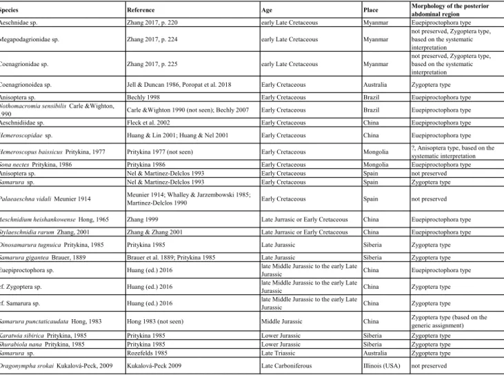
Tab. 1 - List of known palaeo- and mesozoic species (and important specimens) of odonatopteran nymphs (species described from isolated
wing pads are not included).
Tabelle2
Species Reference Age Place Morphology of the posterior abdominal region
Aeschnidae sp. Zhang 2017, p. 220 early Late Cretaceous Myanmar Euepiproctophora type Megapodagrionidae sp. Zhang 2017, p. 224 early Late Cretaceous Myanmar not preserved, Zygoptera type, based on the systematic
interpretation
Coenagrionidae sp. Zhang 2017, p. 225 early Late Cretaceous Myanmar not preserved, Zygoptera type, based on the systematic interpretation
Coenagrionoidea sp. Jell & Duncan 1986, Poropat et al. 2018 Early Cretaceous Australia Zygoptera type Anisoptera sp. Bechly 1998 Early Cretaceous Brazil Euepiproctophora type
Nothomacromia sensibilis Carle &Wighton,
1990 Carle &Wighton 1990 (not seen); Bechly 2007 Early Cretaceous Brazil Euepiproctophora type Aeschnidiidae sp. Fleck et al. 2002 Early Cretaceous China Euepiproctophora type
Hemeroscopidae sp. Huang & Lin 2001; Huang & Nel 2001 Early Cretaceous China Euepiproctophora type
Hemeroscopus baissicus Pritykina, 1977 Pritykina 1977 (not seen) Early Cretaceous Mongolia ?, Anisoptera type, based on the systematic interpretation
Sona nectes Pritykina, 1986 Pritykina 1986 Early Cretaceous Mongolia Euepiproctophora type
Anisoptera sp. Nel & Martinez-Delclos 1993 Early Cretaceous Spain not preserved
Samarura sp. Nel & Martinez-Delclos 1993 Early Cretaceous Spain Zygoptera type
Palaeaeschna vidali Meunier 1914 Meunier 1914; Whalley & Jarzembowski 1985; Martinez-Delclos 1990 Early Cretaceous Spain not preserved
Aeschnidium heishankowense Hong, 1965 Zhang 1999 Late Jurrasic or Early Cretaceous China Euepiproctophora type
Stylaeschnidia rarum Zhang, 2001 Zhang & Zhang 2001 Late Jurrasic or Early Cretaceous China Euepiproctophora type
Dinosamarura tugnuica Pritykina, 1985 Pritykina 1985 Late Jurassic Siberia Zygoptera type
Samarura gigantea Brauer, 1889 Brauer et al. 1889; Pritykina 1985 Late Jurassic Siberia Zygoptera type
Euepiproctophora sp. Huang (ed.) 2016 late Middle Jurassic to the early Late Jurassic China Euepiproctophora type cf. Zygoptera sp. Huang (ed.) 2016 late Middle Jurassic to the early Late Jurassic China Zygoptera type cf. Samarura sp. Huang (ed.) 2016 late Middle Jurassic to the early Late Jurassic China Zygoptera type
Samarura punctaticaudata Hong, 1983 Hong 1983 (not seen) Middle Jurassic China Zygoptera type (based on the generic assignment)
Karatwia sibirica Pritykina, 1985 Pritykina 1985 Lower Jurassic Siberia Zygoptera type
Shurabiola nana Pritykina, 1985 Pritykina 1985 Lower Jurassic Siberia Zygoptera type
Samarura sp. Rozefelds 1985 Late Triassic Australia Zygoptera type
Dragonympha srokai Kukalová-Peck, 2009 Kukalová-Peck 2009 Late Carboniferous Illinois (USA) not preserved
of Dicondylia (Zygentoma and Pterygota). Mayflies are the only pterygotan ingroup with two instars that have the ability to fly actively (‘subimago’ and ‘ima-go’). Though the genitalia are fully developed in the pharate subimago, the subimago never procreates, but moults to the imago after a short time (this can hardly be termed adult moulting). The reduction of this (sub-) adult moulting is generally thought to be apomorphic for Metapterygota (Kristensen 1981) along with character states of the mandible muscu-lature (Staniczek 2000).
The presence of the terminal filum is also sometimes included in phylogenetic analyses (e.g. Beutel & Gorb 2006). However, as discussed by Klass (2009) this is problematic because it was shown (Bechly et al. 2001) that in one Carboniferous odonatopteran a structure comparable to the termi-nal filum is present (see discussion below).
In this study we present new information about the character evolution of the terminal filum within the odonatopteran lineage based on one of the so far oldest larvae preserved in amber. Two lar-vae are described, presumably representing two sub-sequent instars of the same individual.
Methods
Material. The amber piece containing the fossils is currently
held in the collection of Patrick Müller (Zweibrücken) under the col-lection number BUB2823. Export permits are available upon request.
Documentation methods. Micrographic images were
re-corded using two microscope setups. A Keyence VHX-6000 digital microscope was used for standard incident light microscopy - in-cluding the use of cross polarized light (Bengtson 2000; Haug et al. 2013) and stereoscopic photography (Hörnig et al. 2016; Schädel et al 2019). To reduce surface reflections the amber piece was partly wet-ted with a drop of water and covered with a small glass cover slide. For the images resulting from that setup in most cases the included focus-'stacking' method (factually producing a stack of images and fusing it digitally to one sharp image, so below) was applied (compare Haug et al. 2011).
For fluorescence micrographs a Keyence BZ-9000 epi-fluo-rescence inverted microscope was used. Incident light with a median wavelength of 470 nm (generally used for green-fluorescent-protein markers, 'GFP' filter cube) was used to excite the samples. For the inverse epi-fluorescence microscopy the amber piece was completely submerged in water.
Focus stack processing was done using CombineZP (Alan Hadley, GPL) and EnfuseGUI (Enfuse, GPL). Panoramic alignment was done in TrakEM (Fiji, GPL). Further post-processing of the images, including contrast and colour optimisation, was done with GIMP (GPL) and also the 3D red-cyan stereo anaglyphs were created in GIMP. All drawings (including the colouration) were prepared in Inkscape (GPL). Measurements were made from scaled 2D images in ImageJ (Fiji, GPL).
Terminology of abdominal structures. Some terms,
re-ferring to the structures in the posterior abdominal region of odona-tans, are commonly applied in the literature, but are problematic from a theoretical aspect, especially when these structures are compared to corresponding structures in different animal groups. The terms 'epiproct' and 'paraproct' should refer to the unpaired supraanal lobe and the paired subanal lobes (periproct). However, 'epiproct' and 'paraproct' are widely used for posterior abdominal structures of odonatopterans. Median and lateral posterior abdominal processes are not derivatives of the periproct but are simultaneously present in larval odonatans (Matsuda 1976).
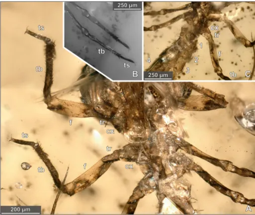
Neither the median nor the lateral posterior abdominal pro-cesses are derivatives of appendages. Consequently, we refrain to use Fig. 2 - Holotype and paratype of Arcanodraco filicauda n. sp., BUB-2823, specimen 1 (paratype) and specimen 2 (holotype, see numbers in the
figure). A) Overview image with specimen 2 (holotype) in ventral view, white light microscopy. B) Overview image with specimen 2 (holotype) in dorsal view, white light microscopy.
the term 'appendage' because it is often used to address arthropod appendage-derived body parts. Also we did choose not to call the posterior abdominal processes 'gills' because the respiratory function cannot be deduced simply by the assumed homology of the struc-tures in the fossils to posterior abdominal processes in extant zy-gopteran larvae for which the respiratory function is suggested by experimental studies (Sesterhenn et al. 2013).
r
esultsDescription and Measurements
The fossils treated in this study are inclusions within a single piece of Burmese amber. The am-ber piece is about 17 mm long at its greatest extent. Two similar small arthropodan fossils are located near the centre of the amber piece and are in close proximity to each other (Fig. 2).
Description of specimen 1 (dark, smaller) Body organisation: The body is organised in a head and a trunk. The trunk is further subdivided into a thorax (bearing the walking appendages) and an abdomen (without prominent externally visible appendages).
Head: The shape of the head can be described as a compressed sphere, being longer in antero-pos-terior axis than in dorso-ventral axis. The head
makes up about one fifth of the overall length of the specimen. The head is distinctly larger in dor-soventral dimension than the posterior-subsequent (thoracic) segments. With prominent large com-pound eyes (Fig. 5A–D) occupying most of the lat-eral side of the head.
Thorax: The thorax comprises three segments (prothoracic, mesothoracic and metathoracic seg-ment). The thorax segments are markedly narrower than the head and of about the same width (Fig. 5A). The thorax bears long papillae-like surface structures (Fig. 5A–D). There are no lateral dorsal extension, i.e. wing pads, visible in the mesothoracic or meta thoracic segments.
All three thoracic segments bear one pair of walking appendages each. Each appendage consists of six elements: coxa, trochanter, femur, tibia, tar-sus and pretartar-sus (from proximal to distal). Coxae not distinct in this specimen. The trochanters are constricted at about the half of their length and dis-tally attached to the femur, trochanter-femur joint oblique to the main axis of the leg.
The femora are somewhat flattened and bear short, delicate setae; prothoracic femur with prom-inent row of setae on the dorsal side; meta- and mesothoracic femur without prominent row of
se-Fig. 3 - Details of the head in the holotype of Arcanodraco filicauda n. sp., BUB-2823,
specimen 2. A) head in dorsal view, white light mi-croscopy. B) head in ventral view, white light microscopy. C–E) labium in ventral view. c white light microscopy. D - epifluorescence microsco-py. E - same image as D but with coloured prementum (prm blue) and labial palps (lp yellow).
lg= ligula; mh= movable hook; ps= palpal spines.
tae (Fig 5D). In the proximal region the tibiae have a bulge-like wider part that is close to the proximal joint and distally followed by a constriction; tibiae distally with strong setae (Figs. 5A & 6A). The tar-si are not subdivided in multiple distinct elements; two (pre-) tarsal claws distal to each tarsus; pretarsal claws with curved tips (Figs. 5A & 6A).
Abdomen: The abdomen is composed of 10 distinct segments (each with a free tergite and cor-responding sternites/sternal structures); posterior to abdominal segment 10 there are further struc-tures (likely abdominal segment 11). All abdominal segments bearing long papilla-like ornamentation on the entire surface; abdominal segments 3 to 7 relatively short and subequal without any gill-like structures. Abdominal segments 8 and 9 longer and more slender than the further anterior segments; no external genital structures visible in abdominal segments 8 and 9. Abdominal segment 10 roughly cylindrical in shape and about of the same length as abdominal segment 9, but distinctly wider. Paired tuft-like evaginations on the ventral side (abdominal segment 10 or 11; Fig. 5B–F).
Posterior to abdominal segment 10 there is one long median process and two (paired) much shorter processes on the ventral side. The base of the median process is almost as broad as the diam-eter of abdominal segment 10. The proximal part
of the median process is relatively translucent com-pared to the distal part. The proximal part is subdi-vided into at least six elements that are separated by slight constrictions (Fig. 5B–C). The width of the median process decreases drastically from its base towards the anterior-most constriction (Fig. 5B–F). The distal part is less translucent and no subdivi-sions are visible. (seta?; Fig. 5B–C). The complete median process bears short setae and few long se-tae; distally with a distinct tip (Fig. 5B–C & Fig. 8D). Lateral processes arising from the ventral side; broad on the proximal end; distally decreasing in width; distal parts of the paired processes lateral-ly diverging. Lateral processes with distal tip similar to the tip of the median process (Fig. 5B–C).
Measurements of specimen 1 (no z-correction): Complete body length (including posterior abdominal processes) 2.10 mm; complete body length (without posterior abdominal processes) 1.41 mm; head length 0.39 mm; head width 0.32 mm; thorax length 0.3 mm; pro-thoracic femur length 0.28 mm; thoracic tibia length 0.40 mm; pro-thoracic tarsus length 0.10 mm; meso-pro-thoracic femur length 0.26 mm; meso-thoracic tibia length 0.31 mm; meso-thoracic tarsus length 0.13 mm; abdominal segments 1–10 length 0.79 mm; abdominal segment 11 (posterior abdominal processes) length 0.72 mm.
Fig. 4 - Details of the thorax in the holotype of Arcanodraco filicauda n. sp., BUB-2823,
specimen 2. A) Thorax in ventral view, white light microscopy. B) Meso- and metathoracic leg of the left body side in ventral view, epifluorescence microscopy. C) Meso- and metathoracic as well as anterior abdominal region in dorsal view, white light microscopy.
cx= coxa; f= femur; tb= tibia; tr= trochanter; ts= tarsus.
Description of specimen 2 (bright, larger) Head: The head is 1.38 times broader than long and dorsally and bears very short setae (indi-cated by small dark spots on). The postero-median part of the dorsal head surface is rather bald com-pared to the rest of the dorsal head surfaces. The posterior margin of the head (in dorsal projection) is convex.
The eyes are large and roughly half circular (in dorsal projection) and oriented antero-laterally with an rough angle of 25° to the mid-line (sagittal plane). Anterior margin of the labrum with short, robust setae (Fig. 3D–E).
The labium is elongated, consisting of two long elements and paired palps on the distal ele-ment. both elements are stacked on top of each other as the joint between the elements is entirely flexed. The proximal element (postmentum; con-joined mentum and submentum) is only partially visible as it is dorsally covered by the rest of the head and ventrally by the distal element (premen-tum). In ventral view, only the distal part of the postmentum is visible at the joint between post-mentum and prepost-mentum which is located well
pos-terior to the rest of the head at about the level of the posterior end of the prothoracic segment. The prementum is medially continuous up to nearly the distal-most point of the labium, leaving only a very short and unsuspicious median cleft on the median plane through the labium; glossae and paraglossae are not discernible in ventral view but conjoined to an unpaired median lobe (‘ligula’). The prementum (incl. ligula) is 1.87 times longer than wide. The proximal margin of the prementum at the post-mentum-prementum joint is concave (Fig. 3D). The lateral margins of the prementum are straight and distally diverging in an angle of about 17°. The ventral side of the prementum is distally bears few short setae. Ligula with moderately convex distal margin, median cleft very short and indistinct (Fig. 3D).
The labial palps consist of two elements each, a proximal element and a latero-distal ‘mov-able hook’. Proximal element of the palp distally with curved spines (Fig. 3D–E). The proximal el-ement of the palp bears short setae on its ventral side. The movable hook is long and gently curved with a concave median margin (Fig. 3C–E).
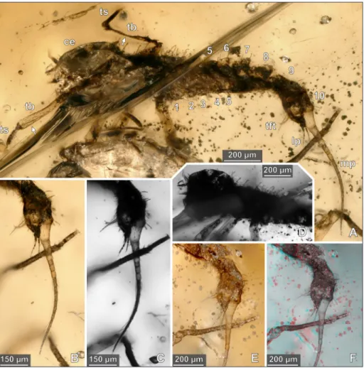
Fig. 5 - Paratype of Arcanodraco fi-licauda n. sp., BUB-2823,
specimen 1. A) Latero-ven-tral view, white light micros-copy, arrows highlight the different preservation qual-ities of the tibiae, numbers refer to abdominal segments 1–10. B) Posterior abdom-inal region in latero-ventral view, white light microsco-py. C) Posterior abdominal region in latero-ventral view, transmitted light micros-copy, numbers refer to ab-dominal segments 1–4. D) Head and thoracic region in latero-ventral view, trans-mitted light microscopy. E) Posterior abdominal region in latero-ventral view, white light microscopy. F) Same image detail as in e, red-cyan stereo anaglyph, white light microscopy.
ce= compound eye; lp= lat-eral process; mp= median process; tb= tibia; tft = tuft; ts= tarsus.
Thorax: The thorax is markedly narrower than the head. The pro-, meso- and metathoracic seg-ments are about equally broad. The thorax bears laterally and dorsally numerous short setae; dorsal-ly additionaldorsal-ly long papillae-like surface structures. There are no wing pads visible in the mesothoracic or meta thoracic segments. Coxae short and conical in shape. The trochanters are constricted at about the half of their length and distally attached to the femur, trochanter-femur joint oblique to the main axis of the leg. The femora are somewhat flattened and bear short, delicate setae. The tibiae have a bulge close to the proximal joint and distally bear strong
setae. The tarsi are not subdivided into multiple dis-tinct elements; tarsi bearing numerous setae. Pretar-sal elements itself not visible; two (pre-) tarPretar-sal claws with curved tips (Figs. 5A & 6A).
Abdomen: The abdomen is composed of 10 free segments (each dorsally forming a distinct tergite). All free segments bearing numerous long papilla-like surface structures (Fig. 2). Anterior-most abdominal segment narrower than the preceding metathoracic segment; anteriormost two abdominal segments increasing in width (Fig. 4A–C); abdom-inal segments 3 to 9 subequal; abdomabdom-inal segment 10 posteriorly decreasing in diameter (Fig. 6F); no
Fig. 6 - Details of the abdominal region in the holotype of Arcanodraco filicauda n. sp., BUB-2823, specimen 2. A) Lateroventral view, body
segments in alternating colours, numbers refer to abdominal segments 1–11, arrows mark the tarsi of the paratype, epifluorescence microscopy. B) Red-cyan stereo anaglyph of the posterior abdominal abdominal region in latero-ventral view, white light microscopy. C) Same image detail, white light microscopy, numbers refer to abdominal segments. D) Posterior abdominal abdominal region and posterior abdominal processes in latero-ventral view, white light microscopy, coaxial light and polarising filter. E) Same image detail as in d but with ring-light illumination. F) Abdominal region and posterior abdominal processes in dorsal view, white light microscopy. G) Posterior abdominal abdominal region in dorsal view, numbers refer to abdominal segments, white light microscopy. H) Same image detail as in g, red-cyan stereo anaglyph, white light microscopy.
external gill-like organs visible; no particular struc-tures on the ventral side of abdominal segment 9 (no external genital structures; Fig. 6 A).
Posterior to abdominal segment 10 (appar-ently part of abdominal segment 11) there is one long median process (60% as long as abdominal seg-ments 1–10 together). Median process with relative-ly narrow base and steadirelative-ly decreasing in length; no subdivision in multiple articles; not translucent (Fig. 6D–F).
Abdominal segment 11 apparently further-more represented by two symmetrical lateral cesses that are much shorter than the median pro-cess (tips of the lateral propro-cesses posteriorly not extending one third of the median process). Lateral processes with close (or touching) broad proximal regions, distally much narrower and diverging with an angle of about 30–33° (Fig. 6B–H). Median pro-cess and lateral propro-cesses each with numerous short spines and with a distinct tip. Tuft-like evaginations on the ventral side of the abdomen at the base of the lateral processes.
Measurements specimen 2 (no z-correction): Com-plete body length (including posterior abdominal processes) 2.61 mm; complete body length (with-out posterior abdominal processes) 1.88 mm; head length 0.42 mm; head width 0.58 mm; prementum length 0.37 mm; prementum width 0.20 mm; thorax length 0.4 mm; pro-thoracic femur length 0.35 mm; pro-thoracic tibia length 0.30 mm; pro-thoracic tar-sus length 0.13 mm; meso-thoracic femur length 0.36 mm; meso-thoracic tibia length 0.29 mm; me-so-thoracic tarsus length 0.13 mm; meta-thoracic femur length 0.35 mm; meta-thoracic tibia length 0.34 mm; meta-thoracic tarsus length 0.14 mm; ab-dominal segments 1-10 length 1.16 mm; abab-dominal segment 11 (posterior abdominal processes) length 0.70 mm.
d
IscussIonConspecifity and ontogenetic stage of the fossils
The two specimens resemble each other in many aspects and also share unique morphological features. However, there are also differences be-tween the two specimens. The subdivision of the median posterior abdominal process is only visible in
specimen 1, whereas in specimen 2 the correspond-ing structure is bare of constrictions and also lacks the translucency that is apparent in specimen 1. The proximal end of the median process in specimen 1 is broad and the diameter of the process is abruptly distally decreasing within one seventh of the total length of the process. In specimen 2 the proximal region of the process is much narrower and the di-ameter of the process distally decreases steadily.
Specimen 1 and 2 are in such a close proximity to each other (partially interlocked, Figs. 2 & 6A) that only allows for the conclusion that both specimen must have been simultaneously embedded in the res-in. Both specimens are also in an rather isolated posi-tion within the amber piece and secluded from other animal inclusions and dense amounts of debris.
Specimen 2 is roughly 25% longer than spec-imen 1 (24% with posterior abdominal processes & 26% without posterior abdominal processes). The size difference – that fits the usual size differences between subsequent instars in eucrustaceans (Mauch-line 1976) – , the striking overall similarity and the morphological differences in the posterior abdomi-nal region are best explained by the specimens being part of an ontogenetic series. The smaller specimen (specimen 1) has also some features that favour its interpretation as an exuvia. The cuticle of one tibia is collapsed while another tibia within the same spec-imen is not collapsed and extremely transparent (ar-rows in Fig. 5A). Also, in general view specimen 1 is much darker. The brighter colour in the presumed later instar (specimen 2) could be explained by a less-er content of pigment in the cuticle as a result of the recent moult.
Following this interpretation, the two speci-mens are best understood as semaphoronts (charac-ter bearing representative of a species at a definite time) of not only the same species, but also of the same individual.
Phylogenetic interpretation (basal delimitation)
The body of the fossil is divided into three functional tagmata (head, thorax & abdomen). Functional ('walking') limbs are only present in the thorax. The appendages of post-ocular segment 5 (maxillae in crustacean terminology) are conjoined, forming an unpaired mouthpart structure (labium). This combination of character states clearly identify the fossils as hexapodans/insects
(Anglo-American/European tradition).
The specialisation of the labium to a raptorial mask (elongation, conjoined glossa and paraglossa, reduction in the number of palp elements, movable hooks on the palps) is apomorphic for the odona-topteran lineage (Odonata and extinct relatives). In extant species the raptorial labium is only seen in larval odonatans (damselflies, dragonflies & Epio-phlebia).
Most fossils of dragonflies in the wider sense (Odonatoptera) that have been described so far are based on adult specimens. Phylogenetic reconstruc-tions for the odonatopteran lineage are heavily de-pendent on characters of the wing venation. For that reason, it is very difficult to include the char-acter evolution of the larval labium into a phyloge-netic framework. Bechly (2002) interpreted the rap-torial mask to be apomorphic for Neodonatoptera (ingroup of Odonatoptera that inter alia comprises Odonata), but stated that the raptorial mask could also be part of the ground pattern of Odonatopte-ra since the labial structure of Eugeropteridae (sis-tergroup of Neodonatoptera) was (and still is) un-known. The elaboration of further groups (nodes) along the lineage from the odonatopteran common ancestor towards Neodonatoptera did not change this issue since no further larvae along this lineage became known since then (Petrulevičius & Gutiér-rez 2016).
The oldest putative fossil of a raptorial mask is from the Pennsylvanian of Illinois (‘Mazon Creek’). Based on the venation in the preserved wing pads, the Carboniferous larva has been interpreted as a meganisopteran (Kukalová-Peck 2009). This would imply that the raptorial mask was already present in the ground pattern of Euodonatoptera (Fig. 9).
Odonatoptera has been established on an apomorphy-based phylogenetic framework (see de Queiroz & Gauthier 1990) for the distinction be-tween node-based, branch-based and apomorphy- based taxon concepts). However, this phylogenet-ic framework is based on characters whphylogenet-ich are not visible in the here described fossil larvae. Nonethe-less, Odonatoptera should herein not be used as a branch-based group as this could likely cause further confusion. Consequently, the interpretation of the fossil larvae can not be as narrow as Odonatoptera. To express the basal delimitation of this phylogenet-ic interpretation correctly, we propose the following nomenclature.
odonatoptera (incl. lin. approx.)
The above abbreviation stands for '[hic] inclu-dit lineam approximatam' (latin for '[this] includes the approximating lineage'). The abbreviation also works for English as well as for other Latin-derived languages. In English it can also be read as 'including the approximating lineage'. The term 'approximat-ing lineage' should refer to the 'direct stem lineage' sensu Ax (Ax 1985) (in contrast to the 'assembled stem lineage'). This means that the fossil does rep-resent a descendant of the oldest common ancestor of (only) odonatopterans, but must not necessarily represent a descendant of Odonatoptera (as a his-torical species/stem species of Odonatoptera).
Ontogenetic stage
The presence of labial mask (larval character) and the absence of externally visible genital struc-tures as well as wings clearly point against an inter-pretation of the presented fossils as adults.
A specialised first instar larva (prolarva) that is present in extant Odonata can also be excluded as possible life stage of the specimens. The legs and the labial mask (visible only in the specimen 2) are well developed and most likely also functional in
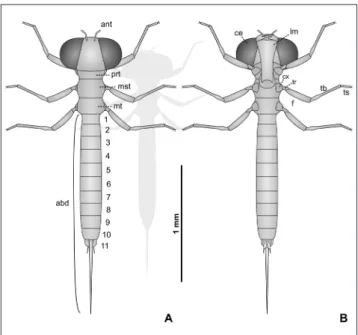
Fig. 7 - Reconstruction of the holotype of Arcanodraco filicauda n.
sp., BUB-2823, specimen 2. A) Dorsal view. B) Ventral view. The shadow in the background is scaled to the size of the paratype (specimen 1).
ant = antenna; prt= prothorax; mst= mesothorax; mt = metathorax; abd= abdomen (numbers refer to abdominal segments); ce= compound eyes; lm= labial mask; cx =coxa; f= femur; tb= tibia; tr= trochanter; ts= tarsus.
the her described fossils, unlike in most prolarvae where the legs are not functional (Tennessen 2009). It should be noted that the prolarva can only be reconstructed to have been present in the ground pattern of Odonata, but not for more basal nodes along the odonatopteran lineage. Hence, this spe-cific stage, with its differentiated morphology could be a novelty of the group Odonata.
Early instars typically have eyes that are rel-atively small compared to the size of the complete head and the relative size of the eyes increases dur-ing the development (Tennessen 2009). Further-more, the head of early stages is typically bloated in its appearance (c.f. figures in Nair & Subramanian 1981; Rowe 2007; Foulkes n.d.). This is in contrast to the larvae described herein, which have relatively large eyes and lack a such bloated appearance of the head. It is thus unlikely that the fossil larvae represent second instar larvae (often misleadingly referred to as first instar, see introduction). Also,
extensive body armour on thorax and abdomen (papilla-like structures present in both specimens) is not seen in first and second instar larvae of extant odonatans.
In representatives of Odonata wingpads de-velop and become visible externally only during the larval development and are usually not present in the earliest instars (Tennessen 2009). Also, the size of the fossils does not support an interpretation as late larval instars. One of the smallest extant odo-natan adults (Agriocnemis kalinga Nair & Subrama-nian, 2014) is about 17 mm in total body length (Nair & Subramanian 1981). The smallest species recorded from Burmese amber (Cretadisparoneura hongi Huang et al., 2015) has a forewing length of 8.1 mm and the abdomen is 11.2 mm long (Huang et al. 2015). Although the aforementioned species is only known from an incomplete specimen it de-ductible that the living animal was about equally large or slightly smaller than Agriocnemis kalinga. The herein described fossils are 1.4 mm and respectively 1.9 mm in total length (without posterior abdominal processes). This enormous size difference cannot solely be explained by the increase in body length during the final moult to the imago. Hence, the size of the herein presented specimens alone strongly points towards an interpretation as comparably ear-ly larval instars.
Posterior abdominal processes
The posterior abdominal processes in the fossil larvae are clearly not related to external gen-ital structures due to the position from which they originate. The elements of the ovipositor originate from abdominal segments 8 & 9 whereas the pro-cesses in the fossil originate posterior to abdominal segment 10.
There are two structures within extant od-onatans that could superficially resemble the un-paired median process of the herein described lar-val specimens. In some lineages the ovipositor can extend far beyond the last abdominal segments (cf. larvae of Aeschnididae: Fleck et al. 2002; Zhang 1999, 2000; Zhang & Zhang 2001). Ovipositors however consists of mainly paired structures (Klass 2009; Matsuda 1976) and the median process in the presented larvae, is undoubtedly a single unpaired structure (Figs. 5B & 6D). The other structure that could resemble the median process is a prolonga-tion of abdominal segment 10 that occurs in some
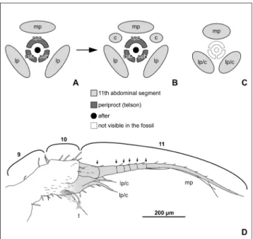
Fig. 8 - Illustration of the posterior part of the abdomen in Arcano-draco filicauda n. sp. and comparison with the same structures
in other odonatopterans. A–C) Schematic visualisation of the posterior part of the abdomen in odonatopterans, styl-ised projection in posterior view; A - early larval instars of odonatans. B - Later larval instars of odonatans. C - Con-dition in both type specimens of Arcanodraco filicauda n. sp.,
the identity of the lateral processes cannot be stated un-ambiguously (either lateral processes or cerci). D) Drawing of the posterior part of the abdomen in the paratype of
Arcanodraco filicauda n. sp. in lateral view, numbers refer to
the abdominal segments, abdominal segment 11 is colour coded (consistent with a-c), on the ventral side the border between segment 10 and 11 is unclear.
mp= median process; lp= lateral process; spa= supraanal lobe; sub= subanal lobe; c= cercus; t= tuft (functional gill?).
anisopteran larvae (e.g. Phyllocycla). The prolon-gation of the posterior abdomen is used for respi-ration whilst the larva is being buried in sediment (Novelo-Gutiérrez et al. 2018). A prolongation of abdominal segment 10 however implies that the posterior abdominal processes are posterior to the prolonged segment. This is clearly not the case for the here presented fossil larvae.
Comparing the fossil larvae to extant odo-natans, but also to other insect groups, there are two structures that could correspond to the lateral processes in the fossil larvae. Those are the cerci and the lateral processes of odonatans ('paraprocts', see introduction). Cerci occur in extant larvae of all groups of Odonata. Unlike the lateral process-es, they are not present in the earliest larval instars (Corbet 1955; Rowe 1992) and, especially, but not only in zygopterans, the cerci are also smaller than the lateral processes (e.g. Asahina 1961; Khelifa 2012; Rowe 1992) (the small size of the cerci com-pared to, for example, mayflies or archaeognathans could be a result of a post-displacement, i.e. a type of heterochrony). This makes it unlikely, though not impossible, that the lateral processes in the de-scribed fossil larvae are cerci.
The more plausible assumption is that the
lat-eral processes correspond to the latlat-eral processes of odonatans ('paraprocts'). Those are present even in the earliest instars and can be traced back to exten-sion of the sternitic region of abdominal segment 11 (Matsuda 1976), which matches the position of the processes in the fossils (Fig. 8D). In zygopterans the lateral posterior abdominal processes are large and often leaf-like. In extant zygopterans they are usually about subequal to the median process but never distinctly shorter than the median process. The extreme opposite morphology however (me-dian process completely reduced) does occur in ex-tant larvae (Xu 2015). In euepiproctophorans the lateral processes are much shorter (compared to the total body legth) and part of the anal pyramid (e.g. Asahina 1961; Rowe 1992).
Zygoptera is mostly characterised by apo-morphic characters in the wing venation, which is reasonable, because this way zygopterans are better distinguished from related extinct lineages (most fossils of these groups are known from wings). Three leaf-like posterior abdominal processes in the larvae have additionally been interpreted as auta-pomorphy for Zygoptera (Bechly 1997; Rozefelds 1985). However, the reconstruction of this possible apomorphy of Zygoptera (within a phylogenetic
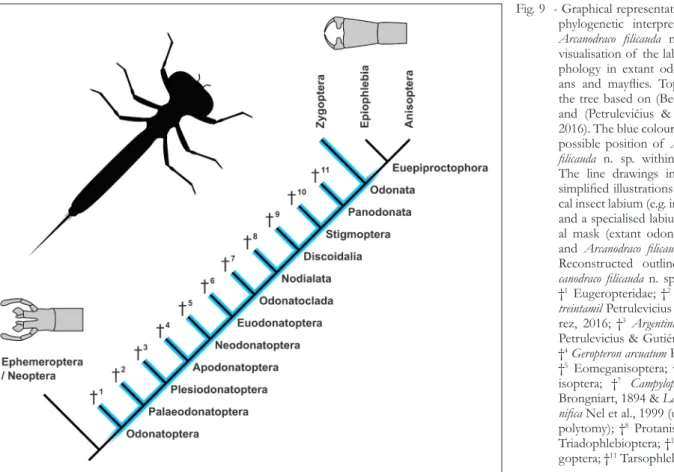
Fig. 9 - Graphical representation of the phylogenetic interpretation for
Arcanodraco filicauda n. sp. and
visualisation of the labium mor-phology in extant odonatopter-ans and mayflies. Topology of the tree based on (Bechly 2002) and (Petrulevičius & Gutiérrez 2016). The blue colour marks the possible position of Arcanodraco filicauda n. sp. within the tree.
The line drawings in grey are simplified illustrations of a typi-cal insect labium (e.g. in mayflies) and a specialised labium raptori-al mask (extant odonatopterans and Arcanodraco filicauda n. sp.).
Reconstructed outline of Ar-canodraco filicauda n. sp. in black.
†1 Eugeropteridae; †2Kirchnerala treintamil Petrulevicius &
Gutiér-rez, 2016; †3Argentinala cristinae
Petrulevicius & Gutiérrez, 2016; †4Geropteron arcuatum Riek, 1984;
†5 Eomeganisoptera; †6
Megan-isoptera; †7 Campyloptera eatoni
Brongniart, 1894 & Lapeyria mag-nifica Nel et al., 1999 (unresolved
polytomy); †8 Protanisoptera; †9
Triadophlebioptera; †10
system that is based on wing characters) is arbitrary at this point. Leaf-like posterior abdominal process-es could well be prprocess-esent in the ground pattern of more inclusive (broader) groups (Bechly 2003) and would then consequently be a plesiomorphy for Zy-goptera.
To our understanding, this is a matter of the polarity of the posterior abdominal processes as a character. There are specialised relatively small processes in Euepiproctophora (Anisoptera and Epiophlebiidae) and leaf-like posterior abdominal processes in most Zygoptera. There are reasons to assume that the latter (leaf-like processes) is the plesiomorphic state for Odonata. Leaf-like pro-cesses occur earlier in the fossil record (Late Trias-sic) (Rozefelds 1985) than the morphology that is seen in euepiproctophorans (uppermost Jurassic to earliest Cretaceous) (Zhang & Zhang 2001). Also, the minimum age of Anisoptera (ingroup of Eue-piproctophora; Middle Jurassic) (Kaur Kohli et al. 2016) is younger than Late Triassic. Additionally, if the correlation between larvae and adults is correct, there are leaf-like processes in Isophlebioptera (in-group of Odonata that is closer related to dragon-flies than to damseldragon-flies) (Pritykina 1985).
If leaf-like processes are assumed to be ple-siomorphic, they cannot be an apomorphy of Zy-goptera, but must have been present earlier in the lineage towards extant damselflies (see also the dis-cussion in Nel et al. 1993). This also means that the larva from the Upper Triassic of Australia can not be used to give a minimum age of Zygoptera (cf. Bechly 1997; Rozefelds 1985). Remarkable is that, except for one Lower Cretaceous fossil (Jell & Dun-can 1986), all Mesozoic larvae with a non-euepiproc-tophoran type of posterior abdominal morphology have short and roundish processes (Samarura-type; 'Samarura' is not a natural group but has been used as a form genus) (Brauer et al. 1889; Nel & Marti-nez-Delclos 1993; Pritykina 1985; Rozefelds 1985). The Samarura-type abdominal processes have been found in fossils of late larval instars. In this way, a Samarura type morphology has been shown for representatives of Isophlebioptera (ingroup of Odonata; sistergroup to Euepiproctophora) (Prity-kina 1985). It is even possible that, compared to the more elongated leaf-like gill pads in Zygoptera, the Samarura-type morphology is plesiomorphic.
Many damselfly larvae have predetermined breaking points in their posterior abdominal
pro-cesses. Those breaking points have been shown to reduce negative effects of unsuccessful moulting (Corbet 1950). Based on the location, the subdi-vision by breaking points into (few) elements and similarities regarding the anatomy (histological sec-tions), Tillyard (1917) concluded that the median process of zygopterans derived by reduction of elements from a 'multisegmented' process (=annu-lated process or process subdivided into numerous elements) as present in mayflies (there called ‘termi-nal filum’). He further assumed the predetermined breaking points in zygopteran gills to be remnants of the subdivision of the terminal filum into multiple elements. The ontogenetic development of the pre-determined breaking points however do not match to the interpretation that they are corresponding to a subdivision into multiple elements as present in mayflies, jumping bristletails (Archaeognatha) and silverfish (Zygentoma). McNeill (1960) pointed out that the predetermined breaking points are not present in the early instars and also the growth-pat-tern of the gills relative to their predetermined breaking points does not match this interpretation. Tillyards interpretation about the origin of the pre-determined breaking points (Tillyard 1917) may be wrong; however, comparative morphological stud-ies have supported that both the median posterior abdominal process in damselfly larvae and the ter-minal filum in mayfly larvae are structures of the tergite of abdominal segment 11 (Matsuda 1976). It is thus most parsimonious to assume that also the median processes in the herein described fossils are structures of the tergite of abdominal segment 11 – especially as the process is located posterior to the tergite of abdominal segment 10 (Fig. 6A). The lo-cation of the median posterior abdominal process, and especially its annulation, points towards the in-terpretation of this structure as the terminal filum.
Gill tufts
Besides the rectal gills in both Zygoptera and Euepiproctophora (Anisoptera and Epiophlebii-dae) and the gill pads in zygopterans there are two further types of gill structures known from odona-tans. In Polythoridae and Euphaeidae (Zygoptera: Calopterygoidea) there are species with tracheal gills on the lateral sides of abdominal segments 2–7 resp. 2–8 (Bitsch 2012). External gill structures more similar to the tufts in the described fossil (Figs. 5B, 6E & 8D) are known from
Amphiptery-gidae (Novelo-Gutierrez 1995; Watson 1966) and Pseudolestidae (Yu & Bu 2011) (also both Calop-terygoidea). In these groups gill tufts are located on the ventral side of abdominal segment 10. For the gill tufts in Amphipterygidae an origin of the tufts from the lamina sub-analis (part of the periproct, see Fig. 8) has been assumed (Watson 1966). This could also be true for the tufts in the fossil larvae judging from their position. However, there is little structural information preserved that could back up the assumption on the same origin of the struc-tures as in Amphipterygidae.
Possible systematic affinities (distal delimitation)
The phylogenetic interpretation is very broad and thus there are many possible affinities for the described fossil larvae. An ingroup position with-in Odonata could be supported by the age of the fossils. With the Late Cretaceous age of Burmese amber (Shi et al. 2012), the fossils are younger than any record of non-odonatan odonatopterans (the youngest record is from the Early Cretaceous) (Nel & Jarzembowski 1998).
Within Odonata, the specimens could be interpreted as representing larvae of a zygopter-an species, yet not without contradictions. Still the high proportion of zygopteran species among the odonatans from Burmese amber (Ross 2018) could be seen as supporting such an assumption, especially as so far no larvae have been described from this locality. Early larval instars of extant zy-gopterans have posterior abdominal processes with almost circular cross sections. Such processes also bear setae and have a distinct distal tip similar to those of the lateral posterior abdominal process-es of the new fossils (Tillyard 1917). Also, there is quite some morphological variation in the mor-phology of the posterior abdominal processes of extant zygopteran larvae. In many zygopteran lin-eages the posterior abdominal processes deviate from the leaf-like shape in various ways (see e.g. Kalkman et al. 2010). Even the complete reduction of the median process occurs in some species (Xu 2015). A morphology similar to that of the here described fossil larvae, where the lateral processes are significantly shorter than the median process, is however not known from extant zygopteran larvae.
Besides Zygoptera and Euepiproctophora there is one other natural group within Odonata
– Isophlebioptera – that could represent a possi-ble affinity for the herein presented fossils. The isophlebiopteran lineage persisted at least until the middle Eocene (Garrouste & Nel 2015). However, as discussed above, isophlebiopteran larvae have been found to have short and very broad leaf-like posterior abdominal processes (Pritykina 1985).
Judging from the morphology in the stud-ied larvae, an affinity not within Odonata but still within Odonatoptera (and its direct stem lineage) seems also likely. If isophlebiopterans indeed pos-sessed leaf-like gills (Pritykina 1985), the most parsimonious reconstruction for the odonatan ground pattern is also with leaf-like gills. The here described fossils posses a terminal filum (discussed above). This likely is plesiomorphic for the odo-natopteran lineage as terminal filaments are also present in mayflies and silverfishes (Zygentoma; sistergroup of Pterygota). With leaf-like abdomi-nal processes presumably being part of the odona-tan ground pattern, it is even more likely that the here described fossils are not ingroup odonatans, because this would imply a reduction and a subse-quent reformation of the terminal filum which is not the most parsimonious assumption (‘Dollo’s law’, Dollo 1893).
As discussed above, the character evolution of the posterior abdominal processes in dragon-fly-related lineages is only sparsely supported by the fossil record. This makes it impossible to re-construct for which node along the odonatopter-an lineage a morphology like in Zygoptera (three similar leaf-like processes) is apomorphic. In con-trast, the condensed morphology of the posterior abdominal processes in Anisoptera can also be re-constructed as apomorphic for the ground pattern of Euepiproctophora (comprises Anisoptera and Epiophlebiidae, all extant non-zygopteran odona-tans). Also, larvae of euepiproctophorans – even early instars – have a much broader and shorter abdomen than the presented fossil larvae (e.g. fig. 2 in Rowe 2007). A reconstruction of the char-acter state of the ground pattern of Epiprocta (comprises Isophlebioptera and Euepiproctopho-ra) is – despite the name of the group – not pos-sible (different morphology within larvae of an epiproctan ingroup, see above).
In this way, the morphology of the posteri-or abdominal abdomen region allows to exclude at least Euepiproctophora from the phylogenetic
in-terpretation of the fossil (distal delimitation). The final phylogenetic interpretation therefore is:
odonatoptera (incl. lin. approx.) nec
euepIproctophora
('nec' is Latin for 'but not')
Of course, among the non-odonatan linaeg-es, those that have a fossil record in the Cretaceous such as Protomyrmeleontidae (Nel et al. 2005) or Tarsophlebiidae (Fleck et al. 2004; Jarzembowski 1990; Huang & Nel 2009) are much more likely as a possible affinity for the herein described fossils, than lineages that lack a Cretaceous or even a Mes-ozoic fossil record.
Species delimitation
From Burmese amber all of the so far for-mally described odonatopterans are odonatans and only adult specimens have been properly described so far (Ross 2018). This leaves either euepiproc-tophorans (Burmaphlebia reifi, Burmastenophlebia flecki and ingroup anisopterans) or ingroup zygopterans (Ross 2018) as, already described, potential adult counterparts of the here described fossils. All eue-piproctophorans can be excluded as possible adult counterparts based on their distinct morphology
in the posterior abdomen region (anal pyramid, see discussion above). Further, no extant zygopteran species has been reported to have a median poste-rior abdominal process that is longer than the lat-eral ones (not even in early stages). Consequently, even though it is not impossible, it is not very like-ly, that the adult counterpart of the here described larva is an already described species.
Of course, the decision whether to describe a new species based on a fossil larva (in a group in which mostly adults have been formally de-scribed) has the consequence of either a later un-der- or overestimation of the species abundance in the fossil fauna. In our view, postulating new species can be seen as the postulation of a hypoth-esis (that the specimen belongs to a species that is not yet described; cf. Pante et al. 2015). Hypoth-eses on species delimitations between fossils do not strictly follow Popper’s concept of hypotheses (statements can not be disproved). However, such hypotheses can be tested for their consistency (Assis 2014). Discussing the possible conspecifity between new Burmese amber specimens and the herein described larvae, represents such a test for consistency. Therefore, we decided to postulate a new species (see below) based on the new speci-men.
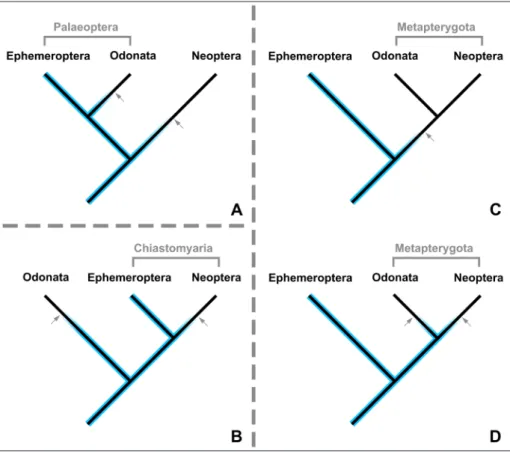
Fig. 10 - Different hypotheses on the relationships between basal pterygotan branches with respect to the implica-tions of Arcanodraco filicauda
n. sp. on the evolution of the terminal filum (median process in this study). Blue colour presence of a termi-nal filum; arrows reductions of the terminal filum. A) Palaeoptera hypothesis. B) Chiastomyaria hypothesis. C) Metapterygota hypothe-sis without the implications of Arcanodraco filicauda n. sp.
taken into consideration. D) Metapterygota hypothesis with the implications of Ar-canodraco filicauda n. sp. taken
Relevance of the fossils in a phylogenetic context
The presence of a terminal filum has already been reported once within Odonatoptera. How-ever, this record is from an adult meganisopteran (Namurotypus sippeli Brauckmann & Zessin, 1989) and the structure that has been claimed to repre-sent a terminal filum is rather club-shaped than filamentous and the subdivision in multiple ele-ments is only visible in the proximal part of this process (Bechly et al. 2001; Brauckmann & Zes-sin 1989). There is little doubt on the homology (evolutionary derivative of the same structure in the common ancestor) of the club-like process in the Carboniferous fossil to the terminal filum of other insect groups. However, this structure dif-fers distinctly from the terminal filum in other in-sect groups with respect to its shape. Due to the proximity to the ovipositor it has been suggested that the process functioned as part of the sexual apparatus (Bechly et al. 2001).
The herein presented fossils represent the youngest occurrence of a (filamentous and subdi-vided) terminal filum within the lineage from the common ancestor of all dragonfly-related lineages towards extant odonatans. Although the presented fossils have a very broad phylogenetic interpre-tation (especially the basal delimiinterpre-tation is prob-lematic, see discussion above), the presence of a raptorial mask and a terminal filum in one animal (although not in one specimen, but in two sem-aphoroths) is unique and delivers a valuable and reliable information on the character evolution of the labium and the terminal filum. The presence of both the raptorial mask as a derived, specialised labium and an ancestral-type terminal filum within the same species (and in this case likely the same individual) allows only for the conclusion that the specialisation of the labium evolved before the terminal filum was reduced (in larvae). It is still possible that the formation of the raptorial mask evolved after the terminal filum was reduced in the adults.
Together with the presence of a terminal filum in adult Namurotypus sippeli (Meganisoptera) (Brauckmann & Zessin 1989), the presence of a larval terminal filum, presented in this study, strongly suggest that the terminal filum was re-duced at least once along the lineage towards the groups that comprise extant dragonflies and
damselflies. This is of special importance with re-spect to the different possible relations between ephemeropterans (mayflies), odonatans (dragon-flies and related species) and neopterans (all other lineages with extant winged insects).
The conflicting hypotheses (Fig. 10) of the basal branching in Pterygota are under debate since decades (see introduction). The terminal filum was often referred as a character that sup-ports the Metapterygota hypothesis (Figs. 10C & 10D) as this hypothesis requires only a single re-duction of the terminal filum (along the lineage to the common ancestor of odonatans and neop-terans; Fig. 10C), while all alternative phylogenetic reconstructions would demand for two such loss-es. With the presence of the terminal filum with-in Odonatoptera (and its direct stem lwith-ineage), all three topologies require at least two independent reductions of the terminal filum (Figs. 10A, 10B & 10D). Consequently, this character no longer sup-ports any of the three hypotheses.
Taphonomy
As discussed above, the two specimens herein presented are likely two subsequent larval instars of the same individual. This interpreta-tion is based on the assumpinterpreta-tion that one of the specimens is an exuvia and the other one repre-sents the carcass. This assumption is backed up by supposedly collapsed body parts (Fig. 5A) and the colour differences between the specimens. Those colour differences are within a very small volume of resin and it is unlikely that they are caused by a simple chemical gradient that is unrelated to the specimens. The colours are probably different due to the different degree in sclerotisation (supposed later instar is brighter; cf. Hörnig et al. 2016).
This taphonomic situation could be ex-plained by three scenarios: 1) the larva just fin-ished the process of moulting when it became embedded in resin; 2) the larva was in the process of moulting when it became embedded in resin and then finished the moulting process whilst al-ready being embedded; 3) the larva was close to the process of moulting (pharate phase) when it became embedded in resin and just then initiat-ed the moulting process (escape moult). Up to know, there are no experimental studies on escape moults in resin and there are only a few amber fossils that would suggest such a process.