1
BIOLOGIA APPLICATA ALLA SALUTE DELL’UOMO
SCUOLA DOTTORALE IN
XXVII
CICLO DEL CORSO DI DOTTORATO
“Nibrin and promyelocytic leukemia proteins in sensing and
signaling of the DNA double strand breaks”
“La nibrina e la proteina della leucemia promielocitica
nel riconoscimentio e segnalazione delle
rotture al doppio filamento del DNA”
DOMENICA CILLI ALESSANDRA DI MASI
Dottorando: Dott.
PAOLO ASCENZI
Docente Guida/Tutor: Prof.
PAOLO VISCA
Coordinatore: Prof.
2
“La mancanza di complessi, una notevole tenacia nel perseguire la strada che ritenevo giusta e la noncuranza per le difficoltà che avrei incontrato nella realizzazione dei miei progetti …… mi hanno enormemente aiutato a far fronte agli anni difficili della vita”
3 INTRODUCTION
The DNA is the most vulnerable among the cell macromolecules and is constantly subjected to different both endogenous and exogenous genotoxic agents action (Kotula et al., 2013). Among these, reactive oxygen and nitrogen species, ionizing radiation (IR), ultraviolet radiation, and chemical mutagens cause different types of damage, from single base mutations to double-stranded breaks, considered the most lethal type of DNA damage (Friedberg et al., 2006; Wanotayan et al., 2015).
The integrity of the genome is of prime importance to ensure its stability and changes in the nucleotide sequence are continually monitored by mechanisms that prevent the occurrence of mutations and chromosomal rearrangements, thus preserving the original message in the DNA; in addition to these sophisticated mechanisms of DNA damage "sensing", the cell has developed others in which signal transducing and effector proteins regulate cell cycle progression, damage repair and apoptosis; overall, these mechanisms are known as "DNA damage response" (DDR) (Zhang et al., 2006; Rupnik et al., 2010; Brandsma and Gent, 2012). In particular, DNA repair is coordinated through the cell cycle checkpoint: in the presence of DNA damage, the activation of specific proteins leads to cell cycle arrest, giving the cell the time required to repair the damage before that DNA replication and mitosis have beginning (Sancar et al., 2004; Bernstein and Rothstein, 2009; Humpal et al., 2009). The repair of DNA in eukaryotic cells requires multiprotein repair complexes recruitment and assembly in situ, controlled through post-translational modifications. Among these, the phosphorylation signal is crucial in the regulation of protein-protein interactions (Williams et al., 2005; Glover, 2006).
Mutations in genes involved in the DDR are at the root of the chromosome instability observed in many human diseases characterized by the presence and/or increased susceptibility to cancer (Khanna and Jackson, 2001; Peterson and Côté, 2004).
1.1 The DNA double strand breaks response
The DNA Double Strand Breaks (DSBs) are the most pernicious forms of DNA damage, because an intact complementary strand is not used as a template for the DNA repair. The DSBs can be formed in response to the action of exogenous agents, such as IR,
radio-4 mimetic drugs, products of oxidative metabolism and mechanical stress (Khanna and Jackson, 2001). However, these breaks also occur as a result of physiological events such as replication, meiotic and V(D)J recombination, immunoglobulins class exchange, apoptosis and retroviral integration (Friedberg et al,. 2006; Jackson and Bartek, 2009). If unrepaired or improperly repaired, the damaged sequences are neither transcribed nor replicated (Yu et al., 1999; Polo and Jackson, 2011). The cells have adapted to tolerate low levels of damage, however, if a single DSB occurs within an essential gene, it determines its inactivation and leads, eventually, to cell death (Yoshida and Miki, 2010; Souliotis and Sfikakis, 2014). Eukaryotic cells have evolved multicomponent macromolecular systems specializing in DNA damage sensing, response and repair (Jeggo and Lӧbrich, 2006; Shrivastav et al., 2008; Bernstein and Rothstein, 2009; Pardo et al., 2009, Gullotta et al., 2010; Lafrance-Vanasse et al., 2015).
The DSBs sensing requests the activation of the signaling pathway to amplify and transduce the damage signal and to generate the appropriate biological responses that include the block of the cell cycle in G1/S phase (G1/S checkpoint), or in the G2/M phase (van den Bosch et al., 2003), the slowdown of DNA synthesis (intra S-phase checkpoint) and the induction of the transcriptional program. For example, the small ribosomal protein RPS3 can regulate ribosome biogenesis in response to DNA damage (Kim et al., 2013) while RPS27a can activate cellular checkpoints via p53 to inhibit cell cycle (Xiong et al., 2011 ).
An example of a macromolecular complex specialized in the DNA damage sensing and in the early response to the DSBs is the MRE11-RAD50-NBN (MRN) complex, formed by dimers of the RAD50 and MRE11 subunits and by the NBN monomer. The MRN complex acts as a specific DSB sensor and effector involved in the cross-talk between the machinery repair and cell cycle checkpoints (Williams et al., 2010). It binds directly the broken DNA ends (Yuan and Chen, 2010) and once stabilized the injury, recruits the Ataxia-Telangiectasia Mutated (ATM) protein kinase, a master regulator of the DNA damage response. In fact ATM coordinates checkpoint activation, DNA repair, and metabolic changes in eukaryotic cells in response to DSBs and oxidative stress (Hopfner, 2014; Paull, 2015). In addition, MRN complex has a role in the processing and repair of the DSBs through the Homologous Recombination Repair (HRR) and the Non Homologous End Joining (NHEJ), in the telomere maintenance, in the reactivation of stalled replication (Slijpecevic,
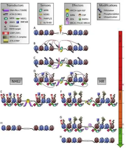
5 2006; Borde, 2007; Williams et al., 2007; Chapman et al., 2012; Srivastava et al., 2015) (Fig. 1.1).
Figure 1.1: Kinetics of DDR protein recruitment and modification at IR-induced DSBs (Vignard et al., 2013).
6 The MRN complex importance is highlighted by several genetic pathologies existence, caused by mutations in genes codifying for MRE11, RAD50 and NBN protein. In individuals with ataxia telangiectasia-like disorder (ATLD; OMIM #604391), mutations in MRE11 have been identified (Stewart et al., 1999; Taylor et al., 2004); in patients with the Nijmegen breakage syndrome (NBS; OMIM #251260) mutations in NBN have been identified (Carney et al., 1998; Matsuura et al., 1998; Varon et al., 1998); finally, only one case of an individual suffering from the syndrome defined as NBS-like disorder (NLD; OMIM #613078), associated with a heterozygous mutation in the RAD50 gene, has been reported (Waltes et al., 2009). All the above-mentioned genetic syndromes are characterized by hypersensitivity to IR, genomic instability and immunodeficiency (The International Nijmegen Breakage Syndrome Study Group, 2000; Dizikiewicz-Krawczyk, 2008).
In response to the DSBs formation, PARP1 protein rapid accumulates on DSBs, induces its own PARylation together with the PARylation of the surrounding chromatin and many DDR actors; it promotes also the recruitment of MRE11 and NBN (Petrini and Stracher, 2003; Lisby and Rothstein, 2004; Haince et al., 2008; Helmink et al., 2009; Porter-Goff and Rhind, 2009; Shikazono et al., 2009; Vignard et al., 2013). The MRN complex localization on the DNA damage, determines ATM auto-phosphorylation at the Ser1981 residue, with the consequent dissociation of ATM dimers and ATM kinase activation (Bakkenist and Kastan, 2003; Kozlov et al., 2006). ATM in the monomeric active form phosphorylates NBN at Ser278 and Ser343 residues, and H2AX histone at the Ser139 residue (the phosphorylated form of H2AX being named γ-H2AX) (Rogakou et al., 1998; Lavin, 2007; Lavin and Kozlov, 2007; Lee, 2007; Vasireddy et al., 2010). MDC1 protein is required to maintain the link between the MRN complex and chromatin and to allow the accumulation (Hari et al., 2010, Tobias et al., 2013). Indeed, through the interaction between the MDC1 Ser-Asp-Thr (SDT) repeated motif (Chapman and Jackson, 2008) and the NBN FHA and BRCT domains (see section 1.2), the MRN complex is recruited on the chromatin to activate additional molecules of ATM kinase, which phosphorylates other molecules of histone H2AX, along several megabases away on both sides from the lesion site. This signal amplification determines the recruitment, on the damage site, of several proteins involved in DDR including 53BP1, BRCA1, p53, CHK1, CHK2, CDC25A, SMC1, RNF8/RNF168/HERC2 (Yuan and Chen, 2010; Hu et al., 2013; Muñoz et al., 2014).
7 The γ-H2AX histone generates an initial signal, on the DNA damage site, represented by Ionizing Radiation Induced Foci (IRIF) formation (Bassing et al., 2003; Celeste et al., 2003), large subnuclear macromolecular aggregates that facilitate the DNA damage repair, being the site where proteins involved in DDR cause cell cycle arrest and DSBs repair (Lowndes and Murguia, 2000; Masi et al., 2008b; Lee et al., 2010; Nakamura et al., 2010; Bauerschmidt et al., 2011).
The HRR and the NHEJ repair systems (Fig. 1.2) are respectively error-free and error-prone and operate optimally in different circumstances (Sancar et al., 2004; Benada et al., 2015; Liu et al., 2015; Wanotayan et al., 2015). In bacteria and unicellular eukaryotes the main mechanism of DSBs repair is the HRR, whereas in mammals is the NHEJ (Christmann et al., 2003; Sancar et al., 2004; Jeggo and Lobrich, 2006; Polo and Jackson, 2011; Brandsma and Gent, 2012). The HRR path predominates in S and G2 phases of the cell cycle and allows a faithful repair of the lesion using the DNA of homologous chromosomes or sister chromatid as template for new strand synthesis (Khanna and Jackson, 2001; van Gent et al., 2001; Symington, 2002; Williams et al., 2007; Woodbine et al., 2014). The NHEJ repair system prevails in the cells that are in the G0/G1 phase (Christmann et al., 2003). In the NHEJ system both broken ends are first recognized and then bound by the heterodimer Ku70/Ku80, which recalls the catalytic subunit of the DNA-dependent protein kinase (DNA-PK) (Ciccia and Elledge, 2010). If the protrusions are not compatible, they can be modified by Artemis nuclease or the Polµ and Polλ DNA-polymerases in order to generate blunt end (Lieber, 2010). Finally, the ligation complex made up of DNA ligase IV (LIG4), XRCC4 and XLF cofactors catalyzes ends joining (Yu et al., 1999; Christmann et al., 2003; Ahnesorg et al., 2006; Buck et al., 2006; Jeggo and Lobrich, 2006). The failure of these repair systems can induce the formation of chromosomal aberrations that can cause aneuploidy and consequently lead to developmental defects, neurodegeneration, immunodeficiency, radiosensitivity, predisposition to cancer, and cell death (Dikomey et al., 1998; Pfeiffer et al., 2000; Lips and Kaina, 2001; van den Bosch et al., 2003; Jackson and Bartek, 2009).
8
Figure 1.2 Schematic representation of the two main repair systems used by eukaryotes to repair DSBs: NHEJ and HR. HR is predominant in S and G2 phases of the cell cycle because of the proximity of the sister chromatid. NHEJ is active throughout the cell cycle, playing a major role during G1 and M phases (Srivastava et al., 2015).
1.2 Nibrin
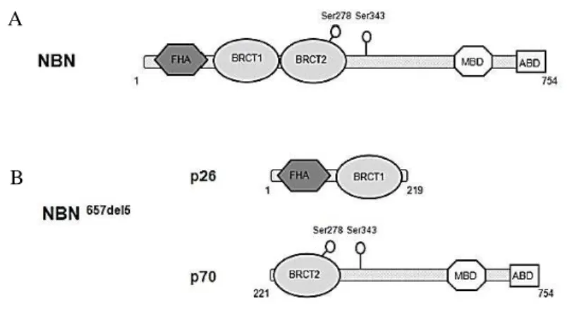
Nibrin, also named NBS1 or p95, is involved in the initial DSBs sensing, but it is also a mediator of the DNA damage response signal because it interacts with downstream effectors involved in DNA repair and cell cycle checkpoint activation. Furthermore it is involved in chromosomal integrity maintenance (Zhang et al., 2006). NBN is localized in the region 21 of the chromosome 8 long arm (8q21) (Carney et al., 1998 and Varon et al., 1998) and is composed of 16 exons (Matsuura et al., 1997). The NBN protein, ubiquitously expressed in different tissues (Kobayashi et al., 2004; Zhang et al., 2006), is long 754 amino acids (aa), has a molecular weight of about 85 kDa and is composed of three functional regions: an N-terminal region, a central region and a C-terminal region (Fig. 1.3).
9
Figure 1.3 NBN main structural components. In N-terminal region, the FHA, BRCT1 and BRCT2 mediate the interaction with phosphoproteins through the binding to pThr (FHA) and pSer (BRCT) residues. The C-terminal region presents sites for MRE11 and ATM recognition. The yellow spheres indicate sites phosphorylated by ATM (Williams et al., 2010).
1.2.1 Nibrin N-terminal and central region
Nibrin N-terminal region (Fig. 1.4) contains the Fork Head Associated Domain (FHA) (aa 24-109) and two Breast Cancer 1 (BRCA1) Carboxy-Terminal (BRCT) domains (BRCT1: aa 114-183; BRCT2: aa 221-291) separated from a linker region of 18 amino acids (Bork et al., 1997; Callebaut and Mornon, 1997; Becker et al., 2006). This region of NBN plays a key role in the recognition of DNA damage through the interaction with γ-H2AX histone and is responsible for the MRN complex localization in IRIF (Tauchi et al., 2001; Zhao et al., 2002; Rupnik et al., 2010) (see section 1.1). In particular some authors argue that the NBN protein possesses the structural determinants (i.e. the BRCT domains) to interact directly with γ-H2AX histone (phosphorylated H2AX) (Kobayashi et al., 2002; di Masi et al., 2008a; di Masi et al., 2012; Mendez et al., 2012;), others suggest that NBN binds directly only MDC1, which in turn, through its tandem BRCT (tBRCT) domains, binds directly to γ-H2AX C-terminal (Lee et al., 2005; Stucki et al., 2005; Coster and Goldberg 2010).
The FHA domain is a protein-protein interaction domain that specifically recognizes the phosphorylated Thr residues of several target proteins involved in numerous biological processes, such as growth and cell division, differentiation, apoptosis, transcription, proteins transport, DNA repair, and protein degradation (Durocher et al., 2000; Li et al., 2000; Liang and Van Doren, 2008). Ohara et al. (2014) suggest that the expression of a FHA-mutated nibrin causes radiosensitization, suggesting a FHA domain role in preventing the generation of DNA mutations caused by ionizing radiations.
10 Furthermore the FHA interacts with phosphorylated Thr residues located in the Ser-Asp-Thr (SDT) motif of MDC1, and is necessary for the IRIF and multiprotein complexes formation that assemble after DSBs formation (Zhao et al., 2002).
The BRCT domains have been identified in many proteins involved in the DDR and may be present in single or multiple copies (Callebaut and Mornon, 1997; Leung and Glover, 2011). Tandem BRCT domains, described for the first time in the BRCA1 protein (Futreal et al., 1994; Miki et al., 1994), function as a single structural unit in the phosphorylated peptides recognition (Mesquita et al., 2011). NBN tBRCT domains are characterized by a globular α/β folding made up of central 4-β filaments flanked by 3 α-helices (α1 and α2 on one side and α3 on the opposite side).
Figure 1.4 Structural details of the NBN N-terminal region (modified from Williams et al., 2009).
11 The large hydrophobic interface that is formed between the BRCT1 domain C-terminal region (helices α1 and α3) and the BRCT2 domain N-terminal (helix α2) represents the recognition site for phosphoproteins and phosphopeptides (Williams et al., 2001; Glover et al., 2004). The residues placed in the α1 and α3 helices and less in the α2 helix, are preserved, suggesting that the packaging "head-tail" is stored in the members of the BRCT superfamily (Finn et al., 2008). Moreover, it is interesting to note that their tight proximity allows the NBN FHA domain and N-terminal BRCT domain to be considered as a single and compact globular structure required for the MRN complex localization on chromatin after DNA damage (Hari et al., 2010). Two conserved Ser residues (Ser278 localized at the BRCT2 C-terminal and Ser343 in the central region) are phosphorylated by ATM in response to IR and are associated with both p53 dependent- apoptosis regulation (Iijima et al., 2008) and the NBN role in the cell cycle S phase checkpoint (Lim et al., 2000; Wu et al., 2000; Zhao et al., 2000; Zhang et al., 2006; Rupnik et al., 2010).
1.2.2 Nibrin C-terminal region
The nibrin C-terminal region contains a motif for the binding with MRE11 (aa 689-691) (Desai-Mehta et al., 2001) and ATM, (aa 734-754) (Falck et al., 2005).
1.2.3 The Nijmegen breakage syndrome
It is fundamental that nibrin preserves its structural integrity since mutations within nibrin are responsible of cancer proneness. In particular mutations at the homozygous status are responsible for NBS, a rare genetic disorder characterized by an autosomal recessive inheritance whose signs are, among the others, immunodeficiency, microcephaly, hypersensitivity to IR, and predisposition to the development of lymphomas and solid tumors (Weemaes et al., 1981; van der Burgt et al., 1996; Chrzanowska et al., 2012). NBS is a rare autosomal recessive disease characterized by chromosomal instability, described for the first time in 1981 in patients living in Nijmegen (Netherlands) (Demuth and Digweed, 2007). The NBS is manifested at birth with microcephaly, dysmorphic facial features that become more apparent with age, immune deficiency and growth retardation. Late complications involve malignant tumors
12 development, especially of hematic origin, and infections in various organs (Chrzanowska et al., 2012). From several years NBS diagnostic markers are known: 1) hypomorphic biallelic mutations in NBN; 2) spontaneous chromosomal instability in peripheral T lymphocytes, with rearrangements like inversions and translocations, especially involving 7 and 14 chromosomes; 3) cellular sensitivity to IR or radiomimetic agents demonstrated in vivo; 4) RDS. The 90% of NBS patients present the NBN 657del5 founder mutation, which occurs mainly in Slav populations (Weemaes et al., 1981; Carney et al., 1998; Varon et al., 1998; Maser et al., 2001; Chrzanowska et al., 2012). NBS was also found in other European countries (including Italy), in North America, Morocco, and New Zealand (Chrzanowska et al., 2012).
1.2.3.1 The 657del5 founder mutation
The 657del5 mutation consists of a 5 bp deletion in the linker region connecting the two NBN BRCT domains. The resultant shift of the reading phase involves the expression of two truncated proteins of 26 kDa (p26) and 70 kDa (p70) and causes the interruption of the two tBRCT. In particular p26 contains the FHA region and the first BRCT domain, and is the product of a protein synthesis premature termination, while the p70 fragment, which includes the second BRCT domain and the entire NBN C-terminal region, originates from an alternative initiation of translation at a cryptic upstream start codon (Maser et al., 2001; Williams et al., 2002; Becker et al., 2006; Alster et al., 2014). With the exception of the first 18 amino acids, the protein p70 has a sequence identical to that of wild-type NBN. It is known that the p26 peptide, preserving the NBN FHA/BRCT1 domains that are essential for both the physical interaction with γ -H2AX and for the re-location of RAD50 and MRE11 near the site of DNA damage, appears to retain ability to recognize γ-H2AX (Kobayashi et al., 2002). These events do not seem to take place efficiently in cells from patients expressing both p26 and p70 fragments, as demonstrated by unrepaired DSBs persistence 24 hours after treatment with IR (di Masi et al., 2008a). However truncated p70-nibrin can form the MRN complex (Kruger et al., 2007; Maser et al., 2007). This aspect suggests that the fragments present a residual retention of the pivotal protein interactions, generally established by wild-type nibrin.
To date, in NBN ten other mutations have been identified (Fig. 1.5) in the homozygous or compound heterozygous status and considered
13 responsible for the syndrome development (Tupler et al., 1997; Varon et al., 1998; Maraschio et al., 1986 and 2001; Nakanishi et al., 2002; Resnick et al., 2002; Gennery et al., 2004; New et al., 2005).
Figure 1.5 NBN gene schematic representation and localization of the mutations responsible for NBS development (Modified from Demuth and Digweed, 2007).
In NBS patients with the 657del5 mutation, an NBN loss of function is observed. The loss of an efficient DSBs repair mechanism, observed in cells prepared from NBS patients, causes severe clinical characteristics in NBS patients, displaying the important NBN role in DSBs signaling and repair, fundamental for the cellular homeostasis maintenance. In particular, the cancer beginning are associable with the chromosomal aberrations high frequency (Chrzanowska et al., 2012); the radiosensibility is imputable to the NBN role in the response to the IR-induced damage and to its non-phosphorylation
by ATM (Gatei et al., 2000; Wu et al., 2000); the microcephaly is a characteristic of the syndromes associated
with defects in DDR and may be caused by an increased cell death, associated with an inefficient DNA repair (the microcephaly seems to be caused by a decreased neurogenesis derived from an increased cellular apoptotic death or from the reduced neuronal precursors proliferative capacity (O’Driscroll and Jeggo, 2008)).
14 2.1 PML and PML-Nuclear Bodies
The ProMyelocytic Leukemia (PML) human gene is localized in the q22 region of chromosome 15. It codifies for PML, an oncosuppressor of almost 70 kDa. The PML alternative splicing generates 6 nuclear and one cytoplasmic isoforms (Jensen et al., 2001; Bernardi and Pandolfi, 2007).
The PML-NBs are non-membrane bound spherical organelles of 0.1-1 µm of diameter, made up of an intricate protein system (Melnik et al., 1999). PML is the NBs regulator protein and together with SP100 represents the permanent component of these macromolecular aggregates. The PML-NBs are stable structures and highly dynamic and their distribution changes highly in relation to cell type, cell cycle phase or to the possible exposition to stress factors (Jensen et al., 2001; Bernardi and Pandolfi, 2007).
2.2 PML-NBs post-translational modifications
The PML post-translational modifications regulate several PML-NBs functions, as also their assembly or disassembly during the cell cycle phase. The PML phosphorylation events involve mostly the N-terminal region of the protein and are associated with PML-NBs role in DDR.
Figure 2.1 Particular of a PML-NB formation. At first SUMO-1 was distributed more sparsely and also more aggregated than PML. A partial colocalization of PML and SUMO-1 in the same spherical shell was evident. By contrast, SUMO-2/3 was located also in the interior of the PML-NB. Sp100 was distributed similarly to PML in the outer shell of spherical shape (Lang et al., 2010)
15 The PML phosphorylation sites are localized near the Real Interesting New Gene (RING) domain, in the Nuclear Localization Signal (NLS) sequence, and in the SUMO-Interacting Motif (SIM) (Bernardi and Pandolfi, 2007; Gresko et al., 2009; Schmitz and Grishina, 2012). During the DDR, the kinases ATM, ATR and CHK2 act phosphorylating these PML sites, regulating their stability and of the proteins constituting PML-NBs (Dellaire et al., 2006). The PML SUMOilation regulates PML stability, dissociation of the transcription factors from PML-NBs, PML regulation of apoptosis, PML-NBs structure maintainance, protein-protein interaction (Ishov et al., 1999; Zhong et al., 2000; Nacerddine et al., 2005; Geiss e Melchior, 2007; Imani-Saber et al., 2014). The PML-NBs formation, also known as nucleation, happens during cell cycle interphase and disappears in mitosis when the activity level of SUMO enzymes lowers and the PML-NBs become more little (Everett et al., 1999) (Figure 2.1).
2.3 PML-NBs role in DNA damage sensing and repair
PML-NBs are constituted by a complex protein network represented by a “basket”, in which are concentrated several proteins, involved also in DNA damage repair (D’Orazi et al., 2002; Hofmann et al., 2002; Dellaire et al., 2006; Yang et al., 2006;). These proteins, being concentrated in the environment delimited by PML-NBs, are readily available for an efficient and rapid DNA repair (Carbone et al., 2002; Dellaire and Bazett-Jones, 2004; Bernardi and Pandolfi, 2007; Kepkay et al., 2011). PML-NBs integrity allows tumor suppressive functions via diverse biologic mechanisms such as growth inhibition, apoptosis induction and suppression of migration and angiogenesis (Chen et al., 2012).
It is important to underline that changes in PML-NBs morphology and number have been observed in response to DNA damage (Mirzoeva and Petrini, 2001; Carbone et al., 2002; Hofmann et al., 2002; Kurki et al., 2003; Seker et al., 2003; Conlan et al., 2004; Eskiew et al., 2004). In fact the stress induced in the chromatin is transmitted to PML-NBs that reply breaking and forming more little nuclear bodies named microbodies (Eskiew et al., 2004; Dellaire et al., 2006). Several DDR proteins are localized in the PML-NBs both constitutively and conditionally, including the DSBs sensing proteins (e.g. MRN complex, ATM, ATR, BRCA1, CHK2, p53, and TOPBP1) and the proteins participating to HRR (Yeager et al., 1999;
16 Henson et al., 2002; Dellaire et al., 2003; Xu et al., 2003; Spardy et al., 2008). Since the repair proteins are localized within PML-NBs, it has been suggested that the PML-NBs may have a role in DSBs sensing and in DNA damage repair events coordination (Dellaire and Bazett-Jones, 2004). It is important to consider that after DSBs induction the Ser824 residue phosphorylation of the KAP1 protein associated to the heterochromatin actives ATM kinase (Kruhlak et al., 2006; Ziv et al., 2006), with the consequent PML-NBs number increment. It has been suggested that this phosphorylation, inducing other nuclear bodies’ formation, facilitates the repair factors access to damage site on chromatin (Kepkay et al., 2011). Also CHK2 kinase protein has a key role in the DNA damage transduction. It is phosphorylated by ATM at Thr68 residue, depending from NBN, and phosphorylates PML protein localized in the nuclear bodies (Stolz et al., 2011).
2. 4 PML-RARα and the Acute Promyelocytic Leukemia
The Acute Promyelocytic Leukemia (APL) is a rare malignant tumour characterized by hemorrhagic manifestation caused by altered blood coagulation (Breccia and Lo Coco., 2014). The disease evolves rapidly with unlucky prognosis. The main disease cause is the differentiation block of the myeloid precursor cells, at the promyelocytes stadium, that so proliferate in an uncontrolled way till they invade bone marrow (de Thè et al., 1991; Kakizuka et al., 1991; Nisole et al., 2013).
APL patients are characterized by PML-RARα gene fusion, caused by the t(15;17) (q22;q21) chromosomal translocation, at the heterozygous status, where RARα codifies for the Retinoic Acid Receptor Alpha localized on chromosome 17 (Rowely et al., 1977; de Thè et al., 1990; de Thè et al., 1991, Brown et al., 1997). The translocation involves one of the three breakpoint chromosomal regions (bcr), located within PML gene and the chromosome 17 breakpoint located within the RARα gene second intron (Miller et al., 1992; Pandolfi et al., 1992). The resultant isoforms maintain the PML DNA binding domain and the binding domain to co-activators/co-repressors, ligands and RARα Retinoid-X-receptor (RXR) (Brown et al., 1997) (Figure 2.2). In APL the PML-NBs are substituted by little nuclear bodies named microspeckles (Melnick
and Licht, 1999). While in normal condition RXR (the other retinoic acid nuclear
17 receptor class (Bushue e Wan, 2010)) binds RARα and the RXR-RARα complex is associated to the Retinoic Acid Response Element (RARE) gene sequence, regulating the expression of the genes involved in the myeloid cells last differentiation in granulocytes (de Thé et al., 1991; Brown et al., 2009), on the contrary, the PML-RARα homodimerizes, binds the DNA, blocking the PML-RARα dependent expression of differentiation genes, and forms aggregates with PML wild-type isoforms, forbidding the normal PML interaction (through a competitive way) (Melnik et al., 1999; Brown et al., 2009; Yeung et al., 2012). The loss of these functions contributes to leukemic promyelocytes proliferation and survival (Brown et al., 2009). The PML-RARα dominant negative effect on the PML-NBs formation and damage repair is responsible for a high chromosomal instability in APL patients (Lanotte et al., 1991; Bishof et al., 2001; Yeung et al., 2012). In fact, it has been demonstrated that the HR mechanism to repair DNA depends on the wild type PML protein presence and on its correct organization in PML-NBs (Yeung et al., 2012).
2. 5 APL treatments
The APL patients are cared with All-Trans Retinoic Acid (ATRA) and/or Arsenic Triosside (ATO) (Khanna-Gupta et al., 1994; Yang et al., 2006; Brown et al., 2009; de Thé et al., 2012; Yeung et al., 2012). The two molecules destroy the PML-RARα oncoprotein, induce the promyelocytes differentiation and the transient reversion to wild type phenotipe, suppressing the dominant negative effect induced by the fusion protein expression (di Thé et al., 2012; Vitaliano-Prunier et al., 2014) (Figure 2.2). In particular, ATRA binds the AF-2 motif in the RARα protein C-terminal region, determining the dissociation of PML-RARα complexes bound to DNA RARE sequences (Tate et al., 1994; de Thé et al., 2012). Furthermore, ATRA restores the RARα target genes activation. ATRA treatment allows new normal NBs formation, from PML proteins produced by non-mutated allele (de Thè et al., 2012). The importance of the effect of PML-NBs integrity loss on the DNA damage signal transduction has allowed to study this relationship and to discover that PML-RARα suppresses CHK2 kinase protein phosphorylation, inhibiting the CHK2-p53 apoptotic pathway in APL. On the contrary the ATO treatment of cell lines prepared from APL patients determines promyelocytes maturation and/or apoptosis through the direct binding with PML protein and PML-RARα
18 (Zhang et al., 2010). The ATO in fact stimulates the fusion protein degradation, promoting its iperSUMOilation (Lallemand-Breitenbac h et al., 2001; Lallemand-Breitenbach et al., 2008; Thatam et al., 2008; Jeanne et al., 2010). The arsenic induces also new PML-NBs formation.
Recent studies performed in mice have been demonstrated that the ATRA and ATO combination as care for APL increases the complete reversion and the number of survivors. For this reason APL is among the first cancer example for which a care with a specific target exists (de Thè et al., 2012). Finally another valid therapy for the care of acute promyelocytic leukemia derives from a study of Luo et al. (2014) that tested a siRNA targeting PML-RARα mRNA, demonstrating that the cell growth of siRNA treated groups was inhibited, and the apoptosis of APL human cell line NB4 could be induced.
Figure 2.2 In a) PML-RARα fusion protein and the main PML and RARα functional domains are represented. The ATRA target is localized in a RARα motif, while the arsenic target is localized in a PML motif. The black triangles indicate the different fusion points. In b) the leukemic cells differentiation after ATRA treatment are observed. In c) the action model relative to the differentiation or transcriptional control through gene activation (up) or repression mediated by degradation (low) is schematized (modified from de Thè et al., 2012).
19 REFERENCES
Ahnesorg P, Smith P, Jackson SP (2006) XLF interacts with XRCC4-DNA ligase IV complex to promote DNA nonhomologous end-joining. Cell 124: 301-313.
Akuttsu N, Iijima K, Hinata T, and Tauchi H (2007) Characterization of the plant homolog of Nijmegen breakage syndrome 1: involvement in DNA repair and recombination. Biochem Biophys Res Commun. 353: 394-398.
Alster O, Bielak-Zmijewska A, Mosieniak G, Moreno-Villanueva M, Dudka-Ruszkowska W, Wojtala A, Kusio-Kobiałka M, Korwek Z, Burkle A, Piwocka K, Siwicki JK, Sikora E. (2014) The role of nibrin in doxorubicin-induced apoptosis and cell senescence in Nijmegen Breakage Syndrome patients lymphocytes. PLoS One 9(8): e104964.
Antoccia A, Sakamoto S, Matsuura S, Tauchi H, and Komatsu K (2008) NBS1 prevents type aberrations through ATM-dependent interaction with SMC1. Radiat. Res. 170: 342-52.
Bakkenist CJ and Kastan MB (2003) DNA damage activates ATM through intermolecular autophosphorylation and dimer dissociation. Nature 421: 499-506.
Bassing CH, Suh H, Ferguson DO, Chua KF, Manis J, Eckersdorff M, Gleason M, Bronson R, Lee C, Alt FW (2003) Histone H2AX: a dosage-dependent suppressor of oncogenic traslocations and tumors. Cell. 114: 359-70.
Bauerschmidt C, Woodcock M, Stevens DL, Hill MA, Rothkamm K, and Helleday T (2011) Cohesin phosphorylation and mobility of SMC1 at ionizing radiation-induced DNA double-strand breaks in human cells. Exp. Cell Res. 317:330-337.
Becker E, Meyer V, Madaoui H, and Guerois R (2006) Detection of a tandem BRCT in Nbs1 and Xrs2 with functional implications in the DNA damage response. Bioinformatics. 22: 1289-1291.
20 Benada J, Burdová K, Lidak T, von Morgen P, Macurek L. (2015) Polo-like kinase 1 inhibits DNA damage response during mitosis. Cell Cycle 14(2): 219-31.
Bernardi R and Pandolfi PP (2007) Structure, dynamics and functions of promyelocytic leukaemia nuclear bodies. Nat Rev Mol Cell Biol. 8: 1006-1016.
Bernstein KA and Rothstein R (2009) At loose end: resecting a double-strand break. Cell. 137: 807-810.
Bishof O, Kim SH, Irving J, Beresten S, Ellis NA, and Campisi J (2001) Regulation and localization of the bloom syndrome protein in response to DNA damage. J Cell Biol. 153: 367-380.
Borde V (2007) The multiple roles of the Mre11 complex for meiotic recombination. Chromosome Res 15: 551-563.
Bork P, Hofmann K, Bucher P, Neuwald AF, Altschul SF, and Koonin EV (1997) A superfamily of conserved domains in DNA damage-responsive cell cycle checkpoint proteins. FASEB J. 11: 68-76.
Brandsma I and Gent DC, (2012) Pathway choice in DNA double strand break: observation of balancing act. Genome integrity 3: 9. Breccia M, Lo Coco F. (2014) Thrombo-hemorrhagic deaths in acute promyelocytic leukemia. Thromb Res. 2014 133 Suppl 2: S112-6. Brown D, Kogas S, Lagasse E, Weissman I, Alcalay M, Pelicci PG, Atwater S, Bishop JM (1997) A PML RAR alpha transgene initiates murine acute promyelocytic leukemia. Proc Natl Acad Sci USA. 94: 2551-2556.
Brown NJM, Ramalho M, Pedersen EW, Moravcsik E, Solomon E, Gromwade D (2009) PML nuclear bodies in the pathogenesis of acute promyelocytic leukemia: active players or innocent bystanders? Frontiers in Bioscience 14: 1684-1707.
Buck D, MalivertL, de Chasseval R, Barraud A, Fondaneche MC, Sanal O, Plebani A, Stephan JL, Hufnagel M, leDeist et al (2006).
21 Cernunnos, a novel nonhomologous end-joining factor, is mutated in human immunodeficiency with microcephaly. Cell 124: 287-299. Bushue N, Wan YJ. (2010) Retinoid pathway and cancer therapeutics. Adv Drug Deliv Rev 62(13): 1285-98.
Callebaut I and Mornon JP (1997) From BRCA1 to RAP1: a widespread BRCT module closely associated with DNA repair. FEBS Lett. 400: 25-30.
Carbone R, Pearson M, Minuccu S, Pellicci PG (2002) PML NBs associated with the Mre11 complex and p53 at sites of irradiation induced DNA damage. Oncogene 21: 1633-1640.
Carney JP, Maser RS, Olivares H, Davis EM, Le Beau M, Yates JR, III, Hayas L, Morgan WF, and Petrini JH (1998) The hMre11/hRad50 protein complex and Nijmegen breakage syndrome: linkage of double-strand break repair to the cellular DNA damage response. Cell 93: 477-486.
Celeste A, Difilippantonio S, Difilippantonio MJ, Fernandez-Capetillo O, Pilch DR, Sedelnikova OA, Eckhaus M, Ried T, Bonner WM, and Nussenzweig A (2003) H2AX haploinsufficiency modifies genomic stability and tumor susceptibility. Cell. 114: 371-383. Chapman JR and Jackson SP (2008) Phospho-dependent interactions between NBS1 and MDC1 mediate chromatin retention of the MRN complex at sites of DNA damage. EMBO Rep. 9: 795-801.
Chapman JR, Taylor MR, Boulton SJ (2012) Playing the end game: DNA double-strand break repair pathway choice. Mol Cell 47: 497-510.
Chen RH, Lee YR, Yuan WC (2012). The role of PML ubiquitination in human malignancies. J Biomed Sci, 19: 81.
Christmann M, Tomicic MT, Roos WP, Kaina B (2003). Mechanism of human DNA repair: an update. Toxicology. 193: 3-34.
Chrzanowska HK, Gregorek H, Dembowska-Baginska B, Kalina MA and Digweed M (2012) Nijmegen breakage syndrome (NBS). OJRD 7: 13.
22 Ciccia A and Elledge SJ (2010). The DNA damage response: making it safe to play with knives. Mol Cell. 40: 179-204.
Conlan LA, McNees CG, Heierhorst J (2004) Proteasome-dependent dispersal of PML NBs in response to alkylating DNA damage. Oncogene 23: 307-310.
Coster G and Goldberg M (2010) The cellular response to DNA damage: A focus on MDC1 and its interacting proteins. Nucleus 1: 166-178.
d’Adda di Fagagna F, Reaper PM, Clay-Farrace L, Fiegler H, Carr P, Von Zglinicki T, Saretzki G, Carter NP and, Jackson SP (2003) A DNA damage checkpoint response in telomere-initiated senescence. Nature 426: 194–198.
D’Orazi G, Cecchinelli B, Bruno T, Manni I, Higashimoto Y et al., (2002) Homeodomain-interacting protein Kinase-2 phosphorylates p53 at Ser46 and mediates apoptosis. Nat Cell Biol. 4:11-19.
de Stanchina E, Querido E, Narita M, Davuluri RV, Pandolfi PP, Ferbeyre G, Lowe SW (2004) PML is a direct p53 target that modulates p53 effector functions. Mol Cell. 13: 523-535.
de Thé H, Lavau C, Marchio A, Chomienne C, Degos L, and Dejean A (1991) The PML-RAR alpha fusion mRNA generated by the t(15;17) traslocation in acute promyelocytic leukaemia encodes a functionally altered RAR. Cell 66: 675-684.
de Thé H, Le Bras M, Lallemand-Breitenbach V. (2012) The cell biology of disease: Acute promyelocytic leukemia, arsenic, and PML bodies. J Cell Biol. 198(1): 11-21.
de Thè, Chomienne C, Lanotte M, Degos L, Dejean A (1990) The t(15;17) translocation of acute promyelocytic leukaemia fuses the retinoic acid receptor alpha gene to a novel transcribed locus. Nature 347: 558-561.
Dellaire G and Bazett-Jones DP (2004) PML nuclear bodies: dynamic sensors of DNA damage and cellular stress. BioEssays. 26: 963-977.
23 Dellaire G, Ching RW, Ahmed K, Jalali F, Tse KC, Bristov RG et al., (2006a) Promyelocytic leukemia nuclear bodies behave as DNA damage sensors whose response to DNA double-strand breaks is regulated by NBS1 and the kinases ATM, Chk2 and ATR. J Cell Biol. 175: 55-66.
Dellaire G, Farral R, Bickmore WA (2003) The nuclear protein database (NPD): Sub-nuclear localization and functional annotation of the nuclear preteome. Nucleic Acid Res. 31: 328-330.
Demuth I and Digweed M (2007) The clinical manifestation of a defective response to DNA double-strand breaks as exemplified by Nijmegen breakage syndrome. Oncogene 26: 7792-7798.
Desai-Metha A, Cerosaletti KM, and Concannon P (2001) Distinct functional domains of nibrin mediate Mre11 binding, focus formation, and nuclear localization. Mol Cell Biol. 21: 2184-2191. di Masi A, Bernardinelli F, and Antoccia A (2008b) Nijmegen Breakage Syndrome: A DNA Double Strand Break Repair Defective Disorder; In: Genetic Inheritance Patterns, in Genetic Inheritance Patterns (Ren Kimura ed) pp 21-73, Nova Science Publisher, Happauge NY.
di Masi A, Cilli D, Leboffe L, Antonini G. (2012) Do tandem BRCT domains of NBN mediate the interaction with γ-H2AX? Curr Topics Biochem. Res. 14(2): 73-83.
di Masi A, Viganotti M, Polticelli F, Ascenzi P, Tanzarella C, and Antoccia A (2008a) The R215W mutation in NBS1 impairs gamma-H2AX binding and affects DNA repair: molecular basis for the severe phenotype of 657del5/R215W Nijmegen breakage syndrome patients. Biochem. Biophys. Res. Commun. 369: 835-840.
Digweed M and Sperling K (2004) Nijmegen breakage syndrome: clinical manifestation of defective response to DNA double-strand breaks. DNA Repair (Amst). 3: 1207-1217.
Dikomey E, Dahm Daphi J, Brammer I, Martensen R, Kaina B (1998) Correlation between cellular radiosensitivity and non-repaired double-strand breaks studied in nine mammalian cell lines. Int. J. Radiat. Biol. 73: 269-278.
24 Dizikiewicz-Krawczyk A (2008) The importance of making ends meet: mutations in genes and altered expression of proteins of the MRN complex and cancer. Mutat Res. 659: 262-273.
Durocher D, Smerdon SJ, Yaffe MB, and Jackson SP (2000) The FHA domain in repair and checkpoint signaling. Cold Spring Harb. Symp. Quant. Biol. 65: 423-431.
Eskiew CH, Dellaire G, Bazett-Jones DP (2004) Chromatin tributes to structural integrity of PML bodies to through a SUMO1-independent mechanism. J Biol Chem. 279: 9577-9585.
Everett RD et al., (1999) A dynamic connection between centromeres and ND10 proteins. J Cell Sci. 112: 3443-3454.
Falck J, Coates J, and Jackson SP (2005) Conserved modes of recruitment of ATM, ATR and DNA-PKcs to sites of DNA damage. Nature 434: 605-611.
Finn RD, Tate J, Mistry J, Coggill PC, Sammut SJ, Hotz HR, Ceric G, Forslund K, Eddy SR, Sonnhammer EL, and Bateman A (2008) Tha Pfam protein families database Nucleic Acids Res. 36 D281-8. Friedberg EC, Aguilera A, Gellert M, Hanawalt PC, Hays JB, Lehmann AR, Lindahl T, Lowndes N, Sarasin A, Wood RD (2006) DNA repair: from molecular mechanism to human disease. DNA Repair (Amst) 5: 986-996.
Futreal PA, Liu Q, Shattuck-Eidens D, Cochran C, Harsham K, Tavtigian S, Bennet LM, Haugen-Strano A, Swensen J, Miki Y et al. BRCA1 mutation in primary breast and ovarian carcinomas. (1994) Science 266: 120-2.
Gatei M, Young D, Cerosaletti KM, Desai-Mehta A, Spring K, Kozlov S, Lavin MF, Gatti RA, Concannon P, Khanna K (2000) ATM-dependent phosphorylation of nibrin in response to radiation exposure. Nat Genet. 25: 115-9.
Geiss-Friedlander R, and Melchior F (2007) Concepts in sumoylation: a decade on. Nat.Rev.Mol.Cell Biol. 8: 947–956. Gennery AR, Statter MA, Bhattacharya A, Barge D, Haigh S, O’Discroll M, Coleman R, Abinun M, Flood TJ, Cant AJ, and Jeggo
25 PA (2004) The clinical and biological overlap between Nijmegen Breakage Syndrome and Fanconi anemia. Clin Immunol. 113: 214-219.
Glover JN, Williams RS, and Lee MS (2004) Interactions between BRCT repeats and phosphoproteins: tangled up in two. Trends Biochem. Sci. 29: 579-585.
Glover JN. (2006) Insights into the molecular basis of human hereditary breast cancer from studies of the BRCA1 BRCT domain. Fam. Cancer 5: 89-93.
Gresko E, Ritterhoff S, Sevilla-Perez J, Roscic A, Frobius K, Kotevic I, et al., (2009). PML tumor suppressor is regulated by HIPK2-mediated phosphorylation in response to DNA damage. Oncogene 28: 698–708.
Gullotta F, De Marinis E, Ascenzi P, and di Masi A (2010) Targeting the DNA double strand break repair for cancer therapy. Curr.Med.Chem. 17: 2017-2048.
Guo A, Salomoni P, Luo J, Shih A, Zhong S, Gu W, Pandolfi PP (2000) The function of PML in p53-dependent apoptosis. Nat Cell Biol. 2(10): 730-736.
Haince JF, McDonald D, Rodrigue A, Dery U, Masson JY, Hendzel MJ, Poirier GG (2008) PARP1-dependent kinetics of recruitment of MRE11 and NBS1 proteins to multiple DNA damage sites. J Biol Chem 283: 1197-1208.
Hari FJ, Spycher C, Jungmichel S, Pavic L, and Stucki M (2010) A divalent FHA/BRCT-binding mechanism couples the MRE11-RAD50-NBS1 complex to damaged chromatin. EMBO Rep. 11: 387-392.
Helmink BA, Bredemeyer AL, Lee BS, Huang CY, Sharma GG, Walker LM, Bednarski JJ, Lee WL, Pandita TK, Bassing CH, and Sleckman BP (2009) MRN complex function in the repair of chromosomal Rag-mediated DNA double strand breaks. J. Exp. Med 3: 669-79.
26 Henson JD, Neumann AA, Yeager TR, Reddel RR (2002) Alternative lengthening of telomeres in mammalian cells. Oncogene. 21: 598-610.
Hofmann TG, Moller A, Sirma H, Zentgraf H, Taya Yet al., (2002) Regulation of p53 activity by its interaction with homeodomain-interacting protein kinase-2. Nat Cell Biol. 4: 1-10.
Hopfner KP. (2014) ATP puts the brake on DNA double-strand break repair: a new study shows that ATP switches the Mre11-Rad50-Nbs1 repair factor between signaling and processing of DNA ends. Bioessays. 36 (12): 1170-8.
Hu Y, Wang C, Huang K, Xia F, Parvin JD, Mondal N. (2013) Regulation of 53BP1 protein stability by RNF8 and RNF168 is important for efficient DNA double-strand break repair. PLoS One 8(2): e57953.
Humpal SE, Robinson DA, Krebs JE (2009) Marks to stop the clock: histone modifications and checkpoint regulation in the DNA damage response. Biochem Cell Biol 87: 243-253.
Iijima K, Ohara M, Seki R, and Tauchi H (2008) Dancing on damaged chromatin: Functions of ATM and the RAD50/MRE11/NBS1 complex in cellular responses to DNA damage. J. Radiat. Res. 49: 451-464.
Imani-Saber Z, Ghafouri-Fard S. (2014) Promyelocytic leukemia gene functions and roles in tumorigenesis. Asian Pac J Cancer Prev. 15(19): 8021-8.
Ishov AM, Sotnikov AG, Negorev D, Vladimirova OV, Neff N, Kamitani T, Yeh ET, Strauss JF 3rd, Maul GG. (1999) PML is critical for ND10 formation and recruits the PML-interacting protein daxx to this nuclear structure when modified by SUMO-1. J Cell Biol. 147(2): 221-34.
Jackson SP and Bartek J (2009) The DNA-damage response in human biology and disease. Nature. 461: 1071–1078.
Jeanne M, Lallemand-Breitenbach V, Ferhi O, Koken M, Le Bras M, Duffort S, Peres L, Berthier C, Soilihi H, Raught B, and de Thé H
27 (2010) PML/RARA oxidation and arsenic binding initiated the antileukemia response of As2O3. Cancer Cell. 18: 88-98.
Jeggo PA and Lӧbrich M (2006) Contribution of DNA repair and cell cycle checkpoint arrest to the maintenance of genomic stability. DNA repair 5: 1192-1198.
Jensen K, Shiels C and Freemont PS (2001) PML protein isoforms and the RBCC/TRIM motif. Oncogene 20: 7223-7233.
Kakizuka A, Miller WHJ, Umesono K, Warrell RPJ, Frankel SR, Murty VV, Dmitrovsky E, Evens RM (1991) Chromosomal traslocation t(15;17) in human acute promyelocytic laekemia fuses RAR alpha with a novel putative transcription factor, PML. Cell 66: 663-674.
Kang J, Bronson RT, Xu Y (2002) Targeted distruption of NBS1 reveals its roles in mouse development and DNA repair. EMBO J. 21: 1447-1455.
Kepkay R, Attwood KM, Ziv Y, Shiloh Y, and Dellaire G (2011) KAP1 depletion increases PML nuclear body number in concert with ultrastructural changes in chromatin. Cell Cycle. 10: 308-322. Khanna KK, Jackson SP (2001) DNA double-strand breaks: signaling, repair and the cancer connection. Nat Genet 27: 247-254. Khanna-Gupta A, Kolibaba K, Zibello TA, Berliner N (1994) NB4 cells show bilineage potential and an aberrant pattern of neutrophil secondary granule protein gene expression. Blood 84: 294-302. Kim Y, Kim HD, Kim J. (2013) Cytoplasmic ribosomal protein S3 (rpS3) plays a pivotal role in mitochondrial DNA damage surveillance. Biochim Biophys Acta. 1833(12): 2943-52.
Kitagawa R, Bakkenist CJ, McKinnon PJ, and Kastan MB (2004) phosphorylation of SMC1 is a critical downstream event in the ATM-NBS1-BRCA1 pathway. Genes Dev. 18: 1423-38.
Kobayashi J, Antoccia A, Tauchi H, Matsuura S, and Komatsu, K (2004) Nbs1 and its functional role in the DNA damage response. DNA Repair (Amst) 3: 855-61.
28 Kobayashi J, Tauchi H, Chen B, Burma S, Tashiro S, Matsuura S, Tanimoto K, Chen DJ, and Komatsu K (2009) Histone H2AX participates the DNA damage-induced ATM activation through interaction with NBS1. Biochem. Biophys. Res. Commun. 380: 752-757.
Kobayashi J, Tauchi H, Sakamoto S, Nakamura A, Morishima K, Matsuura S, Kobayashi T, Tamai K, Tanimoto K, and Komatsu K (2002) NBS1 localizes to gamma-H2AX foci through interaction with the FHA/BRCT domain. Curr.Biol. 12: 1846-1851.
Kotula E, Faigle W, Berthault N, Dingli F, Loew D, Sun J-S, Dutreix M, Quanz M. (2013) DNA-PK Target Identification Reveals Novel Links between DNA Repair Signaling and cytoskeletal Regulation. PlosOne 8(11): e80313.
Kozlov SV, Graham ME, Peng C, Chen P, Robinson PJ, Lavin MF. (2006) Involvement of novel autophosphorylation sites in ATM activation. EMBO J 25(15): 3504-14.
Kruger L, Demuth I, Neitzel H, Varon R, Sperling K, Chrzanowska KH, Seemanova E, Digweed M (2007) Cancer incidence in Nijmegen breakage syndrome is modulated by the amount of a variant NBS protein. Carcinogenis 28: 107-111.
Kruhlak MJ, Celeste A, Dellaire G, Fernandez-Capetillo O, Muller WG, McNally JG, et al., (2006) Changes in chromatin structure and mobility in living cells at site of double-strand breaks. J Cell Biol. 172: 823-34.
Kurki S, Latonen L, Laiho M (2003) Cellular stress and DNA damage invoke temporally distinct Mdm2, p53 and PML complexes and damage-specific nuclear relocalization. J Cell Sci. 116: 3917-3925.
Lafrance-Vanasse J, Williams GJ, Tainer JA (2015) Envisioning the dynamics and flexibility of Mre11-Rad50-Nbs1 complex to decipher its roles in DNA replication and repair. Prog Biophys Mol Biol. Lallemand-Breitenbach V and de Thé H (2010) PML nuclear bodies. Cold Spring Harb. Perspect Biol. 2: a000661.
29 Lallemand-Breitenbach V, Jeanne M, Benhenda S, Nasr R, Lei M, Peres L, et al., (2008) Arsenic degrades PML or PML-RAR alpha through a SUMO-triggered RNF4/ubiquitin-mediated pathway. Nat Cell Biol. 10: 547-555.
Lallemand-Breitenbach V, Zhu J. Puvion F, Koken M, Honore N et al., (2001) Role of promyelocytic leukemia (PML) simulation in nuclear body formation, 11S proteasome recruitment, and As2O3-induced PML or PML/retinoic acid receptor alpha degradation. J Exp Med. 193: 1361-1371.
Lang M, Jegou T, Chung I, Richter K, Munch S, Udvarhelyi A, Cremer C, Hemmerich P, Engelhardt J, Hell SW, and Rippe K (2010) Three-dimensional organization of promyelocytic leukemia nuclear bodies. J Cell Sci. 123: 392-400.
Lanotte M, Martin-Thovenin V, Najman S, Balerini P, Valenzi F and Berger R (1991) NB4, a maturation inducible cell line with t(15;17) marker isolated from a human acute promyelocytic leukemia (M3). Blood 77: 1080-1086.
Lavin MF, Kozlov S. (2007) ATM activation and DNA damage response. Cell Cycle. 6(8): 931-42.
Lavin MF. (2007) ATM and the Mre11 complex combine to recognize and signal DNA double-strand breaks. Oncogene 26(56):7749-58.
Lee JH, Goodarzi AA, Jeggo Pa, and Paull TT (2010) 53BP1 promotes ATM activity through direct interactions with the MRN complex. EMBO J. 29:574-585.
Lee JS. (2007) Activation of ATM-dependent DNA damage signal pathway by a histone deacetylase inhibitor, trichostatin A. Cancer Res Treat 39(3): 125-30.
Lee M, R, Edwards R, Thede G, Glover J (2005) Structure of the BRCT repeat domain of MDC1 and its specificity for the free COOH-terminal end of the gamma-H2AX histone tail. J Biol. Chem. 280: 32053-6.
Leung CCY and Glover JNM (2011) BRCT domains: Easy as one, two, three. Cell Cycle 10: 2461-2470.
30 Li J, Lee GI, Van Doren SR, and Walker JC (2000) Tha FHA domain mediates phosphoprotein interactions. J. Cell Sci. 113: Pt 23: 4143-4149.
Liang X and Van Doren SR (2008) Mechanistic insights into phosphoprotein-binding FHA domains. Acc. Chem. Res. 41: 991-999.
Lieber MR (2010) The mechanism of double-strand DNA break repair by the nonhomologous DNA end-joining pathway. Ann Rev Biochem 79: 181-211.
Lim DS, Kim ST, Xu B, Maser RS, Lin J, Petrini JH, Kastan MB (2000) ATM phosphorylates p95/nbs1 in an S-phase checkpoint pathway. Nature. 6: 613-7.
Lips J and Kaina B (2001) DNA double-strand breaks trigger apoptosis in p53-deficient fibroblasts. Carcinogenesis. 22: 579-585. Lisby M and Rothstein R (2004) DNA damage checkpoint and repair centers. Curr. Opin. Cell Biol. 3: 328-34.
Liu EY, Xu N, O'Prey J, Lao LY, Joshi S, Long JS, O'Prey M, Croft DR, Beaumatin F, Baudot AD, Mrschtik M, Rosenfeldt M, Zhang Y, Gillespie DA, Ryan KM. (2015) Loss of autophagy causes a synthetic lethal deficiency in DNA repair. Proc Natl Acad Sci U SA 112 (3): 773-8.
Lo Coco F, Diverio D, Falini B, Biondi A, Nervi C, and Pelicci PG (1999). Genetic Diagnosis and Molecular Monitoring in the Management of Acute Promyelocytic Leukemia. Blood. 1: 12-22. Lou Z, Minter-Dykhouse K, Franco S, Gotissa M, Rivera MA, Celeste A, Manis JP, van Deursen J, Nussenzweig A, Paull TT, Alt FW, and Chen J (2006) MDC1 maintains genomic stability by participating in the amplification of ATM-dependent DNA damage signals. Mol. Cell. 21: 187-200.
Lowndes NF and Murguia JR (2000) Sensing and responding to DNA damage. Curr. Opin. Genet. Dev. 10: 17-25.
31 Luo H, Pan Y, Hao X, Cao X. (2014) Effect of siRNA targeting PML-RARa fusion gene on activity of the acute promyelocytic leukemia cell line NB4. Sheng Wu Yi Xue Gong Cheng Xue Za Zhi 31(4): 850-4.
Maraschio P, Danesino C, Antoccia A, Ricordy R, Tanzarella C, Varon R, Reis A, Besana D, Guala A and Tiepolo L (2001) A novel mutation and novel features in Nijmegen breakage syndrome. J.Med.Genet. 38: 113-117.
Maraschio P, Peretti D, Lambiase S, Lo CF, Caufin D, Gargantini L, Minoli L and Zuffardi O (1986) A new chromosome instability disorder. Clin Genet 30: 353-365.
Maser RS, Wong KK, Sahin E, Xia H, Naylor M, et al. (2007) DNA-dependent protein kinase catalytic subunit is not required for dysfunctional telomere fusion and checkpoint response in the telomerase-deficient mouse. Mol Cell Biol 27: 2253–2265.
Maser RS, Zinkel R and Petrini JH (2001) An alternative mode of translation permits production of a variant NBS1 protein from the common Nijmegen breakage syndrome allele. Nat.Genet. 27: 417-421.
Matsuura S, Tauchi H, Nakamura A, Kondo N, Sakamoto S, Endo S, Smeets D, Solder B, Belohradsky BH, Der Kaloustian VM, Oshimura M, Isomura M, Nakamura Y, Komatsu K. (1998) Positional cloning of the gene for Nijmegen breakage syndrome. Nat Genet 19(2): 179-81.
Matsuura S, Weemaes C, Smeets D, Takami H, Kondo N, Sakamoto S, Yano N, Nakamura A, Tauchi H, Endo S, Oshimura M, and Komatsu K (1997) Genetic mapping using microcell-mediated chromosome transfer suggests a locus for Nijmegen breakage syndrome at chromosome 8q21-24. Am. Hum. Genet. 60: 1487-1494. Maul GG, Negorev D, Bell P, Ishov AM (2000) Proprieties and assembly mechanisms of ND10, PML bodies or PODs. J Struct Biol. 129: 278-287.
Melnick A and Licht JD (1999) Deconstructing a disease: RAR alpha, its fusion partners, and their roles in the pathogenesis of acute promyelocytic leukemia. Blood 93: 3167-3215.
32 Melnick SJ. (1999) Acute lymphoblastic leukemia. Clin Lab Med. 19(1): 169-86.
Mendez G, Cilli D, Berardinelli F, Viganotti M, Ascenzi P, Tanzarella C, Antoccia A, and di Masi A (2012) Cleavage of the BRCT Tandem Domains of Nibrin by the 657del5 Mutation Affects the DNA Damage Response Less Than the Arg215Trp Mutation. IUBMB Life 10: 853-61.
Mesquita RD, Woods NT, Seaebra ESJ, and Monteiro ANA (2011) Tandem BRCT domains: DNA’s praetorian Guard. Genes & Cancer 1: 1140-1146.
Michel B, Ehrlich SD, and Uzest M (1997) DNA double-strand breaks caused by replication arrest. EMBO J. 16: 430-438.
Miki Y, Swensen J, Shattuck-Eidens D, Futreal PA, Harsham K, Tavtigian S, Liu Q, Cochran C, Bennett LM, Ding W, Bell R, Rosenthal J, Hussey C, Tran T, McClure M, Frye C, Hattier T, Phelps R, Haugen-Strano A, Katcher H, Yakumo K, Gholami Z, Shaffer D, Stone S, Bayer S, Wray C, Bodgen R, Dayananth P, Ward J, Tonin P, Narod S, Bristow P, et al. (1994) A strong candidate for the breast and ovarian cancer susceptibility gene BRCA1. Science 266: 66-71.
Miller WHJ, Kakizuka A, Trankel SR, Warrell RPJ, De Blasio A, Levine K, Evans RM, Dmitrovsky E (1992) Reverse transcription polymerase chain reaction for the rearranged retinoic acid receptor alpha clarifies diagnosis and detects minimal residual disease in acute promyelocytic leukemia. Proc Natl Acad Sci USA. 89: 2694-2698.
Mirzoeva OK and Petrini JH (2001) DNA damage-dependent nuclear dynamics of the Mre11 complex. Mol Cell Biol. 21: 281-288. Muñoz MC1, Yanez DA2, Stark JM3. (2014) An RNF168 fragment defective for focal accumulation at DNA damage is proficient for inhibition of homologous recombination in BRCA1 deficient cells. PLoS One. 9(10): e110522.
Nacerddine K, Lehembre F, Bhaumik M, Artus J, Cohen-Tannoudji M, Babinet C, Pandolfi PP, Dejean A. (2005) The SUMO pathway is
33 essential for nuclear integrity and chromosome segregation in mice. Dev Cell. 9(6): 769-79.
Nakamura K, Kogame T, Oshiomi H, Shimohara A, Sumimoto Y, Agama K, Pommier Y, Tsutsi KM, Tsutsi K, Hartsuiker E, Ogi T, Takeda S, and Taniguchi Y (2010) Collaborative action of Brca1 and CtIP in elimination of covalent modification from double-strand breaks to facilitate subsequent break repair. PLOS. Genet. 6: e1000828.
Nakanishi K, Taniquchi T, Ranganathan V, New HV, Moreau LA, Stotsky M, Mathew CG, Kastan WB, Weaver DT, D’Andrea AD (2002) Interaction of FANCD2 and NBS1 in the DNA damage response. Nat Cell Biol 4: 913-20.
Negorev D, Maul GG (2001) Cellular proteins localized at and interacting within ND10/PML nuclear bodies/PODs suggest functions of a nuclear depot. Oncogene 20: 7234-7242.
New HV, Cale CM, Tischkowitz M, Jones A, Telfer P, Veys P, D’Andrea A, Mathew CG, and Hann I (2005) Nijmegen breakage syndrome diagnosed as Fanconi anemia. Pediatr. Blood Cancer 44: 494-499.
Nisole S, Maroui MA, Mascle XH, et al (2013). Differential Roles of PML Isoforms. Front Oncol, 3, 125.
O’Discroll M and Jeggo PA (2008) The role of the DNA damage response pathways in brain development and microcephaly: Insight human disorders. DNA Repair 7: 1039-1050.
Ohara M, Funyu Y, Ebara S, Sakamoto Y, Seki R, Iijima K, Ohishi A, Kobayashi J, Komatsu K, Tachibana A, Tauchi H (2014) Mutations in the FHA-domain of ectopically expressed NBS1 lead to radiosensitization and to no increase in somatic mutation rates via a partial suppression of homologous recombination Journal of Radiation Research, 55: 690–698.
Pandolfi PP (2001) In vivo analysis of the molecular genetics of acute promyelocytic leukemia. Oncogene. 20: 5726-5735.
Pandolfi PP, Alcaly M, Fagioli M, Zangrilli D, Mercarelli A, Diverio D, Biondi A, Lo Coco F, Rambaldi A, Grignani F, Rochette-Egly C,
34 Gaube MP, Chambon P, Pelicci PG (1992) Genomic variability and alternative splicing generate multiple PML/RAR alpha trascripts that encode aberrant PML proteins and PML/RAR alpha isoforms in acute promyelocytic leukemia. EMBO J. 11: 1397-1407.
Pardo B., Gómez-Gónzalez, B, and Aguilera A (2009) DNA repair in mammalian cells: DNA double-strand break repair: how to fix a broken relantionship. Cell Mol. Life Sci 6: 1039-56.
Paull TT. (2015) Mechanisms of ATM Activation. Annu Rev Biochem
Peterson CL and Côté (2004) Cellular machinaries for chromosomal DNA repair. Genes Dev 6: 602-616.
Petrini JH and Stracher TH (2003) The cellular response to DNA double-strand breaks: defining the sensors and mediators. Trend Cell Biol. 9: 458-62.
Pfeiffer P, Goedecke W, Obe G (2000) Mechanisms of DNA double-strand break repair and their potential to induce chromosomal aberration. Mutagenesis 15: 289-302.
Polo ES and Jackson SP (2011) Dynamics of DNA damage response proteins at DNA breaks: a focus on protein modifications. Gene Dev. 25: 409-433.
Porter-Goff ME and Rhind N (2009) The role of MRN in the S-phase DNA damage checkpoint is independent of its Ctp1-dependent roles in double strand break repair and checkpoint signaling. Mol Biol Cell 7: 2096-107.
Resnick IB, Kondratenko I, Togoev O, Vasserman N, Shagina I, Evgrafov O, Tverskaya S, Cerosaletti KM, Gatti RA, and Concannon P (2002) Nijmegen breakage syndrome: clinical characteristics and mutation analysis in eight unrelated Russian families. J.Pediatr. 140: 355-361.
Rogakou EP, Pilch DR, Orr AH, Ivanova VS, and Bonner WM (1998) DNA double-stranded induce histone H2AX phosphorilation on Ser139. J Biol Chem. 273: 5858-5868.
35 Rowely DH, Golomb M, and Dougherty C (1977) 15/17 translocation, a consistent chromosomal change in acute promyelocytic leukaemia. Lancet. 309: 549–550.
Rupnik A, Lowndes NF and, Granon M (2010) MRN and the race to the break. Chromosoma 119: 115-135.
Sancar A, Lindsey-Boltz LA, Unsal-Kacmaz K, and Linn S (2004) Molecular mechanism of mammalian DNA repair and the DNA damage checkpoints. Annu. Rev. Biochem. 73: 39-85.
Schmitz LM and Grishina I (2012) Regulation of the tumor suppressor PML by sequential post-traslational modification. Mol Cell Onc. 2: 1-12.
Seker H, Rubbi C, Linke SP, Bowman ED, Garfield S, Hansen L, Borden KL, Milner J, Harris CC. (2003) UV-C-induced DNA damage leads to p53-dependent nuclear trafficking of PML. Oncogene. 22(11): 1620-8.
Shikazono N, Noguchi M, Fujii K, Urushibara A. and Yokoya A (2009) The yield processing and biological consequences of clustered DNA damage induced by ionizing radiation. J. Radiat. Res 1: 27-36.
Shiloh Y (2003) ATM and related protein kinases: safeguarding genome integrity. Nature Rev. Cancer 3: 155-168.
Shrivastav M, De Haro LP, and Nickoloff JA (2008) Regulation of DNA double-strand break repair pathway choice. Cell.Res. 18: 134-147.
Slijpecevic P (2006) The role of DNA damage response proteins at telomeres: an “integrative” model. DNA repair (Amst) 5: 1299-1306. Souliotis VL, Sfikakis PP. (2014) Increased DNA double-strand breaks and enhanced apoptosis in patients with lupus nephritis. Lupus. pii: 0961203314565413.
Spardy N, Duensing A Hoskins EE, Wells SI, Duensing S (2008) HPV-16 E7 Reveals a link between DNA replication stress, fanconi anemia D2 protein, and alternative lengthening of
telomere-36 associated promyelocytyc leukemia bodies. Cancer Res. 68: 9954-9963.
Srivastava M, Raghavan SC. (2015) DNA Double-Strand Break Repair Inhibitors as Cancer Therapeutics. Chem Biol. doi: 10.1016/j.chembiol.2014.11.013.
Stewart GS, Maser RS, Stankovic T, Bressan DA, Kaplan MI, Jaspers NG. Raams A, Byrd PJ, Petrini JH, and Taylor AM (1999) The DNA double strand-break repair gene hMRE11 is mutated in individuals with an ataxia-telangectasia-like disorder. Cell 99: 577-587.
Stolz A, Ertych N, Bastians H. (2011) Tumor suppressor CHK2: regulator of DNA damage response and mediator of chromosomal stability. Clin Cancer Res 17(3): 401-5.
Stucki M, Clapperton JA, Mohammad D, Yaffe MB, Smerdon SJ, Jackson SP. (2005) MDC1 directly binds phosphorylated histone H2AX to regulate cellular responses to DNA double-strand breaks. Cell 123(7): 1213-26.
Symington LS (2002) Role of RAD52 epistatis group genes in homologous recombination and double-strand break repair. Microbiol. Mol. Biol. Rev. 66: 630-70.
Tate F, Allenby G, Janocha R,, Kazmer S, Speck J, Sturzenbecker LJ, Abarzua P, Levin AA, and Grippoi JF (1994) Distinct Binding Determinants for 9-cis Retinoic Acid Are Located within AF-2 of Retinoic Acid Receptor α. Mol Cell Biol. 14: 2323-2330.
Tauchi H, Kobayashi J, Morishima K, Matsuura S, Nakamura A, Shiraishi T, Ito E, Masnada D, Delia S, and Komatsu K (2001) The forkhead-associated domain of NBS1 is essential for nuclear foci formation after irradiation but not essential for hRAD50[middle dot] hMRE11 [middle dot] NBS1 complex DNA repair activity. J. Biol. Chem. 276: 12-15
Tauchi H, Kobayashi J, Morishima K, van Gent DC, Shiraishi T, Verkaik NS, vanHeems D, Ito E, Nakamura A, Sonoda E et al (2002) Nbs1 is essential for DNA repair by homologous recombination in higher vertebrate cells. Nature 420: 93-98.