biology
ArticleThe Anti-Oxidant Defense System of the Marine
Polar Ciliate Euplotes nobilii: Characterization of the
MsrB Gene Family
Francesca Ricci1, Federico M. Lauro2, Joseph J. Grzymski3, Robert Read3, Rigers Bakiu4, Gianfranco Santovito5, Pierangelo Luporini1and Adriana Vallesi1,*
1 School of Biosciences and Veterinary Medicine, University of Camerino, Camerino 62032, Italy;
[email protected] (F.R.); [email protected] (P.L.)
2 Singapore Centre for Environmental Life Sciences Engineering (SCELSE), Nanyang Technological University,
60 Nanyang Drive, SBS-01N-27, Singapore 637551, Singapore; [email protected]
3 Division of Earth and Ecosystem Sciences, Desert Research Institute, Reno, NV 89512, USA;
[email protected] (J.J.G.); [email protected] (R.R.)
4 Department of Aquaculture and Fisheries, Agricultural University of Tirana, Tirana 1019, Albania;
5 Department of Biology, University of Padova, via U. Bassi 58/B, Padua 35100, Italy;
* Correspondence: [email protected]; Tel.: +39-073-740-3256 Academic Editor: Chris O’Callaghan
Received: 2 November 2016; Accepted: 7 January 2017; Published: 18 January 2017
Abstract: Organisms living in polar waters must cope with an extremely stressful environment dominated by freezing temperatures, high oxygen concentrations and UV radiation. To shed light on the genetic mechanisms on which the polar marine ciliate, Euplotes nobilii, relies to effectively cope with the oxidative stress, attention was focused on methionine sulfoxide reductases which repair proteins with oxidized methionines. A family of four structurally distinct MsrB genes, encoding enzymes specific for the reduction of the methionine-sulfoxide R-forms, were identified from a draft of the E. nobilii transcriptionally active (macronuclear) genome. The En-MsrB genes are constitutively expressed to synthesize proteins markedly different in amino acid sequence, number of CXXC motifs for zinc-ion binding, and presence/absence of a cysteine residue specific for the mechanism of enzyme regeneration. The En-MsrB proteins take different localizations in the nucleus, mitochondria, cytosol and endoplasmic reticulum, ensuring a pervasive protection of all the major subcellular compartments from the oxidative damage. These observations have suggested to regard the En-MsrB gene activity as playing a central role in the genetic mechanism that enables E. nobilii and ciliates in general to live in the polar environment.
Keywords: methionine sulfoxide reductase; MsrB proteins; oxidative stress; polar microbiology; ciliate nano-chromosomes; Euplotes
1. Introduction
Among the abiotic stresses that affect the life in polar coastal seawaters, high oxygen concentrations and enhanced UV radiations play a central role. They are general causes of increased production of reactive oxygen species (ROS). At protein level, methionine is one of the most oxidation-sensitive amino acids [1–3], and its oxidation by ROS to Met-sulfoxide results in the formation of two, R and S, stereoisomers at the sulfur atom. Organisms repair their methionine-oxidized proteins through the enzymatic activity of two structurally distinct classes of methionine sulfoxide reductases, designated as MsrA (EC 1.8.4.11) and MsrB (EC 1.8.4.12). The former catalyzes the
reduction of the methionine-S-sulfoxide (Met-S-SO), and the latter catalyzes the reduction of the methionine-R-sulfoxide (Met-R-SO) [4].
In a previous comparative study between two phylogenetically closely allied ciliate species, namely Euplotes nobilii which is distributed in both Antarctic and Arctic coastal waters [5–8] and E. raikovi dwelling in temperate mid-latitude seas [9], it was observed that E. nobilii not only recovers more quickly from UV damages, but also withstands much more effectively noxious concentrations of hydrogen peroxide [10]. The implication relevant to these observations that the E. nobilii adaptation to the polar environment involves a particularly potent activity of its antioxidant genes is supported here by showing that it constitutively expresses a family of four distinct MsrB genes, each encoding a protein that takes a distinct localization within the cell. One of these genes was previously identified by PCR amplification and cloning of DNA preparations [11]. The three other genes have now been identified by an in silico analysis of a draft assembly of the transcriptionally active, sub-chromosomic genome that resides in the cell somatic nucleus (macronucleus) and includes only gene-size DNA molecules (or nano-chromosomes) each amplified to hundred or even thousand copies [12].
2. Materials and Methods 2.1. Cells
The E. nobilii wild-type strain 4Pyrm4, isolated from a coastal site at Pyramiden (Svalbard Islands) [7,8], was used. It was cultivated in a cold room, at 4–6◦C, with a daily cycle of 12 h of dark and 12 h of very weak light. The green alga Dunaliella tertiolecta was the standard food source.
2.2. DNA Isolation and Sequencing
Preparations of DNA were obtained from cell cultures according to standard protocols [13], and 10-µg aliquots were used to construct libraries of 250 bp-inserts. These libraries were sequenced by a HiSeq 2000 Illumina platform (Illumina Inc, San Diego, CA, USA) and assembled de novo using Trinity in paired-end mode, with a maximal memory usage of 10 Gb [14]. The assembly generated 37,420 contigs with an N50 score of 2333 bp for a total of 57,810,252 bp. The total GC content of the assembly was 38.07%. To contain the generated contigs, a custom BLAST database was created according to Altschul et al. [15].
2.3. RNA Extraction and Gene Expression Analysis
Total RNA was extracted from cell cultures using the TRIzol plus RNA purification kit (Life Technologies, Carlsbad, CA, USA). To avoid DNA contamination, a DNase-digestion step was performed during RNA purification, according to the on-column DNase treatment protocol (Life Technologies).
For the cDNA synthesis, 2 µg-aliquots of total RNA were reverse-transcribed with the Super Script III Reverse Transcriptase (Life Technologies) in a 20-µL volume, using oligo(dT)-AP (Table1) or random hexamers to start the reaction in the presence of 40 units of RiboLock (Thermo Fisher Scientific, Waltham, MA, USA), as recommended by the manufacturer (Life Technologies). Aliquots (1 µL) of each reaction were directly used as templates for PCR amplifications, which were run in an Eppendorf Ep-gradient Mastercycler (Eppendorf AG, Hamburg, Germany) with oligonucleotides synthesized by Invitrogen (Life Technologies) as primers (Table1). Amplification cycles consisted of a 94◦C denaturation step for 30 s, a 58◦C annealing step for 30 s, and a 72◦C elongation step for 30 s. For each primer combination, the products of two separate amplifications were purified using the NucleoSpin PCR clean-up kit (Macherey-Nagel GmbH, Duren, Germany) and sequenced at the BMR Genomics Centre (Padua, Italy).
For semi-quantitative PCR analysis (sqRT-PCR), 1 µL-aliquots of cDNA were amplified using gene specific primers (Table 1) designed to obtain products of 200–250 bp. A negative control containing RNA, instead of cDNA, was run in parallel. Thirty-five PCR cycles were run to reach the exponential phase of cDNA amplification. Equivalent volumes of amplification products were
Biology 2017, 6, 4 3 of 13
separated by gel electrophoresis, visualized by ethidium bromide staining and quantified using the ImageJ software (NIH, Bethesda, MD, USA). Amplicons of 18S rRNA were used to normalize the levels of gene expression.
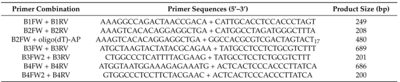
Table 1.Primer combinations used in PCR amplifications and relative dimensions of PCR products.
Primer Combination Primer Sequences (5’–3’) Product Size (bp)
B1FW + B1RV AAAGGCCAGACTAACCGACA + CATTGCACCTCCACCCTAGT 249 B2FW + B2RV AAAGTCACACAGGAGGCTGA + CATGGCCTAGATGGGCTTTA 208 B2FW + oligo(dT)-AP AAAGTCACACAGGAGGCTGA + GGCCACGCGTCGACTAGTACT17 480 B3FW + B3RV ATGCTAAGTACTATACGCAGAA + TATGCCTCCTCTGCGTCTTT 689 B3FW2 + B3RV CTGGCCCTCATTTTACGAAG + TATGCCTCCTCTGCGTCTTT 201 B4FW + B4RV ATGGTAATGGAAAGAGAAATG + ACTCACTCCCACCCTTATCA 686 B4FW2 + B4RV GTGGCCCTCCTTCTACGAAC + ACTCACTCCCACCCTTATCA 200 2.4. Sequence Analysis and Phylogenetic Relationships
The nucleotide gene sequences and the functional domains of the deduced amino acid sequences were analyzed with the SECISearch tool (http://genome.unl.edu/SECISearch.html) and ScanProsite (http://prosite.expasy.org/), respectively. The amino acid sequences used to establish the En-MsrB phylogenetic relationships were aligned with the T-Coffee multiple sequence alignment package [16]. The statistical selection of the best-fit models was carried out with ProtTest 3 [17]. The phylogenetic relationships were assessed using 122 candidate models, and adopting the ‘Bayesian Information’ (BI) criterion along with the ‘Akaike Information’ criterion used also in the corrected version. The best-fit (clock, no-clock, or relaxed clock) model was selected as previously described [18], by assessing the marginal model likelihoods by means of the stepping-stone method that samples a series of distributions representing different mixtures of the posterior and prior distributions [19]. It was applied to the dataset of the MsrB amino acid sequences using 510,000 generations with a diagnostic frequency of 2,500 in two independent runs for each model.
The phylogenetic tree was built with the BI method implemented in MrBayes 3.2 (Department of Biodiversity Informatics, Swedish Museum of Natural History, Stockholm, Sweden) [20], and displayed with the FigTree v1.3 software (Institute of Evolutionary Biology, Edinburgh, UK). Four independent runs, each with four simultaneous ‘Markov Chain Monte Carlo’ chains, were performed for 106generations and sampled every 103. In applying the maximum likelihood (ML) method implemented in PhyML 3.0 (Méthodes et Algorithmes pour la Bioinformatique, LIRMM, Centre National de la Recherche Scientifique, Université de Montpellier, Montpellier, France) [21], bootstrap analyses were performed on 104trees and the tree topology was improved with both the ‘Nearest Neighbor Interchange’ and the ‘Subtree Pruning and Re-grafting’.
3. Results
3.1. Gene Identification and Nucleotide Sequences
The amino acid sequence of the previously identified E. nobilii MsrB protein [11], hereafter designated as En-MsrB1, was used as query to search for new genes encoding Msr of type B on the BLAST database of a draft of the E. nobilii macronuclear genome. In addition to the En-MsrB1 gene, three new genes were identified and designated En-MsrB2, En-MsrB3 and En-MsrB4 (Figure1).
The four En-MsrB genes have the typical organization of the Euplotes macronuclear genes, containing a single open reading frame (ORF) flanked with 5’ leader and 3’ trailer non-coding regions capped with telomeric C4A4and G4T4repetitions. They are characterized by a 5’ leader region which is rather uniform in extension and rich in A and T repetitions arranged to form canonical TATA boxes and less common TAATA motifs (in the En-MsrB1 gene) specific for the transcription regulation. In contrast, the 3’ trailer region is extremely variable in length (from only 36 bp in the En-MsrB3 gene to 404 bp in the En-MsrB2 gene) and devoid of canonical AATAAA polyadenylation signals for the termination of
transcription, except that in the En-MsrB2 gene which contains one such signal. The ORF is identified with the first ATG start codon, except that in the En-MsrB4 gene which includes TAA or TAG stop codons immediately downstream of the first five ATG codons. The ORF of this gene thus coincides with the sixth ATG.
Biology 2017, 5, 4 4 of 13
TAA or TAG stop codons immediately downstream of the first five ATG codons. The ORF of this gene thus coincides with the sixth ATG.
Figure 1. Nucleotide sequences of the En-MsrB genes and deduced amino acid sequences. In the
nucleotide sequences, the telomeric repeats are in italics, the 5’ leader and 3’ trailer regions in lower case letters and the open reading frame in capital letters. The putative transcriptional regulation and polyadenylation signals are underlined, and the in-frame TGA codons highlighted in grey. In the amino acid sequences, the CXXC motifs and the catalytic sites are highlighted in yellow and grey, respectively, and the catalytic and the resolving cysteines are highlighted in red and green, respectively. En-MsrB1 ccccaaaaccccaaaaccccaaaaccccttaatatccagcttttgtagatacattaattaataatttacattaaatcaATGAGTGAAGAGACTAAAGAT M S E E T K D GACCTAAAGGCCAGACTAACCGACATGCAGTACAATGTGACCCAAAAAGCAGCAACTGAGCCACCATTTGTTGGTAAATATAATGAATTTGATGAGGAA D L K A R L T D M Q Y N V T Q K A A T E P P F V G K Y N E F D E E GGCCATTACAACTGTGTTGTATGTGGAGACCTACTATTCAACTCCACCCAGAAGTTCAAGTGTGACTGTGGGTGGCCAGCATTCAATGATAAGGCAGGT G H Y N C V V C G D L L F N S T Q K F K C D C G W P A F N D K A G GTTATTAAGGAGATATCTGATACTACACATGGTATGACTAGGGTGGAGGTGCAATGTGATAAGTGTACTGCTCATCTTGGCCATATCTTCAATGATGGC V I K E I S D T T H G M T R V E V Q C D K C T A H L G H I F N D G CCAGCCCCAACCTACGTCAGATACTGCATCAACTCAGCAAGTATAGATTTTGTCGCTAAGTAAttgtcatgccaaatttagggtgaacgttccatgcaa P A P T Y V R Y C I N S A S I D F V A K - atcattttcaatacgtaagatatgaataggggttttggggttttggggttttgggg 551 bp En-MsrB2 ccccaaaaccccaaaaccccaaaaccccatttttaacaattataattgataattttagtagcaaaATGTCAGAAGAGGTTAAATATGATATTAAGGGGA M S E E V K Y D I K G AGCTAACAGAGCTACAGTATAAAGTCACACAGGAGGCTGATACAGAACCTGCGTTTACAGGGGAGTACTACCAACATTTGTTTCAGTCTGATCATAAAT K L T E L Q Y K V T Q E A D T E P A F T G E Y Y Q H L F Q S D H K ATAAGAAAAATAGTGGCTGGCCTGCGTTTCATGACAATAGCGGGAATGTAACAGAGGTCCCTGTTCCCAACAGTGGAGTAAAAAGGATGGAGGTGGTCT Y K K N S G W P A F H D N S G N V T E V P V P N S G V K R M E V V GATCAAATTGTAAAGCCCATCTAGGCCATGTCTCAAAGGATAATTCATCACCATCAGGTATACGGTATTGTGTAAACTCAGCAAGTCTGAACTTTAGTG C S N C K A H L G H V S K D N S S P S G I R Y C V N S A S L N F S AGAATTAAtattttgcattaacctgaacgttcaatgtaaaagtatcttcaaatattcgctgatttaaggttctctttgaagaaatagtgatgtttatag E N - catcattatttctaattagtataataaaaataaaactttgatgagttggaagtctctgagtaaacttttgaaaatatggtttttgggaacaatactatt tagcagggaaacatgttcttattcttgagagtttcgacaacacccggcctttaggtcaagattgaagtaaaatattaatataatttctgttcattactt gataatttgcagtgaacttttataaacggtcagtaaacttttacaatgtgtgtatataattaatgattttttaatttgtgcattatataaaatttcaac ctcgggtatttactaaggggttttggggttttggggttttgggg 835 bp En-MsrB3 ccccaaaaccccaaaaccccaaaaccccatttagttttataataattgtatcttaaatATGCTAAGTACTATACGCAGAATTAGGCAAAAATTATCAAC M L S T I R R I R Q K L S T TACCACAATCACAAGATCTGCGGCCATTACCCACCCAAAGTCACACCTCAAGAACCTTCAAAATGAATCAGAACAGCCATCAGCATCCTATAAACCATA T T I T R S A A I T H P K S H L K N L Q N E S E Q P S A S Y K P Y CTTTATGTCATCTCTATCAGCCATGTATCAATTCAATATCCTCGCATTCTTCTTATTATCAATATCCCTATCAAAACAAGTAACAGCTGCTGAGACATC F M S S L S A M Y Q F N I L A F F L L S I S L S K Q V T A A E T S CCACGACCCCAGATTTCATAAATGGAGATCTCAGTCACCTGCTAGTTATATTTACTTATTTGCCCATGAATCAGACGCTGATCTTCAAAGTCGTCTGAC H D P R F H K W R S Q S P A S Y I Y L F A H E S D A D L Q S R L T CCCTTTACAATATGAGGTCACCCAGAATCATGGTACTGAACCTCCTTTCAAAAATGAGTACAACGGTAATAAAGATAAAGGTGACTATAAGTGTGTAGT P L Q Y E V T Q N H G T E P P F K N E Y N G N K D K G D Y K C V V ATGTGGTGCTCTCTTATTCCACTCATCCCACAAATATGACTCAGGGAGCGGCTGGCCCTCATTTTACGAAGCTGACGATGAAATCAAAGAGGTGGTGGA C G A L L F H S S H K Y D S G S G W P S F Y E A D D E I K E V V D TGAGTCCCATGGCATGTCTAGAAAAGAGGTACAGTGCAACAGCTGTGGAGCACACCTCGGCCACGTCTTCCCTGACGGCCCCATGCCTACAGGCAAGAG E S H G M S R K E V Q C N S C G A H L G H V F P D G P M P T G K R GTACTGCATGAACTCAGCCAGTATGCTCTTCGTAAAAGACGCAGAGGAGGCATAAtacatttcaactttttcaatacttaaatttggattaggggtttt Y C M N S A S M L F V K D A E E A - ggggttttggggttttgggg 812 bp En-MsrB4 ccccaaaaccccaaaaccccaaaaccccatgatttagagattatattgcaagatataaatatatatggtaatggaaagagaaatgtcttatctgtaagt ctaagagctagattacactaactttatgtagattacattctATGTTTATAATCTTTAGTAGCGAAAATGCAGAGTTAACTTATTGATTGTTTAGAGTCT M F I I F S S E N A E L T Y C L F R V CAAAATTCGAAAAAGTGTGAGGTAATATCAGACTTAAGGACAAGATAGGGATATTTGCATTATTGTGAATTTTATGCATGGTACACATTGTTGAATGCC S K F E K V C G N I R L K D K I G I F A L L C I L C M V H I V E C ATGAAATTAGCCTAAATCCTGATTTTGACACTCACCAGAGTCCTTATGACATATTATACATCTTAGAAGGTGAAGAAAAGCACAAACTCCGTGAATCCC H E I S L N P D F D T H Q S P Y D I L Y I L E G E E K H K L R E S TCTCATCTCTCCAATTCGAAGTAACCCAAAATGACTTCACTGAACCACCTTACCGCAATGACTACTATGACCTAAAAGACAAAGGTGACTACAAGTGCG L S S L Q F E V T Q N D F T E P P Y R N D Y Y D L K D K G D Y K C TAATCTGAGGCCACAACCTATTCAAATCTGACACCAAATATGACTCAGGTTCAGGGTGGCCCTCCTTCTACGAACCTGATGGTGAGGTAGTAGAGAGGC V I C G H N L F K S D T K Y D S G S G W P S F Y E P D G E V V E R CAGATACCTCATCAAATAGGAATAGGACGGAGGTGGTGTGAGGCAACTGTGGAGCCCATTTAGGCCATCTGTTTAAGGATGGGCCTGAGGATAAGACTG P D T S S N R N R T E V V C G N C G A H L G H L F K D G P E D K T GTATGAGGTACTGTATTAACTCAGCTAGCTTGAAGTTTGATAAGGGTGGGAGTGAGTAAagcctgtttttaattttttcaatgattggggtaggatg G M R Y C I N S A S L K F D K G G S E - aggggttttggggttttggggttttgggg 819 bp
Figure 1. Nucleotide sequences of the En-MsrB genes and deduced amino acid sequences. In the nucleotide sequences, the telomeric repeats are in italics, the 5’ leader and 3’ trailer regions in lower case letters and the open reading frame in capital letters. The putative transcriptional regulation and polyadenylation signals are underlined, and the in-frame TGA codons highlighted in grey. In the amino acid sequences, the CXXC motifs and the catalytic sites are highlighted in yellow and grey, respectively, and the catalytic and the resolving cysteines are highlighted in red and green, respectively.
Biology 2017, 6, 4 5 of 13
In-frame TGA stop codons distinguish the ORF of the En-MsrB2 and En-MsrB4 genes. However, they do not act as effective stop signals because Euplotes uses the TGA codon to specify cysteine [22], or even selenocysteine [23]. The co-translational incorporation of this amino acid into the nascent protein chain is directed by specific mRNA secondary structures designated selenocysteine insertion sequence (SECIS) elements, which are responsible for the mRNA binding to selenocysteine-specific translation factors [24]. The En-MsrB2 and En-MsrB4 genes lack any SECIS element, which excludes a selenoprotein nature of their products.
3.2. Amino Acid Sequences
The proteins (En-MsrB1 to En-MsrB4) encoded by the four En-MsrB genes markedly vary from one another in chain length (112 amino acid residues in En-MsrB2, 229 in En-MsrB3) and degree of sequence similarity (42.6% identity in 122 residue-overlaps between En-MsrB2 and En-MsrB3, 56.8% identity in 139 residue-overlaps between En-MsrB3 and En-MsrB4) (Figure2). However, in spite of these variations, they all uniformly show the MsrB-specific ‘catalytic Cys’ lying within a highly conserved Arg-Tyr-Cys-Met/Val/Ile-Asn-Ser-Ala-Ser sequence segment of the carboxyl-terminal region.
Biology 2017, 5, 4 5 of 13
In-frame TGA stop codons distinguish the ORF of the En-MsrB2 and En-MsrB4 genes. However, they do not act as effective stop signals because Euplotes uses the TGA codon to specify cysteine [22], or even selenocysteine [23]. The co-translational incorporation of this amino acid into the nascent protein chain is directed by specific mRNA secondary structures designated selenocysteine insertion sequence (SECIS) elements, which are responsible for the mRNA binding to selenocysteine-specific translation factors [24]. The En-MsrB2 and En-MsrB4 genes lack any SECIS element, which excludes a selenoprotein nature of their products.
3.2. Amino Acid Sequences
The proteins (En-MsrB1 to En-MsrB4) encoded by the four En-MsrB genes markedly vary from one another in chain length (112 amino acid residues in En-MsrB2, 229 in En-MsrB3) and degree of sequence similarity (42.6% identity in 122 residue-overlaps between En-MsrB2 and En-MsrB3, 56.8% identity in 139 residue-overlaps between En-MsrB3 and En-MsrB4) (Figure 2). However, in spite of these variations, they all uniformly show the MsrB-specific ‘catalytic Cys’ lying within a highly conserved Arg-Tyr-Cys-Met/Val/Ile-Asn-Ser-Ala-Ser sequence segment of the carboxyl-terminal region.
Figure 2. Amino acid sequence alignment of the En-MsrB proteins. The alignment was maximized by
gap insertion and residues identical in three or four sequences are highlighted in grey. The CXXC motifs are in bold and italics. The resolving and catalytic cysteines are indicated by grey and filled arrowheads, respectively. The conserved catalytic site is boxed. Signals for mitochondrial targeting, nuclear translocation, transmembrane domain and ER retention are highlighted in green, red, blue and yellow, respectively.
According to the TargetP (Center for Biological Sequence Analysis, Technical University of Denmark, Lyngby, Denmark) and PSORT II softwares (Human Genome Center, Institute for Medical Science, University ot Tokyo, Japan) [25,26], each En-MsrB protein takes an its own specific subcellular localization: (i) the cytoplasm for En-MsrB1, which lacks any signal-peptide motif necessary for the protein transport across membranes; (ii) the nucleus for En-MsrB2, in which the motif His43-Lys-Tyr-Lys-Lys47 (canonically formed by a repetition of basic amino acids) forms a mono-partite nuclear localization signal [27]; (iii) mitochondria for En-MsrB3, in which the first 20 amino acids of the amino-terminal region form a mitochondrial signal sequence; and (iv) the endoplasmic reticulum (ER) for En-MsrB4 which, in addition to including a putative transmembrane domain (positions 35–51) inside its amino-terminal region, also possesses a ER-specific retention signal provided by the Lys199-Gly-Gly-Ser202 motif in the carboxyl terminal region (Figure 2).
3.3. Phylogenetic Relationships En-MsrB1 --- En-MsrB2 --- En-MsrB3 MLSTIRRIRQKLSTTTITRSAAITHPKSHLKNLQNESEQPSASYKPYFMSSLSAMYQFNI 60 En-MsrB4 ---MFIIFSSENAELTYCLFRVSKFEKVCGNIRLKDK 34 En-MsrB1 ---MSEETKDDLKARLTDMQYNVT 21 En-MsrB2 ---MSEEVKYDIKGKLTELQYKVT 21 En-MsrB3 LAFFLLSISLSKQVTAAETSHDPRFHKWRSQSPASYIYLFAHESDADLQSRLTPLQYEVT 120 En-MsrB4 IGIFALLCILCMVHIVECHEISLNPDFDTHQSPYDILYILEGEEKHKLRESLSSLQFEVT 94 En-MsrB1 QKAATEPPFVGKYNEFDEEGHYNCVVCGDLLFNSTQKFKCDCGWPAFNDKAGVIKEISDT 81 En-MsrB2 QEADTEPAFTGEYYQH---LFQSDHKYKKNSGWPAFHDNSGNVTEVPVP 67 En-MsrB3 QNHGTEPPFKNEYNGNKDKGDYKCVVCGALLFHSSHKYDSGSGWPSFYEADDEIKEVVDE 180 En-MsrB4 QNDFTEPPYRNDYYDLKDKGDYKCVICGHNLFKSDTKYDSGSGWPSFYEPDGEVVERPDT 154 En-MsrB1 THGMTRVEVQCDKCTAHLGHIFNDGPAP-TYVRYCINSASIDFVAK---- 126 En-MsrB2 NSGVKRMEVVCSNCKAHLGHVSKDNSSP-SGIRYCVNSASLNFSEN---- 112 En-MsrB3 SHGMSRKEVQCNSCGAHLGHVFPDGPMP-TGKRYCMNSASMLFVKDAEEA 229 En-MsrB4 SSNRNRTEVVCGNCGAHLGHLFKDGPEDKTGMRYCINSASLKFDKGGSE- 203
Figure 2.Amino acid sequence alignment of the En-MsrB proteins. The alignment was maximized by gap insertion and residues identical in three or four sequences are highlighted in grey. The CXXC motifs are in bold and italics. The resolving and catalytic cysteines are indicated by grey and filled arrowheads, respectively. The conserved catalytic site is boxed. Signals for mitochondrial targeting, nuclear translocation, transmembrane domain and ER retention are highlighted in green, red, blue and yellow, respectively.
According to the TargetP (Center for Biological Sequence Analysis, Technical University of Denmark, Lyngby, Denmark) and PSORT II softwares (Human Genome Center, Institute for Medical Science, University ot Tokyo, Japan) [25,26], each En-MsrB protein takes an its own specific subcellular localization: (i) the cytoplasm for En-MsrB1, which lacks any signal-peptide motif necessary for the protein transport across membranes; (ii) the nucleus for En-MsrB2, in which the motif His43-Lys-Tyr-Lys-Lys47(canonically formed by a repetition of basic amino acids) forms a mono-partite nuclear localization signal [27]; (iii) mitochondria for En-MsrB3, in which the first 20 amino acids of the amino-terminal region form a mitochondrial signal sequence; and (iv) the endoplasmic reticulum (ER) for En-MsrB4 which, in addition to including a putative transmembrane domain (positions 35–51) inside its amino-terminal region, also possesses a ER-specific retention signal provided by the Lys199-Gly-Gly-Ser202motif in the carboxyl terminal region (Figure2).
3.3. Phylogenetic Relationships
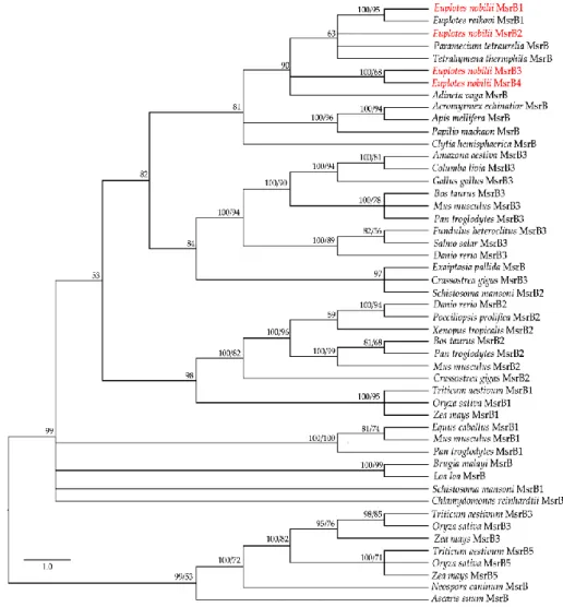
The application of the BI and ML methods to a selected panel of eukaryotic MsrB amino acid sequences available from the GenBank database (MsrBs’ accession numbers can be found in AppendixA, TableA1) generated closely similar trees (Figure3), in which the four En-MsrBs group together into a statistically well supported clade including MsrB sequences of other ciliates and a MsrB sequence described as of non-metazoan origin from the bdelloid rotifer, Adineta vaga [28]. Within this clade, En-MsrB1 finds its counterpart with a MsrB sequence from E. raikovi, while En-MsrB2 associates with MsrBs from Paramecium and Tetrahymena, and En-MsrB4 and En-MsrB4 form their own branch.
Biology 2017, 5, 4 6 of 13
The application of the BI and ML methods to a selected panel of eukaryotic MsrB amino acid sequences available from the GenBank database (MsrBs’ accession numbers can be found in Appendix, Table A1) generated closely similar trees (Figure 3), in which the four En-MsrBs group together into a statistically well supported clade including MsrB sequences of other ciliates and a MsrB sequence described as of non-metazoan origin from the bdelloid rotifer, Adineta vaga [28]. Within this clade, MsrB1 finds its counterpart with a MsrB sequence from E. raikovi, while En-MsrB2 associates with MsrBs from Paramecium and Tetrahymena, and En-MsrB4 and En-MsrB4 form their own branch.
Figure 3. Phylogenetic relationships of the En-MsrB proteins (in red) with MsrBs from other
organisms. Tree reconstruction based on both BI and ML methods. Posterior probability (first number) and boostrap values (second number, when present) are indicated at the nodes. The scale for the branch length is shown on the left.
3.4. Gene Expression
First, we analyzed whether each En-MsrB gene is effectively expressed by preparing cDNA from RNA extracted from growing cells, and amplifying aliquots of cDNA via PCR with primer combinations specific to sequence segments adjacent to the extremities of each gene ORF (Table 1). Amplicons of each gene were obtained and directly sequenced (Figure 4A). No intron was found to interrupt the ORF sequence of the four genes. Also, no intron sequence was identified within the exceptionally long (404 bp) 3’ trailer region distinctive of the En-MsrB2 gene. Evidence for this conclusion was obtained by amplifying Figure 3.Phylogenetic relationships of the En-MsrB proteins (in red) with MsrBs from other organisms. Tree reconstruction based on both BI and ML methods. Posterior probability (first number) and boostrap values (second number, when present) are indicated at the nodes. The scale for the branch length is shown on the left.
3.4. Gene Expression
First, we analyzed whether each En-MsrB gene is effectively expressed by preparing cDNA from RNA extracted from growing cells, and amplifying aliquots of cDNA via PCR with primer combinations specific to sequence segments adjacent to the extremities of each gene ORF (Table1). Amplicons of each gene were obtained and directly sequenced (Figure4a). No intron was found to interrupt the ORF sequence of the four genes. Also, no intron sequence was identified within the exceptionally long (404 bp) 3’ trailer region distinctive of the En-MsrB2 gene. Evidence for this conclusion was obtained by amplifying cDNA preparations with a forward primer specific to
Biology 2017, 6, 4 7 of 13
a sequence internal to the En-MsrB2 ORF and a reverse primer provided by the same oligo (dT)-AP used for cDNA synthesis, and observing that the 3’ untranslated region of each transcript fully matched the uninterrupted 134-bp gene sequence comprised between the TAA stop codon of the ORF and the G nucleotide located 9-bp downstream the AATAAA polyadenylation signal.
Biology 2017, 5, 4 7 of 13
cDNA preparations with a forward primer specific to a sequence internal to the En-MsrB2 ORF and a reverse primer provided by the same oligo (dT)-AP used for cDNA synthesis, and observing that the 3’ untranslated region of each transcript fully matched the uninterrupted 134-bp gene sequence comprised between the TAA stop codon of the ORF and the G nucleotide located 9-bp downstream the AATAAA polyadenylation signal.
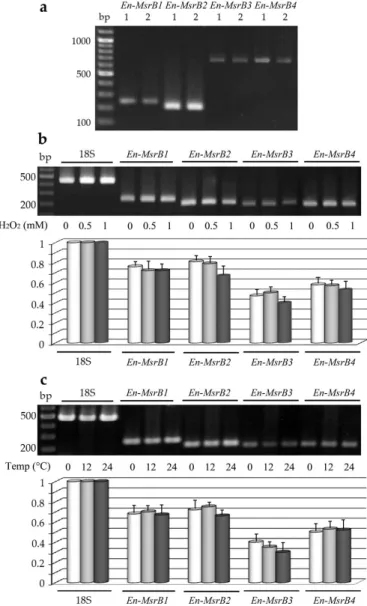
Figure 4. Expression of the E. nobilii En-MsrB genes. (A) Gel electrophoresis of PCR products obtained
by amplifying DNA (lanes 1) or cDNA (lanes 2) preparations with En-MsrB gene-specific primers. (B) Electrophoretic separation (upper panel) and relative transcript abundance (lower panel) of sqRT-PCR products obtained from cells treated for 30 min with increasing concentrations of H2O2. (C)
Electrophoretic separation (upper panel) and relative transcript abundance (lower panel) of sqRT-PCR products obtained from cells exposed for 30 min at increasing temperatures. In both (B) and (C), values were calculated taking the 18S-rRNA PCR fragments as value 1 and represent the means (± SD) of three independent experiments.
Second, we analyzed whether the expression of each En-MsrB gene can be induced to increase in cells exposed to oxidative and thermal stress conditions. The gene expression in response to the oxidative stress was analyzed in cells suspended for 30 min in fresh seawater with H2O2 added in increasing Figure 4. Expression of the E. nobilii En-MsrB genes. (a) Gel electrophoresis of PCR products obtained by amplifying DNA (lanes 1) or cDNA (lanes 2) preparations with En-MsrB gene-specific primers. (b) Electrophoretic separation (upper panel) and relative transcript abundance (lower panel) of sqRT-PCR products obtained from cells treated for 30 min with increasing concentrations of H2O2. (c) Electrophoretic separation (upper panel) and relative transcript abundance (lower panel) of
sqRT-PCR products obtained from cells exposed for 30 min at increasing temperatures. In both (b) and (c), values were calculated taking the 18S-rRNA PCR fragments as value 1 and represent the means (±SD) of three independent experiments.
Second, we analyzed whether the expression of each En-MsrB gene can be induced to increase in cells exposed to oxidative and thermal stress conditions. The gene expression in response to the oxidative stress was analyzed in cells suspended for 30 min in fresh seawater with H2O2added in increasing concentrations. The gene expression in response to the thermal stress was analyzed in cells suspended for 30 min at their standard cultivation temperature of 4◦C and at increased temperatures of 12 and 24◦C. Preparations of cDNA obtained from each cell suspension were amplified by sqRT-PCR
and the relative amounts of the amplified gene transcripts were compared between stressed and not-stressed cells (Figure4b,c). The En-MsrB genes revealed different levels of expression, with the En-MsrB3 gene systematically characterized by the production of appreciably lower amounts of transcript. However, no En-MsrB genes showed a significant quantitative difference in the transcript abundance between stressed and not-stressed cells implying that they behave as constitutive genes, each transcribed at a relatively constant level.
4. Discussion
Like numerous other eukaryotic organisms, E. nobilii relies on the activity of multiple En-MsrB genes to repair Met-oxidized proteins. However, while in animals and plants the expression of these genes is generally responsive to environmental change [29,30], in E. nobilii the expression of all four En-MsrB genes has been found to remain substantially unchanged in cells exposed to oxidative and thermal stresses. Consistently with their constitutive expression, the En-MsrB genes have a short 5’ leader non-coding region devoid of cis-active enhancer motifs directly involved in up-regulating the genetic response in stressed organisms, such as the CCCCT ‘stress-response element’ of Saccharomyces cerevisiae [31], the TGAG/CNNNGC ‘antioxidant responsive elements’ of mammalian cells [32], and the TGACNNN ‘half antioxidant response elements’ that are common in other organisms including the ciliate Tetrahymena thermophila [18,33,34].
The constitutive expression of the En-MsrB genes is likely fundamental for the efficiency of the E. nobilii physiological mechanism of ROS scavenging, that in oxidizing environments has been proposed to be constantly activated by a methionine oxidation/reduction cycling [35]. However, lack of appropriate information on the constitutive, or inducible nature of Msr genes in other Euplotes species suggests caution in concluding that the constitutive expression of the En-MsrB genes reflects a specific adaptive trait of E. nobilii the to the stressful conditions of the polar environment. Only in E. raikovi two Msr genes have been identified and initially analyzed for their expression. One gene encoding a protein structurally related to En-MsrB1 is constitutively expressed like the En-MsrB genes [11]. The other gene encoding a MsrA protein seems to be an inducible gene [10]. However, it does not appear to be endogenous to E. raikovi. It was supposed to derive from Alphaproteobacteria through a phenomenon of lateral gene transfer [36].
The proteins synthesized by the four En-MsrB genes showed remarkably diversified amino acid sequences. The functionally more important variations reside in the number of the CXXC motifs that cooperate for the binding of the zinc ion, and the presence/absence and localization of a Cys residue, known as ‘resolving cysteine’, that is involved in the mechanism of MsrB regeneration (Figure 5). En-MsrB1 and En-MsrB3 are the only two proteins that, like many other eukaryotic MsrBs, orthodoxically contain two CXXC motifs for the zinc-ion binding (i.e., Cys45-Val-Val-Cys48and Cys92-Asp-Lys-Cys95in En-MsrB1; Cys144-Val-Val-Cys147and Cys191-Asn-Ser-Cys194in En-MsrB3). A single CXXC motif (i.e., Cys78-Ser-Asn-Cys81) is distinctive of En-MsrB2 which, as a consequence, should be unable to bind the zinc ion. The crucial role that this ion plays in the stabilization of the MsrB molecular structure [37–39], thus suggests that En-MsrB2 may rely on an increased structural flexibility to interact more effectively with carrier proteins on the way to its subcellular nuclear localization. En-MsrB4 is another structurally rather eccentric MsrB protein because it includes a third CXXC motif (i.e., Cys42-Ile-Leu-Cys45) in the amino terminal region. The role, if any, played by this additional motif remains enigmatic.
With regard to the presence/absence of the resolving cysteine, En-MsrB1 is unique because it includes both this cysteine (Cys63) and the catalytic one (Cys115) in the carboxyl-terminal region. This inclusion makes it a member of the so-called ‘2-Cys MsrBs’, in which the resolving and catalytic cysteines form an intra-chain disulfide bridge destined to be reduced by thioredoxin through disulfide exchange [40–42]. Instead, all other En-MsrBs are characterized by the replacement of the canonical resolving cysteine with a Ser residue. However, while in the recycling process En-MsrB4 may use one of the three additional Cys residues included in its amino-terminal region as resolving cysteine,
Biology 2017, 6, 4 9 of 13
En-MsrB2 and En-MsrB3 do not possess any additional Cys residue to replace the loss of the canonical resolving cysteine. These two proteins thus belong to the group of the so-called ‘1-Cys MsrBs’, whose regeneration avoids the formation of an intra-chain disulfide bond and involves two alternative mechanisms. In one mechanism, glutathione is the reductant of the sulfenic acid formed on the catalytic cysteine after the substrate reduction, and the deglutathionylation step is then achieved by the activity of glutaredoxins [43]. In the second mechanism, thioredoxins directly interact with oxidized 1-Cys MsrBs and regenerate their activity without the help of any other thiol compound [44,45]. Which of the two mechanisms is used by E. nobilii to regenerate its En-MsrB2 and En-MsrB3 proteins remains to be investigated.
Biology 2017, 5, 4 9 of 13
the canonical resolving cysteine. These two proteins thus belong to the group of the so-called ‘1-Cys MsrBs’, whose regeneration avoids the formation of an intra-chain disulfide bond and involves two alternative mechanisms. In one mechanism, glutathione is the reductant of the sulfenic acid formed on the catalytic cysteine after the substrate reduction, and the deglutathionylation step is then achieved by the activity of glutaredoxins [43]. In the second mechanism, thioredoxins directly interact with oxidized 1-Cys MsrBs and regenerate their activity without the help of any other thiol compound [44,45]. Which of the two mechanisms is used by E. nobilii to regenerate its En-MsrB2 and En-MsrB3 proteins remains to be investigated.
Figure 5. Schematic representation of the En-MsrB protein structure. The positions of the zinc-ion
binding CXXC motifs, the catalytic and resolving cysteines, and the additional Cys residues specific of the En-MsrB4 amino-terminal region are indicated by bars. Boxes of different color indicate the relative positions and length of the signals for mitochondrial targeting (green), ER retention (yellow), nuclear localization (red) and trans-membrane domain (blue).
In addition to the four En-MsrB genes, the scanning of the E. nobilii genome identified a fifth gene encoding a ‘MsrB-like’ protein and at least three gene sequences encoding Msr enzymes of type A. Although the MsrB-like protein contains a zinc-ion binding site represented by a double CXXC motif, it lacks the MsrB-specific catalytic cysteine which has been replaced with a Thr residue. Because of this substitution it thus more closely recalls the structure of the ‘SelR’ proteins with unknown function of Oxytricha, Stylonychia, Paramecium and Tetrahymena which similarly possess a double CXXC motif and a canonical catalytic cysteine replaced with a Ser residue.
With regard to the MsrA-coding gene sequences, we can anticipate that they show an origin which is clearly not endogenous to E. nobilii, because they lack the telomeric nucleotide C4A4 repeats that are the distinctive structural marker of every Euplotes macronuclear gene [46]. Instead, they unmistakably cluster close to sequences of prokaryotic MsrA genes, in particular to MsrA gene sequences that are specific of species of the bacterium Francisella. These bacteria live as common and pervasive cytoplasmic endosymbionts of Euplotes species [47,48], suggesting that E. nobilii demands the reduction of its methionine-sulfoxide S-forms to the exogenous enzymatic activity of its bacterial endosymbionts.
5. Conclusions
The four structurally distinct anti-oxidant MsrB genes that have been identified in the somatic (macronuclear) genome of the polar ciliate E. nobilii behave as constitutive genes, in contrast with the inducible response that is commonly shown by Msr genes in other organisms. Their products are markedly diversified in the number and location of the Cys residues involved in the regeneration and zinc-ion binding mechanisms, and include sequence domains specific to localize and be active in distinct subcellular compartments.
Figure 5. Schematic representation of the En-MsrB protein structure. The positions of the zinc-ion binding CXXC motifs, the catalytic and resolving cysteines, and the additional Cys residues specific of the En-MsrB4 amino-terminal region are indicated by bars. Boxes of different color indicate the relative positions and length of the signals for mitochondrial targeting (green), ER retention (yellow), nuclear localization (red) and trans-membrane domain (blue).
In addition to the four En-MsrB genes, the scanning of the E. nobilii genome identified a fifth gene encoding a ‘MsrB-like’ protein and at least three gene sequences encoding Msr enzymes of type A. Although the MsrB-like protein contains a zinc-ion binding site represented by a double CXXC motif, it lacks the MsrB-specific catalytic cysteine which has been replaced with a Thr residue. Because of this substitution it thus more closely recalls the structure of the ‘SelR’ proteins with unknown function of Oxytricha, Stylonychia, Paramecium and Tetrahymena which similarly possess a double CXXC motif and a canonical catalytic cysteine replaced with a Ser residue.
With regard to the MsrA-coding gene sequences, we can anticipate that they show an origin which is clearly not endogenous to E. nobilii, because they lack the telomeric nucleotide C4A4repeats that are the distinctive structural marker of every Euplotes macronuclear gene [46]. Instead, they unmistakably cluster close to sequences of prokaryotic MsrA genes, in particular to MsrA gene sequences that are specific of species of the bacterium Francisella. These bacteria live as common and pervasive cytoplasmic endosymbionts of Euplotes species [47,48], suggesting that E. nobilii demands the reduction of its methionine-sulfoxide S-forms to the exogenous enzymatic activity of its bacterial endosymbionts.
5. Conclusions
The four structurally distinct anti-oxidant MsrB genes that have been identified in the somatic (macronuclear) genome of the polar ciliate E. nobilii behave as constitutive genes, in contrast with the inducible response that is commonly shown by Msr genes in other organisms. Their products are markedly diversified in the number and location of the Cys residues involved in the regeneration and zinc-ion binding mechanisms, and include sequence domains specific to localize and be active in distinct subcellular compartments.
Acknowledgments:Research financially supported by the ‘Programma Nazionale di Ricerche in Antartide’ (PNRA). Author Contributions:Francesca Ricci performed the expression experiments; Federico Lauro, Joseph J. Grzymski, and Robert Read assembled the genomic data and created the BLAST database; Rigers Bakiu performed phylogenetic analyses; Gianfranco Santovito analyzed the data; Pierangelo Luporini and Adriana Vallesi conceived and designed the experiments, prepared cells, analyzed data and wrote the manuscript with contributions of all authors.
Conflicts of Interest:The authors declare no conflict of interest. Appendix A
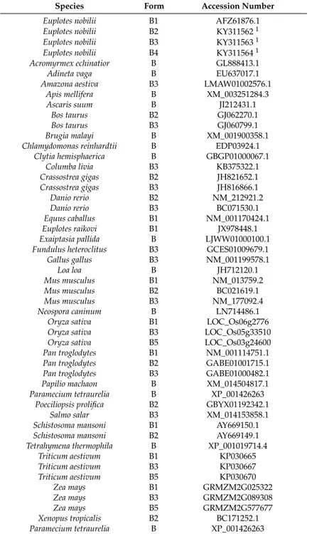
Table A1.MsrB proteins included in the phylogenetic analysis: Source, form and GenBank accession number.
Species Form Accession Number
Euplotes nobilii B1 AFZ61876.1 Euplotes nobilii B2 KY3115621 Euplotes nobilii B3 KY3115631 Euplotes nobilii B4 KY3115641 Acromyrmex echinatior B GL888413.1
Adineta vaga B EU637017.1
Amazona aestiva B3 LMAW01002576.1 Apis mellifera B XM_003251284.3
Ascaris suum B JI212431.1
Bos taurus B2 GJ062270.1
Bos taurus B3 GJ060799.1
Brugia malayi B XM_001900358.1 Chlamydomonas reinhardtii B EDP03924.1
Clytia hemisphaerica B GBGP01000067.1 Columba livia B3 KB375322.1 Crassostrea gigas B2 JH821652.1 Crassostrea gigas B3 JH816866.1 Danio rerio B2 NM_212921.2 Danio rerio B3 BC071530.1 Equus caballus B1 NM_001170424.1 Euplotes raikovi B1 JX978448.1 Exaiptasia pallida B LJWW01000100.1 Fundulus heteroclitus B3 GCES01009679.1 Gallus gallus B3 NM_001199578.1 Loa loa B JH712120.1 Mus musculus B1 NM_013759.2 Mus musculus B2 BC021619.1 Mus musculus B3 NM_177092.4 Neospora caninum B LN714486.1 Oryza sativa B1 LOC_Os06g2776 Oryza sativa B3 LOC_Os05g33510 Oryza sativa B5 LOC_Os03g24600 Pan troglodytes B1 NM_001114751.1 Pan troglodytes B2 GABE01001715.1 Pan troglodytes B3 GABE01000482.1 Papilio machaon B XM_014504817.1 Paramecium tetraurelia B XP_001426263
Poeciliopsis prolifica B2 GBYX01192342.1 Salmo salar B3 XM_014153858.1 Schistosoma mansoni B1 AY669150.1 Schistosoma mansoni B2 AY669149.1 Tetrahymena thermophila B XP_001019714.4 Triticum aestivum B1 KP030665 Triticum aestivum B3 KP030667 Triticum aestivum B5 KP030670 Zea mays B1 GRMZM2G025322 Zea mays B3 GRMZM2G089308 Zea mays B5 GRMZM2G577677 Xenopus tropicalis B2 BC171252.1 Paramecium tetraurelia B XP_001426263 1This work.
Biology 2017, 6, 4 11 of 13
References
1. Vogt, W. Oxidation of methionyl residues in proteins: Tools, targets, and reversal. Free Radic. Biol. Med. 1995, 18, 93–105. [CrossRef]
2. Friguet, B. Oxidized protein degradation and repair in ageing and oxidative stress. Febs Lett. 2006, 580, 2910–2916. [CrossRef]
3. Stadtman, E.R.; Levine, R.L. Protein oxidation. Ann. N. Y. Acad. Sci. 2000, 899, 191–208. [CrossRef] [PubMed] 4. Moskovitz, J. Methionine sulfoxide reductases: Ubiquitous enzymes involved in antioxidant defense, protein regulation, and prevention of aging-associated diseases. Biochim. Biophys. Acta 2005, 1703, 213–219. [CrossRef] [PubMed]
5. Valbonesi, A.; Luporini, P. Description of two new species of Euplotes and Euplotes rariseta from Antarctica. Polar Biol. 1990, 11, 47–53. [CrossRef]
6. Di Giuseppe, G.; Erra, F.; Dini, F.; Alimenti, C.; Vallesi, A.; Pedrini, B.; Wüthrich, K.; Luporini, P. Antarctic and Arctic populations of the ciliate Euplotes nobilii show common pheromone-mediated cell-cell signaling and cross-mating. Proc. Natl. Acad. Sci. USA 2011, 108, 3181–3186. [CrossRef] [PubMed]
7. Di Giuseppe, G.; Barbieri, M.; Vallesi, A.; Luporini, P.; Dini, F. Phylogeographical pattern of Euplotes nobilii, a protist ciliate with a bipolar biogeographical distribution. Mol. Ecol. 2013, 22, 4029–4037. [CrossRef] [PubMed]
8. Di Giuseppe, G.; Dini, F.; Vallesi, A.; Luporini, P. Genetic relationships in bipolar species of the protist ciliate, Euplotes. Hydrobiologia 2015, 761, 71–83. [CrossRef]
9. Jiang, J.M.; Zhang, Q.Q.; Warren, A.; Al-Rasheid, K.A.S.; Song, W.B. Morphology and SSU rRNA gene-based phylogeny of two marine Euplotes species, E. orientalis spec. nov. and E. raikovi Agamaliev, 1966 (Ciliophora, Euplotida). Eur. J. Protistol. 2010, 46, 121–132. [CrossRef] [PubMed]
10. Dobri, N.; Ngueng Oumarou, E.E.; Alimenti, C.; Vallesi, A. Polar and non-polar species of the protozoan ciliate Euplotes behave differently in response to environmental oxidative stress. Ital. J. Zool. 2014, 81, 409–414. [CrossRef]
11. Dobri, N.; Ngueng Oumarou, E.E.; Alimenti, C.; Ortenzi, C.; Luporini, P.; Vallesi, A. Methionine sulfoxide reduction in ciliates: Characterization of the ready-to-use methionine sulfoxide-R-reductase genes in Euplotes. Gene 2013, 515, 110–116. [CrossRef] [PubMed]
12. Jahn, C.L.; Klobutcher, L.A. Genome remodeling in ciliated protozoa. Ann. Rev. Microbiol. 2002, 56, 489–520. [CrossRef] [PubMed]
13. Vallesi, A.; Di Pretoro, B.; Ballarini, P.; Apone, F.; Luporini, P. A novel protein kinase from the ciliate Euplotes raikovi with close structural identity to the mammalian intestinal and male-germ cell kinases: Characterization and functional implications in the autocrine pheromone signaling loop. Protist 2010, 161, 250–263. [CrossRef] [PubMed]
14. Grabherr, M.G.; Haas, B.J.; Yassour, M.; Levin, J.Z.; Thompson, D.A.; Amit, I.; Adiconis, X.; Fan, L.; Raychowdhury, R.; Zeng, Q.D.; et al. Full-length transcriptome assembly from RNA-Seq data without a reference genome. Nat. Biotechnol. 2011, 29, 644–652. [CrossRef] [PubMed]
15. Altschul, S.F.; Madden, T.L.; Schäffer, A.A.; Zhang, J.; Zhang, Z.; Miller, W.; Lipman, D.J. Gapped BLAST and PSI-BLAST: A new generation of protein database search programs. Nucleic Acids Res. 1997, 24, 3389–3402. [CrossRef]
16. Notredame, C.; Higgins, D.G.; Heringa, J. T-coffee: A novel method for fast and accurate multiple sequence alignment. J. Mol. Biol. 2000, 302, 205–217. [CrossRef] [PubMed]
17. Darriba, D.; Taboada, G.L.; Doallo, R.; Posada, D. ProtTest 3: Fast selection of best-fit models of protein evolution. Bioinformatics 2011, 27, 1164–1165. [CrossRef] [PubMed]
18. Ferro, D.; Bakiu, R.; De Pittà, C.; Boldrin, F.; Cattalini, F.; Pucciarelli, S.; Miceli, C.; Santovito, G. Cu, Zn superoxide dismutases from Tetrahymena thermophila: Molecular evolution and gene expression of the first line of antioxidant defenses. Protist 2015, 166, 131–145. [CrossRef] [PubMed]
19. Xie, W.; Lewis, P.O.; Fan, Y.; Kuo, L.; Chen, M.H. Improving marginal likelihood estimation for Bayesian phylogenetic model selection. Syst. Biol. 2011, 60, 150–160. [CrossRef] [PubMed]
20. Ronquist, F.; Teslenko, M.; Mark, P.V.D.; Ayres, D.L.; Darling, A.; Höhna, S.; Larget, B.; Liu, L.; Suchard, M.A.; Huelsenbeck, J.P. MrBayes 3.2: Efficient Bayesian phylogenetic inference and model choice across a large model space. Syst. Biol. 2012, 61, 539–542. [CrossRef] [PubMed]
21. Guindon, S.; Dufayard, J.F.; Lefort, V.; Anisimova, M.; Hordijk, W.; Gascuel, O. New algorithms and methods to estimate maximum-likelihood phylogenies: Assessing the performance of PhyML 3.0. Syst. Biol. 2010, 59, 307–321. [CrossRef] [PubMed]
22. Meyer, F.; Schmidt, H.J.; Plümper, E.; Hasilik, A.; Mersmann, G.; Meyer, H.E.; Engström, A.; Heckmann, K. UGA is translated as cysteine in pheromone 3 of Euplotes octocarinatus. Proc. Natl. Acad. Sci. USA 1991, 88, 3758–3761. [CrossRef] [PubMed]
23. Turanov, A.A.; Lobanov, A.V.; Formenko, D.E.; Morrison, H.G.; Sogin, M.L.; Klobutcher, L.A.; Hatfield, D.L.; Gladyshev, V.N. Genetic code supports targeted insertion of two amino acids by one codon. Science 2009, 323, 259–261. [CrossRef] [PubMed]
24. Su, D.; Li, Y.; Gladyshev, V.N. Selenocysteine insertion directed by the 30-UTR SECIS element in Escherichia coli. Nucleic Acids Res. 2005, 33, 2486–2492. [CrossRef] [PubMed]
25. Emanuelsson, O.; Brunak, S.; Heijne, G.V.; Nielsen, H. Locating proteins in the cell using TargetP, SignalP and related tools. Nat. Protoc. 2007, 2, 953–971. [CrossRef] [PubMed]
26. Nakai, K.; Horton, P. PSORT: A program for detecting sorting signals in proteins and predicting their subcellular localization. Trends Biochem. Sci. 1999, 24, 34–36. [CrossRef]
27. Lange, A.; Mills, R.E.; Lange, C.J.; Stewart, M.; Devine, S.E.; Corbett, A.H. Classical nuclear localization signals: Definition, function, and interaction with importin α. J. Biol. Chem. 2007, 282, 5101–5105. [CrossRef] [PubMed]
28. Hespeels, B.; Flot, J.F.; Derzelle, A.; Van Doninck, K. Evidence for Ancient Horizontal Gene Acquisitions in Bdelloid Rotifers of the Genus Adineta. In Evolutionary Biology: Genome Evolution, Speciation, Coevolution and Origin of Life; Pontarotti, P., Ed.; Springer International Publishing: Cham, Switzerland, 2014; pp. 207–225. 29. Zhu, J.; Ding, P.; Li, Q.; Gao, Y.; Chen, F.; Xia, G. Molecular characterization and expression profile of
methionine sulfoxide reductase gene family in maize (Zea mays) under abiotic stresses. Gene 2015, 562, 159–168. [CrossRef] [PubMed]
30. Liu, F.; Gong, Z.H.; Zhang, W.X.; Wang, Y.; Ma, L.T.; Wang, H.F.; Guo, X.Q.; Xu, B.H. Identification and characterization of a novel methionine sulfoxide reductase B gene (AccMsrB) from Apis cerana cerana (Hymenoptera: Apidae). Ann. Entomol. Soc. Am. 2015, 108, 575–584. [CrossRef]
31. Kobayashi, N.; McEntee, K. Identification of cis and trans components of a novel heat shock stress regulatory pathway in Saccharomyces cerevisiae. Mol. Cell. Biol. 1993, 13, 248–256. [CrossRef] [PubMed]
32. Reddy, S.P. The antioxidant response element and oxidative stress modifiers in airway diseases. Curr. Mol. Med. 2008, 8, 376–383. [CrossRef] [PubMed]
33. Ferro, D.; Franchi, N.; Mangano, V.; Bakiu, R.; Cammarata, M.; Parrinello, N.; Santovito, G.; Ballarin, L. Characterization and metal-induced gene transcription of two new copper zinc superoxide dismutases in the solitary ascidian Ciona intestinalis. Aquat. Toxicol. 2013, 140, 369–379. [CrossRef] [PubMed]
34. Franchi, N.; Ferro, D.; Ballarin, L.; Santovito, G. Expression of genes involved in glutathione biosynthesis in the solitary tunicate Ciona intestinalis exposed to heavy metals. Aquat. Toxicol. 2012, 114, 14–22. [CrossRef] [PubMed]
35. Luo, S.; Levine, R.L. Methionine in proteins defends against oxidative stress. Faseb J. 2009, 23, 464–472. [CrossRef] [PubMed]
36. Dobri, N.; Candelori, A.; Ricci, F.; Luporini, P.; Vallesi, A. Evidence for methionine-sulfoxide-reductase gene transfer from Alphaproteobacteria to the transcriptionally active (macro) nucleus of the ciliate, Euplotes raikovi. BMC Microbiol. 2014, 14. [CrossRef] [PubMed]
37. Kumar, R.A.; Koc, A.; Cerny, R.L.; Gladyshev, V.N. Reaction mechanism, evolutionary analysis, and role of Zinc in Drosophila methionine-R-sulfoxide reductase. J. Biol. Chem. 2002, 277, 37527–37535. [CrossRef] [PubMed]
38. Boschi-Muller, S.; Olry, A.; Antoine, M.; Branlant, G. The enzymology and biochemistry of methionine sulfoxide reductases. BBA-Proteins Proteom. 2005, 1703, 231–238. [CrossRef] [PubMed]
39. Olry, A.; Boschi-Muller, S.; Yu, H.; Burnel, D.; Branlant, G. Insights into the role of the metal binding site in methionine-R-sulfoxide reductases B. Protein Sci. 2005, 14, 2828–2837. [CrossRef] [PubMed]
40. Neiers, F.; Kriznik, A.; Boschi-Muller, S.; Branlant, G. Evidence for a new sub-class of methionine sulfoxide reductases B with an alternative thioredoxin recognition signature. J. Biol. Chem. 2004, 279, 42462–42468. [CrossRef] [PubMed]
Biology 2017, 6, 4 13 of 13
41. Kim, H.Y.; Gladyshev, V.N. Selenium and methionine sulfoxide reduction. In Selenium: Its Molecular Biology and Role in Human Health; Hatfield, D.L., Berry, M.J., Gladyshev, V.N., Eds.; Springer: New York, NY, USA, 2011; pp. 481–492.
42. Tarrago, L.; Laugier, E.; Rey, P. Protein-repairing methionine sulfoxide reductases in photosynthetic organisms: Gene organization, reduction mechanisms, and physiological roles. Mol. Plant 2009, 2, 202–217. [CrossRef] [PubMed]
43. Tarrago, L.; Laugier, E.; Zaffagnini, M.; Marchand, C.; Le Maréchal, P.; Rouhier, N.; Lemaire, S.D.; Rey, P. Regeneration mechanisms of Arabidopsis thaliana methionine sulfoxide reductases B by glutaredoxins and thioredoxins. J. Biol. Chem. 2009, 284, 18963–18971. [CrossRef] [PubMed]
44. Kim, H.Y.; Kim, J.R. Thioredoxin as a reducing agent for mammalian methionine sulfoxide reductases B lacking resolving cysteine. Biochem. Biophys. Res. Commun. 2008, 371, 490–494. [CrossRef] [PubMed] 45. Tarrago, L.; Laugier, E.; Zaffagnini, M.; Marchand, C.H.; Marechal, P.L.; Lemaire, S.D.; Rey, P. Plant thioredoxin
CDSP32 regenerates 1-cys methionine sulfoxide reductase B activity through the direct reduction of sulfenic acid. J. Biol. Chem. 2010, 285, 14964–14972. [CrossRef] [PubMed]
46. Oumarou, N.; Emilie, E. Mechanisms of Repairing Age-Induced Protein Oxidation in the Protozoan Ciliate Euplotes: Characterization of the Methionine Sulfoxide Reductase Genes. Ph.D. Thesis, University of Camerino, Camerino, Italy, 2011.
47. Schrallhammer, M.; Schweikert, M.; Vallesi, A.; Verni, F.; Petroni, P. Detection of a novel subspecies of Francisella noatunensis as endosymbiont of the ciliate Euplotes raikovi. Microb. Ecol. 2011, 61, 455–464. [CrossRef] [PubMed]
48. Schrallhammer, M. Towards an Understanding of the Evolution and Ecology of Host-Symbiont Interactions of Ciliates and Their Intracellular Bacteria with Perspectives on Cell Biology And Nanobiotechnology. Ph.D. Thesis, Biologisches Institut der Universität Stuttgart, Stuttgart, Germany, 2010.
© 2017 by the authors; licensee MDPI, Basel, Switzerland. This article is an open access article distributed under the terms and conditions of the Creative Commons Attribution (CC-BY) license (http://creativecommons.org/licenses/by/4.0/).