https://doi.org/10.1038/s41409-017-0079-z
A R T I C L E
Secondary malignancies after high-dose chemotherapy in germ cell
tumor patients: a 34-year retrospective study of the European
Society for Blood and Marrow Transplantation (EBMT)
Andrea Necchi1●Salvatore Lo Vullo 1●Simona Secondino2●Giovanni Rosti2●Manuela Badoglio 3●
Patrizia Giannatempo1●Daniele Raggi1●Francesco Lanza4●Christian Chabannon 5●Chiara Bonini6●
Luigi Mariani1●Paolo Pedrazzoli2on behalf of the European Society for Blood and Marrow Transplantation,
Cellular Therapy & Immunobiology Working Party– Solid Tumor sub-committee
Received: 1 October 2017 / Revised: 12 December 2017 / Accepted: 14 December 2017 / Published online: 24 January 2018 © Macmillan Publishers Limited, part of Springer Nature 2018
Abstract
We aimed to assess the incidence and risk factors of secondary malignancy (SM) in the young adult patients who received high-dose chemotherapy (HDCT) for germ cell tumors (GCT). The EBMT database was interrogated. Criteria for patient selection included adult male GCT and HDCT administered in any line of therapy. Cumulative incidence methods were used to estimate the time-to-SM diagnosis. Univariable Fine and Gray proportional hazard regression evaluated risk factors of SM occurrence. From 1981 to 2015, 9153 autografts were identified. Among 5295 patients, 59 cases of SM, developed after a median follow-up of 3.8 years, were registered. Of these patients, 23 (39%) developed hematologic SM, 34 (57.6%) solid SM (two patients had uncoded SM). Twenty-year cumulative incidence of solid versus hematologic SM was 4.17% (95% CI: 1.78–6.57) versus 1.37% (95% CI: 0.47–2.27). Median overall survival after SM was significantly shorter for patients who developed hematologic SM versus solid SM (8.6 versus 34.4 months, p= 0.003). Age older than 40 years at the time of HDCT was significantly associated with hematologic, but not solid, SM development (p = 0.004 versus p = 0.234). SM occurrence post-HDCT showed different patterns of incidence and mortality in GCT. These data may be important to optimize patient selection, counseling and follow-up after HDCT.
Introduction
Germ cell tumors (GCT) are classified among the rare solid tumors of the adults, although they are typically diagnosed
in young adult men of 25–35 years of age [1]. Their esti-mated incidence is of 3.29 patients per 100,000 annually in Europe [2]. In patients presenting with metastatic GCT, first-line cisplatin-based chemotherapy, with or without consolidation surgery or radiotherapy according to the his-tologic subtype, is associated with an overall survival (OS) >90% in most cases [1]. For the few patients who progress after platinum-based chemotherapy, no therapeutic standard exists. Since the last few decades, there is an ongoing debate among the GCT experts as to whether high-dose chemotherapy (HDCT) improves survival compared to standard-dose chemotherapy asfirst salvage therapy [3]. As a matter of fact, HDCT demonstrated to be an effective treatment, and is being steadily used worldwide as a salvage therapy option for advanced GCT, mainly as second or subsequent salvage therapy and always with the use of tandem or triple course of carboplatin and etoposide [4–7]. Although, HDCT is thought to improve survival of GCT patients with poorest prognosis, the long-term-side effects of treatment may significantly impair the quality of life in * Andrea Necchi
1 Fondazione IRCCS Istituto Nazionale dei Tumori, Milano, Italy 2 Fondazione IRCCS Policlinico San Matteo, Pavia, Italy 3 EBMT Study Office, EBMT, Paris, France
4 Hospital of Ravenna, Ravenna, Italy 5 Institut Paoli-Calmettes, Marseille, France
6 Università Vita-Salute San Raffaele and IRCCS San Raffaele
Hospital, Milano, Italy
Electronic supplementary material The online version of this article (https://doi.org/10.1038/s41409-017-0079-z) contains supplementary material, which is available to authorized users.
123456789
patients who attain long-term remission. The risk of developing secondary malignancies (SM) in the field of GCT, after either chemotherapy or radiotherapy, was assessed by several authors through population-based stu-dies and prospective trials [8–20]. The main focus of these studies was on patients diagnosed with stage I GCT who received adjuvant treatment or on those with advanced GCT who receivedfirst-line bleomycin, etoposide, and cisplatin (BEP) chemotherapy. The negative impact of either treat-ment options, causing a higher risk of developing solid SM compared to the untreated population, is well documented: the 20-year cumulative incidence of solid SM ~5%, whereas the risk of developing hematologic SM is below 2% [14, 18]. Conversely, very few information is available regarding the effect of HDCT on the risk of developing SM in GCT patients. The European Society for Blood and Marrow Transplantation (EBMT) systematically collect transplant data of patient from European and from a few non-European countries, and then represents a well suited platform for retrospective studies. We promoted a retro-spective study aimed at evaluating the incidence and risk factors for the development of SM in GCT patients who received HDCT as part of their therapeutic strategy.
Patients and methods
Study design and patient population
The EBMT database was interrogated to identify suitable patients for this study. Uniform data field, comprising baseline characteristics and pathology information, and HDCT regimen were collected using an Excel sheet, after approval of the Institutional Review Board (IRB) and ethics committee at each participating institution. Patient selection, data extraction, quality control, and check for consistency were made at the EBMT Office in Paris. Inclusion criteria for the current analysis were: administration of the first HDCT course until 2015, male gender, minimum age of 18 at first transplant, either gonadal or extragonadal primary tumor origin. Receipt of any HDCT regimen was allowed, followed by hematopoietic stem-cell rescue. Contralateral and familial testicular cancers were excluded, as they were all cases who developed concomitant GCT and non-GCT malignancy, given the likelihood that these cases harbored teratoma with malignant transformation. Leukemia and myelodysplastic syndromes were jointly analyzed as hematologic SM.
Statistical analyses
The primary study objective was to analyze the incidence and the risk factors of SM development after HDCT in GCT
recipients. The secondary objective was to assess the out-come after the diagnosis of SM, being OS the secondary endpoint.
Patient, disease, and outcome characteristics were sum-marized using descriptive statistics, with frequencies and percentages used for categorical variables and medians and range or interquartile range (IQR) for continuous variables. Cumulative incidence methods were used to estimate the time-to-SM diagnosis. To estimate the probability of developing SM, the crude cumulative incidence was cal-culated for all patients in a competing risk framework, including death as the competing event. The date of first HDCT course was taken as reference. Univariable, competing-risk regression analysis was performed, as described by Fine and Gray [21], and described as pro-portional hazard regression rates, to identify the effect of prognostic factors on the cumulative incidence function for competing risks data. Multivariable analyses were not undertaken due to the limited number of events. The potential risk factors evaluated by univariable analysis were: age at the time of HDCT delivery, GCT histology, source of stem cells, the number of HDCT cycles, and the conditioning regimen. The Gray test was used to compare the cumulative incidence curves of any risk factor among different groups in the presence of a competing risk (always defined as death from any other cause than hematologic or solid tumors) [22].
Complete case analysis was performed, and no imputa-tion was performed for missing data. Kaplan–Meier curves were used to estimate OS since the diagnosis of SM, and the comparison of OS according to the type of SM was made via the log-rank test. All tests were two-sided and statistical significance was defined as a p-value of 0.05 or less. The analyses were conducted using SAS (SAS Institute Inc., Cary, NC, USA) and R software (http://www.r-project.org/, accessed 30 June 2017).
Results
Patient identi
fication and characteristics
The flow of the case-selection is shown in Supplementary Figure 1. A total of 5295 patients were found in the registry, 5218 of whom suitable for analyses, treated with HDCT in the years 1981–2015. As expected, there was a ratio of 4:1 between non-seminomatous and pure seminomatous cases. Finally, 59 patients, who developed SM in the period 1984–2014, registered from 33 countries, constituted the study population. Patient characteristics are described in Table1, for both the screened and selected patients. Median age at the time of HDCT delivery was 32 years (interquartile range [IQR]: 25–38) in the study group, whereas the median
age at the time of SM diagnosis was 41 years (IQR 36–49). SM was represented by 23 hematologic SM and 34 solid SM (2 cases had SM unclassified). The median time-to-solid SM development was 61.8 months (IQR: 13.8–129.5) and the median time-to-hematologic SM occurrence 38.8 months (IQR: 14.5–81.1). The median follow-up duration was 3.8 years (IQR: 0.93–9.18), and the median OS from HDCT was 8.5 years (IQR: 0.8–not reached).
Several solid tumors were recorded and are shown in Supplementary Table 1. Information on prior to radio-therapy delivery was largely missing in the screened population, as shown in Supplementary Table 2, but it was made available in all cases with SM. Only two of them (3.4%) had received radiotherapy, after first-line che-motherapy, and none of them received post HDCT radio-therapy. In regards to the available conditioning regimen information, about half of the screened patients received the current standard, i.e., high-dose carboplatin and etoposide.
Cumulative incidence of SM and competing-risk
regression analyses
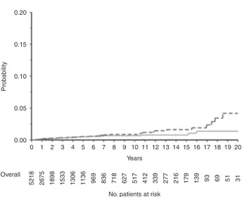
The crude cumulative incidence of SM by type of malig-nancy is shown in Table2 and Fig. 1, whereas the cumu-lative incidence of death from any cause and of SM (any type) is provided in Supplementary Figure 2. The incidence was similar until the 10-year cutoff, and diverged at 20 years, being 4.17% (95% CI: 1.78–6.57) for solid SM, and 1.37 (95% CI: 0.47–2.27) for hematologic SM.
We then stratified patients according to the arbitrary cutoff of 40 years of age at the time offirst HDCT course (Supplementary Table 3). The difference remained numerically significant, and the CCI of solid SM at 20 years for patients aged≥40 years was 6.85% (95% CI: 0–15.47, Supplementary Figure 3A). The results of the univariable competing-risk analyses are shown in Table 3. Older age was the only statistically significant factor associated with SM development (HR for the class“≥40 years” versus “<40 years”: 2.28, 95% CI: 1.35–3.84, p = 0.004). The associa-tion was significant for hematologic SM (HR: 3.48, 95% CI: 1.54–7.86, p = 0.004), whereas it was lost for solid SM (HR: 1.59, 95% CI: 0.76–3.32, p = 0.234; the correspond-ing estimates of CCI are provided in Supplementary Fig-ures 3B, 3C).
Overall survival after SM occurrence
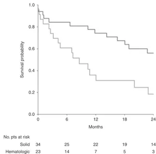
Kaplan–Meier OS curves since the diagnosis of SM are shown in Fig.2. Patients developing solid SM had longer survival compared to those with hematologic SM. Median OS with solid SM was 34.4 months (IQR: 11.8–not estimable) versus 8.6 months (IQR: 2.6–20.0) after hema-tologic SM diagnosis (p= 0.003).
Discussion
To the authors’ knowledge, we have reported an analysis of incidence of SM from the largest population of GCT patients who have received HDCT to date. The adminis-tration of high-dose carboplatin and etoposide for chemor-efractory GCT has become increasingly popular during the last 10 years, owing to the results from large retrospective analyses [5], and it represents the only consolidated indi-cation of HDCT as standard practice in adult solid tumors. In general, HDCT is a highly effective option for relapsed, high-risk patients with GCT, and the proportion of patients who can benefit with long-term remission (i.e., cure of the disease) varies according to the presence of prognostic factors, and may be >50% in good-risk patients [6]. Con-sequently, long-term toxicities of chemotherapy are well-recognized among GCT experts. These side effects may have important implications for follow-up planning and duration, and the available recommendations for the follow-up after salvage therapy or HDCT in these patients remain elusive. SM in particular is a potentially life-threatening late effect of GCT therapy. Regarding the risk of developing SM after treatment for GCT, most of the population-level studies evaluated the effect of adjuvant treatment in clinical stage I patients, and only a few of them reported the effects of chemotherapy for advanced disease. In these studies, the increased risk of developing solid tumors was largely attributed to radiotherapy, especially in older patients, accounting for a sizeable increment of SM development compared to the age-matched general population [23–25]. In regard to chemotherapy effect, some population-based data and retrospective analyses are available to provide guidance for patient counseling [16–20]. These long-dated studies assessed the risk of developing secondary leukemia after HDCT, and identified the total cumulative dose of etoposide >2 gr/m2 as potentially associated with an acceptable risk of about 2% of developing acute myeloid leukemia. Regarding the risk of developing solid SM, it remains unclear whether HDCT administration portends an increased risk compared to standard chemotherapy. Overall, the present CCI results are consistent with the available data, and the cumulative incidence curves of solid SM are close to those reported with standard-dose chemotherapy.
As a strength of our analyses, we were able to show that the trends of SM occurrence after HDCT were different between solid and hematologic tumors. The former showed a delayed increment to above 4% after 20 years, and this observation is originally reported here for HDCT, whereas the latter showed a less significant raise to about 1.4%, which is in line with the published data [16–20]. Despite this, we were unable to run multivariate models due to the small number of SM events, but our observations are unlikely biased by the effect of age, which was not
associated with the risk of developing solid neoplasms. Conversely, as an additional interesting finding, older age was significantly associated with the risk of developing hematologic cancers or myelodysplasia. This finding is likely the result of a secondary effect of HDCT. Further-more, we were able to report the prognostic impact of solid versus hematologic SM development in these patients. These outcomes pose additional warning on the adminis-tration of HDCT in patients older than 40 years, and warrant additional studies.
There are of course some critical limitations to acknowledge in our study. First, the EBMT database lack of potentially important baseline data. For example, the risk of hematologic cancers occurring in primary mediastinal non-seminoma is well acknowledged in the literature [26], and the information of the site of the primary tumor could have allowed us to more finely delineate the characteristics of patients at higher risk of developing hematologic SM. Additional data were unaccounted for in this study: the subtype of acute leukemia, including the information on the underlying cytogenetic alterations, and most importantly the total burden of chemotherapy administration (i.e., number of chemotherapy regimens the patients received for GCT, added to HDCT), that might have had an impact on the risk Table 1 Baseline characteristics of the total analyzed patients from the
EBMT database and of those who developed SM event only (study group) Total screened patients SM patients (N= 5218) (N= 59) Period of HDCT administration 11/1981–12/2015 08/1984–02/2014 1981–1989 175 (3.4) 2 (3.4) 1990–1999 1447 (27.8) 23 (39.0) 2000–2009 2136 (40.9) 29 (49.1) 2010–2015 1460 (27.9) 5 (8.5) Characteristic No. (%)
Age at the time of HDCT—years
Median (interquartile range) 32 (26–39) 32 (25–38) Histology of GCT
Non-seminoma 1972 (37.8) 29 (49.2)
Seminoma 486 (9.3) 8 (13.6)
GCT unclassified 2760 (52.9) 22 (37.2) Source of hematopoietic stem cells
Peripheral blood 4573 (88.4) 51 (86.4) Bone marrow 599 (11.6) 8 (13.6) Missing 46 — Number of HDCT courses 1 2639 (50.6) 31 (52.5) 2 1237 (23.7) 19 (32.2) >2 1337 (25.7) 9 (15.3) Missing 5 — HDCT conditioning regimen CBDCA-VP16 1310 (25.1) 8 (13.6) Other 1805 (34.6) 23 (39.0)a Missing 2103 (40.3) 28 (47.4) Country of origin Germany 1831 (35.1) 21 (35.6) France 957 (18.3) 12 (20.2) United Kingdom 481 (9.2) 5 (8.5) Italy 454 (8.7) 3 (5.1) Switzerland 197 (3.8) 4 (6.8) Austria 176 (3.4) 2 (3.4) Spain 131 (2.5) — Sweden 131 (2.5) 2 (3.4) Turkey 128 (2.4) — Poland 115 (2.2) 1 (1.7) The Netherlands 115 (2.2) 5 (8.5) Belgium 85 (1.6) — Czech Republic 77 (1.5) 1 (1.7) Israel 44 (0.8) — Finland 43 (0.8) 1 (1.7) Denmark 39 (0.7) — Ireland 38 (0.7) 1 (1.7) Table 1 (continued) Total screened patients SM patients (N= 5218) (N= 59) Greece 33 (0.6) — Lithuania 27 (0.5) 1 (1.7) Portugal 22 (0.4) — Australia 16 (0.3) — Croatia 16 (0.3) — Bulgaria 13 (0.2) — Jordan 9 (0.2) — New Zealand 9 (0.2) — Russia 8 (0.2) — Lebanon 6 (0.1) — Argentina 4 (0.08) — Iran 4 (0.08) — Serbia 3 (0.06) — Romania 2 (0.04) — Slovenia 2 (0.04) — South Africa 2 (0.04) —
CBDCA carboplatin, GCT germ cell tumor, HDCT high-dose chemotherapy, SM secondary malignancy, VP16 etoposide
aConsisting of the following regimens: carboplatin, etoposide,
ifosfa-mide (n= 14), carboplatin, etoposide, paclitaxel (n = 2), carboplatin, etoposide, melphalan (n= 1), carboplatin, etoposide, cyclophosphamide (CarboPec, n= 3), cyclophosphamide, and etoposide (n = 3)
of developing SM. The total dose of the drugs used as induction regimens was not recorded as well. However, the total doses of carboplatin and etoposide administered in the standard regimens, as well as in the other mixed regimens published in the literature, is largely comparable [3]. The effect of additional drugs administration on the risk of SM development remains elusive.
Second, despite our study covered a 34-year period of HDCT administration, the quality of the follow-up for alive patients was suboptimal, resulting in a median follow-up duration of<5 years which may have substantially limited our possibility to observe additional cases. Consequently, the number of patients at risk at long-term follow-up was small, thus affecting the reliability of the cumulative inci-dence estimates. Third, we were unable to analyze the age factor, which resulted significantly associated with hema-tologic SM occurrence, as a continuous variable due to the small numbers. For this reason, and based on the observed distribution of age (the third quartile being set at 38 years in the study group), we arbitrarily set the cutoff at 40 year to tackle the effect of older age atfirst transplant, like we did in a previous study from the EBMT database [27]. This cutoff may be further refined with additional studies. Of note, the analyses comprised a huge number of patients from diverse countries: for these reasons, and due to the fact that the overall incidence of SM was rather low in our
study, we did not rely on the incidence data of SM from the National or European registries in order to provide the standardized incidence ratios, and any attempt to perform age-matched cohort analyses is very hard and not suf fi-ciently reliable in our view. Finally, other unexplained environmental risk factors (e.g., smoking) could have con-tributed to the risk of developing SM, especially solid SM. In conclusion, in our large, multinational series of GCT patients treated with HDCT, there was not an exceedingly high occurrence of hematologic SM during the follow-up period, but an association with age older than 40 at the time of HDCT delivery was found. While the small sample size of patients at risk does not allowfirm conclusions, it would appear that there is an increased risk of solid tumors at after 15 years of follow-up, similar to standard-dose che-motherapy. Our observation dictates the need for long-term surveillance in these patients (i.e., at least 20 years), although special follow-up protocols do not seem necessary after HDCT. Conversely, caution is needed for the Table 2 Crude cumulative
incidence of SM Type of SM 5 y CCI, % (95% CI) 10 y CCI, % (95% CI) 20 y CCI, % (95% CI) Solid SM 0.52 (0.27–0.77) 0.86 (0.49–1.24) 4.17 (1.78–6.57) Hematologic SM 0.51 (0.26–0.76) 0.68 (0.36–0.99) 1.37 (0.47–2.27) CCI crude cumulative incidence, CI confidence interval, SM secondary malignancy
0.20 0.15 0.10 Probability 0.00 0.05 0 5218 2675 1898 1533 1306 1136 969 836 718 627 517 412 339 277 216 179 139 93 69 51 31 1 2 3 4 5 6 7 8 9 10 Years
No. patients at risk Overall
11 12 13 14 15 16 17 18 19 20
Fig. 1 Cumulative incidence of secondary malignancy with death as a competing risk. Red curve: incidence of hematologic secondary malignancy; dotted blue curve: incidence of solid secondary malig-nancy. (Colorfigure online)
Table 3 Univariable competing-risk regression model to analyze the association of clinical factors with SM occurrence
Factor HR 95% CI p-value*
Age (years) 0.004
● ≥40 versus <40 2.28 1.35–3.84
GCT histology 0.613
● Non-seminoma Reference Reference
● Seminoma 1.25 0.57–2.72
● Unclassified 0.84 0.49–1.44
Source of stem cells 0.286
● Bone marrow versus peripheral blood 0.67 0.33–1.38 Number of HDCT courses 0.223 ● 1 Reference Reference ● 2 1.53 0.86–2.70 ● >2 0.82 0.39–1.71 HDCT conditioning regimen 0.886
● CBDCA-VP16 Reference Reference
● Other 1.22 0.55–2.70
● Unclassified 1.14 0.53–2.48
CBDCA carboplatin, CI confidence interval, GCT germ cell tumor, HDCT high-dose chemotherapy, HR hazard ratio, SM secondary malignancy, VP16 etoposide
administration of HDCT in patients older than 40 years, pending validation of ourfindings with additional studies with longer median follow-up.
Notes
Data were presented in part in a poster session, at the 2017 Genitourinary Cancers Symposium, February 16-18th, 2017, Orlando (FL), USA.
Data were presented in part in a poster session, at the 2017 Annual meeting of the European Association of Urology (EAU), March 24-28th, 2017, London, United Kingdom.
Data were presented in part in a poster session, at the 2017 Annual Meeting of the American Society of Clinical Oncology (ASCO), June 2-6th, 2017, Chicago (IL), USA.
Compliance with ethical standards
Conflict of interest The authors declare that they have no conflict of interest.
References
1. Horwich A, Shipley J, Huddart R. Testicular germ-cell cancer. Lancet. 2006;367:754–65.
2. Gatta G, Capocaccia R, Botta L, Mallone S, De Angelis R, Ardanaz E, et al. Burden of centralised treatment in Europe of rare tumours: results of RARECAREnet-a population-based study. Lancet Oncol. 2017;18:1022–39.
3. Necchi A, Lanza F, Rosti G, Martino M, Farè E, Pedrazzoli P. High-dose chemotherapy for germ cell tumors: do we have a model? Expert Opin Biol Ther. 2015;15:33–44.
4. Einhorn LH, Williams SD, Chamness A, Brames MJ, Perkins SM, Abonour R. High-dose chemotherapy and stem-cell rescue for metastatic germ-cell tumors. N Engl J Med. 2007;357:340–8. 5. Adra N, Abonour R, Althouse SK, Albany C, Hanna NH, Einhorn
LH. High-dose chemotherapy and autologous peripheral-blood stem-cell rescue for metastatic germ-cell tumors: the Indiana University experience. J Clin Oncol. 2017;35:1096–102. 6. Feldman DR, Sheinfeld J, Bajorin DF, Fischer P, Turkula S, Ishill
N, et al. TI-CE high-dose chemotherapy for previously treated germ cell tumors; results and prognostic factor analysis. J Clin Oncol. 2010;28:1706–13.
7. Necchi A, Miceli R, Bregni M, Bokemeyer C, Berger LA, Oechsle K, et al. Prognostic impact of progression to induction chemotherapy and prior paclitaxel therapy in patients with germ cell tumors receiving salvage high-dose chemotherapy in the last 10 years: a study of the European Society for Blood and Marrow Transplantation Solid Tumors working party. Bone Marrow Transplant. 2016;51:384–90.
8. Fung C, Fossa SD, Milano MT, Oldenburg J, Travis LB. Solid tumors after chemotherapy or surgery for testuicular non-seminoma: a population-based study. J Clin Oncol. 2013;31:3807–14.
9. Travis LB, Fossa SD, Schonfeld SJ, McMaster ML, Lynch CF, Storm H, et al. Second cancers among 40,576 testicular cancer patients: focus on long-term survivors. J Natl Cancer Inst. 2005;97:1429–39.
10. van den Belt-Dusebout AW, de Wit R, Gietema JA, Horenblas S, Louwman MEW, Ribot JG, et al. Treatment-specific risks of second malignancies and cardiovascular disease in 5-year survi-vors of testicular cancer. J Clin Oncol. 2007;25:4370–8. 11. Wanderas EH, Fossa SD, Tretli S. Risk of subsequent non-germ
cell cancer after treatment of germ cell cancer in 2006 Norwegian male patients. Eur J Cancer. 1997;33:253–62.
12. Bokemeyer C, Schmoll HJ. Secondary neoplasms following treatment of malignant germ cell tumors. J Clin Oncol. 1993;11:1703–9.
13. Richiardi L, Scelo G, Boffetta P, Hemminiki K, Pukkala E, Olsen JH, et al. Second malignancies among survivors of germ cell cancer testicular cancer: a pooled analysis between 13 cancer registries. Int J Cancer. 2006;120:623–31.
14. Kier MG, Hansen MK, Lauritsen J, Mortensen MS, Bandak M, Agerbaek M, et al. Second malignant neoplasms and cause of death in patients with germ cell cancer: a Danish nationwide cohort study. JAMA Oncol. 2016;2:1624–7.
15. Chamie K, Kurzrock EA, Evans CP, Litwin MS, Koppie TM, Wotton-Gorges SL, et al. Secondary malignancies among non-seminomatous germ cell tumor cancer survivors. Cancer. 2011;117:4219–30.
16. Kollmannsberger C, Beyer J, Droz JP, Harstrick A, Harttmann JT, Biron P, et al. Secondary leukemia following high cumulative doses of etoposide in patients treated for advanced germ cell tumors. J Clin Oncol. 1998;16:3386–91.
17. Wierecky J, Kollmannsberger C, Boehlke I, Kuczyk M, Schlei-cher J, SchleuSchlei-cher N, et al. Secondary leukemia after first-line high-dose chemotherapy for patients with advanced germ cell cancer. J Cancer Res Clin Oncol. 2005;131:255–60.
18. Kollmannsberger C, Hartmann JT, Kanz L, Bokemeyer C. Therapy-related malignancies following treatment of germ cell cancer. Int J Cancer. 1999;83:860–3.
19. Travis LB, Andersson M, Gospodarowicz M, van Leeuwen FE, Bergfeld K, Lynch CF, et al. Treatment-associated leukemia fol-lowing testicular cancer. J Natl Cancer Inst. 2000;92:1165–71.
1.0 0.8 0.6 Sur viv a l probability 0.4 0.2 0.0 0 34 23 25 14 22 7 19 5 14 3 Solid Hematologic 6 12 Months No. pts at risk 18 24
Fig. 2 Kaplan–Meier curves of overall survival after occurrence of secondary malignancy, according to the type of secondary malignancy. Red curve: hematologic secondary malignancy; blue curve: solid secondary malignancy. (Colorfigure online)
20. Bajorin DF, Motzer RJ, Rodriguez E, Murphy B, Bosl GJ. Acute nonlymphocytic leukemia in germ cell tumor patients treated with etoposide-containing chemotherapy. J Natl Cancer Inst. 1993;85:60–62.
21. Fine JP, Gray RJ. A proportional hazards model for the sub-distribution of a competing risk. J Am Stat Assoc. 1999;94:496–509.
22. Gray RJ. A class of K-sample tests for comparing the cumulative incidence of a competing risk. Ann Stat. 1988;16:1141–54. 23. Horwich A, Fossa SD, Huddart R, Dearnaley DP, Stenning S,
Aresu M, et al. Second cancer risk and mortality in men treated with radiotherapy for stage I seminoma. Br J Cancer. 2014;110:256–63.
24. Hauptmann M, Fossa SD, Stovall M, van Leeuven GFE, Johan-nesen TB, Rajaraman P, et al. Increased stomach cancer risk
following radiotherapy for testicular cancer. Br J Cancer. 2015;112:44–51.
25. Hauptmann M, Borge Johannesen T, Gilbert ES, Stovall M, van Leeuwen FE, Rajaraman P, et al. Increased pancreatic cancer risk following radiotherapy for testicular cancer. Br J Cancer. 2016;115:901–8.
26. Nichols CR, Roth BJ, Heerema N, Griep G, Tricot G. Hemato-logic neoplasia associated with primary mediastinal germ-cell tumors. N Engl J Med. 1990;322:1425–9.
27. Necchi A, Lo Vullo S, Rosti G, Badoglio M, Giannatempo P, Raggi D, et al. Administration of high-dose chemotherapy with stem cell support in patients 40 years of age or older with advanced germ cell tumours: a retrospective study from the Eur-opean Society for Blood and Marrow Transplantation database. Bone Marrow Transplant. 2017;52:1218–20.