IN VIVO EVALUATION OF THE
CARDIOPROTECTIVE
ACTIVITY OF
VEGF‐B
PhD Thesis
Scuola Normale Superiore di Pisa
Uday Puligadda
Supervisor: Professor Mauro Giacca
“Live as if you were to die tomorrow. Learn as if you were
to live forever.”
- Mahatma Gandhi
I TABLE OF CONTENTS Synopsis 1 1 Introduction 3 1.1 Myocardial ischemia (MI) 5 1.2 Peripheral artery disease 8 1.3 Mechanisms of blood vessel formation 9 1.3.1 The Vascular endothelial growth factor (VEGF) family and their receptors 11 1.3.1.1 VEGF‐A 12 1.3.1.2 VEGF‐B 17 1.3.1.3 VEGF Receptors (VEGFRs) 21 VEGFR‐1 22 VEGFR‐1 signaling 24 VEGFR‐2 24 VEGFR‐3 26 Neuropilins 27 Neuropilin1 (NP1) 28 1.4 Heart Failure (HF) 28 1.4.1 Morphological changes in the heart leading from myocardial infarction to HF 29 1.4.1.1 Compensatry and pathological hypertrophy 30 1.5 Gene therapy strategies for cardio vascular disorders 33 1.5.1 AAV 34 Structure 35 Preparation and purification of AAV 36 1.5.2 Gene therapy for the induction of therapeutic angiogenesis 37
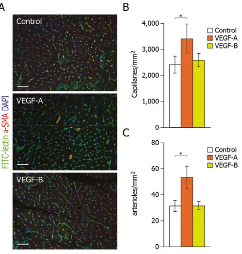
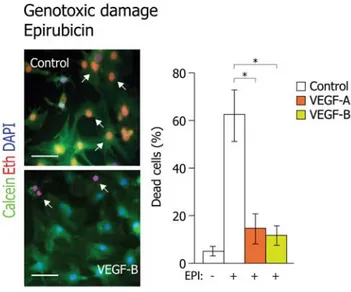
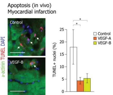
1.5.3 Gene therapy strategies for the treatment of HF 42 2 Results I 46 2.1 Persistent expression of AAV‐2‐mediated VEGF‐A and VEGF‐B in normal rat myocardium 46 2.2 Angiogenic effect of VEGF factors in the normal rat myocardium 47 2.3 Prolonged expression of VEGF‐B, a specific VEGFR‐1 ligand preserves myocardial function: analysis of cardiac function after myocardial infarction by echocardiography 50 2.4 Analysis of cardiac tissues by histo‐pathological studies: morphometric analyses of infarct samples 55 2.5 Analyses of vector genomes and their incorporated transgenes in the injected myocardium 56 2.6 Angiogenic effect of VEGF factors in infarcted hearts 58 2.7 Expression levels of VEGF receptors in vitro 59 2.8 Quantitative analyses of VEGF receptors in‐vitro 61 2.9 In vivo expression of VEGF receptors in the heart 62 2.10 VEGF‐A and VEGF‐B protect cardiomyocytes from apoptosis 64 2.11 VEGF‐A and VEGF‐B activate expression of genes involved in myocardial metabolism and Contractility 66 2.12 VEGF‐B overexpression counteracts induction of genes associated with pathological LV remodelling after MI 68 3 Results II 70 3.1 Localized hypertrophic response in cardiomyocytes 71 3.2 Protective role of VEGF‐B against cardiomyocyte apoptosis 72 3.3 Activation state of Akt, GSK‐3β and FoxO3a 76
III 4 Discussion 79 4.1 Cardiac gene transfer of VEGF‐A and VEGF‐B exerts beneficial activities after myocardial infarction in rats and in heart failure in dogs 79 4.2 Non angiogenic functions of VEGF family members 81 4.3 AAV vectors for cardiac gene transfer 83 4.4 Effects of VEGF‐B on cardiomyocytes in vitro and in vivo 84 4.5 Conclusions 86 5 Materials and Methods 87 5.1 Production, purification and characterization of rAAV vectors 87 5.2 Preparation of neonatal cardiomyocytes 87 5.2.1 Preparation of cardiomyocyte cultures 87 5.2.2 Preparation of non‐myocyte cultures 88 5.3 Isolation and treatment of neonatal rat ventricular cardiomyocytes 88 5.4 Quantification of mRNA by real‐time PCR 89 5.5 Immunoprecipitation and Western blot analysis 91 5.6 Animal studies and echocardiography 92 5.6.1 Surgical procedure for MI 92 5.6.2 Echocardiography analysis 93 5.7 Histo‐pathological studies 94 5.7.1 Morphometric analysis of infarct size 94 5.7.2 Massonʹs trichrome staining 94 5.7.3 Infarct size calculation 94 5.7.4 Immunoflorescence 94 5.7.5 TUNEL assay for apoptosis 95 5.8 Statistical analysis 95
6 Bibliography 96 7 Appendix 121
1
SYNOPSIS
Members of the vascular endothelial growth factor (VEGF) family and their receptors are important regulators of vasculogenesis, angiogenesis, and vessel maintenance in both embryos and adults. In particular, the 165‐aa isoform of VEGF‐A exerts its highly potent angiogenic and arteriogenic activity mainly through binding to VEGFR‐2 in spite of its high affinity binding to VEGFR‐1. However, the biological significance of VEGF interaction with VEGFR‐1 remains elusive. One of our previous studies in dogs unveiled an unexpected cardioprotective activity of VEGF‐A, which was already evident at 48 hours after the induction of a myocardial infarction and was even more marked at 4 weeks. Multiple recent evidences suggested that VEGF‐B, a VEGFR‐1 exclusive ligand, could be selectively active in the myocardium. However the available data did not define clearly the cardiac benefits of VEGF‐B. Therefore, in the present study we wanted to investigate and compare the cardioprotective role of both VEGF‐A (and in particular its 165 aminoacid isoform) and VEGF‐B (167 aminoacid isoform) in a rat model of myocardial infarction through intracardiac injection of the respective genes. For the efficient and prolonged expression of these factors, we exploited viral vectors based on the Adeno‐ Associated Virus (AAV), as an efficient tool for in vivo gene transfer. By analyzing cardiac function by echocardiography, we observed that the prolonged expression of both VEGF factors induced a marked improvement in cardiac contractility, preserving viable cardiac tissue, and preventing left ventricular remodeling over time. In addition, we performed histopatho‐ logical studies on a subset of representative samples. Consistent with the functional outcome, we observed a more potent cardioprotection using the VEGFR‐1 selective ligand, VEGF‐B, despite its inability to induce angiogenesis. We then tried to dissect the molecular mechanisms by which VEGF‐B might act on cardiomyocytes both in vitro and in vivo. We found that the action of VEGF‐B was mainly mediated by the upregulation of VEGFR‐1 and that stimulation of this receptor exerted positive inotropic and anti‐apoptotic effects on cardiomyocytes. In addition, VEGF‐B elicited a gene expression pattern reminiscent to the one observed in compensatory
heart hypertrophy, both in cultured cardiomyocytes and in infarcted hearts. We further expanded our investigation on the cardio protection conferred by VEGF‐B167 in a pacing‐ induced hear failure model in canines. Interestingly, the findings obtained in this model were similar and totally in keeping with the observations done in the myocardial ischemic model in rats, again showing that VEGF‐B167 exerted cardio protective effects in the absence of angiogenesis or pathological hypertrophy. Instead, it delayed the progression towards heart failure and prevented cardiomyocytes from apoptosis by controlling pro‐apoptotic intracellular mediators, such as Akt and its downstream targets GSK‐3β and FoxO3a.
3
INTRODUCTION
In the early periods of the 20th century, cardiovascular disorders (CVDs) were not the major cause of death, and accounted for less than 10% of deaths in the population globally. At the end of the century, however, according to the statistics of 2005, CVDs had become one of the major causes of mortality, being responsible for at least 30% of deaths in the general population. It is now estimated that CVDs cause 17 million deaths every year worldwide (World Health Report, 2006). In Europe, CVDs account for 49% of all deaths (Rayner et al., 2003), corresponding to nearly 1.9 million people. In the United States, nearly 2400 individuals die of CVD every day, with an average of 1 death every 37 seconds. It is predicted that, by 2030, almost 23.6 million people will die from CVDs, which will then remain as the single leading causes of death (www.who.int/mediacentre/factsheets/fs317/en/index.html).
The incidence rate of CVDs is higher in Western countries than in developing countries, irrespective of industrialization, rapid progress in medical technologies, advancements in research and development of early prognostic methods or treatments. Current estimates indicate that CVDs incidence is about 50% in developed countries and about 28% in low and middle income countries (Mathers et al., 2006). However, in some developing countries, such as India, the burden of CVD is rising. The estimated prevalence of coronary heart disease is around 3–4% in rural areas and 8–10% in urban areas among adults older than 20 years, representing a twofold rise in rural areas and a six‐fold rise in urban areas over the past four decades (Reddy et al., 2005).
Regarding the economic burden, the expenditure on CVD in the United States was nearly 350 billion USD, in 2003, comprising both direct and indirect costs (Hodgson et al., 2001).
The major common cause of CVD is atherosclerosis, which can affect various districts of the circulatory system and yields distinct clinical manifestations. The pathology of atherosclerosis initiates with the formation of early focal lesions (called as fatty streaks) in the intima of arteries,
essentially consisting of lipoproteins deposition, which bind to the extracellular matrix components, such as proteoglycans. At a later stage, chemical modifications such as lipoptotein oxidation and non enzymatic glycation occur. In lipoprotein oxidation, the lipoproteins undergo oxidative modifications and forms hydroperoxides, lysophospholipids, oxysterols and chlorotyrosyl moieties, whereas in non enzymatic glycation, which is especially relevant in diabetic patients with sustained hyperglycemia, apolipoproteins and other structural arterial proteins undergo glycation and lose their natural protective function. These events further accelerate atherogenesis. After accumulation of extracellular lipids, leukocytes such as lymphocytes and monocytes are recruited within fatty streaks. This is favored by the constituents of oxidatively modified LDL molecules, as well as by a few cytokines, like intereukin‐1(IL‐1) and tumor necrosis factor (TNF‐), which stimulate expression of adhesion molecules and receptors on the surface of endothelial cells. Lymphocytes and monocytes penetrate the endothelial layer and take up residence in the intima. There, monocytes differentiate into macrophages and transform into lipid‐laiden foam cells through the uptake of lipoprotein particles by receptor‐mediated endocytosis. Some of the foam cells may die as result of apoptosis and result in the formation of a lipid rich center, called the necrotic core. Mononuclear phagocytes release a number of growth factors, such as platelet derived growth factor (PDGF), fibroblast growth factor (FGF) and other cytokines, which stimulate the proliferation of smooth muscle cells migrated from tunica media into the intima. This event in turn stimulates the additional deposition of extracellular matrix. The lesions with accumulation of macrophage‐derived foam cells are transformed into fibro‐fatty lesions with the accumulation of fibrous tissue and smooth muscle cells. These plaques also accumulate calcium, bound to bone‐associated proteins, such as osteocalcin, osteopontin and bone morphogenic proteins. The process of atherosclerosis has been recently reviewed in detail by many reviewers (Insull 2009, Falk 2006, Virmani et al., 2002, Glass et al., 2001, Libby 2001, Lusis 2000).
Several risk factors contribute to plaque formation. These are classified into two categories: modifiable and unmodifiable. The modifiable risk factors can be modulated by life style (physical activity, healthy diet with high HDL intake, smoking cessation) or pharmacological
5
therapy for the predisposing conditions such as hypertension, hyper‐cholestrolemia and diabetes mellitus. The unmodifiable factors include age, gender (men age ≥ 45years, women age ≥ 55years) and family history of premature coronary heart disease.
Given the social, economic, and sanitary burden of CVD, the identification of novel therapeutic strategies interfering with the molecular mechanisms of disease onset and progression is absolutely required. In this context, the appreciation of the potential value of gene therapy has been steadily growing over the last few years. The utilization of nucleic acids as therapeutic tools for CVD is currently envisaged in at least three main areas: i) treatment of cardiac and peripheral ischemic conditions by the induction of neoangiogenesis; ii) treatment of heart failure to preserve or improve cardiac function; iii) prevention of restenosis after percutaneous coronary intervention (PCI).
1.1 MYOCARDIAL ISCHEMIA (MI)
The term ischemia was first used by the German pathologist Rudolf Virchow. It derives from the Greek word “ischein” (to restrain) and “haima” (blood). Thus, ʺischemiaʺ refers to conditions of insufficient blood supply to a tissue (Bohuslav Ošt̕ádal et al., 1999).
Myocardial ischemia specifically refers to a condition in which there is insufficient blood flow to the myocardium. Patients with myocardial ischemia fall into one of two large groups: patients with chronic coronary artery disease (CAD), with stable angina pectoris. These patients experience discomfort or pain in the chest or left arm, induced by physical activity or in stress, which can be relieved by rest and/or sublingual nitroglycerin. A second group consists of patients which present with acute coronary syndromes (ACSs), divided into patients with acute myocardial infarction (MI) with ST‐segment elevation (STEMI) at their presenting electrocardiogram and patients with unstable angina or non‐ST‐segment elevation MI (UA/NSTEMI), accompanied by elevated levels of serum enzymes troponin T or I, and creatine phosphokinase. MI is an emergency condition treated by either percutaneous coronary angioplasty (with or without stent placement) or thrombolysis.
Myocardial ischemia is diagnosed by different tests, including: 1) electrocardiography (ECG), which records the electrical conduction activity of the heart; 2) exercise tolerance test, to evaluate the functional reserve of the heart; 3) echocardiography, to evaluate the condition of valves and chambers of the heart, and measure cardiac function; 4) contrast angiography (CT angiography/MR angiography), to assess the presence of atherosclerotic plaques in the coronary arteries; 5) nuclear ventriculography, to analyse the size of the heart chambers and their blood pumping ability using radioactive tracers (Hunt et al., 2005); 6) electron‐beam computed tomography (EBCT), to detect coronary calcification observed in early stages of CAD (Haberl et al., 2001).
Patients with CAD are currently treated by pharmacological therapy to reduce morbidity and to prevent complications. The line of treatment consists of the use of drugs such as anti‐platelet agents (aspirin, dipiridamole or clopidrogrel) to prevent thrombus formation, anti‐anginal drugs (amlodipine or bepridil) to minimise the rate of occurrence as well as severity of angina attacks, inhibitors of angiotensinogen converting enzyme (ACE inhibitors, such as benazepril, captopril or ramipril) to lower blood pressure and protect the heart, beta‐blockers (metoprolol or propranolol) to lower heart rate, blood pressure, and oxygen use by the heart, calcium channel antagonists (amlodipine, diltiazem or verapamil) to relax arteries, lowering blood pressure and reducing strain on the heart, diuretics to lower blood pressure and treat congestive heart failure, nitrates (nitroglycerin or isosorbide) to stop chest pain and improve blood supply to the heart; statins to lower blood cholesterol.
PCI is widely used in patients with symptomatic CAD and evidence of ischemia due to stenosis of one or two vessels, and even in selected patients with three‐vessel disease. Coronary artery Bypass Grafting (CABG) is highly preferred in patients with stenosis of the left main coronary artery and in those with three‐vessel CAD, especially with diabetes and/or impaired left ventricular function, who require revascularization.
Compared to pharmacological therapy, CABG shows better prognostic efficacy (increase in life expectancy) in only a selected group of patients with high risk atherosclerotic lesions, localized
7
in specific anatomical segments of the coronary arteries. However, CABG usually decreases symptoms and thus improves quality of life in most patients. PCI instead (defined as ʺelectiveʺ PCI to contrast it with ʺprimaryʺ PCI, performed in patients with acute MI) entails the use of an arterial catheter containing a deflated balloon at its extremity. The catheter is inserted through the femoral artery and the balloon is positioned, under angiographic guidance, at the level of the atherosclerotic plaque in the coronary artery. The balloon is then inflated to mechanically destroy the plaque and restore vessel perviousness. Concomitant with angioplasty, an endovascular tubular prosthesis (stent) is usually positioned in correspondence with the artery segment where the balloon was inflated in order to maintain patency of the vessel to prevent restenosis. When compared to pharmacological therapy, PCI does not seem to offer significant survival advantage. However, it significantly improves the patient’s quality of life (episodes of angina, dyspnea and limitations of exercise capacity). PCI thus represents an extremely valuable alternative to CABG in the treatment of angina; as already reported above, CABG is only superior to PCI in a selected group of patients with high risk lesions.
In certain conditions, rupture of the atherosclerotic plaque causes acute MI through the sudden formation of a thrombus obstructing an atherosclerotic coronary artery. About 25‐35% of patients with MI die before receiving medical assistance, usually due to ventricular fibrillation. For those who are rapidly hospitalized, prognosis has very significantly improved through the revascularization of the occluded artery. Revascularization can be obtained by mechanical of pharmacological treatment. The former consists in primary PCI, that is balloon angioplasty performed in emergency conditions, with or without stent placement. Primary PCI usually restores blood flow in more than 90% of patients. If this procedure is delayed more than 90 minutes from the initial medical contact, pharmacological thrombolysis can be attempted. This is achieved by using thrombolytic agents containing plasminogen activators (streptokinase or tissue plasminogen activator ‐ tPA). These are enzymes that transform plasminogen into plasmin and thus degrade the fibrin network within the thrombus, eventually leading to dissolution of the thrombus. Collectively, patients with MI who are rapidly hospitalized and are
mechanically or pharmacologically revascularized have today a very high survival rate (90‐ 95%). 1.2 PERIPHERAL ARTERY DISEASE Peripheral artery disease (PAD) is caused by obstruction of large arteries in the arms and legs, in particular the iliac, femoral, popliteal and tibial arteries. The obstruction of large arteries can result from atherosclerosis, embolism or thrombus formation, or any other inflammatory process lead to stenosis. Patients may present with acute or chronic ischemia.
Similar to CAD, there are several predisposing factors to PAD, such as smoking, obesity, diabetes mellitus, dyslipidemia and hypertension. The disease is mostly observed in people over 50‐55 years of age. The National Health and Nutrition Examination Surveys (NHANES) during the years 1999‐2000 reported that approximately 5 million adults were affected by PAD and that the prevalence increased with age and disproportionately affected blacks (Selvin et al., 2004). Fewer than 50% patients with PAD are asymptomatic, and diagnosis is only made after targeted investigation (typically, analysis of blood flow by echo‐color‐doppler). About 50% of cases have variable leg symptoms; of these, 10% of cases have classic symptoms of intermittent claudication (Hirsch et al., 2001, Crigui et al., 1985): patients, while walking, are forced to stop repeatedly due to leg pain, typically in the calves. Pain is due to the accumulation of lactic acid, which stimulates peripheral pain receptors ‐ consequent to muscle activity in anaerobiosis, since the arterial stenosis does not allow proper supply of oxygenated blood. If the blood supply is diminished critically, patients may experience pain even at rest, in particular at night when legs are raised in bed. The pain initially develops in toes and in feet and then passes to calves. Ulcers may develop under feet and toes. In few cases, necrosis and then gangrene develops, which is irreparable. This is the end stage of PAD. In these cases, amputation of foot or leg or entire limb is unavoidable.
9
1.3 MECHANISMS OF BLOOD VESSEL FORMATION
The mechanism of blood vessel formation involves three processes: vasculogenesis, angiogenesis and arteriogenesis (Storkebaum et al., 2004). The term vasculogenesis traditionally refers to the de novo synthesis of blood vessels during embryonic development. In this process, a primitive capillary network is formed from angiogenic progenitor cells, called angioblasts, which differentiate into endothelial cells and coalesce to form an initial vascular plexus (Risau et al., 1995, Carmeliet 2003). Antigenic markers common to angioblasts and hematopoietic stem cells have been identified, consistent with the possibility that a common precursor (hemangioblast) might exist and be able to originate both hematopoietic and endothelial cells. The budding of new capillary branches from existing blood vessels into new blood vessels is named angiogenesis. This process is usually observed during both embryonic development and in the adult life. The process is initiated by the metabolic activation, proliferation and migration of endothelial cells, concomitant with vast remodeling of the extracellular matrix. At subsequent times, the newly formed capillaries progressively mature by the addition of mural cells (pericytes and smooth muscle cells) allowing proper functionality, and a vascular network formed by larger vessels (arterioles and venules) is eventually formed.
Finally, in addition to vasculogenesis and angiogenesis, a third process of blood vessel formation exists, known as arteriogenesis. This process consists in the remodeling of existing arteries to increase their luminal diameter in response to physiological stimuli of vasoconstriction or vasodilatation. A classic example of arteriogenesis in the adult is the formation of a collateral network, visible upon angiography, in response to the progressive occlusion of an arterial vessel by an atherosclerotic plaque. In this process, vessels are generated to become medium and large arteries with properly developed tunica media (to control patency of vascular lumen), in order to functionally meet the demand of tissue perfusion (Heli et al., 2006).
If we observe the anatomy of blood vessels, capillaries are made up of a single layer of endothelial cells with supporting vascular pericytes, whereas arteries and veins consist of multiple layers (from inside to outside): tunica intima, the inner most layer surrounding the
lumen composed of endothelial cells, pericytes and basement membrane; tunica media, the middle layer composed of smooth muscle cells and extracellular matrix; tunica adventitia, which is particular prominent in large vessels, consisting of fibroblasts and their extracellular‐ matrix (Figure 1.1).
Figure 1.1 During vasculogenesis, endothelial progenitor cells develop into primitive vascular network, which expands further with the recruitment of SMC and pericytes and develops into fully functional vascular network. Lymphatic network emerges from transdifferentiation of veins (Carmeliet 2005).
In physiological conditions, the major stimulus for the process of angiogenesis is hypoxia. In hypoxic conditions, cells in the tissue start sensing the reduced O2 concentration and initiate vascular responses to increase expression of genes encoding angiogenic growth factors. In this response pathway, the central element is HIF‐1, a heterodimeric transcription factor able to activate expression of a series of genes. Among the induced genes are several angiogenic cytokines and their receptors. Among these, members of the vascular endothelial growth factor (VEGF) family and their receptors (VEGFR) are the most powerful, initial responsive elements in the angiogenic cascade, which are required in all phases of angiogenesis in the adult and vasculogenesis in the embryo (Wang et al., 1995, Carmeliet 2003, Ferrara et al., 2003).
11
1.3.1 The Vascular Endothelial Growth Factor (VEGF) family and their receptors
The mammalian family of the vascular endothelial growth factor (VEGF) includes 5 structurally related proteins: VEGF‐A, VEGF‐B, VEGF‐C, VEGF‐D and placental growth factor (PlGF). There are two additional VEGF family members of non mammalian origin: VEGF‐E and VEGF‐F. VEGF‐E was identified in the Orf virus, which causes extensive vascular proliferation in human skin upon infection (Lyttle et al., 1994, Meyer et al., 1999, Ogawa et al., 1998). VEGF‐F, instead, is a VEGF‐like molecule isolated from the Trimeresurus flavoviridis Habu snake venom, which is able to induce vascular permeability preferentially through VEGFR‐1 signaling (Takahashi et al., 2004).
The function of the different VEGF family members are mainly mediated by the cell surface tyrosine‐kinase receptors VEGFR‐1 (flt‐1), VEGFR‐2 (KDR/flk‐1) and VEGFR‐3 (flt‐4), whose relative affinity and distribution mediate the biological effect (Carmeliet et al., 1999). More specifically, VEGF‐A binds VEGFR‐1 and ‐2; VEGF‐B and PlGF selectively bind VEGFR‐1, while VEGF‐C and VEGF‐D bind VEGFR‐3. The last two factors mainly induce a lymphangiogenic response, leading to the formation of lymphatic rather than hematic vessels (Karkkainen et al., 2004, Stacker et al., 2001). The characteristics of VEGF‐A and VEGF‐B, which are the subject of research of this thesis, are reported in detail in the following sections (Figure 1.2).
Figure 1.2 The multiple splicing isoforms of VEGF‐A bind VEGFR‐1 and VEGFR‐2, whereas VEGF‐B and PlGF only bind VEGFR‐1. The Orf virus VEGF‐E factor is a selective ligand for VEGFR‐2. Of the other members of the VEGF family, VEGF‐C and VEGF‐D bind VEGFR‐2 and VEGFR‐3 and participate in the formation of the lymphatic network. Specific isoforms of VEGFs factors are also able to bind the co‐receptor NP‐1 and NP‐2, which can heterodimerize with either VEGFR‐1 or VEGFR‐2 (Hicklin et al., 2005). 1.3.1.1 VEGF‐A
VEGF‐A is by far the best characterized member of VEGF family and the first one to be discovered. The need for timely and precisely regulated VEGF‐A gene expression is highlighted by the remarkable observation that deletion of a single VEGF‐A gene allele (Ferrara et al., 1996, Carmeliet et al., 1996) or modest gene overexpression (Miquerol et al., 2000) result in embryonic lethality.
VEGF‐A has been originally described as Vascular Permeability Factor (VPF), due to its ability to increase vascular permeability and to disrupt vascular barrier integrity (Weis and Cheresh, 2005).
The purified protein molecule of the human VEGF‐A gene, also referred to as VEGF, is 46‐kDa in weight. It dissociates upon reduction into two apparently identical 23 kDa subunits (Ferrara
13
et al., 1989). The human gene is located on chromosome 6 (p12) and is composed by 8 exons and 7 introns, (Tischer et al., 1991), with a coding region of about 14 kb. The VEGF pre‐mRNA undergoes alternative splicing to produce at least eight different protein isoforms, composed of 121, 165, 189 (Tischer et al., 1991), 145 (Poltorak et al., 1997), 162 (Lange et al., 2003), 165b (Bates et al., 2002), 183 (Jingjing et al., 1999) and 206 (Houck et al., 1991) amino acids, respectively. Among these isoforms: VEGF‐A121, VEGF‐A145, VEGF‐A148, VEGF‐A165, VEGF‐A183, VEGF‐A189 and VEGF‐A206, are generated by alternative splicing of exons 6 and 7, whereas the, VEGF165b isoform is generated by exon 8 distal splice site selection, thus differing from the canonical VEGF165 only in the carboxy‐terminal six amino acids (resulting in change of the amino acid sequence form CDKPRR to SLTRKD) (Harper and Bates, 2008). Surprisingly, this isoform seems to bind VEGFR‐2 with the same affinity as VEGF165, but it lacks any angiogenic property, and is therefore defined as antiangiogenic (Woolard et al., 2004). In mice, all isoforms are one amino acid shorter than their human counterparts (Figure 1.3).
Figure 1.3 Comparitive mRNA structures of VEGF‐A isoforms A) Schematic representation of the VEGF‐A gene intron and exon organization. Arrows indicate transcriptional start site. The position of translation initiation and termination codons are shown B) mRNA with exon 5/8 junction c) mRNAs including exon 7 D) mRNAs including exon 6 and 7 E) mRNA including exon 6 F) mRNA contain alterative exon 8b.
15
VEGF‐A binds with high affinity to two tyrosine kinase receptors: Flt‐1(fms‐related tyrosine kinase 1) or VEGFR‐1 and Flk‐1(Fetal liver kinase 1)/KDR(kinase insert domain receptor) or VEGFR‐2, as well as to a non tyrosine kinase co‐receptor: neuropilin‐1 (NP‐1) (Ferrara, 2004). All the isoforms (with the exception of VEGF121, which is acidic and freely soluble) are highly basic, poorly diffusible in nature. In particular, the longest isoforms, once released into the extracellular environment, bind to heparin sulfate proteoglycans (HSPGs) on the cell membrane and to the extracellular matrix at various degrees, with the only exception of VEGF121, as it lacks the heparin binding domain. VEGF‐A165, which is the predominant isoform in vivo, has an intermediate capacity to bind HSPGs (Ferrara, 2004).
The regulation of VEGF‐A production occurs at multiple levels, including transcription, splicing, mRNA stability, translation and subcellular localization of the various factor isoforms. At the transcriptional level, many stimuli, including growth factors, p53 mutation, nitric oxide (NO), hormones, cytokines and cellular stress control VEGF expression (Takahashi and Shibuya, 2005). In particular, hypoxia, via Hypoxia Inducible Factor 1 (HIF‐1) is the major positive regulator of VEGF expression. Hypoxic conditions induce accumulation of the highly instable subunit of HIF‐1, leading to the formation of an active transcriptional activator that binds to the Hypoxia Responsive Elements (HRE) in the 5ʹ flanking region of the VEGF promoter (Pages and Pouyssegur, 2005). Hypoxia is a key factor in stabilizing VEGF mRNA by binding of regulatory proteins to 3’ untranslated region (UTR), and in translating VEGF mRNA via Internal Ribosomal Entry Site sequences present in the 5ʼ UTR (Stein et al.,1998). VEGF is expressed in most of the cell types including vascular smooth muscle cells, chondrocytes, epithelial cells, neutrophils, macrophages, monocytes, mesenchymal cells, pituitary and pancreatic endocrine cells, hepatocytes and even many tumor cells types. The VEGF165 isoform is the one preferentially expressed by most tissues (followed by VEGF121 and VEGF189), it is secreted as a homo‐dimer with moderate affinity for heparin and is a powerful inducer of endothelial cell migration, proliferation, survival and vascular permeability (Leung et al., 1989; Senger et al., 1983). How the differential splicing of these isoforms is regulated in vivo is however largely unknown.
VEGF plays an important role in two major processes of vascular development: vasculogenesis and angiogenesis. The non redundant role of the different VEGF isoforms in vessel development is highlighted by the observation that VEGF164 mice (which only express the VEGF164 isoform) are normal and healthy, while VEGF120 puppies (which only express the VEGF121 isoform) exhibit serious vascular remodelling defects, including defective branching (Stalmans et al., 2002).
Even the quantity of VEGF‐A is critical in order to obtain normal vessel development, since VEGF‐A is haploinsufficient (Carmeliet et al., 1996; Ferrara et al., 1996). Recent data have pointed out the importance of VEGF also in vessel maintenance: VEGF produced by ECs is crucial for vascular homeostasis, through a cell‐autonomous VEGF signaling mechanism (Lee et al., 2007); this observation confirms the fundamental and complex role of VEGF in vessel biology.
VEGF is considered as survival factor for endothelial cells, based on the experiments conducted both in vitro and in vivo (Gerber et al., 1998, Alon et al., 1995). In vitro, VEGF prevents endothelial cell apoptosis in serum starved conditions by binding selectively to its Flk‐1 receptor but not to its Flt‐1 receptor, followed by the activation of the phosphatidylinositol 3‐ kinase (PI3 kinase)/Akt pathway (Gerber et al., 1998). The major indications of the capacity of VEGF to act as a survival factor for endothelial cell in vivo come from experiments performed by exposing the retinal vasculature of newborn mice to hyperoxia in order to reduce VEGF levels. This results in a gradual regression of the retinal capillary vasculature, which can be prevented by the intraocular injection of VEGF, thus unequivocally identifying this protein as a potent pro‐survival factor (Alon et al., 1995).
Beyond its peculiar activity on endothelial cells, recent evidence indicates VEGF as a pleiotropic molecule. For instance, VEGF has direct action on a variety of neural cells. Its neuronal protective function was observed in mice with reduced VEGF levels within the nervous system, as these mice developed motor neuron degeneration. Further studies revealed its important role in preventing neuronal death after acute spinal cord or cerebral ischemia (Storkebaum et al., 2004). In addition, VEGF also displayed a potent activity in promoting survival and
17
regeneration of skeletal muscle cells (Arsic et al., 2004; Germani et al., 2003) and seems to have a role in liver homeostasis as well (LeCouter et al., 2003).
As far as disease conditions are concerned, up‐regulation of VEGF‐A was implicated in the pathogenesis of tumors, age related‐macular degeneration, proliferative retinopathy, rheumatoid arthritis, psoriasis, polycystic ovary syndrome, ascites and brain edema. In contrast, lower levels of VEGF‐A associated with amyotrophic lateral sclerosis and pre‐eclampsia (Ferrara 2004).
Finally, in our laboratory we observed an effect of VEGF‐A165 on bone marrow (BM) derived CD11b+ cells, as these cells expressed VEGF receptors. In particular we observed the induction of VEGF‐dependent BM cells migration, proliferation and secretion of cytokines able to trigger smooth muscle cell recruitment (Zacchigna et al., 2008).
1.3.1.2 VEGF‐B
VEGF‐B was first cloned and characterized by Grimmond et al. (1996) and designated as VEGF related factor (VRF) from a human fetal brain library and from normal and tumor tissues. Two alternatively spliced isoforms, having 186 and 167 amino acids, have been identified. These isoforms differ at their carboxy‐terminal ends, as a consequence of a shift in the open reading frame (ORF). Both have a high degree of homology at the amino terminus with VEGF‐A, and the shorter form contains all the 16 cysteine residues present in VEGF‐A165. Since the VEGF‐B gene also contains a signal peptide, it is considered a secreted factor. The gene presents a ~5 kb coding region, which comprises 8 exons. It is located at the D11S750 locus on Human chromosome 11q13. The alternative spliced forms arise from either the inclusion or exclusion of exon 6; the two transcripts appear to be ubiquitously expressed in both normal and malignant tissues.
The human VEGF‐B167 mRNA is 1.4 kb in length and shares 88% sequence identity with mouse VEGF‐B167. It differs from VEGF‐B186 in lacking exon 6 and its C‐terminus contains a strongly basic cystein rich heparin binding domain, similar to VEGF‐A and other related growth factors, which is absent in VEGF‐B186. VEGF‐B186 is indeed less basic, more hydrophobic and highly
soluble in nature (Olofsson et al. 1996a). However, Northern blot analysis of human tissues showed that both isoforms are similarly abundant in heart, skeletal muscle, pancreas, and prostate (Olofsson et al. 1996b).
More recently, an additional spliced form, VEGF‐B155, was discovered. It contains 155 amino acids and has same C‐terminal sequence of VEGF‐B 167, but lacks exon 5 and 6 (Gollmer et al., 2000)(Figure 1.4).
Figure 1.4 Comparative structures of VEGF‐B isoforms. The number of each exon is given below for each gene structure. Splicing of exon 6 and 5 introduces a 1‐base frameshift and a different reading frame in VEGF‐B167 and VEGF‐B155 respectively compared to longest isoform VEGF‐B186. The STOP codon of VEGF‐B167 and VEGF‐B155 are located 41 bases downstream of the STOP codon of VEGF‐B186
The different VEGF‐B isoforms are secreted as disulfide‐linked homodimers, but can form heterodimers with other VEGF members and still act as endothelial cell growth factors (Olofsson et al., 1996b).
VEGF‐B has a wide range of tissue distribution. It is highly expressed in heart, brain, testis and kidney cells (Lagercrantz et al., 1996) and less in developing muscle, bone, pancreas, adrenal gland, and in the smooth muscle cell layer of several larger vessels (Olofsson 1996a).
From in vitro experiments, it was found that VEGF‐B186 has a peculiar regulatory function in endothelial extracellular matrix degradation, adhesion and migration during the process of angiogenesis by binding selectively to VEGFR‐1 (Olofsson et al., 1998). In vivo, VEGF‐B plays an important role in pathologic vascular remodeling and synovial angiogenesis of arthritis. This
19
was studied in two different mice models of arthritis: antigen induced arthritis (AIA) and collagen induced arthritis (CIA). In both conditions, VEGF‐B‐/‐ mice displayed significant reduction in synovial inflammation with increase in vessel density and swollen knee joints after 7 days of intra‐articular injection of the inflammatory stimulus (methylated bovine serum albumin (mBSA) in AIA and chick type II collagen (CII) in CIA) (Mould et al., 2003).
In addition to this phenotype, VEGF‐B‐/‐ mice displayed vascular dysfunction, reduction in heart size, thickened ventricular wall and impairment in recovery of energy metabolism after coronary occlusion even though they are healthy and fertile, thus indicating a specific role of VEGF‐B in development or function of the coronary vasculature (Bellomo et al., 2000). When the heart function was analyzed by ECG, VEGF‐B knockout mice had atrial conduction abnormalities with prolonged PQ interval. From these data, it was concluded that VEGF‐B is important for normal cardiac function in adults and has no essential role during the development of the cardiovascular system. In these mice, there is no evidence of up‐regulation of VEGF‐A or PIGF mRNA levels to compensate the loss of VEGF‐B (Aase et al., 2001).
Additional gene targeted experiments showed that VEGF‐B stimulates angiogenesis in ischemic myocardium whereas it has a minor role in vessel growth in skin, retina, lung and ischemic limb (Li et al., 2008). Mice expressing cardiac specific VEGF‐B167 transgene showed cardiac hypertrophy with increase in size of blood vessels and have lower heart rate and blood pressure. However, this cardiac hypertrophy had no affect on normal cardiac function. Interestingly, these mice had alteration in lipid metabolism, indicating a role of VEGF‐B on this function (Karpanen et al., 2008).
Louzier et al. (2003) assessed the effects of VEGF‐B in chronic hypoxic pulmonary hypertension by investigating VEGF‐B‐/‐ mice and over‐expressing both VEGF‐B isoforms through adenoviral‐mediated delivery of the genes to the rat lungs. No significant differences were found in the pulmonary hemodynamics, right ventricular hypertrophy, distal vessel muscularization, or vascular density after 3 weeks of hypoxia between VEGF‐B‐/‐ mice and control mice. The protective effects of both isoforms were similar to human VEGF A165 except an
increase in eNOS expression and vascular permeability in the lungs after 5 days of VEGF‐A overexpression.
Adenoviral mediated intra‐myocardial gene transfer of VEGF‐B186 isoform was reported to induce angiogenesis and arteriogenesis in ischemic myocardium of pigs and rabbits. These effects were reported to be mediated through G‐protein signaling pathway via activation of neuropilin receptor‐1. Cardiomyocytes were protected from apoptosis and had metabolic effects by inducing expression of fatty acid transport protein‐4 and lipid and glycogen accumulation (Lahteenvuo et al., 2009). The results of these experiments, however, were potentially blurred by the pro‐inflammatory response to first‐generation adenoviral vectors, which were used to delivery the VEGF‐B186 gene to the myocardium.
Again related to lipid metabolism, VEGF‐B was reported to have a role in endothelial targeting of lipids to peripheral tissues. VEGF‐B is highly expressed in heart and brown adipose tissue; both these tissues are enriched with mitochondria to metabolize fatty acids, a major energy source (Hagberg et al., 2010). In VEGF‐B‐/‐ mice, lipids are accumulated in these tissues instead of being transported into white adipose tissue, and the tissue lipid uptake appeared compromised. By comparing the expression pattern of VEGF‐B with the distribution of mitochondrial proteins, there was strong correlation, different from VEGF‐A and PIGF. However, a deeper analysis of this VEGF‐B‐/‐ phenotype indicated that VEGF‐B does not regulate or influence mitochondrial gene expression, but its co‐expression with mitochondrial proteins produced a novel regulatory mechanism with tight coordination between endothelial uptake and mitochondrial lipid utilization. In particular, these resulted supported the conclusion that VEGF‐B affects the uptake of long chain fatty acids (LCFAs) by endothelial cells from the circulation and their further transport to the surrounding tissues for mitochondrial processing; this regulation would be mediated by interaction with VEGFR‐1 and neuropilin‐1 (Hagberg et al., 2010).
Similar to VEGF‐A, also VEGF‐B186 appears to have a direct protective effect on neurons, as observed in SOD1 (superoxide dismutase‐1) mutant rats, which represent an accepted model of
21
ALS (amyotrophic lateral sclerosis). It also protected cultured primary motor neurons against degeneration and the magnitude of protection was remarkably higher when studied comparatively with VEGF‐A and other classical neurotrophic factors, such as brain derived neurotrophic factor (BDNF), and ciliary neurotrophic factor (CNTF) (Poesen et al., 2008). In addition, VEGF‐B regulates neurogenesis in the adult brain in both in vitro (Sun et al., 2004) and in vivo (Sun et al., 2006). Finally, VEGF‐B isoforms are abundantly expressed in all grades of human astrocytomas, even more than VEGF165 and VEGF121 ; of notice, in cultured glioblastomas cells, the levels of expression of VEGF‐B appear insensitive to conditions like hypoxia and glucocorticoid treatment (in contrast, dexamethasone is known to reduce VEGF‐A expression) (Gollmer et al., 2000).
Li et al. (2008b) observed that VEGF‐B is a potent apoptosis inhibitor by suppressing the expression of the BH3 (Bcl‐2 homology domain 3)‐only proteins (Bmf, Hrk, Bad, Bid, Bim) and other apoptotic related genes (TNF‐α, Trp53inp1, Casp8, Bak, Bax), through the activation of VEGFR‐1, both in the brain and in the retina, without inducing angiogenesis. Its antiapoptotic activity was tested in two conditions such as axotomy‐induced neuronal apoptosis and NMDA (N‐methyl‐D‐aspartic acid)‐induced neuronal death of retina in which the neurons were rescued after intra‐vitreal administration of VEGF‐B167.
1.3.1.3 VEGF Receptors (VEGFRs)
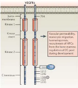
A key element in the complex regulation of VEGF activity is represented by the VEGF receptors (VEGFR): VEGFR‐1 (Fms‐related tyrosine kinase; Flt1), VEGFR‐2 (Kinase insert Domain Receptor;KDR or Fetal liver kinase; Flk1) and VEGFR‐3 (Flt4), expressed by several cell types (Figure 1.5). VEGFRs belong to the Receptor Tyrosine Kinase (RTK) super family. Members of this family consist of an extracellular domain composed by seven immuno globulin (Ig)‐like domains, a short transmembrane and a juxtamembrane segment, and are characterized by a split intracellular tyrosine kinase domain interrupted by a 70 aa long kinase insert domain (Carmeliet, 2005b; Matsumoto and Claesson‐Welsh, 2001; McTigue et al., 1999; Olsson et al., 2006; Roskoski, 2008; Shibuya and Claesson‐Welsh, 2006).
Figure 1.5 Intracellular domains of dimerized and activated VEGFR‐1 is shown with tyrosine‐phosphorylation sites, which are indicated by numbers. Circled R indicates use of the phosphorylation site. Dark blue square indicates the position of tyrosine residue. Binding of signaling molecule; PLCγ, phospholipase C‐γ (dark blue oval) to certain phosphorylation site (boxed number), initiates the signaling cascade. Final biological outcome that is coupled to the respective receptor is indicated in pink box. (Olsson et al., 2006)
VEGFR‐1
VEGFR‐1(Flt‐1) has high affinity towards VEGF‐A, VEGF‐B, PIGF and VEGF‐F (De Vries et al., 1992, Olofsson et al., 1998, Park et al., 1994, Takahashi et al., 2004). Northern blot analysis showed that it is highly expressed in placenta and, at lower levels, in liver, muscle, kidney and choriocarcinoma cells. In rats, it is abundant in tissues and is expressed highly in lungs (Shibuya et al., 1990). Its expression was observed on the surface of different cell types such as endothelial cells (LeCouter et al., 2003), monocytes (Barleon et al., 1996), stem cells (Hattori et al., 2002), pericytes (Yamagishi et al., 1999), cancer cells (Wey et al., 2005), astrocytes (Krum et al., 2002) neurons (Sun et al., 2006), megakaryocytes, smooth muscle cells, osteoclasts (Casella et
23
al., 2003, Sawano et al., 2001) and microglia (Forstreuter et al., 2002); in several of these different cell types, the receptor plays an important role in their activation, proliferation and migration. Even if VEGFR‐1 and VEGFR‐2 are structurally similar, the function of VEGFR‐1 is rather different and multifaceted. VEGFR‐1 has a 10‐fold higher affinity for VEGF‐A compared to VEGFR‐2 (Waltenberger et al., 1994), however its activator function on endothelial cells is very limited or absent. For this reason, it was suggested that the receptor might act as a negative regulator of angiogenesis by acting as a decoy receptor, subtracting VEGF‐A from binding to VEGFR‐2. This function was supported by the early embryonic phenotype of VEGFR‐1 knockout mice. These mice displayed abnormal vascular channels and died in utero at embryonic day 9.5, indicating that VEGFR‐1 is essential for the organization of the embryonic vasculature, normal endothelial cell‐cell or cell‐matrix interactions, but not for endothelial cell differentiation (Fong et al., 1995). In contrast, VEGFR‐2 null mutant mice exhibited impaired endothelial and hematopoietic cell development. Knocking out the tyrosine kinase domain of VEGFR‐1 in mice had no affect on vascular development, further indicating that Flt‐1 differs from Flk‐1 in showing higher binding affinity to VEGF, but having lower kinase activity (Hiratsuka et al., 1998). Therefore, it has been assumed that VEGFR‐1 acts as a VEGF‐A trap, preventing excessive VEGFR‐2 activation during embryonic development (Shibuya, 2001). A physiological role of this endogenous VEGF‐A trap was shown to be be physiologically present in adult life: a soluble VEGFR‐1VEGFR‐1, known as sFlt1 and expressed also by human placenta (Shibuya et al., 1990), is essential in order to preserve corneal avascularity (Ambati et al., 2006; Ambati et al., 2007).
Beside interacting with VEGFR‐2, thus either synergising or inhibiting VEGF‐A activity, VEGFR‐1 can also interact with NP‐1. This latter event plays an active role in the migration of monocytes and macrophages (Barleon et al., 1996; Zacchigna et al., 2008c) and mediates migration of haematopoietic bone marrow progenitors to initiate the pre‐metastatic niche in mouse models of tumor metastasis (Kaplan et al., 2005).
VEGFR‐1 signaling
Despite its ability to bind VEGF‐A with more than 10‐fold higher affinity than VEGFR‐2, VEGFR‐1 only undergoes weak phosphorylation, even if all kinase motifs are conserved (Waltenberger et al., 1994). Relevant to this issue, VEGFR‐1 and VEGFR‐2 share a 43% of overall homology, lower in the extracellular domain (33%) and higher in the kinase domains (70%). Nevertheless, the mechanism responsible for VEGFR‐1 kinase‐impaired activity is still debated. Several VEGFR‐1 tyrosine residues were identified as potentially phosphoryla‐ ted, together with their interacting partners (among others, SH2 domain containing protein, p38/PI3K, Growth Factor Receptor ‐bound protein 2 (Grb2) and Nkc) (Olsson et al., 2006; Shibuya, 2006).
Takahashi et al. (2003) reported that binding of VEGF to its receptor VEGFR‐1stimuates signal transduction of both PI3K/Akt and ERK/mitogen‐activated protein kinase (MAPK) in activated hepatic stellate cells in vitro. In another report, it was observed that activation of VEGFR‐1 through VEGF‐B has strong anti apoptotic action in rescuing neurons in the retina and brain of mouse models of ocular neurodegenerative disorders and stroke, respectively. This effect is predominantly mediated by intracellular singaling of ERK1/2 or Akt pathways and inhibition of proapoptotic BH3‐only proteins and other apoptosis‐ and cell death–related proteins, including p53 and members of the caspase family via VEGFR‐1(Li et al., 2008b). From these studies it is evident that VEGFR‐1 is strongly associated with AKT and ERK/MAPK pathways in its regulation.
VEGFR‐2
The gene for VEGFR‐2 or KDR (kinase insert domain receptor) was cloned from a human endothelial cells cDNA library (Terman et al., 1991). Activation of VEGFR‐2 induces endothelial cell proliferation, migration, angiogenesis, tube formation, vascular permeability and promotes their survival (Olsson et al., 2006). This receptor is also expressed, albeit at lower levels, in other cell types, such as various types of stem cells (Rafii et al., 2003), CD34/AC133‐positive bone‐ marrow precursors, neuronal cells (Asahara et al., 1999), in which it mainly induces
25
proliferation, recruitment, growth and survival (Sondell et al., 2000, Jin et al., 2006). By acting as the main receptor for VEGF‐A, it plays a major role in both angiogenesis and vasculogenesis (Millauer et al., 1993).
Extensive experimental evidence showed that VEGFR‐2 signaling is required for cardio vascular development (Shalaby et al., 1995) and it plays a major role in neovasculari‐ zation in both physiological and pathological conditions. During development, VEGFR‐2 expression is detectable from E7.5 in mesodermal cells, which then migrate and differentiate into primitive ECs (Shalaby et al., 1995). During adult life, VEGFR‐2 is expressed mostly in vascular and lymphatic ECs, even if lower levels of VEGFR‐2 are detected in haematopoietic stem cells, neurons, osteoblasts as well as megakaryocites (Matsumoto and Claesson‐Welsh, 2001). Yamashita et al. (2000) observed that FLK positive cells derived from embryonic stem cells can act as vascular progenitors; these cells can differentiate into endothelial and mural cells and form mature vessels in three‐dimensional cultures and in chick embryos
In several organs, the activities of this receptor extend beyond angiogenesis. For instance, the interaction between VEGF‐A and KDR plays a pivot role for the normal development of the ocular vasculature at early developmental stages but also for nonvascular retinal developmental functions, such as coordination of neural retinal development (Gogat et al., 2004).
In contrast with the canonical pro‐angiogenic role of VEGF‐A/VEGFR‐2 signaling, Greenberg et al. (2008) defined a role for VEGF‐A mediated activation of VEGFR‐2 as an inhibitor of neovascularization, on the basis of its capacity to disrupt vascular smooth muscle cell function. VEGF‐A can indeed suppress the assembly of a receptor complex consisting of platelet derived growth factor receptor‐B (PDGFRB) and VEGFR‐2, thus decreasing pericyte coverage, preventing vessel maturation and promoting endothelial cell proliferation. Another report showed that a secreted form of VEGFR‐2 (sVEGFR‐2) can inhibit developmental and reparative lymphangiogenesis induced by VEGF‐C. Tissue‐specific loss of sVEGFR‐2 in mice at birth induced spontaneous lymphatic invasion in a developed mouse model of alymphatic cornea and hyperplasia of skin lymphatics without affecting blood vasculature. In contrast,
administration of sVEGFR‐2, inhibited the process of lymphangiogenesis but not hemangiogenesis when tested in different models such as mouse model of corneal transplantation, corneal allograft and suture‐induced corneal neovascularization where lymphangiogenesis is the major process (Albuquerque et al., 2009).
VEGFR‐3
VEGFR‐3/Flt4 constitutes the receptor for the VEGF‐C and VEGF‐D family members. The observation that, in adults, VEGFR‐3 expression is restricted to the lymphatic endothelium, together with the availability of genetic models, essentially linked this receptor to lymphatic development and maintenance (Kaipainen et al., 1995, Veikkola et al., 2001). In particular, VEGFR‐3 signaling is required for lymphatic EC sprouting, as well as for lymphatic vessel maintenance through the inhibition of apoptosis (Alitalo et al., 2005). Nonetheless, VEGFR‐ 3 gene knockout mice exhibit defects in arterial‐venous remodelling of the primary vascular plexus, leading to embryonic lethality from day E9.5 (Dumont et al., 1998). Therefore, during embryonic development, VEGFR‐3 activity is not restricted to lymphatics, but has an important function in blood vessel development as well. Recent data extended the role of VEGFR‐ 3 in angiogenesis also during adult life; VEGFR‐3 was found to be highly expressed in angiogenic sprouts, while targeting of VEGFR‐3 signaling resulted in decreased sprouting, vascular density, vessel branching and EC proliferation in different mouse angiogenesis models (Tammela et al., 2008).
VEGFR‐3 is also found to be up‐regulated in the tumor microvasculature, thus opening the possibility to exploit VEGFR‐3 targeting agents to inhibit tumor growth (Saharinen et al., 2004). The extension of the VEGFR‐3 role not only to lymphatic biology, but also to pathological and embryonic development, warrants further studies to define the molecular mediators of these diverse activities, still incompletely explored.
As already mentioned in the case of VEGFR‐1, VEGFR‐3 signaling can be modulated by the interaction with VEGFR‐2 and other co‐receptors, such as NP2. The formation of these complexes is biologically relevant and, for example, VEGFR‐2‐VEGFR‐3 heterodimers
27
might form in vivo both in lymphatic cells and subtypes of ECs, resulting in differential phosphorylation sites and eventually differential signaling (Dixelius et al., 2003).
All these observations suggest that VEGFR‐3, besides its fundamental role in lymphatic vessel development and maintenance, also acts as a regulator of vascular network formation and therefore may constitute an additional target of anti‐angiogenic therapies.
Neuropilins
Neuropilins (NP1 and NP2), initially identified as receptors for class 3 semaphorins, in association with plexin family receptors, mediate repulsive axon guidance in the developing nervous system (Fujisawa, 2004). Even if NP1 and NP2 share only 44% homology, they have similar structural features but different binding and signaling properties (Neufeld et al., 2002). In particular, Semaphorin3A (SEMA3A) only binds NP1, SEMA3F and SEMA3G interact only with NP2, while SEMA3B, SEMA3C and SEMA3D bind both receptors. Additionally, NP1 and NP2 also display specific and mutually selective binding to factors not belonging to the SEMA family, such as VEGF‐A, VEGF‐B, VEGF‐C, PlGF, PDGF‐bb, FGF2, TGF β, Hepatocyte Growth Factor (HGF) and galectin. Moreover, neuropilins differ in VEGF‐A isoform binding, as NP2 binds to VEGF‐A165 and VEGF‐A145 , while NP1 mainly binds VEGF‐A165 (Shraga‐Heled et al., 2007, Pan et al., 2007).
The biological difference between NP1 and NP2 is enforced by their non‐redundant role in development, as NP1 deficient mice die during mid‐gestation with defects in the heart, vasculature, and nerve projection (Kawasaki et al., 1999), while NP2 knockout mice are viable and display only defects in nerve projection (Chen et al., 2000; Giger et al., 2000). Interestingly, the double NP1/NP2 knockout mouse had a more severe abnormal vascular phenotype than either NP1 or NP2 single knockouts, resembling the phenotypes of VEGF‐A and VEGFR‐2 knockouts (Takashima et al., 2002).