Pisa
CLASSE DI SCIENZE MATEMATICHE, FISICHE E NATURALI
CORSO DI PERFEZIONAMENTO IN NEUROBIOLOGIA
2007-2009
Tesi di perfezionamento
Factors that control ocular dominance plasticity
in the rat visual cortex
CANDIDATA RELATORI
Chiara Cerri Dr. Matteo Caleo
Prof. Lamberto Maffei
INDEX
1 INTRODUCTION... 1
THE VISUAL SYSTEM: A PARADIGMATIC MODEL FOR STUDYING PLASTICITY ... 1
Anatomy of the visual system... 1
The rat visual system ... 3
The corpus callosum: a role in cortical binocularity?... 4
Anatomy and physiology of the Corpus Callosum ... 4
Role of callosal connections in cortical binocularity: a matter of controversy... 8
EXPERIENCE-DEPENDENT PLASTICITY IN THE VISUAL CORTEX ... 10
Critical period for ocular dominance plasticity ... 10
Synaptic mechanisms of ocular dominance plasticity ... 12
Homosynaptic mechanisms ... 13
Role of inhibition... 16
Homeostatic plasticity... 20
Molecular basis of OD plasticity ... 22
Neurotrophins ... 23
NMDA receptors ... 25
Intracellular pathway and gene expression ... 26
Extracellular environment ... 28
Callosal plasticity in the visual cortex ... 30
Corpus callosum and visual experience... 30
Role of callosal connections in cortical maturation ... 31
Experience-dependent plasticity in adulthood... 33
Promoting adult plasticity... 34
EXPERIENCE-DEPENDENT STRUCTURAL PLASTICITY... 35
Dendritic spines and sensory experience... 38
Molecular mechanisms of spine plasticity... 40
The Small Rho GTPases... 40
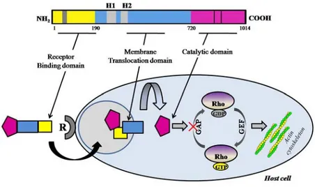
CNF1 activates RhoGTPases... 44
CNF1 structure, internalization and enzymatic activity ... 44
Effects of CNF1 treatment... 45
2 AIM OF THE THESIS ... 47
3 MATERIALS AND METHODS ... 49
CNF1 preparation ... 49
Animal treatment and surgical procedures ... 50
Electrophysiology ... 50
Geniculate recordings... 50
Visual cortex recordings ... 51
Pull-Down Assay ... 53
Dendritic spine analysis... 54
Immunoblotting for vGLUT1 and GAD65/67 ... 54
Anatomical analysis... 55
Statistical Analysis... 56
4 RESULTS ... 57
CALLOSAL CONTRIBUTION TO OCULAR DOMINANCE IN RAT PRIMARY VISUAL CORTEX ... 57
TTX EXPERIMENT... 57
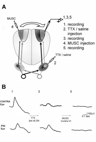
Experimental protocol ... 57
TTX and muscimol injections silence activity within the geniculate and cortex ... 59
Contra/Ipsi VEP ratio before and after silencing of the geniculocortical
pathway... 61
VEP amplitude and latency for the contralateral and ipsilateral eye after silencing of the geniculocortical pathway ... 63
Binocularity of cortical units before and after silencing of the geniculocortical pathway... 65
MUSCIMOL EXPERIMENT ... 66
Experimental protocol ... 66
Cortical binocularity before and after silencing of callosal connections... 67
Peak discharge rates of cortical units before and after muscimol injection... 69
ROLE OF CALLOSAL CONNECTIONS IN OCULAR DOMINANCE PLASTICITY ... 70
Acute silencing of callosal inputs restores binocularity in monocularly deprived animals... 70
Callosal inputs suppress deprived eye responses... 72
Role of the callosum in adult MD... 73
ACTIVATION OF RHOGTPASES TRIGGERS DENDRITIC SPINE REMODELING AND FUNCTIONAL PLASTICITY IN THE ADULT VISUAL CORTEX ... 75
Effects of CNF1 injection into the visual cortex ... 75
CNF1 reactivates ocular dominance plasticity in the adult visual cortex... 79
5 DISCUSSION ... 86
CALLOSAL CONTRBUTION TO OCULAR DOMINANCE IN RAT PRIMARY VISUAL CORTEX ... 86
Callosal connections and binocularity: a review of the literature... 86
Callosal pathways contribute to binocularity by providing ipsilateral eye input to visual cortex... 87
ROLE OF CALLOSAL CONNECTIONS IN OCULAR DOMINANCE PLASTICITY ... 90
ACTIVATION OF RHOGTPASES TRIGGERS DENDRITIC SPINE REMODELING AND FUNCTIONAL PLASTICITY IN THE ADULT VISUAL CORTEX... 94
REFERENCES... 97
INTRODUCTION
THE VISUAL SYSTEM: A PARADIGMATIC MODEL FOR STUDYING PLASTICITY
Plasticity is the ability of the brain to reorganize its connections (at both the structural and functional level) in response to changes in sensory experience. Plasticity is fundamental for the development of neuronal circuitry, enables the brain to adapt to its environment and plays a crucial role in normal brain functions such as learning and memory. Experience-dependent refinements are particularly prominent during well defined periods in early life, the so called critical periods (CPs). During CPs neural circuits display a heightened sensitivity to certain environmental stimuli and proper experience is required to set in motion a cascade of functional and anatomical events in the brain, which ultimately consolidate synaptic connections into their final wiring patterns.
Understanding the mechanisms involved in the development and plasticity of connections is an issue of great interest for neuroscientists. This topic has important implications not only for elucidating how neural circuitry is formed, but also for identifying therapeutic approaches to developmental disorders or strategies to promote recovery after injuries in adulthood, when the brain is normally less plastic. The visual cortex has long been an established model for the study of experience-dependent plasticity because of the relatively easy manipulation of visual inputs. In particular ocular dominance (OD) plasticity triggered by monocular eyelid suture is a classic paradigm to study experience-dependent changes in neural connectivity. Classic experiments have been performed in cats and primates, while rodents have become popular quite recently due to the advent of gene manipulating techniques and to the possibility of combining physiology with biochemical and molecular analysis.
Anatomy of the visual system
The basic anatomical organization of the visual system is highly conserved amongst mammals. The sensory structures are represented by the eyes: light enters the eye by first passing the cornea and finally reaching the very back of the eye, the retina. The retina is responsible for converting light into neural signals that can be relayed to the brain. The retina is a very specialized sensory structure, consisting of a group of
different types of neurons whose role is to collect light, extract basic information and pass the pre-processed image to visual structures in the brain.
These cell types are photoreceptors, bipolar cells, horizontal cells, amacrine cells, and ganglion cells. They are arranged within the retina in precise layers. Axons from the ganglion cells bundle together to form the optic nerves. Fibers from the nasal half of each retina turn towards the opposite side of the brain in a point called optic chiasm, while the fibers from the temporal half of each retina do not cross. Past the chiasm, retinal ganglion cell axons run within the two optic tracts.
Retinal inputs terminate within two major subcortical visual structures, the superior colliculus (SC) and the dorsal geniculate nucleus (dLGN), a portion of the thalamus. Retinal connections are topographically organized: neighboring ganglion cells project to nearby target neurons setting up a retinotopic map in the dLGN. The dLGN is the structure that relays input to visual cortex. In primates and humans, the dLGN contains six layers, each of which receives inputs from one eye only. Indeed, retinal axons coming from the two eyes terminate in adjacent but not overlapping eye-specific layers that are strictly monocular (Hickey and Guillery, 1974).
Projections of neurons in dLGN reach the primary visual cortex, or V1, in the occipital portion of the brain. The adult visual cortex, like all neocortices, consists of six cellular layers between the pial surface and the underlying white matter and contains a complete representation of the contralateral visual hemifield. The majority of inputs from dLGN terminate in layer IV, then neurons in layer IV relay their information to layers II/III, that in turn communicate to layer V-VI. In carnivores and primates, inputs of each eye reach layer IV into alternating stripes, the ocular dominance (OD) columns (LeVay et al., 1975). The columnar systems of the visual cortex communicate together by means of long-range horizontal connections. These connections allow individual cells to integrate information from a wide area of cortex (Gilbert, 1992). Many mammals have binocular vision, and their visual cortical neurons can respond to stimulation of both eye, even if the response to one eye can be predominant (eye preference). Hubel and Wiesel (1962) recorded cells from cat visual cortex and classified them according to their relative response to the stimulation of the two eyes (Ocular Dominance, OD). They indicated with the class 1 the cells that respond only to the stimulation of the contralateral eye, with class 7 the cells driven exclusively by the ipsilateral eye and with class 4 the cells equally driven
by the two eyes. The other classes correspond to cells with an intermediate degrees of dominance of each eye (Hubel and Wiesel, 1962).
Primary visual cortex contains two main types of neurons: pyramidal cells are projection neurons, while non-pyramidal cells represent local interneurons. There are several types of pyramidal cells and interneurons characterized by distinct morphology, physiological properties, and synaptic connectivity patterns.
On a functional level, visual cortical neurons share important response properties which were described for the first time by Hubel and Wiesel (1962). Neurons are selective for several spatial and temporal variables of the visual stimulus such as orientation, direction and velocity of movement, spatial and temporal frequency (Maffei and Fiorentini, 1973; Fregnac and Imbert, 1984).
Once basic processing of visual space has occurred in V1, the visual signal goes to secondary visual cortex, V2, which surrounds V1. Primary and secondary visual areas are connected with associative areas as well as with other sensory cortices.
The rat visual system
The rat, a nocturnal rodent, has a refined and effectively functioning visual system. Its eyes are laterally positioned and therefore the binocular field is relatively small. In relation to eyes position, the proportion of uncrossed optic axons amounts to only 3% of all the retinal ganglion cells and most ganglion cells project to the contralateral side of the brain. In albinos the ipsilateral projection is even smaller (about 1.5%; Lund, 1965; Dreher et al., 1985; Ahmed et al., 1996). The largest retinal projection terminates in the superior colliculus (SC); 40% of ganglion cells project to the dorsal LGN (dLGN) that lacks the lamination typical of other mammals. Indeed, in rodents the dLGN contains two patches, each receiving eye-specific input (Godement et al., 1984; Reese, 1988). The inner core is ipsilateral, surrounded by an outer shell representing the contralateral patch.
In rodent primary visual cortex, no clear anatomical indication of ocular dominance columns can be found, and afferents serving the two eyes converge on the same postsynaptic target cells at the level of layer IV (Antonini et al., 1999). However, Thurlow and Cooper (1988) found hints of a patchy organization of ipsilateral and contralateral inputs in the visual cortex of the rat, using a functional mapping by
means of deoxyglucose (Thurlow and Cooper, 1988). This was confirmed by Caleo and coworkers (1999) with electrophysiological recordings (Caleo et al., 1999a). The physiological properties of the visual cortical neurons of the rat are immature at eye opening (postnatal day 15, P15) and develop gradually during the first month of postnatal life. For example, properties like selectivity for orientation and for the direction of movement are absent at P17 and increase progressively to reach adult values at around P30 (Fagiolini et al., 1994). Binocularity is also immature at P17 and the OD distribution gradually takes the adult shape (Fagiolini et al., 1994).
It has been estimated that in the lateral segment of V1 (mapping the central part of the visual field) 80% of cortical neurons are binocular (Fagiolini et al., 1994; Caleo et al., 1999a; Caleo et al., 1999b; Di Cristo et al., 2001; Caleo et al., 2007). This percentage is surprisingly high considering the very low amount of ipsilateral projection in rat visual system. Indeed, based on this last evidence the input from the ipsilateral eye should be small and the fraction of binocularly driven cells be consequently much lower than 80%. Despite of this, a previous work by Montero et al. (1973) showed that the inputs from ipsilateral eye are not so restricted as expected. Indeed, the area of the visual cortex over which gross potentials can be evoked by stimulation of the ipsilateral eye is quite extensive (Montero, 1973). Understanding how binocular responses are determined in the rodent primary visual cortex has important implications for the studies of OD plasticity.
One possibility is that a contribution to binocularity is provided by the Corpus Callosum which links retinotopically corresponding positions of the two hemispheres (Jacobson, 1970; Cusick and Lund, 1981; Mizuno et al., 2007).
The corpus callosum: a role in cortical binocularity?
Anatomy and physiology of the Corpus Callosum
The corpus callosum (CC) is the largest and one of the most important connection systems of the brain because it provides the main link between the neocortical areas of the two cerebral hemispheres. It is an evolutionary innovation restricted to placental mammals because it is absent in the brain of non-mammalian vertebrates as well as of non-placental mammals.
Many studies have assigned to the CC an important role in integrating and unifying the activities of the two hemispheres and in the development of hemispheric asymmetry (Bloom and Hynd, 2005). The nature and function of the CC has long
been investigated not only for its interesting role in interhemispheric cooperation, but also for the evidence that alterations in this structure are frequent in psychiatric and developmental disorders. For example, abnormalities in the size of the CC have been found in patients diagnosed with schizophrenia, autism, mental retardation, Down’s syndrome, Attention Deficit Hyperactivity Disorder, developmental dyslexia and developmental language disorders (Bloom and Hynd, 2005). Despite the amount of research devoted to the CC, the basic nature and physiology of interhemispheric integration is not fully understood. The primary visual cortex is a good model to address this issue essentially for two reasons. First, visual perception requires interaction between the two hemispheres. Indeed, each hemisphere receives information from the contralateral half of the visual field. Second, the physiological properties of V1 cells are well known and extensively characterized.
Visual callosal connections mature late in humans, at around one month of age in cats and at P15 in rodents. The CC first enlarges caudally then develops rostrally. Similarly myelinization occurs slowly and with a caudal-rostral development (Bloom and Hynd, 2005).
In primary sensory areas, interhemispheric projections link essentially homotopic zones. As mentioned above, the cortical representation of the visual field is split along the vertical midline, with the left and the right hemifields projecting to separate hemispheres. There is a central region of overlap that is represented in both hemispheres, and amounting to 1 degree (deg) of visual angle in humans (foveal region) and to about 10 deg in rodents. The functional continuity of the visual field is re-established by interhemispheric connections that reciprocally connect cortical zones where the representation of the vertical midline of the visual field is located. Indeed, anatomical and electrophysiological studies in cat show that callosal connections form a dense stripe along the border of areas 17 and 18 (Payne, 1994). Neurons in this boundary have receptive fields mapping the vertical midline, together with a smaller portion of the ipsilateral hemifield (Blakemore et al., 1983; Payne, 1990; Payne and Siwek, 1991; Payne, 1994). Recordings from split-chiasm cats in which the genicolo-cortical and transcallosal pathway can be differently activated by stimulation of one or the other eye, reveal that the inputs of both pathways converging onto a given target neuron are remarkably similar in terms of receptive field size and orientation selectivity (Berlucchi and Rizzolatti, 1968; Milleret et al.,
1994). Furthermore, the “callosal” and “genicolo-cortical” receptive fields lie in corresponding points of the two halves of the visual field in close contact with the vertical meridian, forming together a receptive area that crosses the vertical meridian (Berlucchi and Rizzolatti, 1968).
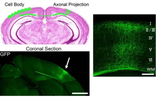
In rodents, the projection field of callosal axons seems to be wider with respect to cats or higher mammals (Olavarria and Van Sluyters 1985; Lewis and Olavarria, 1995; Houzel and Milleret, 1999). Indeed, the entire mediolateral extent of striate cortex contains callosal cells; however, callosal terminals are still quite concentrated at the border between area V1 and V2 (Cusick & Lund, 1981; Mizuno et al., 2007; Olavarria and Van Sluyters, 1983). The spatially organized pattern of callosal projections is characterized by a dual connectivity scheme. As in cat, GFP-labeled callosal axons from one hemisphere project densely to a narrowly restricted region at the border between areas 17 and 18 in the contralateral hemisphere, in which they terminated in layers II–III and V (Mizuno et al., 2007; Fig. 1.1). Here callosal connections link cortical loci sharing the same receptive fields along the vertical meridian, as in higher species. Experiments with retrograde tracers have revealed a second pattern of callosal projections. In the medial-most region of the area 17 there are callosal afferents that link symmetric cortical regions mapping symmetric positions of the extreme periphery of the visual field (Houzel et al., 2002). These projections are likely to be involved in processing requiring large-scale integration of features across the entire visual field such as the detection of symmetric shapes. Callosal connections also play a role in certain binocular functions such as depth perception (Berardi et al., 1988). Experiments in cats, monkey and humans have also revealed the spatial and temporal characteristics of the visual information transmitted through the callosum. At least in adults, the callosum behaves as a low pass filter. Indeed, high spatial and temporal frquencies are attenuated. The sensitivity to contrast is also reduced (Berardi et al., 1988).
The population of cortical neurons projecting to the corpus callosum (less than 10% of all cortical neurons) has been characterized genetically, biochemically, morphologically and electrophysiologically. Most callosally projecting neurons can be classified as pyramidal neurons, although their phenotypic variability is considerable (Vercelli and Innocenti, 1993). The few non-pyramidal neurons which project to the corpus callosum in adult animals are spiny stellate, but also smooth
stellate and fusiform cells which use the synaptic transmitter gamma-aminobutyric acid (GABA) and are presumably inhibitory to their targets. Reported percentages of GABAergic neurons in total populations of callosally projecting neurons vary from 3-5% in rats (Gonchar et al., 1995), and 8% in cats (Fabri and Manzoni, 2004).
Consistent with these percentages, electrophysiological evidence in experimental animals indicates that most of callosal fibers are mainly excitatory to their direct target neurons in the cortex (Matsunami and Hamada, 1984; Karayannis et al., 2007). Callosal fibers activate monosynaptically corticofugal pyramidal neurons, as well as local non-pyramidal neurons, including spiny excitatory and aspiny inhibitory interneurons. Through these last inhibitory GABAergic interneurons the callosal input can inhibit pyramidal neurons in the opposite hemisphere (Carr and Sesack, 1998; Karayannis et al., 2007). Callosal fibers use the glutamate transmitter for monosynaptic excitation (Cisse et al., 2004; Ziskin et al., 2007), while the disynaptic inhibition from callosal inputs is mediated by both GABAA and GABAB receptors
(Kawaguchi, 1992; Chowdhury et al., 1996; Chowdhury and Matsunami, 2002).
Figure 1.1 Distribution of interhemispheric axon projections in the mouse visual cortex visualized using GFP. GFP-labeled callosal axons projected densely to a restricted region of the contralateral cortex, at the border between areas 17 and 18 (arrow). Scale bar: 1 mm. The right panel represents the layer-specific innervation patterns of callosal axons in the visual cortex. Callosal axons terminate in layers II-III and V. Scale bar: 200 μm (Mizuno et al., 2007).
Role of callosal connections in cortical binocularity: a matter of controversy
As I discussed above, the transcallosal pathway of visual cortex has been implicated in a variety of functions, including fusion of the two visual hemifields (Hubel and Wiesel, 1967), extension of receptive fields across midline (Antonini et al., 1983) and depth sensitivity (Berardi et al., 1988). In addition, the corpus callosum seems to be involved in further binocular tasks such as interocular alignment (Elberger, 1979). To perform these functions we may expect that the binocularity of neurons in the area 17/18 border region is strongly influenced by callosal input. The contribution of callosal input to cortical binocularity has been studied by cutting the optic chiasm (split chiasm preparation) or the optic tract on one side, by transection of the corpus callosum and by cooling the area 17/18 border of one hemisphere. The results obtained from these studies are controversial and the role of callosal connections in cortical binocularity is far to be clear.
One of the first evidence of the influence of callosal connections on binocular response comes from experiments by Berlucchi and Rizzolatti (1968). They reported that some neurons at the 17/18 border could be driven binocularly after midsagittal transection of the optic chiasm. This result clearly demonstrates a callosal input to these cells, since visual information from the contralateral eye could only come through the callosum (Berlucchi and Rizzolatti, 1968).
Other studies reported that after cutting the optic tract on one side in the cat, some neurons in the visual cortex ipsilateral to the lesion could still be activated by visual stimulation trough the callosal connections (Choudhury et al., 1965; Vesbaesya et al., 1967). Conversely, others reports indicated that callosal input is modest in the deafferented visual cortex of similarly operated cats (Yinon et al., 1982; Podell et al., 1984).
A direct demonstration of the importance of callosal connections for binocularity in primary visual cortex arises from induced lesions of the corpus callosum in cats. Payne and colleagues (1980) showed that removal of callosal input significantly reduces the numbers of binocularly driven simple and complex cells (Payne et al., 1980) in cat visual cortex. Similar results were reported by Yinon et al. (1992) that demonstrated a reduction in cortical binocularity after CC transaction in cat and kitten (Yinon et al., 1992). Moreover, inactivation by cooling of the contralateral hemisphere gave rise to a substantial increase in the proportion of monocular cells
with a consequent destruction of binocularity (Blackmore et al., 1983). All these findings are in contradiction with those of other groups reporting no changes in binocularity after callosotomy in adult cats (Minciacchi and Antonini, 1984; Elberger and Smith, 1985). In one of this studies it has been found that alterations in binocularity occurred only when section of the callosum was performed during an early phase of development (Elberger and Smith, 1985).
The discrepancies between these different reports likely arise as a consequence of technical aspects, including age at which the callosal section is performed, and time elapsed between surgery and recording.
In rodents, the studies on the role of callosal connections in binocularity are also controversial (Drager, 1975; Diao et al., 1983; Coleman et al., 2009).
Recent work by Coleman et al. (2009) argues aganist an involvement of the CC in binocularity. Using morphometric measures, they suggested that OD in the primary visual cortex of the mouse can be solely accounted by the relative density of feed-forward geniculocortical inputs from the two eyes. Indeed, the contralateral eye pathway exhibits a higher degree of convergence on geniculate neurons than the ipsilateral eye pathway (Coleman et al., 2009).
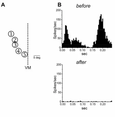
Conversely, electrophysiological experiments based on inactivation of callosal connections suggested an important role for interhemispheric connections in determining cortical OD. Diao et al. (1983) recorded single units in primary visual cortex of adult albino rats before and after inactivation by cooling of the contralateral hemisphere. After cooling ipsilateral responses were reduced and the cumulative OD distribution shifted towards the contralateral eye (Diao et al., 1983).
It is important to stress that a clarification of the role of callosal connections in binocularity is fundamental for the interpretation of studies on OD plasticity. Indeed, rodents have become the most popular model for studies of cortical plasticity at the physiological and molecular level. One of the aims of the present thesis is exactly an assessment of the role of the CC in the construction of normal OD and in OD plasticity.
EXPERIENCE-DEPENDENT PLASTICITY IN THE VISUAL CORTEX
Before eye opening, the initial formation and the development of anatomical and physiological features of the visual system are controlled by intrinsic factors like genetic programs and spontaneous activity (Crowley and Katz, 1999, 2002; Sur and Rubenstein, 2005). Successive aspects of brain development require experience-dependent activity to reach complete maturation (Zhang and Poo, 2001; Sengpiel and Kind, 2002). Indeed, total absence of sensory input leads to a delay in the maturation of the visual cortex. In animals reared in darkness (Dark Rearing) from birth, cortical neurons display immature properties such as reduced orientation and direction tuning, larger receptive field sizes, and lower visual acuity (Fregnac and Imbert, 1978; Timney et al., 1978; Benevento et al., 1992; Fagiolini et al., 1994; Pizzorusso et al., 1997; Gianfranceschi et al., 2003). A total lack of visual experience also affects neuronal structure: dendritic spine are increased in size, changed in morphology, and reduced in density (Wallace and Bear, 2004). Some developmental processes seem to be restored once the animals are exposed to light (Buisseret et al., 1978, 1982). For example, spine size is recovered by light exposure while spine density remains abnormally small (Wallace and Bear, 2004). Deprivation of vision during development also perturbs the regulation of age-specific gene sets suggesting that experience is implicated in the expression of genes required for brain maturation (Majdan and Shatz, 2006).
Visual experience is particularly crucial during the critical period (CP). During this period of heightened plasticity, experience can produce permanent and extensive modifications of cortical organization.
Critical period for ocular dominance plasticity
A classic paradigm to study experience-dependent changes in neural connectivity is ocular dominance (OD) plasticity: the rapid changes in visual cortex circuitry which result from unbalanced inputs from the two eyes (Tropea et al., 2009).
Hubel and Wiesel (1963) first demonstrated that in cat cortical neurons had an eye preference, and cells driven by the same eye were grouped together, originating the columns of ocular dominance. Blocking input from one eye by lid suture (monocular deprivation, MD) during development leads to a loss of physiological responses to that eye and to a dramatic increase in the number of neurons responding
preferentially to stimuli presented to the open eye (Hubel and Wiesel, 1963). This ocular dominance shift results as a consequence of the heightened plasticity present in cortical circuits during CP. Indeed, susceptibility to MD changes with age: it begins 5-10 days after onset of vision, it is most robust during CP, then it declines and it is absent or minimal in the adult age (Wiesel and Hubel, 1963a; Fagiolini et al., 1994; Gordon and Stryker, 1996). Age-dependent OD shift induced by alteration of visual experience has been reported in all mammals studied like monkeys (Horton and Hocking, 1997), ferrets (Issa et al., 1999) and rats (Maffei et al., 1992; Fagiolini et al., 1994). Interestingly, critical period duration is tightly correlated with average life expectancy. The concept of an early CP for the effect of MD is well established for carnivores, primates, and for the rat. The mouse seems to display OD plasticity also outside the classical CP depending on the type of anesthesia and the method used to assess OD. However, an age-dependent decline of OD is also observed in mice and fully adult mice are not sensitive to MD (Lehmann and Lowel, 2008).
In all mammals examined MD leads to anatomical and functional effects. Anatomical changes comprise an expansion of territories driven by open eye, and a subsequent reduction of those driven by deprived eye (Katz and Shatz, 1996). Moreover, geniculate neurons receiving input from deprived eye are shrunken (20-25%) and those driven by open eye are hypertrophic (10-15%) (Sherman and Spear, 1982). Studies by Stryker and colleagues showed that anatomical changes at thalamocortical level occur days after detection of the functional effects. Indeed, an OD shift is already detectable after a short period of MD (1-3 days), while changes in thalamocortical arborization are visible only after 4 days of MD (Antonini and Stryker, 1993, 1996; Antonini et al., 1998). Moreover, experiments by Trachtenberg and coworkers (2000) showed that functional OD shift occurs first in the supragranular layers and then they guide changes at the geniculocortical synapse. Rapid OD plasticity in the upper layers of the cortex is accompanied by similarly rapid anatomical changes in the long-range horizontal connections between OD columns in these layers (Trachtenberg and Stryker, 2001). This evidence suggested that horizontal connections could represent a structural correlate for functional OD shift.
In mouse, as in higher mammals, MD promotes growth of the open eye’s geniculocortical connections and arrest of growth of deprived arbors (Antonini et al.,
1999). Interestingly, mouse OD shift has been found in all layers but the shift was more pronounced in extragranular layers than in layer IV, with the greatest shift in layer V. This finding suggests that in the mouse, as in other species, intracortical as well as geniculocortical synapses undergo plasticity with MD (Gordon and Stryker, 1996).
In addition to the shift in OD, MD impairs the animal’s behaviour by reducing visual acuity of the deprived eye and affecting stereoscopic vision (Medini and Pizzorusso, 2008). Remarkably, physiological responses in the deprived retina and thalamus remain completely unaffected (Wiesel and Hubel, 1963b; Sherman and Stone, 1973). An imbalance in binocular vision during childhood affects visual acuity also in humans leading to a pathological condition designated amblyopia or “lazy eye” (Medini and Pizzorusso, 2008). Amblyopia is clinically important because it is the most frequent cause of vision loss in infants and young children, occurring naturally in about 2-4% of the population (Levi, 2006).
Synaptic mechanisms of ocular dominance plasticity
The shift in ocular preference induced by MD has been originally thought to be the outcome of a process of activity-dependent competition between the synaptic terminals driven by the two eyes for connection with the postsynaptic neuron. This idea was supported by the fact that binocular lid suture was not effective to alter OD columns in mammals (Wiesel and Hubel, 1965; Sherman and Spear, 1982; Gordon and Stryker, 1996). In favour of a competitive view, Chapman and coworkers showed that an imbalance in the electrical activities of the two retinas is sufficient to shift OD also in visual deprivation conditions (Chapman et al., 1986). In addition, reversible blockade of intrinsic cortical activity by intracortical infusion of tetrodotoxin (TTX) or muscimol has a remarkable impact on OD shift (Reiter et al., 1986; Reiter and Stryker, 1988). Interestingly, the results obtained with these two manipulations are extremely different. Activity blockade by TTX infusion completely prevents the OD shift following MD (Reiter et al., 1986). By contrast, the administration of the inhibitory neurotransmitter agonist muscimol, that selectively blocks postsynaptic cells discharges, causes an OD shift in the direction of the less active, deprived eye (paradoxical shift; Reiter and Styker, 1988). One possible explanation of this result is that the activity of deprived thalamocortical terminals is
now better correlated with that of the inhibited postsynaptic cell, therefore leading to synapse strengthening. Thus, the correlation between pre and postsynaptic activity appears to be very important for the expression of OD plasticity.
Binocular competition was originally related to heterosynaptic mechanisms by which open eye inputs drive down the synaptic efficacy of the deprived inputs (Miller et al., 1989; Harris et al., 1997). Indeed, active geniculate neurons from the open eye become functionally and structurally strengthened because they compete better for postsynaptic space than less active afferents from deprived eye. It has been suggested that inputs from the two eyes compete for the acquisition of a neurotrophic factor from target structures (Maffei et al., 1992). An alternative view is that OD plasticity is due to different homosynaptic mechanisms, whereby inputs from the two eyes are affected separately by MD (Smith et al., 2009). For example, in rodents, modifications in the monocular zone of the visual cortex (where no competition can occur) that resemble those taking place in the binocular region have been demonstrated after contralateral MD (Pham et al., 1999; Heynen et al., 2003).
Homosynaptic mechanisms
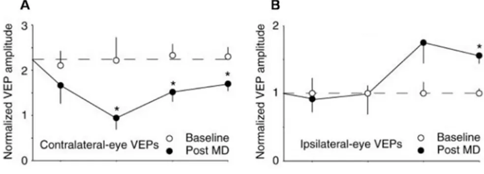
In support of the homosynaptic view recent data suggest that the OD shift is the result of two forms of synaptic plasticity: an initial depression of deprived eye inputs and only later a potentiation of responses from the open eye (Fig. 1.2).
Figure 1.2 Two responses to monocular deprivation. Absolute VEP amplitudes (normalized to average baseline ipsilateral-eye VEP) before (open symbols) and after (filled symbols) various periods of visual deprivation. (A) Depriving the contralateral eye of vision by lid closure causes a rapid depression of contralateral-eye VEP amplitude that reaches statistical significance after 3 days of MD and remains significant at 5 and 7 days. (B) Depriving the contralateral eye of vision by lid closure
leads to a delayed potentiation of the ipsilateral-eye VEPs, significant after 7 days (Frenkel and Bear, 2004).
It is worth noting that the quality of visual experience through the open eye is not changed during MD, therefore its response potentiation has to be ascribed to modification in cortical circuitry. This aspect of OD plasticity is theoretically predicted by the influential Bienenstock, Cooper and Munro (BCM) model (Bienenstock et al., 1982). According to this theory, the reduction in overall cortical activity caused by closing the contralateral eyelid decreases the value of the modification threshold, θm thereby facilitating potentiation of correlated inputs
(reviewed by Bear 2003; Smith et al., 2009).
Several lines of evidence indicate that homosynaptic depression occurs only at active synapses. Indeed, lid suture was more efficient in shifting OD towards open eye than blockade of all residual retinal activity by intravitreal TTX (Frenkel and Bear, 2004). In particular, retinal silencing prevents depression of the deprived eye inputs but enhances potentiation of responsiveness to the open eye (Rittenhouse et al., 1999). In the last years researchers have tried to elucidate the cellular and molecular mechanisms underlying this bidirectional kinetics of OD plasticity. To this end strengthening and weakening of inputs from the eyes has been related to the mechanisms involved in long term potentiation (LTP) and long term depression (LTD) at central synapses.
Various line of evidence strongly suggest that LTD-like mechanisms influence depression of deprived-eye responses. Brief MD at the peak of the critical period induces the AMPA receptor internalization and the same phosphorylation pattern of glutamate receptor 1 (GluR1) subunit that occurs after LTD induction in vitro (Heynen et al., 2003). Interestingly MD occluded further LTD, causally linking LTD-like mechanisms in the loss of responsiveness observed after MD (Heynen et al., 2003). Another study by Bear’s group showed that in MD mice, blocking the internalization of GluR2, that is necessary for LTD, deprived eye depression and ocular dominance shift are prevented (Yoon et al., 2009). This effect is restricted to layer IV suggesting that LTD is layer specific (Crozier et al., 2007; Yoon et al., 2009).
Other lines of evidence support the idea that long-term potentiation (LTP) of the synapses driven by the open eye is important for OD plasticity. For instance, αCaMKII activity is required for both LTP in vitro and OD plasticity in vivo (Kirkwood et al., 1997; Taha and Stryker, 2002). Moreover, one form of LTP (white matter-layer II-III) in the visual cortex is developmentally regulated with a decline over time that mirrors that of the critical period (Kirkwood et al., 1996). Other forms of NMDA-dependent LTP (layer IV- layer II-III) are present in visual cortical slices of adult rat.
Further suggestion comes from the recently discovered phenomenon of stimulus-selective response potentiation: in juvenile mice, the magnitude of visually driven thalamo-cortical responses in layer IV increases following repeated presentation of an oriented stimulus and this potentiation is dependent on NMDAR activation. Moreover, it has been shown that GluR1 delivery to synapses, that is crucial for LTP, is required for visual experience-dependent plasticity (Frenkel et al., 2006).
Remarkably the capability of use-dependent potentiation remains relatively intact in the adult visual cortex, as shown by the fact that potentiation of visually driven responses has been described in vivo in the adult rodent visual cortex after tetanic stimulation of the dLGN (Heynen and Bear, 2001). Important findings in this field come from Sawtell and coworkers (2003). They found that in adult mouse depriving the dominant contralateral eye of vision leads to a persistent NMDA receptor-dependent potentiation of the weak ipsilateral eye (Sawtell et al., 2003). Moreover these data are one of the first demonstration that adult mouse visual cortex has a greater potential for experience-dependent plasticity than previously appreciated. It is important to note that the proprieties of OD plasticity vary significantly with age. In adulthood the rapid depression of deprived eye response is absent while delayed potentiation of the open eye inputs continues to occur. According to these data, in slices of mouse visual cortex LTP can be elicited beyond P35, but NMDAR-dependent long-term depression (LTD) cannot (Kirkwood et al., 1997). Furthermore LTD occlusion and AMPA receptor modifications are not observed in adult animals subjected to MD (Heynen et al., 2003). These evidences suggest that the capability to depress a deprived input is developmentally regulated.
Other studies indicate that LTD- and LTP- like processes may not be sufficient to fully describe naturally occurring plasticity observed in vivo. For example, a mutation that disrupts LTD dependent on metabotropic glutamate receptor (mGluR) does not alter the normal OD shift in response to MD (Renger et al., 2002). Furthermore, OD plasticity is blocked by overexpression of the protein phosphatase calcineurin, the only known Ca2+/calmodulin-activated protein phosphatase in the brain, but LTD appears normal in these animals (Yang et al., 2005). Brain-derived neurotrophic factor (BDNF) prevents LTD in V1, but BDNF-overexpressing mice are sensitive to MD at least during an early phase of postnatal development (Huang et al., 1999; Jiang et al., 2003). Finally, GAD65 knockout (KO) mice, which lack OD plasticity, show no deficit in induction of LTP or LTD in layer II/III of mouse binocular visual cortex (Hensch et al., 1998). Thus, it is still unclear whether MD effects can be entirely modeled by the solely LTD/LTP mechanism. Indeed, several alternative hypotheses have been formulated to explain the phenomenology of OD plasticity.
Role of inhibition
Considering that neuronal circuits in the brain are intricately interconnected, changes of thalamocortical inputs are not sufficient to totally explain the process of OD plasticity. In the complicate neuronal network of the visual cortex, inhibitory interneurons have been emerged as a key regulator of neuronal plasticity (Hensch, 2004).
During last years, it has become clear that inhibition not only is a ‘brake’ for excitation but also has an important role in sculpting the pattern of electrical activity (Berardi et al., 2003). Indeed, GABAergic transmission in mammalian forebrain has been implicated in sharpening the temporal signaling in neurons (Pouille and Scanziani, 2001). Thanks to inhibition a postsynaptic target neuron can detect an imbalance of activity between afferents and can consequently be engaged within the plastic process.
In neocortex inhibitory connections are developed later than excitatory connections (Blue and Parnavelas, 1983). It is possible that, excitatory and inhibitory circuit elements reach an optimal balance once in life during which activity-dependent plasticity may occur.
Drastic pharmacological perturbations of excitatory-inhibitory balance, such as hyperexcitation by bicuculline (Ramoa et al., 1988) or total silencing by TTX (Reiter et al., 1986) disrupt OD plasticity. However, these results do not explain how changes in the relative amounts of excitation and inhibition interfere with plasticity. Taking advantage of gene-targeting technology, Hensch and co-workers demonstrated a decisive role for excitatory-inhibitory balance. Mice carrying a deletion of the 65-kDa isoform of glutamic acid decarboxylase (GAD65), the GABA biosynthetic enzyme, exhibit a significant reduction of stimulated GABA release and show no shift in responsiveness toward the open eye following brief MD (Hensch et al., 1998). A direct physiological consequence of excitatory-inhibitory unbalance in GAD65 KO mice is the observation of a prolonged discharge, that is the tendency to continue to fire even after stimuli have passed the cell’s receptive field. This prolonged discharge resemble that seen in wild type mice before CP, when intrinsic inhibition is weak and OD plasticity is absent. When CP starts this discharge drops down. Thus, ocular dominance plasticity and prolonged discharge are tightly co-regulated by inhibition (Fagiolini and Hensch, 2000). Notably, in GAD65 KO mice mechanisms of synapse modification in vitro, as LTP and LTD, are not impaired, demonstrating no general deficit in activity-dependent plasticity (Hensch et al., 1998). Normal OD plasticity in GAD65 KO mice can be rescued if GABA transmission is enhanced in the visual cortex by means of diazepam (Hensch et al. 1998). Remarkably, rescue of plasticity is possible at any age in GAD65 KO mice, which indicates that the critical period is dependent on the proper level of inhibitory transmission (Fagiolini and Hensch., 2000). According to this view, the onset of the critical period can be accelerated by prematurely enhancing inhibition with benzodiazepines just after eye opening (Fagiolini and Hensch, 2000; Iwai et al., 2003). In addition in transgenic mice overexpressing brain-derived neurotrophic factor (BDNF) development of GABA mediated inhibition is accelerated and this results in early opening and closure of the critical period (Huang et al., 1999).
Not all GABA circuits are involved in critical period regulation. Among the many types of GABA-positive interneuron (they account for nearly 20% of cortical neurons), parvalbumin-positive (PV) interneurons seem to be the most important players (Fig. 1.3). Indeed, maturation of parvalbumin-positive interneurons parallels critical period onset (Del Rio et al. 1994) and both events are accelerated by BDNF
overexpression (Huang et al., 1999). Interestingly, only GABA type A (GABAA)
receptor circuits have been found to drive cortical plasticity. In particular, GABAA
receptors carrying the α1 subunit appear to be crucial in CP regulation (Fagiolini et al., 2004). These receptors are preferentially enriched at somatic synapses opposite to terminals of PV large basket cell.
With age, large PV cells are preferentially enwrapped in perineuronal nets (PNN) of extracellular matrix (ECM). When these are disrupted, perisomatic inhibition of their targets is reduced (Saghatelyan et al., 2001), and ocular dominance shifts can be induced by monocular deprivation, even in adulthood (Pizzorusso et al., 2002).
Taken together, these results indicate that during development there are two functional threshold of inhibition in the visual cortex: the first one is necessary to trigger plasticity and the second one causes the end of critical period. Pharmacological reduction of inhibitory transmission by infusion of mercaptopropionic acid (MPA, an inhibitor of GABA synthesis) or Picrotoxin (a GABAA receptor antagonist) into the rat visual cortex effectively reactivates OD
plasticity in adulthood (Harauzov et al., 2010). Moreover, also other manipulations resulting in reductions of cortical inhibition promote adult plasticity (He et al., 2006; Sale et al., 2007; Maya Vetencourt et al., 2008).
Figure 1.3 Heterogeneity of local GABA circuits in the neocortex. Many subtypes of GABA releasing inhibitory interneuron can be identified in the neocortex on the basis of morphology, connectivity, expression of calcium-binding proteins or neuropeptide content. Moreover, specific contacts are preferentially enriched in specific GABAA receptor α-subunits. The perisomatic localization of different subunits is shown next to the central pyramidal neuron. All subunits are found diffusely along the dendrite. ais, axon initial segment; CCK, cholecystokinin expressing; Ch, chandelier cell; CRC, Cajal–Retzius cell; DB, double bouquet cell; M, Martinotti neuron; N, neurogliaform neuron; PV+, parvalbumin positive (Hensch 2005).
Recently the relative contribution of excitation and inhibition in OD plasticity has been addressed with new approaches. By recording from synaptically coupled pairs of neurons in layer IV, it has been shown that brief MD increases the strength of inhibitor GABAA mediated-synaptic responses (Maffei et al., 2006). These data
propose that this novel form of long-term potentiation of inhibition (LTPi) contribute to the deprivation-induced loss of visual responsiveness in the rodent visual cortex (Maffei et al., 2006). It has also been suggested that this inhibitory plasticity is fundamental in modulating cortical circuits refinement during development and may regulate the onset of OD plasticity (Maffei et al., 2010). Other studies, by measuring the early consequences of MD in different classes of cortical neurons in vivo, showed that responses of inhibitory cells are also modified by deprivation (Gandhi et al., 2008; Sugiyama et al., 2009; Kameyama et al., 2010). In particular, Yazaki-Sugiyama and co-workers identified Fast Spiking (FS) interneurons (PV positive large basket cells), as a substrate for the early phase of the OD plasticity process. Intracellular recordings showed that these cells normally exhibit binocular response which following MD first shift paradoxically towards the deprived eye and only later towards the open eye (Yazaki-Sugiyama et al., 2009). These results suggest that the initial enhanced activation of FS interneurons by deprived-eye inputs may trigger an inhibitory suppression of their own responses in the visual cortex (Yazaki-Sugiyama et al., 2009).
A completely different view of the role of inhibition in the expression of OD plasticity has been provided by Khibnik and coworkers (2010). They induced an OD shift with 3 days of MD and then pharmacologically removed the influences of intracortical inhibition and excitation, leaving only afferent thalamocortical synaptic inputs. They found that OD shift induced by brief MD is not influenced by
intracortical inhibition, being fully expressed by modification of excitatory talamocortical inputs. They concluded that inhibition precedes and perhaps influences modification of exitatory synapses but play a marginal role in OD plasticity (Khibnik et al., 2010).
Homeostatic plasticity
The role of other forms of plasticity in OD plasticity has begun to be investigated only recently. Homeostatic synaptic plasticity has emerged as an important complement to Hebbian forms of plasticity in the activity-dependent refinement of synaptic connectivity (Turrigiano and Nelson, 2004; Davis, 2006; Turrigiano, 2008). The term ‘homeostatic’ refers to a form of plasticity that acts to stabilize the activity of a neuron or neuronal circuit against perturbations, such as changes in cell size or in synapse number or strength, that alter excitability (Turrigiano, 2007). Examples of homosynaptic mechanisms described in these years include: activity-dependent regulation of intrinsic neuronal firing properties (Zhang and Linden, 2003); pre and post–synaptic forms of excitatory synaptic plasticity, such as synaptic scaling, that adjust the strength of all excitatory synapses of a neuron up or down to stabilize firing (hence the term “synptic scaling”; Turrigiano and Nelson, 2004); balancing of excitation and inhibition within neuronal networks (Maffei et al., 2004); compensatory changes in synapse number (Wierenga et al., 2006). All these changes seem to act in order to restore the neuronal firing rates to normal levels after perturbation.
From all these findings three principal models by which neurons implement homeostatic plasticity have been postulated. A cell-autonomous mechanism where individual neurons sense their own activity, through, for instance, changes in Ca2+
influx, and then adjust all of their synaptic strength up or down to keep this value relatively constant. A synapse-specific mechanism, in which local synaptic signaling induces compensatory changes in presynaptic and/or postsynaptic function. For example, depolarization induced by glutamate receptors activation might negatively change the number of glutamate receptors on the postsynaptic cell or generate a retrograde signal that blocks vesicle release from the presynaptic terminal. Finally, changes in network activity could lead to altered release of a diffusible ‘activity signal’ that negatively regulates synaptic function.
The first reports of synaptic homeostasis at central synapses suggested that neurons respond to changes in activity by scaling up or down the strength of all of their synapses through a simple change in the accumulation of postsynaptic glutamate receptors (O'Brien et al., 1998; Turrigiano et al., 1998). For example, treatment of neocortical cultures with TTX increases the amplitude but not the frequency of miniature excitatory postsynaptic currents (mEPSC) (Lissin et al., 1998; O'Brien et al., 1998; Turrigiano et al., 1998). Other in vitro studies showed that also changes in presynaptic functions, such as alterations in release probability and in number of release sites, are involved in homeostatic plasticity (Murthy et al., 2001; Burrone et al., 2002; Thiagarajan et al., 2005). Chronic activity blockade in cortical cultures increases the expression of vesicular glutamate transporter vGLUT1, whereas hyperactivity reduces vGLUT expression (De Gois et al., 2005; Erickson et al., 2006).
Despite the large number of studies, the nature of the activity signal that controls synaptic scaling is still debated. Indeed, it is currently unknown whether the activity signal relevant for synaptic scaling is postsynaptic changes in firing, presynaptic changes in release, or local dendritic changes in receptor activation and/or Ca2+ influx. Synaptic scaling has been observed at cortical synapses also in vivo following sensory deprivation and is developmentally regulated. It has been proposed that synaptic scaling is important in regulating cortical excitability during activity-dependent development (Desai et al., 2002; Maffei et al., 2004; Goel et al., 2006). An important issue in homeostasis is how changes in activity are signaled to neurons or synapses. It has been proposed that several activity-dependent molecular mechanisms are involved. Documented examples are BDNF, cytokine tumor-necrosis factor α (TNFα) and the immediate-early gene product Arc (Rutherford et al., 1998; Shepherd et al., 2006; Stellwagen and Malenka, 2006; Kaneko et al., 2008a). BDNF enhances mEPSC amplitude onto excitatory neurons (Copi et al., 2005) but its effect seems to be depend on brain region and developmental stage. TNFα originates from glia and seems to regulates AMPA receptors on neurons in a network-level homeostatic process (Turrigiano, 2007). Finally, Arc overexpression decreases AMPA-receptor mediated transmission and prevents the increase in mEPSC amplitude induces by chronic TTX (Shepherd et al., 2006).
Concerning visual cortex plasticity, a recent study reports evidence for homeostatic mechanisms using two-photon calcium imaging in vivo (Mrsic-Flogel et al., 2007). The authors investigated how MD shifts the magnitude of deprived and non-deprived eye responses in individual neurons. They found that the population of neurons driven only by the deprived eye unexpectedly showed strong responses after MD. These findings demonstrate that the weak input of deprived eye is not able to induce response depression, which instead seems to be dependent on the input of the open eye. In addition the proportion of monocular, closed eye-driven cells remained constant after MD and most neurons in monocular cortex increased their responsiveness. All these findings suggest that in the deprived visual cortex there are compensatory mechanisms that maintain firing rates within a certain range during MD.
Another recent work shows that the pharmacological or genetic blockade of TNFα signalling in visual cortex has no effect on the depression of the deprived eye input but prevents the gain of responsiveness to the non-deprived eye. This result demonstrate that synaptic scaling underlies the enhancement of responsiveness to the non-deprived eye (Kaneko et al., 2008a).
Some studies have addressed the importance of a form of synaptic plasticity designed spike-timing dependent (STDP) for the experience-dependent plasticity. STDP refers to the temporal order of pre- and postsynaptic spiking in eliciting long-term synaptic depression or potentiation (Celikel et al., 2004; Dan and Poo, 2004). It has been proposed that loss of responsiveness of deprived inputs is mediated by STDP (Celikel et al., 2004; Hensch, 2004; Kuhlman et al., 2010) but its role in OD plasticity is not fully clarified. Recently, it has been proposed that MD-induced OD plasticity observed in fast-spiking interneuron is consistent with a STDP rule (Yazaki-Sugiyama et al., 2009).
Molecular basis of OD plasticity
In parallel with the physiological mechanisms described above, a complex and interrelated molecular network sets the basis for OD plasticity. This network involves a large number of molecules whose expression is developmentally regulated
and differentially altered by visual experience. In this chapter I describe some of the molecular mechanisms associated with to visual cortex plasticity.
Neurotrophins
Neurotrophins (NTs) are a family of neurotrophic factors that include: nerve growth factor (NGF), brain derived neurotrophic factor (BDNF), neurotrophin-3 (NT-3) and neurotrophin-4 (NT-4). They support the survival and differentiation of neurons by binding to and activating tyrosine kinase receptors of the Trk family.
In 1990s pioneering studies by Maffei’s group suggested that NTs are important players of plastic processes. The authors demonstrated that exogenous NGF administration prevents the loss of visual acuity and the shift of OD induced by MD in the rat visual system (Domenici et al., 1991; Maffei et al., 1992). Based on these finding, they put forward the idea that competition in OD plasticity might depend on the uptake of NT secreted by cortical neurons in an activity-dependent manner and retrogradely transported by geniculate neurons. Several other subsequent reports confirmed this hypothesis (Harris et al., 1997; McAllister et al., 1999; Pizzorusso et al., 1999). In particular it has been demonstrated that, with the exception of NT-3, all neurotrophins influence MD, but not all factors play an identical role on visual neuron properties (Lodovichi et al., 2000). Indeed, NT-4 and NGF prevent the shift induced by MD without affecting spontaneous or visually-driven activity (Gillespie et al., 2000; Lodovichi et al., 2000). In contrast, BDNF is less effective in preventing OD shift, but it alters both spontaneous and visually-evoked activity of cortical neurons.
Other studies, using a complementary strategy based on antagonizing the action of endogenous NTs, demonstrated that NTs are important for normal visual cortical development and plasticity (Berardi et al., 1994; Domenici et al., 1994; Cabelli et al., 1997). For example, inactivation of NGF signaling by specific antibodies alters visual acuity and binocularity of cortical neurons, induces a shrinkage of geniculate neurons (Berardi et al.,1994) and prolongs the sensitive period for MD in the rat (Domenici et al., 1994).
NT synthesis and release are regulated during development in an experience-dependent mode (Bozzi et al., 1995; McAllister et al., 1999). NTs can modulate electrical activity, at pre and postsynaptic level, increasing neurotransmitter release
and depolarizing neurons but they also act on gene expression (Sala et al., 1998; Kafitz et al., 1999; Lodovichi et al., 2000; Poo, 2001). Moreover, evidence from literature indicates that NTs stimulate dendritic growth and this action is activity-dependent (McAllister et al., 1995; McAllister et al., 1996). Regulation of synaptic plasticity by NTs in the cortex requires afferent electrical activity (Caleo et al., 1999a). Taken together these results indicate that activity and NTs interact reciprocally. Their interaction may provide a mechanism by which active neuronal connections are selectively enhanced.
Striking evidences for the role of NTs in regulating the experience-dependent plasticity came from studies in mice overexpressing BDNF. A precocious BDNF expression accelerates the development of visual acuity and the time course of the critical period. In addition, in these mice maturation of inhibition is accelerated suggesting a close link between BDNF and GABA circuits (Huang et al., 1999). Moreover, mice overexpressing BDNF display a normal functional development even when reared from birth in total darkness (Dark Rearing). Indeed these mice display visual acuity, critical period for OD plasticity and inhibitory transmission identical to those of normal, light-reared mice (Gianfranceschi et al., 2003).
In rodents, BDNF expressed in the retina is transported anterogradely by retinal ganglion cells to the geniculate nucleus. Levels of BDNF in the retina not only influence development of the lateral geniculate nucleus and superior colliculus (Caleo et al., 2000; Caleo et al., 2003), but also have a role in OD plasticity (Mandolesi et al., 2005). Indeed, MD reduces BDNF expression in the deprived retina and intravitreal injection of BDNF into the deprived eye prevents the OD shift induced by MD (Mandolesi et al., 2005). However, other studies provide different insights on the role of BDNF in OD plasticity. Data from heterozygous knockout mice for BNDF demonstrate that a 50% reduction in the BDNF levels has no effect on OD plasticity (Bartoletti et al., 2002). Furthemore, Stryker and colleagues, using a genetic approach to inhibit TrkB signaling, show that OD shift in response to MD does not require TrkB activity (Kaneko et al., 2008b). However, these data do not exclude the important and well recognized role of NTs in visual cortex plasticity. Indeed, the actions of other NTs could compensate for the lack of modification in visual cortex plasticity observed in these mutant mice.
NMDA receptors
NMDA receptors (NMDARs) mediate excitatory synaptic transmission and their expression is widespread in the brain. They have the characteristic of being both transmitter and voltage-dependent, and permitting Ca2+ influx, they are related to the intracellular signaling implicated in plasticity. Evidence for NMDA receptor involvement in visual cortex plasticity comes from experiments showing that blockade of NMDA receptors prevented the effects of MD (Bear et al., 1990; Roberts et al., 1998; Sawtell et al., 2003). NMDA receptors are developmentally regulated and their expression is modified by electrical activity. This activity-dependent regulation seems to be mediated by epigenetic mechanisms (Lee et al., 2008).
In visual cortex, NMDA subunit composition varies over development from an increased expression of receptors containing the NR2B subunit to a progressive addition of the subunit NR2A, with a time course paralleling that of critical period (Roberts and Ramoa, 1999). Dark rearing (which delays CP closure and impairs cortical maturation) delays the expression of NR2A subunit, suggesting that NR2B/NR2A switch is involved in visual cortex development and CP regulation (Berardi et al., 2003). However, in transgenic mice with deletion of NR2A subunit the sensitivity to deprivation is weakened but restricted to the typical critical period (Fagiolini et al., 2003).
A recent study shows that GAD65KO mice not only have altered inhibition but also have alterations in NMDA receptor subunit composition and function. Remarkably, treatment with benzodiazepines, which rescues OD plasticity, increases NR2A levels. These results suggest that changes in either inhibition or excitation would engage mechanisms that converge to regulate NMDA receptors, thereby enabling plasticity (Kanold et al., 2009). Another suggestion of the possible mode by which NMDA receptor are implicated in plastic mechanism is provided by Bear’s group (2009).The deprived-eye responses in NR2A KO mice are unchanged after brief MD (3 days), whereas the non-deprived eye responses dramatically potentiate (Cho et al., 2009). These data suggest that a reduction in the NR2A/NR2B ratio during monocular deprivation is permissive for the compensatory potentiation of non-deprived inputs (Cho et al., 2009).
Intracellular pathway and gene expression
Electrical activity, neurotrophins, and NMDA receptors control plasticity of cortical circuitry operating on three intracellular kinases: cAMP-dependent protein kinase (PKA; Beaver et al., 2001; Cancedda et al., 2003), extracellular-signal-regulated kinase (ERK; Di Cristo et al., 2001; Cancedda et al., 2003), α Ca2+/calmodulin dependent protein kinase II (α CAMKII; Taha et al., 2002). Besides these kinases, the phosphatase calcineurin seems to be involved in OD plasticity (Yang et al., 2005). Thus, OD plasticity may be regulated by the balance between kinases and phosphatases.
All these effectors are mutually interconnected by a complex network of interactions and have targets both in the cytoplasm and the nucleus. In particular, in the cytoplasm substrates crucial for synaptic transmission, neuronal excitability and morphological stabilization are target of phosphorylation, while in the nucleus, the targets are molecules engaged in gene expression (Berardi et al., 2003).
Long lasting modifications in neuronal circuits require gene expression and protein synthesis (Mower et al., 2002; Taha and Stryker, 2002). These mechanisms are necessary also for mediating the action of experience on visual cortex development. Visually-driven activity activate transcriptional factors, such as zif268 or CREB (Caleo et al., 1999b; Pham et al., 1999; Mower et al., 2002) but zif268 is not necessary for OD plasticity (Mataga et al., 2001) while activation of CREB is crucial for OD plasticity (Pham et al., 1999; Liao et al., 2002; Mower et al., 2002). CREB triggers the expression of genes essential for establishment and maintenance of plastic changes and which are under the control of the cAMP-response element (CRE) promoter (Pham et al., 1999; Mower et al., 2002). Studies based on a combination of DNA microarrays, RT PCR and immunohistochemistry show that several sets of genes are modulated by visual experience or deprivation (Majdan and Shatz, 2006; Tropea et al., 2006). For example, expression of a binding protein of insulin-like growth factor-1 (IGF1) is highly upregulated after MD, and exogenous application of IGF1 prevents the physiological effects of MD on OD (Tropea et al., 2006). Moreover, a recent microarray screen indicates that the calcium sensor cardiac troponin C (part of a complex that mediates calcium-dependent actin-myosin interaction) is elevated in visual cortex during the critical period, and is regulated by visual activity (Lyckman et al., 2008).
Recently, it has been proposed that Arc is an effector molecule important for OD plasticity. In visual cortex Arc expression starts only after eye opening and is activated by visual stimulation (Tagawa et al., 2005; Wang et al., 2006). In KO mice the total absence of Arc, renders visual cortical synapses insensitive to the effects of both experience and deprivation (McCurry et al., 2010).
In these years, the regulation of chromatin structure has emerged as one mechanism regulating visual cortex plasticity. Indeed, histone phosphoacetylation has been demonstrated to be involved in OD plasticity (Putignano et al., 2007). The histone phosphoacetylation is mediated by ERK and is developmentally regulated. In adult mice visual experience activate ERK but induction of histone phosphoacetylation and CREB-mediated gene expression are much lower than in juvenile animals. In line with this, a pharmacological-induced histone acetylation restores plasticity in the adult visual cortex (Putignano et al., 2007). Experience-dependent regulation of histone acetylation could be a way to regulate specific sets of genes important to consolidate plastic changes (Medini and Pizzorusso, 2008).
Recently, a novel factor has been added in the list of molecules involved in visual cortex plasticity. Sugiyama and colleagues (2008) have found that Otx2 homeoprotein, an essential morphogen for embryonic head formation, is reused later in life to regulate CP (Sugiyama et al., 2008). Otx2 is transported from the retina to the visual cortex where it is internalized by PV positive GABAergic interneurons. Intracortical delivery of the recombinant Otx2 protein in mice before the onset of the critical period shifts prematurely OD towards the open eye after MD. In contrast, in an Otx2 conditional KO mouse, MD during CP fails to induce OD plasticity. This plasticity defect could be rescued by diazepam treatment, consistent with an immature PV cell function in Otx2 KO mice (Sugiyama et al., 2008). Taken together these observations suggest that Otx2 is a molecular messenger that travels from retina to visual cortex to regulate time-course of CP by promoting the maturation of PV positive interneurons.
Extracellular environment
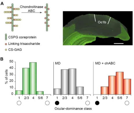
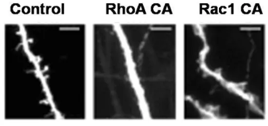
Extracellular and pericellular microenvironment contains important regulators of visual cortical plasticity. One of the first demonstration of the involvement of extracellular components in OD plasticity comes from the evidence that pharmacological inhibition of extracellular protease tissue plasminogen activator (tPA) attenuates the OD shift induced by MD (Mataga et al., 1996). tPA is an immediate early gene induced by electrical activity (Qian et al., 1993) and its proteolytic activity is increased during MD (Mataga et al., 2002). In keeping with these results, tPA KO mice display an impaired MD induced ocular-dominance shift that could be rescued by exogenous tPA (Mataga et al., 2002). A subsequent study indicated that the increase of tPA that occurs after MD is needed for structural plasticity of dendritic spines (Mataga et al., 2004). Thus, tPA, by proteolitically removing extracellular molecules inhibitory for plasticity, enables OD shift induced by MD. Part of the inhibitory action of the extracellular environment resides in components of the extracellular matrix (ECM) called chondroitin-sulfate proteoglycans (CSPGs). These molecules are organized in typical structures, the perineuronal nets (PNNs), around soma and dendrites of PV-positive neurons. Several studies indicate that the developmental increase of PNNs correlates with the end of the classical CP and that Dark Rearing delays the formation of PNNs in the visual cortex (Hockfield et al., 1990; Koppe et al., 1997; Pizzorusso et al., 2002). The inhibitory action of CSPGs for plasticity is demonstrated by the evidence that degradation of CSPGs in adulthood by the bacterial enzyme chondroitinase ABC is able to restore OD plasticity (Fig. 1.4) and to promote recovery from amblyopia (Pizzorusso et al., 2002; Pizzorusso et al., 2006). Interestingly, treatment with chondroitinase ABC increases spine density (Pizzorusso et al., 2006) suggesting that removal of CSPGs may provide a permissive environment for structural plasticity. It is worth noting that ECM proteolysis could also be regulated by particular rearing conditions. Enriched environment promotes amblyopia recovery and decreases PNNs number (Sale et al., 2007).
Figure 1.4 Relationship between chondroitin-sulfate proteoglycans (CSPGs) and adult visual cortical plasticity. (A) Treatment with chondroitinase ABC degrades the chondroitin-sulfate glycosaminoglycans (CS-GAGs) from CSPG. This degradation results in major disruptions to the macromolecular heterophilic interactions that hold the perineuronal net together (left). Immunostaining for the CSPG neurocan shows that the treatment of the adult visual cortex with chondroitinase ABC removes CSPGs from the whole binocular subfield of the adult visual cortex (area Oc1b) and from neighbouring cortical areas (right). (B) In adult rats (> P100), MD ( black and white circles indicate the ocular-dominance class corresponding to the deprived and non-deprived eyes, respectively) does not cause a shift of ocular dominance. When adult rats were treated with chondroitinase ABC (chABC), monocular deprivation elicited a significant shift of ocular dominance towards the non-deprived eye (Berardi et al., 2003).
Another candidate for plasticity regulation has also been recognized in myelin-associated proteins. It is well known that these proteins exert an active inhibitory role in mechanisms of brain repair. Few years ago it has been demonstrated that myelinization not only inhibits recovery from injury, but can also promote the decrease of plasticity observed at the end of the CP. Indeed, maturation of intracortical myelination has been found to correlate with the end of the critical period (McGee et al., 2005). In addition the absence of either NogoA (a growth inhibitor associated to myelin) or its receptor NgR prevents the closure of the critical period and OD plasticity persists well into adulthood (McGee et al., 2005).