DIPARTIMENTO DI MEDICINA TRASLAZIONALE
Corso di Dottorato di Ricerca in Scienze e biotecnologie mediche Ciclo XXXII
Assessment of primary mutations in treatment-naïve HIV-1 Subtype C-infected patients in Malawi
SSD - Settore Scientifico Disciplinare: MED07
Coordinatore PROF MARISA GARIGLIO Tutor PROF GIORGIO BELLOMO
PREFACE
The DREAM Program (Disease Relief through Excellent and Advanced Means) was formed and run by the Community of Sant’Egidio, a catholic international organization born in Italy in 1968. Initially was an HIV and nutrition care NGO, and now is a global health care institution for communicable and non-communicable diseases (DREAM 2.0) with programs on tuberculosis, cervical cancer, hypertension, diabetes, and others. Designed for quality and based on partnerships, is a public health program as well as applied research. All our services are free of charge.
DREAM started in Mozambique in 2002 and is now in 11 other countries with 47 health centers, 24 molecular biology laboratories and 300,000 patients assisted from 2002, 130,000 patients on HAART of whom 15,000 children. Millions of peoples reached by the program and 50,000 children were born healthy in the PMTCT program
The laboratory monitoring is part of an holistic approach that DREAM uses in routine patient care. The laboratories perform Blood count, Biochemistry, CD4 count, HIV-1 viral load, Infant virological testing and, in Malawi and Mozambique, HIV genotyping, initially with Trugene platform (Siemens Health Care Diagnostics), now with a validated in-house method.
The promotion of laboratory medicine in management of HIV positive population in Malawi is one of the major contributions that DREAM Program has impacted to the Country. When DREAM rolled out its program in 2005, Malawi did not have any molecular biology laboratory for routine patient care, but owing to the training, advocacy and setting up of model infrastructures by DREAM, the Country has registered significant developments and now 10 molecular laboratories are spread across the Country for viral load and infant virological testing.
The Malawi ART guidelines recommends an HIV genotyping test for all patients failing second line regimens before switching them to third line regimens. There is also need for genotyping services for surveillance studies. DREAM Malawi program has the only genotyping facility in the Country at its Blantyre laboratory since 2008, which is also available for all public structures. DREAM has a leading role to share expertise in HIV drug resistance services in Malawi even now, that the Country has established a genotyping lab at National HIV Laboratory in Lilongwe.
SUMMARY
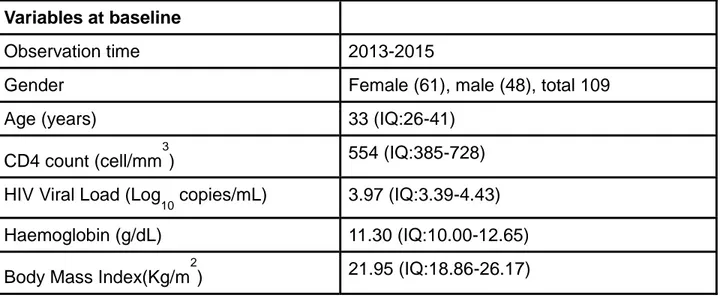
The aim of the PhD study was to describe retrospectively the prevalence of transmitted HIV-1 resistance mutations in a cohort of naïve patients starting treatment at DREAM program health centers in Malawi. In Africa naïve resistance mutations rate is unknown or underestimated.
In Malawi in 2014, 1.8 million people have turned to the services of HTC (HIV testing and counseling) (Malawi National AIDS Commission). There are also important results on the treatment front: as many as 61% of adults with HIV are under anti-retroviral therapy (ART), 67.6% of whom are effective virological suppression. Moreover, the estimate frequency of Transmitted HIV drug resistance (TDR) in the Country is based on a very limited number of specimen, and more studies were needed to support the national surveillance supplying programmatic information in designing education and prevention programs as well as supporting a wise use of antiretroviral drugs by clinicians and policy makers.
Simultaneous to this study, analyzing the viral sequences, we tried to highlight some polymorphism patterns to study them and compare with other patterns related to other HIV-1 subtypes (as B, present in Western Countries, when in Malawi the major subtype described is C ).
Most of the activities were developed in Blantyre DREAM laboratory. An affordable laboratory method is needed in Low Limited Setting (LLS) as Malawi, to obtain the virus sequences, in order to allow national structures to produce data locally and to dramatically improve the number of performed tests. For this reason in the PhD meantime a homemade cost effective method able to detect HIV drug resistances was developed and successfully used.
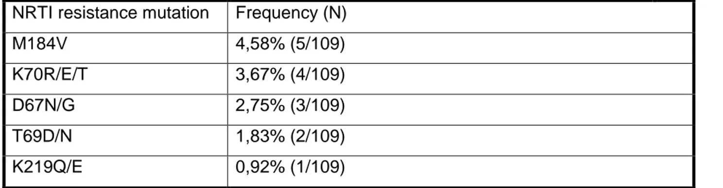
The study provides some important information in understanding the transmitted resistance rate in the Country Out of 109 samples from naïve patients, 40 (36,70%) presented at least one TDR-associated mutation, The observed frequency of nucleoside reverse transcriptase inhibitors (NRTIs) resistance mutations was: M184V (4,58%), K70R/E (3,67%),D67N (2,75%). The prevalence of resistance mutations to non-nucleoside reverse transcriptase inhibitor (NNRTI) was K103N/S (11,93%), G190A (6,42%), Y181C/V (3,67%), V106M (2,75%).
The study showed that there is an increase in NNRTI resistance and that the activity of NNRTIs is compromised because of the high level of NNRTI resistance.
However in developing African countries, the majority of first line ART regimens are nevirapine/efarinenz (NVP/EFV) based, but these drugs are responsible for selection of mutations to the entire class of NNRTI (K103N, Y181C and G190A). The study confirms the need to review the treatment protocols because the use of NNRTIs could be the cause in the increase of resistance.
Given such levels of HIV Drug Resistance (HIVDR) prevalence, HIVDR testing and surveillance capacity in Malawi should be prioritized as scale-up and the adoption of the universal ART eligibility for people living with HIV (PLHIV).
To highlight the polymorphism patterns, of the 216 codons subjected to nucleotide variations found through all the sequencing products, the most represented ones were selected to ensure greater statistical significance, reducing the number to 22 codon.
Many of these polymorphisms were found to be present in very high percentages, making it part of the consensus sequence for the subtype C.
The association of V60I with all three mutations of the NAM-1 pathway, the positive association of L228R and negative of S48T/Q with M41L and L210W are very significant. Very significant associations are found between the L228R and all 4 mutations of the NAM-2 pathway, and the associations of V60I and I135T/M with two mutations each. The negative associations with S48T/Q and K173T are also very evident. The most significant associations are those with I135T / M (positive) and with V245Q (negative). The associations highlighted by a previous study on subtype B have been compared and the only concordances found concern the association of L228R with the mutations of the NAM-2 pathway and of T39A with the mutations for NRTI.
All these findings underline how subtypes B and C are characterized by substantially different mutational profiles.
These associations are even more significant considering that three polymorphisms were found with very high frequency (81.2% for T39E/D, 55.0% for Q174T and 71.3% for R211K) enough to be considered part of the consensus sequence of the subtype C, and resulting associated with NNRTI resistance mutations, one could suspect their role in making the subtype C naturally more likely to develop resistance to NNRTIs.
SOMMARIO
Lo scopo dello studio di dottorato è stato quello di descrivere retrospettivamente la prevalenza delle mutazioni di resistenza HIV-1 trasmesse in una coorte di pazienti naïve che iniziano il trattamento presso i centri sanitari del programma DREAM in Malawi.
In Africa infatti il tasso di mutazioni di resistenza naïve è sconosciuto o sottostimato.
Nel 2014, in Malawi, 1,8 milioni di persone si sono rivolte ai servizi di HTC (test HIV e counselling) (Malawi National AIDS Commission). Ci sono stati importanti risultati sul fronte del trattamento antiretrovirale (ART): ben il 61% degli adulti con HIV è in trattamento con ART, il 67,6% dei quali mostra una efficace soppressione virologica. Inoltre, la frequenza stimata della resistenza trasmessa ai farmaci contro l'HIV (TDR) nel Paese si basa su un numero molto limitato di campioni e sono necessari ulteriori studi per supportare il programma di sorveglianza nazionale, che fornisce informazioni alla progettazione di programmi di educazione e prevenzione e per sostenere un uso saggio dei farmaci antiretrovirali da parte di medici e responsabili politici.
Contestualmente a questo studio, analizzando le sequenze virali, abbiamo provato a mettere in evidenza alcuni pattern di polimorfismi per studiarli e e confrontarli con pattern relativi ad altri sottotipi di HIV 1 (in particolare il B, maggiormente diffuso nei Paesi Occidentali, mentre in Malawi il sottotipo maggiore descritto è C).
La maggior parte delle attività sono state sviluppate nel laboratorio DREAM a Blantyre.
Per ottenere le sequenze di virus è stato necessario sviluppare un metodo di laboratorio a basso costo, per permettere a Paesi come il Malawi di ottenere le sequenze virali in modo sostenibile, al fine di consentire alle strutture nazionali di produrre dati localmente e di aumentare notevolmente il numero di test eseguiti. Per questo motivo nel dottorato di ricerca è stato sviluppato all'interno del laboratorio e utilizzato con successo un metodo economicamente efficace in grado di rilevare le resistenze ai farmaci dell'HIV.
Lo studio fornisce alcune importanti informazioni sulla comprensione del tasso di resistenza trasmessa nel Paese. Su 109 campioni di pazienti naïve, 40 (36,70%) hanno presentato almeno una mutazione associata a TDR. La frequenza osservata delle mutazioni di resistenza agli inibitori della trascrittasi inversa nucleosidica (NRTI) era per M184V (4,58%), K70R/E (3,67%),D67N (2,75%). La prevalenza di mutazioni di resistenza relativamente agli inibitori della trascrittasi inversa non nucleosidica (NNRTI) era K103N/S (11,93%), G190A (6,42%), Y181C/V (3,67%), V106M (2,75%).
Lo studio ha mostrato che c'è un aumento della resistenza NNRTI e che l'attività degli NNRTI è compromessa a causa dell'alto livello di resistenza.
In molti paesi africani in via di sviluppo, la maggior parte dei regimi ART di prima linea sono basati su neviralian/efavirenz (NVP / EFV), ma questi farmaci sono responsabili della selezione delle mutazioni dell'intera classe di NNRTI (K103N, Y181C e G190A). Lo studio conferma quindi la necessità di revisione dei protocolli di trattamento perché l'uso di NNRTI potrebbe essere la causa dell'aumento di resistenza.
Dati tali livelli di prevalenza dell'HIV Drug Resistance (HIVDR), i test di resistenza e la capacità di sorveglianza epidemiologica in Malawi dovrebbero essere prioritari, viste le nuove politiche di accesso universale al trattamento.
Per evidenziare i patterns di polimorfismo, sono stati selezionati i piu' rappresentativi tra i 216 codoni che presentavano variazioni dei nucleotidi, per garantire una maggiore significatività statistica, riducendo il numero a 22 codoni.
Molti di questi polimorfismi sono stati riscontrati in percentuali molto alte, rendendoli parte della sequenza di consenso per il sottotipo C.
L'associazione di V60I con tutte e tre le mutazioni di NAM-1, l'associazione positiva di L228R e negativa di S48T/Q con M41L e L210W sono molto significative.
Associazioni molto significative si trovano tra L228R e tutte le 4 mutazioni del pathway NAM-2 e le associazioni di V60I e I135T/M con due mutazioni ciascuna. Anche le associazioni negative con S48T/Q e K173T sono molto evidenti. Le associazioni più significative sono quelle con I135T/M (positiva) e con V245Q (negativa).
Le associazioni evidenziate da uno studio precedente sul sottotipo B sono state confrontate e le uniche concordanze riscontrate riguardano l'associazione di L228R con le mutazioni del percorso NAM-2 e del T39A con le mutazioni per NRTI.
Tutti questi dati sottolineano come i sottotipi B e C siano caratterizzati da profili mutazionali sostanzialmente diversi.
Queste associazioni sono ancora più significative considerando che questi tre polimorfismi sono stati trovati con altissima frequenza (81,2% per T39E / D, 55,0% per Q174K e 71,3% per R211K) abbastanza da essere considerati parte della sequenza di consenso del sottotipo C, e risultando associati a mutazioni di resistenza, si potrebbe sospettare il loro ruolo nel rendere il sottotipo C naturalmente più probabile che sviluppi resistenza agli NNRTI.
INTRODUCTION
1.1 Characteristics of the human immunodeficiency virus (HIV)
The human immunodeficiency virus (HIV), the etiologic agent of acquired immunodeficiency syndrome (AIDS), belongs to the Retroviridae family.
Currently two HIV serotypes are known: HIV-1, which refers to genetically related viruses found predominantly in different regions of central and southern Africa, Asia, Europe and America (1) and HIV-2 which is a distinct virus prevalent in certain West African countries (2). Although both of these viruses cause AIDS, all individuals infected with HIV-2 have a long period of latency and low mortality.
1.2 Classification of the human immunodeficiency virus
Traditionally, the Retroviruses, which until the beginning of the 80s were known and studied for neoplastic diseases caused only in some species of animals, are classified into three sub-families: Oncovirinae, Spumavirinae and Lentivirinae.
Viruses originally isolated as transforming agents are grouped into oncoviruses. They cause sarcomas, leukemia, mammary tumors and, in some cases, a variable suppression of the immune system in different species of animals.
Two human retroviruses, identified for the first time in 1980 by R.C. Gallo and his collaborators, are also part of this family: Human T Lymphoma viruses I and II (HTLV I-II) associated with T-cell lymphomas and neurological disorders.
Spumaviruses owe their name to the ability to induce vacuolar lesions that give a foamy appearance to cells grown in vitro.
Lentiviruses are associated with long-term diseases but without a direct relationship with the neoplasms. Before HIV-1 was isolated and characterized, Lentiviruses were known for diseases caused in some species of felines and ungulates. From the study of these viruses, much information has been received about the pathogenesis of AIDS.
HIV-1 is classified as a member of the Lentivirus group based on its gene organization, similarity of nucleotide sequences and the type of pathology it entails, characterized by a long phase of clinical latency. Lentiviruses are viruses that induce chronic degenerative diseases
in their hosts, preceded by a long incubation period and by a variable involvement of the immune system and the central nervous system.
Another characteristic that finally distinguishes Lentiviruses is the complexity of the genome. Human-isolated Lentiviruses are grouped into two types named HIV-1 and HIV-2 based on serological and sequence properties (16,17).
A classification based on the env gene sequences, described in the 1998 HIV Compendium (18), recognizes several HIV-1 subtypes or clades (19).
Within each subtype, there is a high degree of variability. Although mutations appear to be the most responsible factor for viral variation, it has been postulated that recombination mechanisms may also occur in individuals infected with viruses of different HIV-1 (19) and HIV-2 clades (20).
Some areas of the world mainly host a single subtype, while two or more subtypes may be prevalent in other populations (19).
Molecular epidemiological studies indicate that the pattern of global distribution and variation is due to viral migration rather than viral mutation.
1.3 Morphology and structure
1.3.1 Structure of the virions
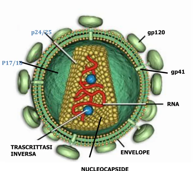
Virions are the extracellular particles produced by cells infected with HIV-1. Figure 1 is a schematic representation of the virus and its components.
Figure 1: HIV structure
gp120 p24/25 TRASCRITTASI INVERSA ENVELOPE RNA gp41 NUCLEOCAPSIDE P17/18
Electron microscopy shows that the virion particles have a spherical shape with a diameter of 110 nm and are formed by an external pericapside, the envelope, consisting of a double phospholipid layer on which virus-specific proteins anchor. In fact, the envelope contains about 72 triangular symmetry protuberances (21,22) each consisting of three or four heterodimers of the glycoprotein encoded by the env gene. Each heterodimer is composed of two subunits, one of which (gp120), which protrudes totally outwards, contains the domains that recognize and bind the CD4 receptor and the specific coreceptor for entry into the different target cells of the virus, while the other one (gp41), which is mostly inserted in the double phospholipidic layer, has a fusogenic activity.
The envelope also contains some cellular proteins, such as histocompatibility antigens (MHC), acquired during the budding process through the cell membrane.
Associated with the internal face of the double phospholipidic layer, a matrix protein structure is observed, consisting of the p17 protein, which forms an internal thickening of the double phospholipid layer.
Inside the viral particle is the nucleocapside or central core with a truncated cone structure. The nucleocapside crosses the entire diameter of the virion and the narrow end of the cone appears to be connected to the lipid double layer with a proteinaceous structure named CEL (core-envelope link) (23). The region between the nucleocapside and the envelope is called the paranucleoid region (24). The composition of CEL and the paranucleoid region must still be determined.
The nucleocapside consists of two single-stranded viral RNA molecules encapsulated by proteins that derive from the precursor polypeptide synthesized by the gag gene. This precursor is cut from the viral protease into four protein products: p24, the main capsid protein, whose function is to package the viral genome in virions; the p17 or matrix protein, which as mentioned is located between the nucleocapside and the virion envelope, the p9, protein of the nucleocapside that binds tightly to the genome and finally the p7 whose location is not yet clear (25). These proteins are assembled according to an icosahedral type cubic symmetry.
Inside the core, in addition to the nominal RNA, there are RNA transfer (tRNA) molecules and some viral enzymes. The tRNAs are used to trigger the replication, while the viral enzymes, which derive from the polypeptide precursor of the pol gene, are the reverse transcriptase (RT), a heterodimer consisting of two polypeptides (p51 / p66) which intervenes in the
replication of the genome, the integrase (p32) which causes the integration of viral DNA into the cellular DNA and the protease (p10) that intervenes in the maturation of the genome. An independent domain of RT carries out an additional enzyme activity of specific ribonuclease (RNAse H) that degrades the RNA in the RNA-DNA hybrid produced during the proviral genome synthesis.
1.4 Structure and organization of the viral genome
Like all retroviruses, HIV-1 presents two genomic forms: a single-stranded RNA in the extracellular phase of the life cycle of the virus and a double-stranded DNA within the cell. In the early stages of infection, virion RNA is converted into the double-stranded DNA form by the RT of the virus and is then integrated into the genome of the host cell (provirus).
The virions contain two identical copies of single-stranded RNA of approximately 9.2 Kb with positive polarity. The two strands are associated in numerous points along their length; in particular, the junction point of greater stability is located near the 5 'end of each genome. The role of diploidy is still obscure.
Viral RNA presents the characteristics typical of most eukaryotic messenger RNAs: the CAP (m7G5'ppp5'Gmp) at the 5 'end and a tail of about 200 adenine residues at the 3' end. Also occasionally adenine residues can be methylated. A tRNAlys molecule is positioned near the 5 'end of each strand and serves as a primer for the synthesis of the DNA negative filament by RT.
The genomic structure of HIV-1 is shown in Figure 2.
Figure 2: the genomic structure Proteine
strutturali
Enzimi
Two repeated sequences, named long terminal repeat (LTR), are placed at the ends of the genome and flank the three major open reading frames (ORFs) of the virus: gag, pol and env and another six smaller ORFs: tat and rev, essential for viral replication, vif, vpr, vpu and nef not essential for replication, also called accessory or auxiliary genes. The presence of these six ORFs gives the virus an extraordinary level of complexity.
1.4.1 Long-terminal repeat
As in the other retroviruses, the LTR region is divided into three functionally distinct domains: U3 (-453 / + 1), R (+ 1 / + 98) and U5 (+ 99 / + 185) (from 5 'to 3').
These functional units are critical for virus integration within the host genome and contain promoters and enhancer elements recognized by viral and cellular transcription factors.
The U3 region extends from the 5 'end of the genome to the site of initiation of transcription (+1) and contains the viral promoter, consisting of three transcription domains: the modulation domain, the enhancer and the core or base domain . The first portion of U3 is the modulation region that contains cis-acting regulator transcription elements to which many cellular factors are bound (i.e. AP-1 and N-FAT-1). Downstream of the modulation region is the enhancer a region formed, in HIV-1, by two sites of 10 bp. These sites bind the cellular factor NF-kB which has the function of increasing the transcription activity of the virus following the activation of the same factor by some cellular regulatory proteins or other viruses. At the 3 'end of the U3 is the core containing a TATAA box region for binding to the cellular RNA polymerase II responsible for viral transcription; the viral transcription initiation site is 22 bp downstream of this region. Sites for binding the SP1 cell transcriptional factor are placed immediately after the TATAA box (26).
The region R encodes a sequence of RNA that forms a hairpin (stem-loop) structure at the 5 'end of the transcript called tat-response element (TAR). This structure binds the Tat protein of HIV-1, a powerful activator of viral transcription.
The R / U5 region is located in the leading sequence of all viral transcripts. The 3 'end of the transcripts is defined by the R / U5 edge of the 3' LTR.
Although the nucleotide sequences of the two LTRs are identical, retroviruses have mechanisms by which the 5 'LTR is used as a transcription promoter while the 3' LTR is a
signal to add the poly-A tail, in fact signals in the U3 and R region they are recognized by cellular factors that add poly-A tails to the 3 'end of viral transcripts (27).
1.4.2 Structural genes
HIV-1, like all retroviruses, has three structural genes essential for replication, called respectively: gag (group specific antigen), pol (polymerase) and env (envelope). The three genes are organized in the genome in the order 5'-gag-pol-env-3 '.
The gene gag represents the first ORF of the HIV-1 genome that encodes a peptide precursor of the internal structural proteins of the virus, of 55 kDa translated by an mRNA not subjected to splicing processes (unspliced) as long as the genome.
The precursor, the polypeptide p55, is subsequently cut from the viral protease to produce the virid capsid protein, p24, the matrix protein, p17, the nucleocapside protein p9 and the p7 previously described.
The pol gene overlaps the gag gene for about 241 bp. This gene is expressed as gag / pol fusion protein from an unspliced mRNA as long as the genome and from the segmentation of this protein the viral enzymes are derived: the protease, p10, with a autocatalytic cut, the integrase, p32, and the etherimer p66 / p51 ie the reverse transcriptase. An independent domain of the RT plays a ribonuclease function.
The env gene encodes a highly glycosylated protein precursor, gp160, translated from a polycistronic mRNA derived from a single splicing process (singly-spliced). The protein is cleaved from a host endopeptidase into two portions: the N-terminal gp120 and the C-terminal gp41 which form, through non-covalent interactions, the surface antigen of HIV-1, which mediates the entrance of the virus. Sequence comparison studies reveal a model, of the env sequence encoding gp120, of 5 variable regions (V1-V5) interspersed in conserved regions while the gp41 region is fairly conserved (28). One of these domains (V3) is highly variable and is the main target of neutralizing antibody activity.
1.4.3 Regulatory genes
The tat and rev regulatory genes control the expression of viral genes at the transcriptional and post-transcriptional levels.
The first coding exon of tat is located in the central region of the viral genome between the vpr gene and the env gene while the second exon is superimposed on the rev reading frame (open reading frame) and the env gp41 (29). The Tat protein is translated from monocistronic transcripts, produced early in the infection, subjected to multiple spliced processes (multi-spliced), and interacts with the TAR site at the 5 'end of each messenger. In this way Tat increases the levels of viral RNA by acting at the beginning of the transcription (30) or during elongation (31).
The rev gene regulates the splicing and transport of transcripts from the nucleus to the cytoplasm (32). The two coding exons of rev partially overlap with tat exons and the rev protein is also translated by multiply-spliced transcripts produced in the early stage of infection.
1.4.4 Ancillary Genes
The accessory genes of HIV-1 are vif, vpr, vpu and nef. Vpr is assembled within the virion and although most studies have not found traces of the other three proteins in the virion, it is possible that small amounts may be incorporated equally. The proteins of these genes are mostly translated from singly spliced transcripts.
Although several studies have been conducted on the function of these genes, many of these are discordant. Presumably such genes play an important role in virus-host relationships. It is believed that the proteins encoded by the vpr and vif genes are both involved in the transmission mechanism of the virus. It seems that the vpr-encoded protein also influences the transmission rate, facilitating the nuclear localization of the viral genome, while the protein produced by the vif gene seems to be essential for virion infectivity. The protein encoded by the vpu gene, could instead be involved in the maturation process of the viral particle, facilitating its exit from the host cell.
The nef gene extends from the 3 'end of env up into the U3 domain of the 3' LTR. The transcription of the gene produces two precocious multiply spliced transcripts, independent of the rev function, one of which is monocistronic while the other is bicistronic and encodes both for nef and for rev (33).
The Nef protein has many important functions among which the activation of the cascade of enzymes that determine the activation state of the cell, in this sense the Nef protein is required for an efficient replication of HIV-1.
1.5 Life cycle of HIV
In the replicative life cycle, shown in Figure 3, of HIV-1, an early and a late phase is recognized. The early phase begins with the binding of the virion both the CD4 cell surface receptor and the coreceptor, with consequent entry of the nucleocapside into the cytoplasm. The virus RT, associated with the nucleocapside, converts the RNA into DNA that is maintained in a nucleoprotein complex and transported within the nucleus. Here the double helix of DNA is integrated into the cellular genome. The late replication phase begins with the transcription and processing of viral RNA derived from the proviral form and ending with the release of new virions from the cell.
1.5.1 Bonding and entry of the virion
The HIV-1 attack on cells is mediated by the initial interaction between the extracellular domain (gp120) of the virus surface antigen and the CD4 receptor located on the cell membrane of T-helper lymphocytes, macrophages, dendritic cells and microgliali (34). CD4 is a member of the immunoglobulin family and consists of four extracellular domains (D1-D4) that have a similar structure to those found in other immunoglobluline families (35). Other cell surface receptors are implicated in the attack and entry of the virion. As already mentioned, among the main molecules that can act as receptors together with CD4, it is possible to distinguish those that function as chemokine receptors; moreover, other receptors would be molecules that can facilitate the infection or that may be involved in the regulation of some steps following viral assembly, such as the fusion between cell and cell, and those that can function as primary receptors in HIV infection -1 in CD4- cells.
The existence of a CD4 co-receptor was supported by the observation that the transfection of human CD4 into murine cells (36) or a human glioma cell line (37) is not sufficient to allow entry of the virus despite the HIV-1 particles or the recombinant gp120 bind efficiently to the cells. In this regard, a protein called fusin (CXCR4) has been identified that, when coexpressed with CD4 in mouse cells, allows the fusion with cells expressing viral envelope proteins on the surface. In addition, antibodies against fusin block fusion in human cells that are normally permissive for HIV-1. This molecule belongs to the superfamily of the G protein-coupled receptors and has a close relationship with the α-chemokine CXC family receptors. It is more specifically implicated in the infection of lymphocytotropic strains and is associated with the viral phenotype SI (Syncitia Inducing).
Other chemokine receptors have been identified as coreceptors in the infection of macrofagotropic strains, in particular some cytokine receptors belonging to the α-chemokine group including CCR1, CCR20, CCR3, CCR5. Among these, CCR5 is considered to be one of the major receptors for CD4 cell infection, allowing membrane fusion and infection for the macrofagotropic HIV-1 strains, this receptor is associated with the NSI phenotype. Recent acquisitions have shown that subjects homozygous for the 32 bp deletion in the gene coding for the coronector CCR5 are frequently, but not always, resistant to HIV infection. This observation supports on one hand the hypothesis that HIV-resistant individuals do not
express CCR5 correctly, and on the other that this molecule is one of the most important HIV receptors.
Moreover, current research has developed a series of models on the possible process of attack of lymphocytotropic and macrophage-viral strains following binding of the CD4 receptor and, respectively, of CXCR4 and CCR5 receptors (34).
Other molecules may play a primary role in regulating critical steps following virus entry and cell-to-cell transmission, such as the fusion of cell membranes of CD4 T cells and HIV-1-infected monocytes and the consequent formation of syncytes . In this regard, recent studies have shown that the CD4 molecule is necessary but not sufficient to mediate the formation of syncs and that this process is regulated by the interaction between the LFA-1 adhesion molecule and its ICAM-1, ICAM-2 receptors and ICAM-3 present on lymphocytes (38). Finally, a possible receptor in the glial and neuronal cells CD4- is the glycolipid galactosyl-ceramide (GalC). In fact, it has been shown that the HIV-1 gp120 binds to this molecule with similar affinity to that of CD4, but the efficiency of the infection is lower in experimental conditions. (39,40).
As already mentioned, an important factor for the understanding of the virus-receptor binding is the genetic variability of HIV-1, a particularly important feature since variations in the nucleotide sequence can determine biologically relevant structural changes in viral proteins by modifying their functional and immunogenic properties ( 41, 42).
Following the recognition and binding of the target cell, the most accredited mechanism of entry of the virion (internalization) in most cells is the direct fusion of the cellular plasma membrane with the viral envelope by a process independent of pH.
The heterodimer gp120 / gp41 is kept within the envelope, through non-covalent interactions, in an oligomeric complex. In the native state, that is before the binding to the cell, each oligomeric complex presents the leucine zipper sequences of the gp41 separated from each other. Afterwards, most likely the binding to CD4 or the proteolytic cut of a membrane protease (CD26) that recognizes and cuts the sequences in the V3 loop at the level of the Arg320, induces a conformational change such that the leucine zippers in the gp41 interact and they form a fusion domain that facilitates the fusion between the two membranes.
Finally, one factor to consider is the different cellular tropism that distinguishes different HIV-1 isolates from the same infected person. Central nervous system HIV-1 isolates preferentially grow in macrophage cultures (macrophage-like strains), whereas those obtained from
peripheral blood mononuclear cells, stimulated with PHA and IL2, propagate better in T lymphocytes (lymphocytotropic strains). Blood dendritic cells and Langherans cells of the skin and mucous membranes also allow replication of many HIV-1 strains with different tropism. In contrast, follicular dendritic cells of the lymph nodes, which present the antigen to T lymphocytes in the lymphatic follicles can only bind HIV-1 to their surface without getting infected.
1.5.2 Synthesis and integration of viral DNA
The model for the reverse transcription of lentiviruses is similar to that proposed for other retroviruses and has been elucidated by analysis in cell-free systems (43). The reverse transcription of the genomic RNA is mediated by the reverse transcriptase (RT, p66 / p51) an enzyme that modulates different activities: DNA polymerase dependent RNA, DNA dependent DNA polymerase and ribonuclease (RNAase H) (44).
It is activated in the cytoplasm by a signal that is not yet well identified. The synthesis of the first DNA strand, ie the negative polarity one, begins with the annealing of the virus tRNAlys at the PBS site (primer binding site) of the RNA mold that acts as primer for the RT. The complete double helix is formed by a rather complex mechanism. After the synthesis of the filament
linear DNA is completed in the cytoplasm, the viral DNA, in the form of a nucleoprotein complex, migrates to the nucleus to be integrated into the genome of the host cell. Although both the structure and the precise composition of this preintegration complex are unknown, it is certain that viral DNA, integrase and matrix protein are part of this complex (45). The transport of the complex in the nucleus is an active process of the host cell that requires ATP but is independent of cell division (46). Lentiviruses have been proposed to replicate in differentiated non-proliferative cells in contrast to the other retroviruses.
Integration is not a random process (48). The analysis of cellular sequences flanking the provirus reveals that HIV-1 is preferentially inserted in or near two classes of repeated elements of DNA in the human genome: L1 and Alu (49). These elements are transposons and show common properties with retroviruses. Thus, the preferential integration of HIV-1 within these elements may reflect the local chromatin structure that is more susceptible to acquiring a transposon. In vitro analyzes of the proviral integration mechanism show that the
intermediate for the covalently linked provirus in the cellular genome is linear and non-circular viral DNA. However, the circular form, which can not replicate, can be found after infection and can remain stable for many days as opposed to the linear form that is rapidly degraded.
1.5.3 Expression and regulation of viral genes
The control of genome synthesis is complex and involves the combined action of viral agents acting in cis, viral transactivators and different cellular proteins. In the nucleus, the integrated proviral DNA is transcribed from the cellular RNA polymerase II to produce RNA precursors of the same length as the genome.
The expression of the provirus leads to the formation of three classes of transcripts: genomic RNA for viral progeny, messenger RNA for translation into the cytoplasm of polyproteins Gag and Gag-Pol and precursors for over 30 alternatively spliced messenger RNAs that are translated into the cytoplasm for produce env glycoproteins and accessory proteins (50). Initially in the absence of the Tat transactivator, the viral specific transcription level is low. The precursor RNAs are transported through a Rev-independent transport mechanism but before transport the spliceosomes remove the introns from the multiply spliced transcripts that are then translated to produce the regulatory Tat, Rev and Nef proteins that return to the nucleus. As the transcription proceeds, Tat levels increase and consequently the transcription activity itself increases. As Rev also accumulates, the precursor RNAs are transported to the cytoplasm by a Rev-dependent mechanism
does not require splicing before transport. In this phase, unspliced and singly spliced viral mRNAs are translated into Gag, Pol, Env, Vpr, Vpu and Vif proteins in the cytoplasm. Rev, therefore, acts as a molecular chaperone interacting with the RRE sequence present in all the transcripts in correspondence of the env gene.
1.5.4 Assembly and release of the virus
The first event in the assembly of virions is the interaction between the precursor polypeptide of the gag gene (pr55), the polypeptide precursor Gag-Pol (Pr160) and the viral genome to produce a nucleoprotein complex. This process can occur in the cytoplasm or on the membrane. The maturation of the Gag polyproteins during assembly is mediated by the
protease domain in Pr160 to produce a mature nucleocapsid. At the same time, the gp120 and gp41 are inserted into the plasma membrane and, following interaction of the matrix protein with the cytoplasmic tail of gp41, the viral nucleoprotein complex extrudes or buds through the membrane to produce a
mature virione. The precise mechanism of budding is unknown.
1.6 Role of viral enzymes
1.6.1 Reverse transcriptase
Retroviruses have a reverse transcriptase that catalyzes the synthesis of proviral DNA using viral RNA as the template. This process is complex, but
in short, it consists of three catalytic passages:
1) The production of a complementary DNA chain from the genomic RNA, which involves the formation of a RNA-DNA hybrid.
2) The removal of the RNA mold (by the ribonuclease activity of the RNase H domain of the RT), leaving the DNA with a single strand.
3) The synthesis of a second DNA chain, complementary to the first.
The double DNA chain is subsequently integrated into the chromosomal DNA by integrase. The proviral DNA becomes the template for the transcription of viral RNA for the formation of new viral particles.
The reverse transcriptase is a heterodimer composed of the subunits p55 and p66 (51)
The polypeptide contains the DNA polymerase domain (440 amino acids) and the RNase-H domain (120 amino acids). The RNase-H domain is located in the C-terminal region of the p66 subunit. Although the p51 and p66 subunits have an identical amino acid sequence, they occupy completely different relative positions. In the p66 subunit there is a split in which the RNA-DNA hybrid takes place. This structure is absent in the p51 subunit, which does not catalyze polymerization. The acid residues (aspartic acid) in position 110, 185 and 186 confer the polymeric activity.
RT activity is an essential component of HIV-1 replication.
A particular feature of the enzyme is the low degree of accuracy in the incorporation of the bases, which is fundamental for the continued survival of the virus. That is, the RT enzyme
has no 'proof reading' capability and, compared to other DNA polymerases, has a higher error frequency. This is the major factor in the generation of mutations in the viral genome. The replacement of a nucleotide can however encode the same amino acid (synonymous mutations), or it can lead to the introduction into the protein of a different amino acid (non-synonymous mutation). The amino acid change can lead to a modification of the structure and some cases of the function of the protein.
1.6.2 Protease
The HIV-1 protease is an aspartic protease that cuts the nascent polyproteotic complex during viral replication. The enzyme is composed of two identical 99 amino acid polypeptides, with a proline at the N-terminal end and a C-terminal phenylalanine. Each monomeric subunit contains two regions with β-sheets structure and a small α-helical region.
The region comprising the active site extends from methionine to position 46 to the lysine in position 55 (52).
The study of a new class of drugs, protease inhibitors, has led to a growing interest in the mechanism of action of the protease and its role in HIV infection. In fact, the maturation events catalyzed by the enzyme are essential for the formation of infectious virions.
1.6.3 Integrase
Similarly to all retroviruses, replication processes depend on the integration of the viral genome into the chromosomes of the host cells. The HIV-1 integration processes are performed by viral integrase after a series of DNA cuts and subsequent re-joining reactions. In recent years PCR-based assays have been developed that can measure viral integration rate in cell systems and allow the identification of compounds capable of specifically blocking integrase in vivo.
1.7 Dynamics of viral replication
The knowledge of the viral replication trend was initially based on limited virological and immunological observations (p24 antigen and CD4 lymphocyte counts) and on the clinical progression of the disease.
My colleagues and I have shown that HIV-1 replication is high and continuous at different stages of infection and that viral load can be used as a prognostic indicator of AIDS development (53). In particular, the plasma levels of the virus are constant and this implies that the virus is produced and eliminated with the same frequency, ie that the system is in equilibrium.
Two important studies published in 1995 provided the first analysis of viral dynamics through the use of antiretroviral therapy (54, 55). They reported an exponential decline in plasma viremia within two weeks of initiation of antiretroviral therapy. The decrease in plasma viraemia after treatment reflected the combination of two separate effects: the elimination of plasma-free virions and the decrease in the cells that produce the virus, resulting in the blocking of new infections.
The average life span of the virus was established by observing the progression of plasma viral decline, assuming that viral replication was completely stopped during antiretroviral therapy. The average life of the virus was about 2 days. In particular, the production of HIV-1 has been estimated at around 0.68 x 10⁹-1.1 x 10⁸ virions per day (54, 55).
In 1996 Perelson and colleagues, using mathematical models and non-linear regressions, calculated the amount of virions eliminated daily and the average life of virus-producing cells (56). The average life of the virus was estimated at 6 hours and therefore the production of virions was 10.3 x 10⁹ virions per day, 15 times higher than previous estimates.
1.8 Genetic variability of HIV-1
One of the most striking features of the HIV-1 virus is its extreme genetic variability that manifests itself not only in the isolates of different individuals, but also in the different isolates of the same individual during the course of the infection.
The molecular bases of viral variation are due to the high frequency of error in the incorporation of nucleotides by the RT. These modifications are the result of mutations in viral
genes that code for these enzymes and derive from the high error frequency of viral retrotranscrypt, of about 3x10⁻⁵ substitutions per replicative cycle (57). Considering that the HIV-1 genome is about 104 bases and that the total viral load in subjects with HIV-1 infections is 109-1010 virions, the probability that in a given patient can occur every single possible nucleotide substitution is around at 104-105 times in one day (58). These data suggest the rapid onset of a genetic divergence within the viral population, generating within each HIV-infected individual a genetically different 'quasispecies', rather than a single viral population.
1.8.1 Mechanisms of viral variation
Viral variation is due to three possible mechanisms: Mutation
The fundamental characteristic of the continuous evolution of HIV-1 is the low degree of accuracy in RT polymerization. This enzyme has a high error rate and has no 'proof-reading' activity and therefore proves to be the most responsible for generating mutations in the HIV-1 genome. These changes are usually the result of point mutations that can be divided into two categories: substitutions and insertions or deletions.
a) Replacement is the change of a nucleotide with a different one during viral DNA formation. The substitution may or may not involve coding for a new protein by providing a different 'fitness' to the virus.
b) Insertion and deletion are additions or deletions of one or more nucleotides. Insertion or deletion of nucleotides may alter the 'frame' of the mRNA reading.
In particular, the structural gene env seems to possess a greater degree of variability compared to other regions of the HIV-1 genome (46-50). Analyzing sequences of env coming from the same patient with HIV-1 infection it is possible to highlight the presence of 'quasispecie', that is of closely related but distinct viral variants, which differ from each other for about 2-5% of the sequence of env (63-65). Considering instead isolates coming from different geographic areas, the genetic variability in env can reach values of 20-30% which decrease to 6-19% for isolates coming from the same area (59-62, 66-69). For this reason, the env gene represents the most frequently used gene in lithogenic analyzes, since its high rate of variability makes it possible to more precisely evaluate the genetic distances between different isolates.
Recombination
Another mechanism of viral variation derives from recombination. This event occurs frequently during retrotranscretion, as there are two genomic RNA chains in each virion. The HIV-1 RT enzyme has the ability to transfer the synthesis of a DNA chain from one template to another.
Superinfection and recombination
Retroviral recombination requires simultaneous infection of the same cell by two different strains (ie superinfection) and the subsequent integration of two parent proviral generations in the same nucleus.
Simultaneous expression and assembly of viral RNA generates a population of first generation heterozygous viral particles. These particles in turn can infect new host cells and be retrotranscribed. In this reaction, the reverse transcript can jump from one template to another. This phenomenon is what is defined as strand-switching activity of the HIV-1 RT. In this way a second chimeric generation of provirus can be formed. The viral particles produced by the second generation will contain the recombinant forms of viral RNA (3).
1.9 Pathogenesis and natural history of infection
The main route of infection transmission is represented by homosexual and heterosexual relationships and the probability of infection depends on the number of sexual partners and the different sexual practices (70). The virus is transmitted through genital secretions, both female and male, and blood (cells, plasma and coagulation factors) (71). A strong transmission incidence is registered among drug addicts due to the use of contaminated needles. The virus can be transmitted from the infected mother to the child either by transplacental route or through childbirth following exposure to the genital tract or following breastfeeding. On the other hand, non-sexual personal contacts, exposure to saliva (72), contact with urine (73) and exposure to insects (74) have never been directly implicated in the transmission of HIV.
Little is known about the early events following HIV-1 infection. The initial target cells are most likely those of the monocyte-macrophage dendritic line such as Langherans cells and lymphocytes in the genital and rectal features. Infection of new cells in the blood and dissemination occurs through the free virus or by cell-cell interaction. The major targets of HIV-1 are the lymphoreticular system, the hematopoietic system and the nervous system. Target cells that are critical for immunopathogenesis are dendritic cells, CD4 T lymphocytes, and monocyte macrophages.
The pathogenesis of neurological diseases of HIV-1 is also unknown. The predominant cells that are infected within the nervous system are monocytes and macrophages. Within the nervous system, infected macrophages are likely to release monochins, the alpha factor of tumor necrosis or the beta factor of transformation, and viral proteins that are toxic to neural cells.
Despite the devastating effects of HIV-1 on host immunity, infected individuals develop a humoral and cellular response against HIV-1-associated antigens. Neutralizing antibodies are produced by most individuals (71) and some researchers have noted a correlation between disease progression and low titers of neutralizing antibodies (74). Envelope glycoproteins are the major targets for neutralizing antibodies and the different epitopes on gp120 have shown the ability to generate neutralizing antibodies in experimental animals. Some of these are in hypervariable regions (75) while others are in conserved areas. The reactivity to the gag gene products sought by the enzyme immunoassay (ELISA) seems to reflect the progression of the infection. In fact the decrease of circulating anti-p24 antibodies and the appearance of circulating p24 antigens often correspond to the decrease in CD4 T cells and are associated with the progression of immune dysfunction and the development of AIDS (76, 77).
Also the cellular response is directed against HIV-1 antigens, in fact, cytotoxic T lymphocytes react with the env, pol, gag and regulatory gene products.
The typical clinical course of HIV-1 infection has been clearly defined following the isolation of HIV-1 from patients with AIDS or clinical signs preceding AIDS (120). It includes a phase of primary infection, with an acute syndrome of varying severity, a prolonged period of clinical latency and a final stage of disease characterized by an increase in susceptibility to opportunistic infections and neoplastic diseases (79). A peculiar feature of HIV-1 infection is the wide variability that can be found between one patient and another in the progression of the disease. In particular, what varies considerably is the duration of the clinical latency
between the different infected persons (80) and the progression to AIDS, which occurs after an average period ranging from eight to ten years (81, 82), although the decline immunological and clinical is much more precocious in a significant proportion of patients. In this regard, it is interesting to note that in a small number (from 2 to 5%) of people infected with HIV-1 (long-term non-progressor subjects) the clinical latency phase and the immunocompetence stage extend over a period of much longer time (75, 83).
In recent years there has been much debate whether CD4 T cell depletion and AIDS pathogenesis are the result of direct cytolytic effects of HIV-1, of T-cell apoptosis by non-specific activation processes or are due to dysregulation phenomena in the production of cytokines and autoimmunity events.
According to biological and immunological molecular tests, the pathogenesis and progression of HIV-1 infection is currently described as a multi-stage process (84), in which a multitude of viral and host factors are potentially implicated. been studied in recent years in vitro and in vivo (84.85).
In this complex scenario, different biological and molecular outcomes (86) have highlighted the correlation between viral replication and infection progression by studying the association between clinical stage and viral load in progressing patients (representing the majority of people infected from HIV-1) and the dynamics of viral activity during the natural course of the disease.
1.9.1 Genetic complexity of the pandemic
The increasing availability of sequences of the entire HIV-1 genome from different continents, made possible by new assays (Heteroduplex Mobility Assay) and new methodologies (PCR-long-range and automatic capillary sequencing), has greatly enriched the sequence database of HIV (HIV Sequence Database, Los Alamos, New Mexico, USA) and has allowed the study of the changes occurred in the last decade in the distribution of distinct subtypes in different geographical areas (3, 4). The sequences from different viral isolates have in fact been classified over time into groups and subtypes based on their phylogenetic relationships.
Each subtype includes sequences that are equidistant to each other and to be assigned to each subtype the strains must be similar to each other and different from other subtypes throughout the length of the genome. To study the evolutionary distances of the sequences,
phylogenetic analysis is used. In particular these distances are represented by phylogenetic trees. They are two-dimensional graphs made up of branches connected to each other via nodes. The terminal nodes, or extremities, represent the current (or contemporary) taxa, while the internal nodes represent the ancestral ones. The phylogenetic relationships are defined through the tree topology: the distribution of the nodes and the length of the branches that connect them provide indications on the evolutionary divergence between the different taxa, allowing to identify monophyletic groups or evolutionary lines (11).
To date, three groups have been defined: M (Major), O (Outlier) and N (New). Within the M group, 11 subtypes have been classified (A, B, C, D, E, F, G, H, I, J and K). The strain E, previously identified on the basis of the env gene as a separate subtype, was found to be a recombinant based on the entire genome whose genes gag and env belong respectively to subtypes A and E. Also subtype I, initially identified in a limited number of env gene sequences has turned out to be a recombinant form in which the gag gene is a sequence composed of parts of subtype A and G, while env is a sequence distinct from the others, ie I (3, 4).
In addition to the subtypes identified to date, the so-called Recombinant Circulating Forms (CRF) are present in different geographical areas. Six have been officially classified (CRF 01-06) and 3 CRF 07-09) have been recently identified (4, 5).
Among the recombinant forms CRF 01AE and CRF_02AG play an extremely important role as they circulate at high frequency respectively in South East Asia and in sub-equatorial Africa (3-5).
One of the major characteristics of the human immunodeficiency virus is its extremely high genetic variability. This heterogeneity is the result of both the high error rate of reverse transcriptase (6), which governs the evolution of subtypes, generating the high inter-subotypic and intra-subtypical variability, and the rapid turnover of virions in individuals infected with HIV. (7, 8). Furthermore, the reverse transcriptase enzyme has recently been recognized as highly recombinogenic (3) and this characteristic appears to be the main cause of CRF generation. The recombination event requires the simultaneous infection of a cell by two different viral strains (superinfection), followed by the assembly of an RNA transcript coming from each provirus within the heterozygous virion. After the subsequent infection of a new cell the reverse transcriptase, through a strand-switching mechanism between the two RNA
templates, can generate a new retroviral DNA sequence that will be recombinant between the two parental genomes (9, 10).
These mosaic viruses are defined as recombinant and this is supported by the fact that distinct recombination points can be recognized between genomic regions belonging to different subtypes (87, 88). Today it is clearly established that recombination is a relatively common occurrence that occurs between different HIV subtypes where high prevalence cocircolane occurs. Recombination generally occurs between strains belonging to different subtypes, but it can also occur between strains belonging to the same subtype.
In 1999 the further increase in the complete genomic sequences of HIV-1 (89, 90), together with epidemiological studies on its variability and its geographical distribution (91), allowed to reconsider the contribution to the pandemic of the different variants and especially of the recombinant forms (92).
On this basis the most recent classification was introduced (92), which provides that:
1) the recombinant forms of HIV-1 with high prevalence in certain populations and geographic areas are called "Recombinant Circulating Forms" (CRF);
2) sub-sub-types can be defined on a geographical basis;
3) at least three complete genomes must be isolated in order to identify a 'new' subtype, a sub-subtype, and a CRF.
If the genome contains sequences originating from more than two subtypes, the letters are replaced by "cpx", which stands for "complex".
The designation of the subtype represented a formidable epidemiological molecular marker to follow the evolution of the HIV-1 pandemic.
On the basis of current knowledge it seems clear that the various subtypes, sub-subtypes and CRFS are the result of specific epidemiological events / phenomena.
The predominant viral forms in the global epidemic are subtypes A and C, followed by subtype B and recombinants CRF02_AG and CRF01_AE (3, 93, 94).
In Africa the subtypes A and C and CRF02_AG are the most common, however in this continent are present all the groups (M, O and N) and all the subtypes, according to the origin of the epidemic. The strains belonging to groups O and N were originally identified in Africa, were not classified into subtypes and some recent reports suggest their penetration in Europe and the United States (4). In Southern and Eastern Africa the subtype C (12, 95) predominates, in West and Central West Africa the majority of the viruses are CRF02_AG
(13). In the United States, Canada, Europe and Australia, subtype B is largely the most represented, although recent studies have shown the presence of many other subtypes belonging to group M and group O viruses (96-103). Subtype B predominates in South America, however subtypes F and C have been identified (104-106). In Asia there are many different subtypes: in India the C and A strains are prevalent today, while in South East Asia, in addition to the B that probably represented the first subtype, the subtype C and the CRF01_AE circulate. In China, further CRFs have recently been identified: CRF07_BC and CRF08_BC (14, 15).
The exact prevalence of recombinant strains is not known in detail, as the studies conducted are limited to some regions and even in these areas they may not be representative of their real distribution. In Africa, preliminary data show that the gag / env discordance, a recombination index, in the samples analyzed can vary from 10 to 40, in agreement with the studied countries or regions (13, 107-111). The recombinant strains detected by these studies are related to the subtypes that co circulate in these regions: for example in Nigeria, where the subtypes A and G circulate to the same extent, the recombinants found are CRF02_AG (107, 108). Similarly, in the Democratic Republic of the Congo, where many subtypes circulate, a high prevalence of recombinants was found among the various subtypes (112). The global distribution of the different forms of HIV-1 is a dynamic process. The likelihood of detecting new recombinant forms will increase with increasing HIV-1 variants that will circulate together in different parts of the world. The patterns of the new mosaicisms will become increasingly complex as the recombination will involve viral strains which are in turn the result of one or more recombination events. Mosaic viruses of CRF02_AG have already been observed in some African countries (113).
The worldwide distribution of distinct HIV-1 subtypes has a major impact on epidemiology, pathogenesis, diagnosis, treatment and prevention of infection.
It has long been hypothesized that the presence of different subtypes in specific areas has important implications for pathogenesis. The subtypes differ in the number or conformation of regulatory elements such as the binding site for NF-kB and the loop of the regulatory region TAR of the genomic RNA; this suggests that this represents respectively both a replicative advantage and a greater response to the pro-inflammatory cytokine TNF-α than an up-regulation of tat-mediated transcription (114-116). On the side of the structural genes the env gene has been studied with particular reference to the use of the coreceptors by the different
subtypes. These studies demonstrated important differences between the different subtypes in coreceptorial use and consequently in cell tropism. The ability to selectively use specific coreceptors and the ability of strains to adapt in vivo may influence the ability of viral 'quasispecies' to colonize new cell types in the long term and may reflect a different course of the disease's natural history (117).
The recent estimates of the evolution of the pandemic in the world indicate that heterosexuals will play a predominant role among the categories at risk of contracting HIV infection (118). However, in both Southeast Asia and Eastern European countries flourishing local economies based on narcotics trafficking are responsible for major outbreaks (119-121).
These epidemics showed two characteristics, perhaps associated with each other, worthy of great interest: a) the emergence and rapid spread of new recombinant strains characterized by complex mosaicism in which the gene transcriptase shows numerous recombinations and b) the extraordinarily low genetic diversity of HIV-1 intrapazient characterizing these new infections when compared to other parenteral HIV-1 outbreaks (pediatric nosocomial infections, an infection in a Scottish prison, infections in Australian hemophiliacs) (3). The factors influencing these phenomena are not clarified. Although rapid diffusion of the 'founding' strain (founder), early detection of the virus after transmission and exposure of these subjects to multiple viruses have been reported, RT mosaicism has been hypothesized viral implies a reduced processivity and an alteration of affinity for the nucleotides to be incorporated, that is a decreased frequency of error in the retrotranscription.
The relationship between specific subtypes and specific disease courses remains largely to be clarified. Although studies conducted in Thailand showed that CD4 cell decline, AIDS progression time and survival time were similar in groups of subjects infected with B strain compared to those with CRF01_AE (122, 123), others Studies conducted in Africa suggest that infection sustained by C strains results in higher RNA levels and lower CD4 lymphocyte counts than infections sustained by subtypes A and D (124, 125). In this regard, new prospective studies are required in seroconverted subjects to clarify the role in disease progression of different subtypes and in particular of recombinant strains such as CRF02_AG which, based on available epidemiological data, seem to have a selective advantage to transmission in Nigeria ( 5).
Molecular methods (RT-PCR, branched DNA and Nucleic Acid Sequenced-Based Amplification) developed in the industrialized countries for the measurement of plasma HIV-1
viremia, which is used both for the evaluation of disease progression and for monitoring response to therapy, used subtype B and showed some limitations in the detection of non-B subtypes (126-129). Although new versions of these essays have recently been introduced, to date it is not known their ability to detect subtypes that circulate at low frequency in industrialized countries (F, H, J and K) and in particular the CRFs that are acquiring a important role in the epidemic in Africa and South East Asia and that have recently been detected in Europe (3).
1.10 Laboratory diagnosis of HIV infection
The laboratory diagnosis of HIV-1 infection is based both on the determination of virus-specific antibodies and on the detection of viral antigens and / or viral nucleic acids, as well as on virus isolation. The serology diagnosis is used for large-scale screening surveys and has continuously improving levels of sensitivity and specificity. The majority of those infected with HIV produce specific antibodies within 6-8 weeks of infection. The period of time between the onset of infection and the production of antibodies is called the "serology window". However, in some cases, the appearance of antibodies may occur within 6 months of infection. Once the anti-HIV antibodies appear, they persist throughout life, especially those directed against envelope proteins (env). The anti-p24 and anti-env antibodies (gp41 and gp160) are the most frequently detected in the early stages of seroconversion.
The isolation of the virus and the search for viral components (genomic RNA, proviral DNA and viral antigens) is essential . For example, the research of proviral DNA is particularly useful in the early stages of infection (serology window) and in the diagnosis of vertical transmission of infection, when the presence of anti-HIV antibodies transmitted from the mother to the newborn does not allow a reliable serological diagnosis (9-12 months from birth). Furthermore, the quantitative determination of viral DNA gnomic RNA has proved to be a particularly reliable virological parameter for monitoring the progression of infection and antiretroviral treatment response. Finally, the evaluation of plasma viral load is an important prognostic parameter for the evaluation of survival and the probability of vertical transmission of the virus. (130, 131)
1.10.1 Serological diagnosis
Since the discovery, in the early eighties, of human retroviruses and of the etiological relationship between human immunodeficiency virus type 1 (HIV-1) and type 2 (HIV-2) and acquired immunodeficiency syndrome (AIDS), investigations serological tests for specific antibodies have represented, and still represent, the fundamental approach to virological diagnosis of HIV infection.
Since HIV infection soon reached epidemic proportions, it soon became necessary to identify new diagnostic systems that would allow for the accurate and rapid screening of an ever-increasing number of sera. Immunoenzymatic methods (ELISA) soon became the main tool for the detection of anti-HIV antibodies, thanks to their speed and ease of execution. In addition to improving the technical characteristics of ELISA assays, linked to the introduction of automated systems, their diagnostic efficiency has been improved thanks to the use of recombinant and synthetic antigens, which have allowed to increase the specificity. The use of recombinant antigens and / or synthetic peptides has also made it possible to address the problem of the diagnosis of infection with HIV-1 strains not belonging to the main subgroup (main, M), but belonging to the O (outlier) subgroup, and from HIV-2 strains.
Nonetheless, false positive results can be obtained. Therefore, the development of confirmatory assays that guarantee the specificity of the reaction between viral antigens and antibodies present in the serum remains an essential priority. The method known as Western Blot is the most widely used confirmatory test used in the diagnosis of HIV infection.
Screening essays
Rapid assays have been introduced for the screening of anti-HIV antibodies in order to have diagnostic tools even in poorly equipped environments, such as small hospitals or first aid services, or frankly inadequate, such as medical centers in countries in development, where the acquisition and maintenance of expensive and delicate equipment for automated diagnosis, such as those required for ELISA systems, are impractical. The main characteristic of these essays is the extreme simplicity and speed of execution and the visual reading of the result. Therefore, any positive result obtained with a screening method must be rechecked and then submitted to a confirmatory test.