I N H O N O R O F L A R R Y H E N C H
Bioactive glasses functionalized with polyphenols:
in vitro interactions with healthy and cancerous
osteoblast cells
M. Cazzola1, E. Verne`1, A. Cochis2, R. Sorrentino2, B. Azzimonti2, E. Prenesti3, L. Rimondini2, and S. Ferraris1,*
1
Department of Applied Science and Technology, Institute of Materials Physics and Engineering, Politecnico di Torino, C.so Duca degli Abruzzi 24, 10129 Turin, Italy
2Department of Health Sciences, Università del Piemonte Orientale UPO, Via Solaroli 17, 28100 Novara, Italy 3
Department of Chemistry, Università degli Studi di Torino, Via Pietro Giuria 7, 10125 Turin, Italy
Received:30 September 2016 Accepted:1 February 2017
Ó
Springer Science+Business Media New York 2017ABSTRACT
Bioactive glasses are widely studied as biomaterials for bone contact applica-tions. In this research work, the opportunity to modify the surface of a bioactive glass with polyphenols (gallic acid, and natural polyphenols extracted from red grape skin and green tea leaves) has been investigated in order to induce a selective anti-tumor activity in vitro. The presence of surface grafted molecules has been optically proved by fluorescence microscopy exploiting their auto-fluorescence. Direct and indirect cytotoxicity assays have been performed with human bone osteosarcoma cells (U2OS) and human fetal pre-osteoblasts (hFOB), as well as the quantification of oxygen and nitrogen reactive species (RONS) engendered from cells in response to the materials. Finally, the DNA damage of U2OS cells upon contact with the bioactive glass has been evaluated in order to verify any selective cytotoxic activity of functionalized materials against cancer cells. Results showed a selective cytotoxic activity of functionalized bioactive glasses toward osteosarcoma cells that was particularly evident when cells were cultivated directly onto glasses surface. Moreover, the presence of grafted polyphenols increased the RONS production and induced a permanent DNA damage on the U2SOS cells while they promote a certain anti-inflammatory action toward hFOB. These preliminary results suggest polyphenols grafted bioactive glasses as promising material for bone substitution in cancer treatment.
E. Prenesti, L. Rimondini and S. Ferraris have co-shared authorship.
Address correspondence toE-mail: [email protected]
DOI 10.1007/s10853-017-0872-5
Introduction
For many decades, the concept of biomaterial has been mainly related to its bio-inertness to minimize the for-mation of fibrous tissue at the interface with the host tissues. This definition has been historically used to identify the so-called first generation biomaterials. When bioactive glasses have been discovered in 1969, a ‘‘second generation biomaterials’’ have been provided, with the ability to give rise to an interfacial bond with the tissues [1]. Today bioactive glasses are widely studied for their bone bonding ability and for their versatility, which allow the tailoring of their composi-tion for specific needs (e.g., bone regeneracomposi-tion, antibacterial activity and angiogenesis) [2–6]. In the field of guided bone growth, bioactive glasses are pro-posed in different forms (bulk, coatings, particles, granules, 3D scaffolds), and they have been defined as osteoinductive materials, being able to provide a bioactive interface that elicits both intracellular and extracellular responses, promoting osteogenic stem cells colonization. The ‘‘genetic design’’ of bioactive glasses has become one of the most challenging fields of bone tissue engineering through the introduction of active ions (Sr, Cu, Fe, B, Zn, etc.) both in the bulk or just on their surface. The aim of this strategy is to stimulate rapid bone regeneration by the controlled release of biologically active ions [2]. This innovative approach determined the evolution from the concept of tissue replacement to tissue regeneration and provided the so-called third generation biomaterials. Moreover, the surface reactivity of bioactive glasses, originally known as the prerequisite to induce the interfacial bonding of the implant with host tissues, is now recognized to allow the exposition of reactive hydroxyls groups that can be employed for biomolecules grafting, either by direct bonding or with the use of spacer molecules [9–12]. This feature allows the introduction on the bioactive surface of a further specific biological bioac-tivity, i.e., the ability not only to regenerate tissues, but also to provide a therapeutic action.
In this context, an increasing interest for natural molecules, and in particular for polyphenols, has been reported in the scientific literature, for their antioxi-dant, anticancer, antibacterial, anti-inflammatory, vasoprotective and bone stimulating activities [13–16]. Among their properties, natural polyphenols are widely studied for their ability to protect from cancer development [17], and a specific anticancer activity has
been reported for grape and green tea extracts against various tumor cells [18–20]. Moreover, a selective action against cancer cells, compared to healthy ones, has been reported [21,22]. It has been observed that polyphenols adsorb on artificial surfaces [23,24] and the possibility to associate polyphenols to various materials has been proposed and explored by some authors in order to stabilize the molecules and control their delivery [23–36]. However, few papers consider the opportunity to combine the properties of bioactive glasses and polyphenols [9–12,37,38].
In previous works [9–12], the authors demonstrated the possibility to graft gallic acid (GA) and polyphenols extracted from red grape skin (GPH) and green tea leaves (TPH) to bioactive glasses maintaining their activity. In particular, a silica-based bioactive glass with pronounced bioactive behavior (named CEL-2) has been surface functionalized with the above mentioned biomolecules and fully characterized in vitro in an acellular environment by means of: (1) compositional analyses (XPS), (2) redox activity of grafted polyphenols (by the Folin&Ciocalteu test), (3) apatite forming ability (by soaking in Simulated Body Fluid, henceforth SBF), (4) surface charge and isoelectric point evaluation, and (5) wettability and radical scavenging activity [9].
The aim of the present research work is to verify the ability of CEL-2 glass samples functionalized with GA, GPH and TPH to selectively affect the viability of healthy and cancerous osteoblast cells. Since the apatite forming ability of the bioactive glass is not reduced, but even enhanced after functionalization, the possibility to offer bioactive substrates for the growth of healthy bone cells and, at the same time, to reduce the viability of cancerous ones is a promising strategy for the development of innovative bone substitutes after tumor resection.
Finally, as a possible explanation of different cells behavior onto control and functionalized glasses, oxygen and nitrogen reactive species (RONS) engen-dered from cells in response to specimens’ delivered molecules were evaluated as well as the DNA damage caused in tumor cells due to reactive species activity.
Materials and methods
Glass preparation and functionalization
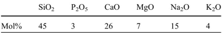
In the present research work, samples of a silica-based bioactive glass CEL-2, with the molar
composition reported in Table1, were prepared by melt and quenching route in bulk form and used as substrates for the surface functionalization.
Glass bars were annealed as reported in [9] and cut in slices (2 mm thick, area of about 1 cm2) which were polished by one side with SiC abrasive papers up to 4000 grit.
Glass samples were washed in acetone (1 time, 5 min) and water (3 times, 5 min) in an ultrasonic bath and let dry under a laminar flow cabinet (FAS-TER CYTOSAFE) in order to expose reactive hydro-xyl groups, as described in [7–9].
Samples were surface functionalized with gallic acid (GA, 97.5–102.5% titration, Sigma-Aldrich) and polyphenols extracted from red grape skin (GPH) and green tea leaves (TPH). GPH and TPH are complex mixtures of polyphenols obtained from dried red grape skins [Barbera variety, Vaglio Serra (AT), Italy] and green tea leaves (Longjing variety, Hangzhou, China) by conventional solvent extraction in a water–ethanol solution (20:80 volume ratio), 1 h at 60 °C, with a solid–liquid ratio of 1:20 and 1:50, respectively, as described in [9].The functionalization process foreseen samples soaking 3 h at 37 °C in the respective solutions, as described in [9]. Direct grafting to the hydroxyl groups exposed on the glass surface can be obtained by this procedure, as described in [9].
Glass slices were steam sterilized (20 min, 121 °C, 1 atm) in autoclave (ASAL 760) before the surface modification process. All the functionalization pro-cess was carried out under a laminar flow cabinet (FASTER CYTOSAFE) previously UV sterilized 20 min.
Molecular release evaluation
Functionalized samples were soaked in 10 ml of ultrapure water at 37 °C for 1 and 7 days that were chosen as representative for biological experiments starting and ending points. At the end of the soaking period, both solutions and samples undergone the Folin&Ciocalteu test, as described in [9, 12],
characterized by UV spectrophotometric determina-tion of the redox activity (related to the amount) of both polyphenols released in water and on the glass surface.
Fluorescence observation
of the biomolecules
Polyphenols auto-fluorescence has been previously reported in the literature [39]; accordingly, bare and functionalized glass specimens were observed by fluorescence microscopy (Leica DM5500 B, Leica Microsystems, IL, USA) in order to determine the presence of the biomolecules grafted on the surface. Finally, obtained images were analyzed by ImageJ software (3D surface plot, NIH, Bethesda, USA) with the aim to determine molecules surface distribution and thickness.
Cells
Cells used for experiments were purchased from the American Type Culture Collection (ATCC, Manassas, USA). Human bone osteosarcoma cells U2OS (ATCC HTB-96) and human fetal pre-osteoblasts hFOB (hFOB 1.19, ATCC CRL-11372) were used for exper-iments as representative for tumor and non-tumor cells, respectively. U2OS cells were cultured in Dul-becco’s modified Eagle medium (DMEM, Sigma) supplemented with 10% fetal bovine serum (FBS, SIGMA) and 1% antibiotics (penicillin/strepto-mycin), while hFOB were cultured in DMEM: Ham’s F12 mixture (50:50, Sigma) supplemented with 10% fetal bovine serum, 1% antibiotics and 0.3 mg/ml neomycin (G418 salt, Sigma). Cells were cultured at 37 °C, 5% CO2 until 80–90% confluence, detached with trypsin–EDTA solution (Sigma) and used for experiments.
Direct cytotoxicity evaluation
Sterile specimens were collected into the wells of a 24-multiwell plate (Nunc Delta, Thermo Fisher Sci-entific), and cells (both U2OS and hFOB) were directly seeded onto each surface in a defined number (2 9 104cells/specimens). After 1, 3 and 7 days, the cells viability was evaluated by the meta-bolic colorimetric 3-(4,5-dimethylthiazol-2-Yl)-2,5-diphenyltetrazolium bromide assay (MTT, Sigma); briefly, at each time point cells were carefully
Table 1 CEL2 bioactive glass molar composition (elements expressed using their oxides as reference compounds)
SiO2 P2O5 CaO MgO Na2O K2O
washed with PBS, and the MTT solution (2 mg/ml in fresh medium) was added to all specimens. Plate was incubated at 37 °C, 5% CO2for 4 h in the dark. Then, supernatants were gently removed and crystal formazans solved by 300 ll of dimethyl sulfoxyde (DMSO, Sigma); 100 ll were then collected, spotted into a 96-multiwell (Nunc Delta, Thermo Fisher Scientific) plate and the optical density (o.d.) eval-uated at 570 nm by spectrophotometer (Spec-traCount, Packard Bell). The o.d. of cells cultivated onto only-washed glasses was used as control and considered as 100% viability; test specimens o.d. were normalized toward controls and expressed as function of them. Experiments were performed in triplicate.
Not direct cytotoxicity evaluation
Sterile specimens were collected into sterile 50 ml tubes and submerged with 10 ml of fresh medium each (10 ml medium/sample). Tubes were stored at 37 °C, 5%CO2, and after 1, 2, 3, 5 and 7 days, 2 ml of supernatant was collected from the tubes and used to cultivate cells previously seeded in a defined number (1 9 104cells/well) into a new 48-well plate. After 24-h cultivation with supernatants, cells viability was evaluated by the MTT assay as previously described for the direct assay. Cells cultivated with fresh medium were used as control and considered as 100% viability; test specimens o.d. were normalized toward controls and expressed as function of them. Experiments were performed in triplicate.
RONS evaluation
In order to quantify oxygen and nitrogen reactive species (RONS) engendered from cells in response to specimens’ delivered molecules, cells (either U2OS or hFOB) were cultivated onto bioactive glass specimens as prior described in 2.5. After 1-, 2- and 3-day cultivation, 100 ll supernatants were col-lected from each well containing specimen, spinned down for 5 min at 12,000 rpm and stored at -80° until use. Finally, RONS were quantified using the OxiSelectTM In Vitro ROS/RNS Assay Kit (Cell Biolabs INC, San Diego, USA) following Manufac-turer’s instructions. Briefly, 50 ll of supernatants were mixed with 50 ll of Catalyst (1:250 in PBS) in each well of a 96-black-bottom-well plate (Sigma)
and stored 5 min at room temperature. Afterward, 100 ll DCFH stabilized solution were added to each well; DCFH is a non-fluorescent probe that can be rapidly oxidized by ROS and RNS becoming the highly fluorescent molecule DCF. DCF formation was evaluated by spectrophotometer (SpectraCount, Packard Bell) at 530 nm wavelength. Finally, fluo-rescent unites (RFU) were converted in RONS concentration by means of a peroxide standard curve.
Apoptosis and DNA damage evaluation
To test whether biomolecules grafting lead to tumorigenic U2OS cells death by DNA damage, cells were cultivated directly onto CEL-2TPH and CEL2 (considered as control) glasses for 3 days as descri-bed in 2.5. Afterward, specimens were collected, washed 3 times with PBS and fixed with ImmunoFix (BioOptica, Milan, Italy) for 5 min, at room temper-ature. Then, after rinsing carefully specimens with PBS, cells were permeabilized 20 min with Triton (0.5% in PBS) working on ice. Primary antibodies anti-53BP1 (Abcamab36823, Cambridge, UK, 1:800 in PBS containing 2% goat serum and 1% bovine serum albumin) and anti-cyclin B1 (Abcamab181593, Cam-bridge, UK, 1:250 in PBS containing 2% goat serum and 1% bovine serum albumin) were then added for 4 h at room temperature. Finally, specimens were co-stained with an appropriate secondary antibody (AlexaFluo488, Immunological Science, Rome, Italy, 1:400 in PBS) and with 40,6-diamidino-2-phenylindole (DAPI; Sigma-Aldrich) to visualize nuclei. Stained bioactive glasses were analyzed by fluorescence microscopy (Leica AF 6500; Leica Microsystems, Basel, Switzerland).
Statistical analysis of data
Statistical analysis of data was performed using the Statistical Package for the Social Sciences (SPSS v.20.0, IBM, Atlanta, GA, USA). Data were statisti-cally compared by one-way ANOVA, followed by Sheffe’s test for post hoc analysis, in the case of independent samples, and by Friedman’s ANOVA followed by Conover’s test, in the case of dependent samples. Two-sample comparisons were done using the Mann–Whitney U test. The significance level was set at p \ 0.05.
Results
Biomolecules surface deposition
Fluorescence images of control (CEL-2), gallic acid (CEL-2 GA) and polyphenols extracted from red grape skin (CEL-2 GPH) or green tea leaves (CEL-2 TPH) grafted glasses are reported in Fig.1, upper panel.
Bare CEL-2 does not report any signal as no bioactive molecules were grafted onto the surface; on the opposite, CEL-2 GA, CEL-2 GPH and CEL-2 TPH show a marked fluorescent signal due to the grafted biomolecules. Moreover, fluorescence images were analyzed by 3D software revealing a similar, homo-geneous and continuous distribution of the biomo-lecules onto each specimen (lower panel).
Molecular release evaluation
A negligible amount of polyphenols (GA, GPH and TPH) has been detected in the release solutions up to 7 days. On the other hand, a certain amount of polyphenols remains in an active state on the samples
surface even after 7 days soaking in water at 37 °C (0.009 ± 8 9 10-5, 0.002 ± 0.001 and 0.002 ± 2 9 10-6 GA equivalent for CEL-2GA, CEL-2GPH and CEL-2TPH, respectively).
Direct cytotoxicity
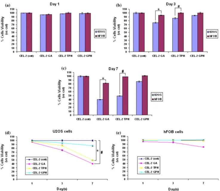
Results of cells cultivated onto specimens’ surface for 1, 3 and 7 days are reported in Fig. 2.
In general, after 1 day of direct cultivation (Fig. 2a), no significant differences (p [ 0.05) have been noticed between controls (CEL-2 cnt) and test speci-mens; in fact, U2OS and hFOB cells viability is between 96–99% in comparison with untreated control.
After 3 days (Fig.2b) of direct cultivation, a certain selection in term of cells viability can be noticed between U2OS and hFOB cells for CEL-2 GA and CEL-2 TPH specimens. Tumor cells viability decrea-ses to 75 and 82% for GA and TPH, respectively, while safe cells viability remains in a range of 94–99% for all test specimens. A significant difference in the viability of U2OS and hFOB cells has been observed for specimens functionalized with gallic acid and
Figure 1 Fluorescence images of CEL-2, CEL-2 GA, CEL-2 GPH and CEL-2 TPH. Coated specimens showed a marked fluorescent signal due to the biomolecules presence (lower panel, stained in red). Accordingly, 3D surface analysis revealed a
homogeneous and continuous biomolecules distribution presenting a comparable thickness (expressed as arbitrary units). Bar scale 50 lm, magnification 920.
green tea leaves polyphenols (p \ 0.05, indicated by * and #, respectively). On the opposite, no significant differences have been noticed for other specimens.
After 7 days (Fig.2c), the selective killing activity of the CEL-2 GA and CEL-2 TPH doped specimens lead to a viability loss of about 60 and 51% for tumor cells, while hFOB results are still in the range of 82–98%.
Accordingly, U2OS and hFOB viability comparison is significant for both CEL-2 GA (p \ 0.05, indicated by *) and CEL-2 TPH (p \ 0.05, indicated by #).
Finally, in Fig.2d, e, the single cells viability modification in function of time is presented. Inter-esting, tumorigenic U2OS cells (d) reported a signif-icant viability decrease after 7 days in contact with CEL-2 GA (p \ 0.05, indicated by #) and CEL-2 TPH (p \ 0.05, indicated by *) in comparison with untreated controls; conversely, hFOB viability is always [80% thus not presenting any significant differences if compared to untreated controls (e, p [ 0.05).
Figure 2 Direct cytotoxicity evaluation. Cells cultivated directly onto specimens’ surface showed a different behavior. In fact, tumorigenic U2OS cells viability was significantly lowered after 3 (b) and 7 (c) days by GA and TPH coating in comparison with results obtained for hFOB cells (p \ 0.05, indicated by * and #, respectively). Thus, as shown in d and e, tumorigenic cells
viability was significantly decreased in comparison with controls when cells were cultivated onto GA and TPH coated glasses (d, p \ 0.05, indicated by * and #, respectively); on the opposite, safe bone cells viability was never lowered in a significant manner in comparison with controls even after 7 days culture (e,p [ 0.05).
Not direct cytotoxicity
The viability of cells cultivated with 1, 2, 3, 5 and 7 days specimens’ supernatants is reported in Fig.3. Even if no differences can be noticed in the first three days (a-b-c, p [ 0.05), after 5 (d) and 7 (e) days culturing, a significant difference in terms of viability (around 15–20%) has been detected for U2OS cells cultivated with CEL-2 GA and CEL-2 TPH super-natants. By comparing hFOB and U2OS cells viabil-ity, a significant difference can be noticed for CEL-2 GA and CEL-2 TPH (p \ 0.05, indicated by * and #, respectively), while no differences have been found for CEL-2 GPH (p [ 0.05). Accordingly, considering U2OS cells viability in comparison with untreated control (f), a significant lowering in terms of viability can be observed after 7 days for 2 GA and CEL-2 TPH (p \ 0.05, indicated by * and #, respectively);
on the opposite, no difference has been found for safe hFOB cells during the 7 days culturing (g, p [ 0.05) in comparison with controls.
RONS evaluation
Oxygen and nitrogen reactive species (RONS) engendered from cells in response to bare and func-tionalized glasses were evaluated and compared in function of time over 3 days. Results are reported in Fig.4.
After 24 h (Fig.4a), no significant differences were noticed by comparing RONS produced by cells and glasses without cells (p [ 0.05); however, a significant difference can be observed by comparing CEL-2 TPH used to cultivate hFOB and U2OS cells: here, a higher RONS signal can be detected for U2OS (p \ 0.05, indicated by*). On the opposite, after 72 h (Fig.4b), more significant differences can be observed; in general, the grafting of biomolecules is effective in lowering RONS amount (p \ 0.05, indicated by §) onto nude glasses. Moreover, when cells are added to test and control glasses, RONS level is higher for U2OS as previously noticed after 24 h (p \ 0.05, indicated by *).
DNA damage evaluation
In order to evaluate the presence of DNA damage due to the activity of CEL-2 TPH, U2OS cells were cultivated for 3 days (2 9 104cells/specimen) onto the surface of the functionalized glasses and control
bFigure 3 Not direct cytotoxicity evaluation. Cells cultivated with
1 (a), 2 (b), 3 (c), 5 (d) and 7 (e) days specimens’ surnatants showed a different behavior as previously noticed for cells cultivated directly onto glasses surfaces. In fact, tumorigenic U2OS cells viability was significantly lowered after 5 and 7 days by GA and TPH molecules released from glasses’ coating in comparison with results obtained for hFOB cells (p \ 0.05, indicated by * and #, respectively). Thus, as shown inf and g, tumorigenic cells viability was significantly decreased in compar-ison with controls when cells were cultivated with GA and TPH surnatants (f,p \ 0.05, indicated by * and #, respectively); on the opposite, safe bone cells viability was never lowered in a significant manner in comparison with controls even after 7 days (g,p [ 0.05).
Figure 4 RONS evaluation after 24 (a) and 72 (b) h. The presence of coated biomolecules was effective in protecting safe hFOB cells form inflammation after 24 and 72 h in comparison with tumorigenic U2OS cells where inflammation due to RONS
was not lowered (p \ 0.05, indicated by *). Moreover, the biomolecules coating was effective in lowering RONS amount (p \ 0.05, indicated by §) onto nude glasses after 24 h (p \ 0.05, indicated by §).
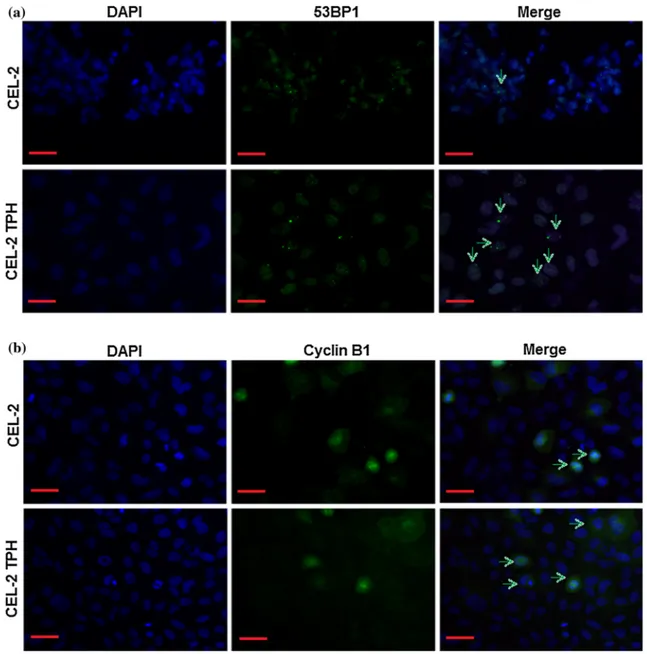
nude CEL-2 ones. Then, the localization of 53BP1 was evaluated by fluorescence; results are reported in Fig.5a. Firstly, by nuclear DAPI staining (blue dye), it is possible to see that U2OS cells are able to form typical tumor cells aggregates in nude CEL-2 glasses, whereas they are single dispersed into CEL-2 TPH. Second, 53BP1 signals (green dye) are mostly found inside nuclei (a, indicated by green arrows) rather than in the cytoplasm, confirming that a DNA dam-age occurred. Finally, to tests whether tumor cells were able to restore DNA damage occurred, the Cyclin B1 staining was applied (b). Interesting, also
Cyclin B1 signals were found to be mostly in the nuclei in, thus demonstrating that tumorigenic cells cultivated onto CEL-2 TPH glasses are not able to restore occurred DNA damage.
Discussion
The feasibility of grafting gallic acid and polyphenols extracted from red grape skin and green tea leaves on CEL-2 bioactive glass has been previously verified by means of XPS analyses and the Folin&Ciocalteu test
Figure 5 Biomolecules DNA damage induction. When tumori-genic cells were cultivated onto CEL-2 TPH glasses, 53BP1 signal was found to be mostly inside nuclei, thus showing the presence of an occurred DNA damage (agreen signals indicated by arrows).
Moreover, when the same cells were marked with Cyclin B1, signals were again noticed inside nuclei (bgreen signals indicated by arrows) suggesting that cells were not able to undergo DNA repair.Bar scale 50 lm, magnification 920.
and showed in our previous work [9]. Here, we fur-ther confirmed the presence of the cited biomolecules on the glass surface after functionalization by means of fluorescence microscopy exploiting their auto-flu-orescence. The biomolecules appear as homoge-neously distributed onto specimens’ surface, and they are well distinguishable from the bulk glass. Also, the thickness of the functionalized layer results as comparable between different types of treatment. Accordingly, the possibility to visualize the presence of gallic acid and natural polyphenols on the surface of bioactive glasses by means of their auto-fluores-cence is reported here for the first time, and it could represent a useful tool to investigate polyphenols grafting.
The release of the grafted molecules from the samples is negligible, but their availability in an active form on the glass surface is measurable up to 7 days. This result confirms the possibility to locally administer polyphenols from the glass surface to bone tissues in an active state. The in situ adminis-tration of polyphenols at the interface between the biomaterial and bone, after a surgical resection of a tumor mass, could be of interest for its protective effect toward tumor-relapse in synergy with a low dose systemic chemotherapy. However, no defined trend can be observed on the various surfaces, and it must be taken into account that the amount of polyphenols, determined indirectly by the Folin&Ciocalteu method, on the surface of the tested materials is affected by the surface alteration upon contact with water-based media (due to the high surface reactivity of the bioactive glass) and by the pH increase of the water media (again associated to the glass reactivity) and the consequent possible alteration of the biomolecules (which are highly pH-sensitive). An in depth study of the molecular release from functionalized bioactive glasses of different degree of reactivity in different buffered solutions (e.g., SBF) and of their stability upon storage in physiological media for different experimental times should be of interest, but it is out of the scope of the present paper and will be investigated in a future work.
The hypothesized activity of the polyphenols grafted onto bioactive glasses toward bone cancerous cells (U2OS) has been investigated by means of their toxicity by the metabolic MTT assay. Moreover, to verify whether this activity was targeted or not, same experiments were performed toward healthy bone
cells (hFOB) to determine any differences in terms of viability. The untreated CEL-2 resulted as in vitro cytocompatible confirming our previous findings [40]: glass surface seeded cells viability (both U2OS and hFOB) resulted as comparable to the one repor-ted by cells cultivarepor-ted onto polystyrene wells (\2% cells viability loss, data not shown). Accordingly, they were considered as control referring to the polyphenols grafted glasses. On the contrary, when gallic acid and TPH or GPH were introduced onto the same CEL-2 glasses, a different trend in cells viability was noticed by comparing U2OS and hFOB results. As reported in the results section, in the direct assay certain selective viability reduction for cancerous osteoblast cells has been observed for all functional-ized samples after 3-day culture; particularly, the growing trend resulted GPH \ TPH \ GA, and it was particularly evident after 7 days of incubation. Conversely, the viability reduction for cancerous osteoblast cells was less evident in the indirect assay, where only a moderate reduction was noticed after 7-day culture. These results might seem in contrast to each other as the same specimens were used; how-ever, the explanation of these data could be found in the molecular release assay results. In fact, by the Folin&Ciocalteu test, it was possible to realize that most of the grafted molecules were still present onto glasses surface after 7 days immersion, while only a small amount was released. So, when cells were directly seeded onto glasses surface, they were in touch with the higher amount of molecules thus undergoing their effect. Conversely, the eluates used for the indirect assay restrained the lower amount of molecules thus little affecting cells viability. Regard-less differences between direct and indirect assays results, these data confirmed the two main goals of this work. The first one is that it was confirmed that peculiar property of polyphenols were preserved upon their grafting onto biomaterials leading to the hypothesis to use them for the local administration of these molecules through implant surfaces. The sec-ond important finding is the ability of functionalized surfaces (especially CEL-2GA and CEL-2TPH) to selectively affect cancer cells viability preserving the healthy ones. This result is in accordance with the data reported in the literature about the selective cytotoxic action of polyphenols against cancer cells [17–22].
To explain the possible mechanism behind polyphenols toxic activity, oxygen and nitrogen
reactive species (RONS) were monitored in culture media, in presence or absence of cells upon the introduction of bare CEL-2 and TPH grafted ones. Tea polyphenols functionalized surfaces have been preselected for RONS evaluation on the basis of the best results obtained in the direct and indirect via-bility assay above discussed and for their natural origin. This latter aspect is of great interest as the use of natural molecules can overcome possible side effects of chemical substances, both on patients and on the environment, and promote a sustainable use of resources. In fact, the here introduced natural extracts can be easily obtained from the by-products of the agri-food production chain in large amount. RONS assay results revealed a certain amount of reactive species registered in presence of cells without mate-rials that can be ascribed to their metabolic activity, as reported in the literature [41]. Similarly, the introduction of bioactive glass samples (CEL-2 and CEL-2TPH) induces a certain amount of reactive species in the medium that can be correlated with materials surface reactivity and that is close to the one caused by the sole cells, without differences between bare and functionalized samples at 24 h. On the other hand, after 3 days, a marked difference between CEL-2 and CEL-2TPH can be registered leading to speculate that tea polyphenols can reduce the RONS production in the culture medium. These findings are similar to those previously reported by authors [9] where it was evidenced the ability of both CEL-2 and CEL-2TPH to scavenge oxygen radicals produced by the photolysis of hydrogen peroxide. Some minor differences can be ascribed to the ferent experimental times considered and to the dif-ference in the test typology and medium.
As far as the evaluation of RONS in cell culture is concerned, in presence of bioactive glasses, an increase in the production of reactive species can be recorded in cancerous cells in presence of CEL-2TPH samples while a protective activity and anti-inflam-matory effect for the healthy ones were noticed. This result shows a sort of selective protective activity toward safe cells due to the biomolecules presence. Conversely, tumorigenic cells seem to display high level of inflammation that can be related to the high death ratio previously observed with MTT assays. These results are not surprising as the high cancer cells sensitivity toward reactive species has been largely demonstrated [18–20,42–44]. For example, in a recent review, Yan et al. [45] proposed that cancer
cells tend to express more aquaporins on their cyto-plasmic membranes, which may cause the H2O2 uptake speed in cancer cells to be faster than in normal cells. Moreover, it is reported in the literature that polyphenols, and among them gallic acid, trans-resveratrol (as one of the major constituent of grape polyphenols) and catechins (as the most abundant molecules in tea extracts), can interfere with different pathways involved in oncogenesis and progression causing the selective apoptosis of several types of cancer cells [46–50].
Finally, we investigated whether polyphenols toxicity was due to a reversible or not DNA damage in U2OS cancerous cells. Accordingly, the nuclear localization of 53BP1 in most of the cells cultivated in direct contact with CEL-2 TPH specimens con-firmed that DNA damage occurred. This genetic harm can be probably related to the polyphenols-derived RONS generation as prior debated. After-ward, by checking a further nuclear localization of Cyclin B1 dye, it was possible to speculate that U2OS cells were not able to self-repair the RONS-induced DNA damage thus leading to cells death (as verified also by MTT assay). Interesting, DAPI staining preliminary suggested also that cells culti-vated onto TPH grafted were not able to grown in tight contact to each others as typically occurs for tumor cells; this effect was not visualized in control CEL-2 specimens where cells were stained as form-ing aggregates. However, any speculation regardform-ing tumor progression inhibition must be deeply inves-tigated in future works.
In conclusion, the effect of polyphenols coupled to a bioactive glass on the viability and RONs produc-tion of healthy and cancerous osteoblast has been reported for the first time in the present paper.
This is a preliminary confirmation that polyphe-nols (e.g., gallic acid and green tea extracts) can effectively exert a selective cytotoxic action against bone cancer cells upon grafting to the surface of a bioactive glass. Moreover, this action can be associ-ated with the production of reactive species in the cancerous cells with a consequent selective and not reversible DNA damage. On the other hand, an anti-inflammatory action has been evidenced on the healthy osteoblast cells.
Altogether, preliminary results reported in the present paper are extremely encouraging for the development of innovative smart bioactive glasses for bone contact applications.
Conclusions
In the present research, the in vitro response of healthy (hFOB) and cancerous (U2OS) osteoblast cells to a bare and polyphenol-grafted bioactive glass has been investigated. A selective cytotoxic activity of gallic acid and tea polyphenol-grafted bioactive glass has been evidenced against U2OS particularly in direct assay. Bioactive glass functionalized with tea polyphenols was able to induce reactive oxygen and nitrogen spe-cies (RONS) production in U2OS cells and, on the other hand, to exert a sort of anti-inflammatory action for hFOB. Finally, a permanent DNA damage, together with a certain difficulty to form tumor aggregates, has been evidenced for U2OS cells cultured on tea polyphenols grafted bioactive glass.
These results highlight that bioactive glasses func-tionalized with polyphenols can be extremely promising as biomaterials for application in bone substitution for cancer treatment.
Compliance with ethical standards
Conflict of interest The authors declare that they have no conflict of interest.
References
[1] Hench LL (2006) The story of BioglassÒ. J Mater Sci Mater Med 17:967–978
[2] Hench LL (2009) Genetic design of bioactive glass. J Eur Ceram Soc 29:1257–1265
[3] Hench LL, Roki N, Fenn MB (2014) Bioactive glasses: importance of structure and properties in bone regeneration. J Mol Struct 1073:24–30
[4] Miguez-Pecheco V, Hench LL, Boccaccini AR (2015) Bioactive glasses beyond bone and teeth: emerging appli-cations in contact with soft tissues. Acta Biomater 13:1–15 [5] Rabiee SM, Nazparvar N, Azizian M, Vashaee D, Tayebi L
(2015) Effect of ion substitution on properties of bioactive glasses: a review. Ceram Int 41:2741–2751
[6] Baino F, Novajra G, Miguez-Pacheco V, Boccaccini AR, Vitale-Brovarone C (2016) Bioactive glasses: special applications outside the skeletal system. J Non-cryst Solids 432:15–30 [7] Verne` E, Vitale-Brovarone C, Bui E, Bianchi CL, Boccaccini
AR (2009) Surface functionalization of bioactive glasses. J Biomed Mater Res 90A:981–992
[8] Verne` E, Ferraris S, Vitale-Brovarone C, Spriano S, Bianchi CL, Naldoni A, Morra M, Cassinelli C (2010) Alkaline
phosphatase grafting on bioactive glasses and glass–ceram-ics. Acta Biomater 6:229–240
[9] Cazzola M, Corazzari I, Prenesti E, Bertone E, Verne` E, Ferraris S (2016) Bioactive glass coupling with natural polyphenols: surface modification, bioactivity and anti-oxi-dant ability. Appl Surf Sci 367:237–248
[10] Zhang X, Ferraris S, Prenesti E, Verne` E (2013) Surface functionalization of bioactive glasses with natural molecules of biological significance. Part I: gallic acid as model molecule. Appl Surf Sci 287:329–340
[11] Zhang X, Ferraris S, Prenesti E, Verne` E (2013) Surface functionalization of bioactive glasses with natural molecules of biological significance. Part II: grafting of polyphenols extracted from grape skin. Appl Surf Sci 287:341–348 [12] Ferraris S, Zhang X, Prenesti E, Corazzari I, Turci F, Tomatis
M, Verne` E (2016) Gallic acid grafting to a ferrimagnetic bioactive glass–ceramic. J Non-cryst Solids 432:167–175 [13] Saiko P, Szakmary A, Jaeger W, Szekeres T (2008)
Resveratrol and its analogs: defense against cancer, coronary disease and neurodegenerative maladies or just a fad? Mutat Res 658:68–94
[14] Piotrowska H, Kucinska M, Murias M (2012) Biological activity of piceatannol: leaving the shadow of resveratrol. Mutat Res 750:60–82
[15] Kang NJ, Shin SH, Lee HJ, Lee KW (2011) Polyphenols as small molecular inhibitors of signaling cascades in carcino-genesis. Pharmacol Therapeut 130:310–324
[16] Petti S, Scully C (2009) Polyphenols, oral health and dis-ease: a review. J Dent 37:413–423
[17] Lewandowska H, Kalinowska M, Lewandowski W, Step-kowski TM, Brzoska K (2016) The role of natural polyphenols in cell signaling and cytoprotection against cancer development. J Nutr Biochem 32:1–19
[18] Nowshehri JA, Bhat ZA, Shah MY (2015) Blessing in dis-guide: bio-functional benefits of grape seed extracts. Food Res Int 77:333–348
[19] Ullah N, Ahmad M, Aslam A, Tahir MA, Aftab M, Bibi N, Ahmad S (2016) Green tea phytocompounds as anticancer: a review. Asian Pac J Trop Dis 6:330–336
[20] Khan N, Mukhtar H (2008) Multitargeted therapy of cancer by green tea polyphenols. Cancer Lett 269:269–280 [21] Schuck AG, Weisburg JH, Esan H, Robin EF, Bersson AR,
Weitschner JR, Lahasky T, Zuckerbraun HL, Babich H (2013) Cytotoxic and proapoptotic activities of gallic acid to human oral cancer HSC-2 cells. Oxid Antioxid Med Sci 2:265–274
[22] Sharma A, Gautam SP, Gupta A (2011) Surface modified dendrimers: synthesis and characterization for cancer tar-geted drug delivery. Bioinorg Med Chem 19:3341–3346
[23] Ball V, Meyer F (2016) Deposition kinetics and electro-chemical properties of tannic acid on gold and silica. Colloid Surf A 491:12–17
[24] Sileika TS, Barrett DG, Zhang R, Lau KHA, Messersmith PB (2013) Colorless multifunctional coatings inspired by polyphenols found in tea, chocolate, and wine. Angew Chem Int Ed 52:10766–10770
[25] Nam JB, Ryu JH, Kim JW, Chang IS, Suh KD (2005) Sta-bilization of resveratrol immobilized in monodisperse cyano-functionalized porous polymeric microspheres. Polymer 46:8956–8963
[26] Bae JH, Shanmugharaj AM, Noh WH, Choi WS, Ryu SH (2007) Surface chemical functionalized single-walled carbon nanotube with anchored phenol structures: physical and chemical characterization. Appl Surf Sci 253:4150–4155 [27] Peng H, Xiong H, Li J, Xie M, Liu Y, Bai C, Chen L (2010)
Vanillin cross-linked chitosan microspheres for controlled release of resveratrol. Food Chem 121:23–28
[28] Kong X, Jin L, Wei M, Duan X (2010) Antioxidant drugs intercalated into layered double hydroxide: structure and in vitro release. Appl Clay Sci 49:324–329
[29] Das S, Ng KY (2010) Colon-specific delivery of resveratrol: optimization of multi-particulate calcium-pectinate carrier. Int J Pharm 385:20–28
[30] Yu SH, Mi FL, Pang JC, Jiang SC, Kuo TH, Wu SJ, Shyu S (2011) Preparation and characterization of radical and pH-responsive chitosan-gallic acid conjugate drug carriers. Carbohyd Polym 84:794–802
[31] Cho YS, Kim SK, Ahn CB, JeJ Y (2011) Preparation, characterization and antioxidant properties of gallic acid-grafted-chitosans. Carbohyd Polym 83:1617–1622
[32] Bozic M, Gorgieva S, Kokol V (2012) Laccase-mediated functionalization of chitosan by caffeic and gallic acids for modulating antioxidant and antimicrobial properties. Car-bohyd Polym 87:2388–2398
[33] Francesko A, Soares da Costa D, Reis RL, Pashkuleva I, Tzanov T (2013) Functional biopolymer-based matrices for modulation of chronic wound enzyme activities. Acta Bio-mater 9:5216–5225
[34] Rawat K, Saxena A, Verma AK, Vohra R, Bohidar HB (2014) Potential of gallic acid loaded polysaccharide-protein (Agar-Gelatin) co-hydrogels in wound healing. J Pharma Res 3:14–17
[35] Forte L, Torricelli P, Boanini E, Gazzano M, Rubini K, Fini M, Bigi A (2016) Antioxidant and bone repair properties of quesrcetin-functionalized hydroxyapatite: an in vitro osteo-blast-osteoclast-endothelial cell co-culture study. Acta Bio-mater 32:298–308
[36] Varoni EM, Rimondini L, Iriti M (2012) Plant products for innovative biomaterials in dentistry. Coatings 2:179–194
[37] Malavasi G, Ferrari E, Lusvardi G, Aina V, Fantini F, Morterra C, Pignedoli F, Saladini M, Menabue L (2011) The role of coordination chemistry in the development of inno-vative gallium-based bioceramics: the case of curcumin. J Mater Chem 21:5027–5037
[38] Dziadek M, Dziadek K, Zagrajczuk B, Menaszek E, Cho-lewa-Kowalska K (2016) Poly(e-caprolactone)/bioactive glass composites enriched with polyphenols extracted from sage (Salvia officinalis L.). Mater Lett 183:386–390 [39] Lavid N, Schwartz A, Yarden O, Tel-Or E (2001) The
involvement of polyphenols and peroxidase activities in heavy-metal accumulation by epidermal glands of the waterlily (Nymphaeaceae). Planta 212:323–331
[40] Verne´ E, Ferraris S, Vitale-Brovarone C, Cochis A, Rimondini L (2014) Bioactive glass functionalized with alkaline phosphatase stimulates bone extracellular matrix deposition and calcification in vitro. Appl Surf Sci 313:372–381
[41] Jebahi S, Oudadesse H, El Feki H, Rebai T, Keskes H, Pellen P, El Feki A (2012) Antioxidative/oxidative effects of strontium-doped bioactive glass as bone graft. In vivo assays in ovariectomised rats. J Appl Biomed 10:195–209 [42] Urruticoechea A, Alemany R, Balart J, Villanueva A, Vin˜als
F, Capella´ G (2010) Recent advances in cancer therapy: an overview. Curr Pharm Des 16:3–10
[43] Ko¨ritzer J, Boxhammer V, Scha¨fer A, Shimizu T, Kla¨mpfl TG, Li YF, Welz C, Schwenk-Zieger S, Morfill GE, Zim-mermann JL, Schlegel J (2013) Restoration of sensitivity in chemo-resistant glioma cells by cold atmospheric plasma. PLoS ONE 8:1–10
[44] Cheng X, Sherman J, Murphy W, Ratovitski E, Canady J, Keidar M (2014) The effect of tuning cold plasma compo-sition on glioblastoma cell viability. PLoS ONE 9:1–9 [45] Yan D, Talbot A, Nourmohammadi N, Sherman JH, Cheng X,
Keidar M (2015) Toward understanding the selective anticancer capacity of cold atmospheric plasma—a model based on aquaporins (review). Biointerphases 10:04080101–04080113 [46] Verma S, Singh A, Mishra A (2003) Gallic acid: molecular
rival of cancer. Environ Toxicol Pharmacol 35:473–485 [47] Athar M, Back JH, Kopelovich L, Bickers DR, Kim AL
(2009) Multiple molecular targets of resveratrol: anti-car-cinogenic mechanisms. Arch Biochem Biophys 486:95–102 [48] Li Y, Ba¨ckesjo¨ C, Haldose´n L, Lindgren U (2009) Resver-atrol inhibits proliferation and promotes apoptosis of osteosarcoma cells. Eur J Pharmacol 609:13–18
[49] Arau´jo JR, Gonc¸alves P, Martel F (2011) Chemopreventive effect of dietary polyphenols in colorectal cancer cell lines. Nutr Res 31:77–87
[50] Sajilata MG, Bajaj PR, Singhal RS (2008) Tea polyphenols as nutraceuticals. Compr Rev Food Sci Food Saf 7:229–254