1
DEPARTMENT OF
NEUROSCIENCE BIOMEDICINE AND MOVEMENT SCIENCE
GRADUATE SCHOOL OF
APPLIED LIFE AND HEALTH SCIENCES
DOCTORAL PROGRAM IN LIFE AND HEALTH SCIENCES
WITH THE FINANCIAL CONTRIBUTION OF ATENEO - UNIVERSITY OF VERONA
Cycle XXX, year 2014-2017
HTLV Tax, HBZ and APH-2 regulatory protein interaction with host cell factors: implications for NF-κB pathway deregulation and tumorigenesis.
S.S.D. BIO13
Coordinator: Prof. Giovanni Malerba
Tutor: Prof.ssa Maria Grazia Romanelli
Doctoral Student: Dott.ssa Stefania Fochi
2 This work is licensed under a Creative Commons Attribution-NonCommercial-
NoDerivs 3.0 Unported License, Italy. To read a copy of the licence, visit the web page:
http://creativecommons.org/licenses/by-nc-nd/3.0/
Attribution — You must give appropriate credit, provide a link to the license, and indicate if changes were
made. You may do so in any reasonable manner, but not in any way that suggests the licensor endorses you or your use.
NonCommercial — You may not use the material for commercial purposes.
NoDerivatives — If you remix, transform, or build upon the material, you may not distribute the modified
material.
HTLV Tax, HBZ and APH-2 regulatory protein interaction with host cell factors: implications for NF-κB pathway deregulation and tumorigenesis.
Stefania Fochi
PhD thesis Verona, 11 Maggio 2018
3 ABSTRACT
Human T-cell leukemia virus type 1 (HTLV-1) is a retrovirus that infects approximately 20 million people worldwide and 5% of them develop adult T-cell leukemia (ATL), a fatal T-cell malignancy with a poor prognosis. The HTLV-1 genome encodes two proteins that play a pivotal role in the oncogenic process, the regulatory protein Tax and the accessory protein HBZ. The expression of HTLV-1 oncoprotein Tax plays a key role in CD4+ T-cell transformation, interacting with host factors and deregulating several cell pathways implicated in the regulation of cell cycle and cell survival. Among them, Tax activates contitutively the NF-κB pathway, which play a primary role in inflammation, immunity, cell proliferation, apoptosis and cancer. HTLV-1 basic zipper protein (HBZ), encoded by the antisense viral genome strand, is essential for viral persistence and promotion of T-cell proliferation, acting in concert with Tax and contributing to ATL development.
The purpose of my PhD research is to expand the knowledge of the molecular mechanism of NF-κB pathway deregulation mediated by the interactions of Tax and HBZ regulatory proteins with selected host factors. We studied the effect of Tax-1 and HBZ on NF-κB promoter activation by comparative analyses with the homologous regulatory proteins expressed by the genetically related HTLV type 2, which is not associated with ATL disease. We focused the experimental analyses on two relevant aspects of the NF-κB deregulation: the interactions of Tax and the antisense proteins with key factors of the NF-κB pathway mediating p65 activation, and their role on the alternative NF-κB pathway modulation. Our data demonstrated for the first time that HBZ and APH-2 differ in the inhibitory mechanism of Tax-dependent NF-κB activation. By confocal microscopy, we observed that APH-2, unlike HBZ, was recruited in Tax-2-cytoplasmic structures containing the NF-κB factors that are essential for the activation of the pathway, the adaptor protein TAB2 and the NF-κB modulator NEMO. The formation of these complexes results in the impairment of p65 transcription factor translocation into the nucleus. The analyses of these complexes showed that TRAF3, a key
4
factor of the alternative NF-κB pathway, interacts with Tax and APH-2. Applying the CRISPR/Cas9 system, we generated a cell model that allowed us to define the contribution of TRAF3 in the inhibition of NF-κB. The results obtained revealed for the first time that the absence of TRAF3 dramatically reduced the Tax-1 transactivating activity of NF-κB.
In conclusion, the results of my PhD thesis identify a new cellular factor essential for the action of HTLV Tax protein in the deregulation of cellular pathways and support the hypothesis that the different molecular mechanism of HBZ and APH-2 in the NF-κB inhibition may reflect divergent effects on HTLV-infected cells survival and probably on leukemogenesis induced by HTLV.
5 SUMMARY/RIASSUNTO
Il virus T-linfotropico umano di tipo 1 (HTLV-1) è stato il primo retrovirus ad essere scoperto nell’uomo. La sua infezione è la causa dello sviluppo, dopo anni d’infezione asintomatica, di un’aggressiva neoplasia conosciuta come leucemia delle cellule T dell’adulto (ATL), per la quale non esiste attualmente una terapia efficace. A oggi rimangono sconosciuti gli eventi che portano alla malattia nel 5% dei soggetti infettati, ma è dimostrato che i primi eventi dell’infezione possono essere determinanti per la persistenza del virus e la conseguente patogenicità. Due proteine codificate dal genoma di HTLV-1, le proteine Tax e HBZ, svolgono un ruolo fondamentale nel processo oncogenico, e sono richieste dal virus per completare il ciclo infettivo (Tax) e rendere persistente l’infezione (HBZ). L'espressione dell’oncoproteina Tax svolge un ruolo chiave nella trasformazione delle cellule T CD4+, interagendo con fattori della cellula ospite e deregolando
diverse vie di segnalazione cellulare che controllano il ciclo cellulare, la risposta immunitaria e la proliferazione. In particolare, la proteina Tax attiva il fattore di trascrizione NF-κB che svolge un ruolo primario nella regolazione della risposta immunitaria, nell'infiammazione, nella proliferazione cellulare, nell'apoptosi e nel cancro. La proteina HBZ è codificata dal filamento antisenso del genoma virale e svolge un ruolo essenziale nella persistenza del virus e nella promozione della proliferazione delle cellule T in concerto con Tax, contribuendo allo sviluppo della patologia ATL.
Lo studio condotto durante il dottorato di ricerca ha avuto come obiettivo principale migliorare le nostre conoscenze sulle interazioni delle proteine virali Tax e HBZ con fattori cellulari ed i loro effetti sul meccanismo molecolare che porta ad attivazione di NF-κB. A questo scopo abbiamo utilizzato l’analisi comparativa degli effetti dell’espressione delle proteine Tax e HBZ con le proteine omologhe prodotte da HTLV di tipo 2 (HTLV-2), un retrovirus geneticamente correlato ad HTLV-1, ma meno patogeno e soprattutto non associato allo sviluppo della malattia ATL. Gli studi comparativi sull'attività funzionale di Tax-1 e HBZ e gli omologhi di HTLV-2, Tax-2 e APH-2,
6
contribuiscono all’identificazione dei processi necessari alla tumorigenesi di HTLV. Nello studio abbiamo focalizzato l’attenzione su due aspetti rilevanti della deregolazione NF-κB: le interazioni di Tax e le proteine antisenso con i fattori chiave della via di segnalazione NF-κB, e il loro ruolo nella modulazione del pathway alternativo di NF-κB. I risultati dello studio hanno dimostrato per la prima volta che HBZ e APH-2 differiscono nel meccanismo molecolare che porta all’inibizione di NF-κB mediata da Tax. Attraverso studi di microscopia confocale, abbiamo osservato che APH-2, diversamente da HBZ, è reclutata in strutture citoplasmatiche in cui è presente Tax-2 e proteine essenziali per l’attivazione della via NF-κB, la proteina adattatrice TAB2 e il modulatore NEMO. La formazione di questi complessi rende inefficace il trasferimento nel nucleo del fattore di trascrizione p65. Dall’analisi dei complessi è derivata l’evidenza che un fattore chiave della via non canonica NF-κB, il fattore TRAF3, interagisce con le proteine Tax e APH-2. Applicando il sistema CRISPR/Cas9, abbiamo generato un modello cellulare che ha permesso di definire il contributo di TRAF3 nell’inibizione del pathway alternativo di NF-κB. I risultati ottenuti hanno dimostrato per la prima volta che l’assenza del fattore TRAF3 riduce drasticamente l’attivazione di NF-κB mediata da Tax-1.
In conclusione, i risultati di questa tesi identificano un nuovo fattore cellulare necessario all’azione di deregolazione dei pathway cellulari mediata dalla proteina Tax di HTLV e supportano l’ipotesi che il diverso meccanismo molecolare d'inibizione di NF-κB mediato da HBZ e APH-2 possa riflettere effetti divergenti sulla sopravvivenza delle cellule infette da HTLV e probabilmente sulla leucemogenesi indotta dal virus.
7
8 PUBLICATIONS
1. Stefania Fochi, Simona Mutascio, Umberto Bertazzoni, Donato Zipeto, Maria Grazia Romanelli (2018). HTLV deregulation of the NF-κB
pathway: an update on Tax and antisense proteins role. Front. Microbiol.
9:285 doi: 10.3389/fmicb.2018.00285
2. Louise Dubuisson, Florence Lormières, Stefania Fochi, Jocelyn Turpin, Amandine Pasquier, Estelle Douceron, Anaïs Oliva, Ali Bazarbachi, Valérie Lallemand-Breitenbach, Hugues De Thé, Chloé Journo, Renaud Mahieux (2018). Stability of HTLV-2 Antisense Protein is Controlled by
PML Nuclear Bodies in a SUMO-Dependent Manner. Oncogene. doi:
10.1038/s41388-018-0163-x.
3. Stefania Fochi, Simona Mutascio, Francesca Parolini, Donato Zipeto, Maria Grazia Romanelli (2017). HTLV antisense proteins role in the
9 CONGRESSES ATTENDED AND ABSTRACTS
ORAL PRESENTATIONS
1. Stefania Fochi, Elisa Bergamo, Michela Serena, Donato Zipeto, Maria Grazia Romanelli. HTLV-1 and HTLV-2 Tax, HBZ and APH-2 interaction
with host factors: their involvement in cellular pathways regulation.
HERN, HTLV European Research Network 2016, Bucarest, Romania. 20-22.05.2016 (oral presentation).
2. Stefania Fochi, Simona Mutascio, Francesca Parolini, Donato Zipeto, Maria Grazia Romanelli. HTLV-1 basic leucin zipper factor and its
homologous APH-2 impair NF-κB activation mediated by the viral oncoprotein Tax. Associazione Italiana di Biologia e Genetica, PhD
Meeting Santa Margherita Ligure, Italy. 11-13.05.2017 (oral
presentation).
CONFERENCES CONTRIBUTIONS
1. Elisa Bergamo, Erica Diani, Stefania Fochi, Pamela Lorenzi, Michela Serena, Donato Zipeto, Maria Grazia Romanelli. The human T-cell
leukemia virus -2 (HTLV-2) antisense protein APH-2 interacts with p65 and inhibits NF-ĸB Tax-2 activation. Società Italiana di Biofisica e
Biologia Molecolare, SIBBM Meeting, Turin, Italy. 01-03.07.2015. 2. Oncogenic Viruses Workshop, Padova, Italy. 25-26.09.2015.
3. Stefania Fochi, Elisa Bergamo, Michela Serena, Pamela Lorenzi, Donato Zipeto, Maria Grazia Romanelli. Functional role of HTLV Tax, HBZ and
APH-2 regulatory proteins on cellular autophagy pathway. PhD-Day
Meeting, University of Verona, Italy. 01.2016.
4. Stefania Fochi, Elisa Bergamo, Michela Serena, Donato Zipeto, Maria Grazia Romanelli. Human T-cell leukemia virus HBZ and APH-2
antisense proteins interaction with host factors and their involvement in NF-κB activation. Società Italiana di Biofisica e Biologia Molecolare,
10
SIBBM Meeting Naples, Italy. From Genomes to Functions. 16-18.06.2016.
5. Stefania Fochi, Elisa Bergamo, Michela Serena, Francesca Parolini, Simona Mutascio, Donato Zipeto, Maria Grazia Romanelli. The interplay
between HTLV proteins and cellular factors: impact on the NF-κB cell signaling. PhD-Day, University of Verona, Italy. 12.2017.
6. Francesca Parolini, Simona Mutascio, Michela Serena, Stefania Fochi, Maria Grazia Romanelli, Donato Zipeto. CRISPR/Cas9 as a powerful tool
for genome editing. Convegno primavera dermatologica, Bucarest,
Romania 29.03-02.04.2017.
7. Stefania Fochi, Simona Mutascio, Francesca Parolini, Donato Zipeto, Maria Grazia Romanelli. A CRISPR/Cas9 based approach to study the
implication of HTLV regulatory proteins in the NF-κB modulation.
Association of Cell Cultures meeting: The future of cancer therapy: the genome editng era, Catanzaro, Italy. 07-09.06.2017.
8. Francesca Parolini, Simona Mutascio, Michela Serena, Stefania Fochi, Maria Grazia Romanelli, Donato Zipeto. CRISPR/Cas9 for the Study of the
Interactions between Viruses and Host. Association of Cell Cultures
meeting: The future of cancer therapy: the genome editng era, Catanzaro, Italy. 07-09.06.2017.
9. Stefania Fochi, Simona Mutascio, Francesca Parolini, Donato Zipeto, Maria Grazia Romanelli. Host-virus interactions: HTLV antisense
regulatory proteins play a role in the dysregulation of NF-κB pathway.
Società Italiana di Biofisica e Biologia Molecolare, SIBBM Meeting, Milan, Italy. From Single Cells to 3D-Cell Culture. 14-16.06.2017.
10. Francesca Parolini, Simona Mutascio, Michela Serena, Stefania Fochi, Maria Grazia Romanelli, Donato Zipeto. CRISPR/Cas9 to study virus-host
interactions. Società Italiana di Biofisica e Biologia Molecolare, SIBBM
Meeting, Milan, Italy. From Single Cells to 3D-Cell Culture. 14-16.06.2017.
11
11. Federica Ferrarini, Francesca Martinetto, Roberta Galavotti, Stefania
Fochi, Pamela Lorenzi, C., Di Gaetano, B., Pardini, A., Naccarati, D. De
Pietri Tonelli, Maria Grazia Romanelli, Patricia M.-J. Lievens.
Characterization of FOXP2-11, a novel alternatively spliced product of FOXP2 gene. Società Italiana di Biofisica e Biologia Molecolare, SIBBM
Meeting, Milan, Italy. From Single Cells to 3D-Cell Culture. 14-16.06.2017.
12. Stefania Fochi, Simona Mutascio, Francesca Parolini, Donato Zipeto, Maria Grazia Romanelli. HTLV antisense proteins role in the NF-κB
modulation. International Virology Conference, Toronto, Canada.
30-31.10.2017.
AWARDS
1. Poster award: Stefania Fochi, Elisa Bergamo, Michela Serena, Donato Zipeto, Maria Grazia Romanelli. Human Tcell leukemia virus HBZ and
APH-2 antisense proteins interaction with host factors and their involvement in NF-κB activation. Società Italiana di Biofisica e Biologia
Molecolare, SIBBM Meeting Naples, Italy. From Genomes to Functions. 16-18.06.2016.
2. Poster award: Stefania Fochi, Elisa Bergamo, Michela Serena, Francesca Parolini, Simona Mutascio, Donato Zipeto, Maria Grazia Romanelli. The
interplay between HTLV proteins and cellular factors: impact on the NF-κB cell signaling. PhD-Day, University of Verona, Italy. 12.2017.
3. Poster award: Stefania Fochi, Simona Mutascio, Francesca Parolini, Donato Zipeto, Maria Grazia Romanelli. A CRISPR/Cas9 based approach
to study the implication of HTLV regulatory proteins in the NF-κB modulation. Association of Cell Cultures meeting: The future of cancer
12 INDEX
ABSTRACT ... 3
SUMMARY/RIASSUNTO ... 5
PUBLICATIONS ... 8
CONGRESSES ATTENDED and ABSTRACTS ... 9
INDEX ... 12
LIST OF FIGURES: ... 14
LIST OF TABLES: ... 16
ABBREVATIONS ... 17
1. INTRODUCTION ... 21
1.1 Human viral oncogenesis ... 21
1.2 Human T-cell leukemia virus (HTLV) and Epidemiology ... 22
1.3 HTLV transmission and disease association ... 24
1.3.1 Adult T-cell Leukemia/lymphoma (ATLL) ... 24
1.3.2 HTLV-1 Associated Myelopathy/Tropical Spastic Paraparesis (HAM/TSP) ... 25
1.3.3 HTLV-2 Associated Disease ... 26
1.4 HTLV Cell to Cell Transmission and Infection ... 26
1.5 HTLV-1 and HTLV-2 Genomic Organization ... 28
1.5.1 The pX region ... 29
1.5.1.1 The accessory proteins ... 29
1.5.1.2 Rex ... 30
1.6.1 Protein Structure of Tax-1 and Tax-2 ... 32
1.6.2 Cellular Localization of Tax-1 and Tax-2 Proteins ... 33
1.6.3 The Transforming Activity of Tax... 35
1.7 HBZ and APH-2 antisense proteins from HTLV-1 and HTLV-2 ... 36
1.7.1 Protein Structure of HBZ and APH-2 ... 36
1.7.2 Cellular Localization of Antisense Proteins ... 37
1.7.3 The Oncogenic Potential of HBZ ... 38
1.8 Effect of Tax, HBZ and APH-2 on NF-κB Signaling Transduction ... 39
1.8.1 NF-κB Pathways ... 40
13
1.8.3 HBZ, APH-2 and NF-κB Signaling ... 44
1.8.4 HTLV proteins deregulation of NF-κB in pathogenesis ... 46
1.8.5 The host factor TRAF3 and the alternative NF-κB pathway ... 47
2. AIM OF THE RESEARCH ... 49
3. MATERIALS AND METHODS ... 51
3.1 Cell culture and transfection ... 51
3.2 Plasmids, antibodies and reagents ... 51
3.3 Co-immunoprecipitation ... 52
3.4 Western blot analysis ... 53
3.5 NF-κB luciferase reporter assay ... 53
3.6 Immunofluorescence and confocal microscopy analysis ... 54
3.7 Development of TRAF3 knock-out cell lines by CRISPR/Cas9... 54
4. RESULTS ... 59
4.1 HBZ and APH-2 interact with p65 and suppress the NF-κB pathway ... 59
4.2 HBZ and APH-2 inhibit the Tax-mediated NF-κB activation... 61
4.3 APH-2 is recruited in cytoplasmic structures in the presence of Tax-2 ... 62
4.4 APH-2 is recruited in cytoplasmic complexes with TAB2 and NEMO in the presence of Tax-2 ... 64
4.5 p65 nuclear translocation is impaired in the presence of APH-2 ... 67
4.6 APH-2 and Tax-2 form complexes with TRAF3 ... 71
4.7 TRAF3 expression is affected by the presence of HBZ and APH-2 ... 76
4.8 HBZ interacts with p52 ... 79
4.9 Analysis of the NF-κB activity in TRAF3-/- cell line ... 79
4.10 Tax-1-mediated NF-κB activation is impaired in the absence of TRAF3 82 4.11 The LXXLL2 domain of APH-2 is required for PML localization... 84
5. DISCUSSION ... 86
6. CONCLUSION AND PERSPECTIVES ... 90
7. REFERENCES ... 92
14 LIST OF FIGURES:
Figure 1. Geographical distribution of the main foci of HTLV-1 infection.
Figure 2. Clinical features of an ATL patient and typical "flower cell" in the peripheral blood of an acute ATL patient.
Figure 3. Schematic representation of the human T-cell leukemia virus.
Figure 4. HTLV life cycle.
Figure 5. Schematic representation of HTLV-1, HTLV-2, HTLV-3, and HTLV-4
genomic organization.
Figure 6. Structural and functional domains of the Tax proteins.
Figure 7. Comparison of Tax-1 and Tax-2 subcellular localization.
Figure 8. Schematic representations of HBZ and APH-2 functional domains.
Figure 9. The NF-κB pathway.
Figure 10. Schematic representation of Tax-1 and Tax-2 interactions with factors
of NF-κB pathway.
Figure 11. Tax and HBZ effect on NF-κB.
Figure 12. The balance between Tax and HBZ expression regulates the outcome
of HTLV-1 infection.
Figure 13. CRISPR/Cas9 workflow.
Figure 14. pSpCas9(BB)-2A-Puro (PX459) V2.0
Figure 15. HBZ and APH-2 interact with p65 and suppress the NF-ĸB pathway.
Figure 16. HBZ and APH-2 inhibit the Tax-mediated NF-ĸB activation.
Figure 17. APH-2 interacts with Tax-2 and it is recruited in cytoplasmic
15 Figure 18. APH-2 is recruited in cytoplasmic complexes with TAB2 in the
presence of Tax-2.
Figure 19. APH-2 is recruited in cytoplasmic complexes with NEMO in the
presence of Tax-2.
Figure 20. p65 nuclear translocation is impaired in the presence of APH-2.
Figure 21. APH-2 suppresses the IκB degradation mediated by Tax-2.
Figure 22. APH-2 and Tax-2 interact with TRAF3.
Figure 23. Tax-M22 and Tax-1 K1-10R interact with TRAF3. Figure 24. HBZ did not form complexes with TRAF3 and Tax.
Figure 25. APH-2, unlike HBZ, form complexes with TRAF3 and Tax.
Figure 26. TRAF3 expression is affected by the presence of HBZ and APH-2.
Figure 27. HBZ reduces TRAF3 expression through both autophagic and
proteasomal degradation.
Figure 28. HBZ interacts with p52.
Figure 29. Generation of TRAF3knockout cell lines.
Figure 30. Analysis of the NF-κB activity in TRAF3-/- cell line.
Figure 31. Tax-1-mediated NF-κB activation is impaired in the absence of
TRAF3.
Figure 32. The LXXLL2 domain of APH-2 is required for PML localization.
Figure 33. Molecular model of APH-2 and HBZ inhibitory mechanism of NF-κB
16 LIST OF TABLES:
Table 1. The human onco-viruses.
Table 2. Summary of main functional and structural differences between Tax-1
and Tax-2.
Table 3. Overview of HBZ and APH-2 regulatory functions.
Table 4. Comparative effect of the HTLV regulatory proteins on the NF-κB
pathway.
17 ABBREVIATIONS
aa: amino acid
AP-1: Activator Protein 1
ATL: Adult T-cell Leukemia
ATLL: Adult T-cell Leukemia/ Lymphoma
AZT: Azidothymidine
BAF: B-cell Activating Factor
bp: base pairs
bZIP: basic leucine zipper
CBP: CREB Binding Protein
CD4, CD8: T-cell mature phenotype
CNS: Central Nerve System
CRE: Cyclic-AMP Response Element
CREB: Cyclic AMP Response Element Binding protein
DMEM: Dulbecco’s Modified Essential Medium
dsDNA: double stranded DNA
EBV: Epstein-Barr Virus
EGFR: Epidermal Growth Factor Receptor
Env: envelope
FBS: Fetal Bovine Serum
GAG: Group-Associated Gene
GFP: Green Fluorescent Protein
GLUT1: Glucose Transporter 1
18 HBV: hepatitis B virus HBZ: HTLV-1 bZIP factor HCC: Hepatocellular Carcinoma HCV: hepatitis C virus HHV-8: Human Herpesvirus-8
HIV: Human Immunodeficiency Virus HPV: Human Papilloma Virus
HRP: Horseradish Peroxidase
HSPG: heparin sulfate proteoglycan
hTERT: human Telomerase Reverse Transcriptase
HTLV: Human T-cell Leukemia Virus
IARC: Internation Agency of Cancer Research
IKK: IκB kinase
ICAM-1: the intercellular adhesion molecule-1
IL-2: Interleukin-2
IL-6: Interleukin-6
IL-15: Interleukin-15
IFN-α: Interferon-α
LC3: Microtubule-associated protein 1A/1B-Light Chain 3
LMP1: Latent infection Membrane Protein 1
LT-β: LymphoToxin B
LTR: Long Terminal Repeat
MAPK: Mitogen-Activated Protein Kinase
19 NB: Nuclear Body
NES: Nuclear Export Signal
NF-κB: Nuclear Factor kappa-light-chain-enhancer of activated B cells
NIK: NF-κB-Inducing Kinase
NLD: Nuclear Localization Determinant
NLS: Nuclear Import Signal
NRP-1: VEGF-165 receptor neuropilin-1
nt: nucleotide
ORF: Open Reading Frame
PBM: PDZ-binding domain motif
PML: Promyelocytic leukemia protein–containing nuclear bodies
PTM: Post-Translational Modification
RANKL: receptor activator of nuclear factor kappa-B ligand Rex: Regulator of viral protein expression
RxRE: Rex response element
SDS-PAGE: Sodium Dodecyl Sulphate Polyacrilamide Gel Electrophoresis
ssRNA+: positive single-stranded RNA
STAT3: Signal Transducer and Activator of Transcription 3
STLV: Simian T cell Leukemia Virus
SUMO: Small Ubiquitin-like Modifier TAB2: TAK1-Binding protein 2
TAK1: Transforming growth factor-β–Activated Kinase 1 Tax: Transcriptional activator of pX region
20 TGFβ: transforming growth factor beta
TNFR1: Tumor Necrosis Factor Receptor 1
TRADD: Tumor necrosis factor Receptor type 1-Associated DEATH Domain
protein
TRAF: Tumor necrosis factor Receptor-Associated Factor
TRE1: Tax-responsive element 1
21 1. INTRODUCTION
1.1 Human viral oncogenesis
In the early 1960s, the concept of human viral oncogenesis was an implausible idea that the medical community accepted at the time of Epstein-Barr Virus identification in Burkitt lymphoma cells before, and of Human T-cell leukemia viruses (HTLV-1) discovery later (Tagaya et al., 2017). Cancers caused by pathogens, including bacteria and viruses, are estimated to be >20%, and human cancer properly related to oncovirus infection is approximately 12/15% (Mesri et al., 2014). Seven human viruses (Table 1) have been included by the International Agency of Cancer Research (IARC) in the Group 1 of biological carcinogenic agents: Epstein-Barr virus (EBV), Human Herpesvirus-8 (HHV-8), high-risk Human Papilloma Viruses (HPV), Hepatitis C Virus (HCV), Hepatitis B Virus (HBV), Human T-Cell Lymphotropic/Leukemia Virus-1 (HTLV-1) and Merkel Cell Polyomavirus (MCV) (Bouvard et al., 2009).
Table 1. The human onco-viruses.
Family and Genome Virus Tumors Related
Herpesviridae (DNA) Epstein-Barr virus (EBV) Burkitt lymphoma, nasopharyngeal carcinoma, Hodgkin lymphoma
Kaposi's sarcoma-associated herpesvirus (KSHV/HHV-8)
Kaposi Sarcoma
Papovaviridae (DNA) Human Papilloma virus (HPV-16,-18)
Cervical cancer
Hepadnaviridae (DNA) Hepatitis B virus (HBV) Hepato cellular carcinoma
Flaviviridae (RNA) Hepatitis C virus (HCV) Hepato cellular carcinoma
Retroviridae (RNA) Human T-lymphotropic virus (HTLV-1)
Adult T-cell leukaemia
Poliomaviridae (DNA) Merkel
Cell Polyomavirus (MCV)
22
Neoplasms arise as pathobiological consequences of viral infection that evolve specific mechanisms to replicate, persist and evade the immune system through the deregulation of host oncogenic signaling (Mesri et al., 2014). HPV type 16 and 18 are responsible for 60/70% of cervical cancer worldwide, but also vaginal, vulvar, penis, anus cancers, and some head and neck cancers (Khan et al., 2005). HBV and HCV infections associated with hepatocellular carcinoma (HCC) cancer in approximately 80% of cases (Rosa et al., 2017). Some oncoviruses required co-factors and/or micro-environmental alterations to facilitate their oncogenic ability. For instance, Burkitt lymphoma EBV-infection associated is coincident with
Plasmodium falciparum infection, the etiological agent of malaria (Rochford et
al., 2005). HHV-8 also contributes to the development of Kaposi's sarcoma and lymphoproliferative disorders in collaboration with the Human Immunodeficiency Virus(HIV) (Bhutani et al., 2015). MCV was recently discovered to cause a rare, aggressive skin tumor, Merkel cell carcinoma, and the induced oncogenic mechanism is still under investigation (Feng et al., 2008).
In 1980s, the retrovirus HTLV-1 was isolated from leukemic cells from a patient with Adult T-cell leukemia/lymphoma (ATLL) (Yoshida et al., 1982). Several evidences indicate that the oncogenesis induced by HTLV-1 may involve many mechanisms: a) chronic inflammation as a consequence of viral persistence; b) the insertion of oncogenes into the genome of the host cell and the expression of tumor suppressor genes; c) host immune response affection to permit the pre-cancerous cells to evade the immune system surveillance (Zhang et al., 2017). For all these properties HTLV-1 represent a useful model to investigate virus-host cell interactions and discover the critical steps that ultimately can lead to cancer (Tagaya et al., 2017).
1.2 Human T-cell leukemia virus (HTLV) and Epidemiology
The discovery of the first human retrovirus, Human T-cell lymphotropic/leukemia virus (HTLV), proceeded quite independently in the United States and Japan. HTLV was first isolated in T-cells from a patient affected by cutaneous T-cell lymphoma, and then in fresh peripheral lymphocytes from a patient with ATL, at
23
the beginning of the 80s (Poiesz et al., 1980a, b; Yoshida et al., 1982). HTLVs originate from the gradual evolution of the simian T-lymphotropic viruses (STLVs) transmitted from monkeys to humans in Africa, approximately 35.000 years ago (Watanabe et al., 1984). To date four types of HTLVs, HTLV1/2/3/4, have been identified. HTLV-1 predominantly infects T lymphocytes and is estimated to infect from 10 to 20 million people worldwide (Gessain and Cassar, 2012). HTLV-1 has spread to different geographic regions with human migration and now is endemic in the Southwestern part of Japan, Saharan Africa and South America and, the Caribbean area. In the Middle East Europe, Romania seems to represent a HTLV-1 endemic region (Figure 1) (Gessain and Cassar, 2012). Human cell lymphotropic virus subtype two (HTLV-2) was isolated from a T-cell variant of hairy T-cell leukemia in 1982, and differently to HTLV-1, a direct link between HTLV-2 infection and neoplasm development has not been identified (Kalyanaraman et al., 1982). HTLV-2, is prevalent among the native Amerindian population in North America as well as South America, and among intravenous drug users in the United States and Europe (Vrielink & Reesink, 2004). HTLV-3 and HTLV-4, have been isolated and characterized in 2005, and currently, their infection has not been associated with any disease (Mahieux and Gessain, 2009).
Figure 1. Geographical distribution of the main foci of HTLV-1 infection.
Epidemiological data of HTLV-1 infected carriers obtained from approximately 1.5 billion of individuals (Gessain and Cassar 2012).
24 1.3 HTLV transmission and disease association
In 1 infected subject free virions are not detected in the serum. The HTLV-1 spread occurs by cell-to-cell transmission (Bangham et al., 20HTLV-17). HTLV-HTLV-1 is generally transmitted via three routes in vivo: sexual intercourse, parenteral transmission (blood transfusion or intravenous drug use) and mother-to-infant transmission (mainly by breast feeding) (Matsuoka et al., 2005). HTLV-1 infection has been associated with two major diseases, Adult T-cell leukemia/lymphoma (ATLL) and HTLV-1 associated myelopathy/tropical paraparesis (HAM/TSP) (Hinuma et al., 1981; Gessain et al., 1985). In addition, HTLV-1 infection has been linked to a number of inflammatory diseases including alveolitis, dermatitis, polymyositis, uveitis, arthritis, and peripheral myopathy (Gonçalves et al., 2010).
1.3.1 Adult T-cell Leukemia/lymphoma (ATLL)
Adult T-cell leukemia/lymphoma (ATLL) is an aggressive peripheral T-cell malignancy with a poor prognosis and no cure currently available. Approximately 6–7% of male and 2–3% of female HTLV-1 carriers develop ATL after a long period of latency, 20–40 years from initial infection (Kogure et al., 2017). Based on the diversity in clinical features and prognosis, ATL has been classified into four clinical type acute, lymphomatous, chronic, and smoldering subtypes; the majority of infected people are asymptomatic throughout their lives. The overexpression of various cytokines and chemokines in ATL patients provides a lymphocytic infiltration in some organs such as the skin, liver, spleen, the gastrointestinal tract, and lungs. ATL patients show numerous erythematous, nodules, and plaques with ulceration (Figure 2, A). The acute stage of ATL is characterized by lymphocytes with multilobulated nuclei (flower cells) (Figure 2,
25
Figure 2. (A) Clinical features of an ATL patient (www.medicaljournals.se); (B)
Typical "flower cell" in the peripheral blood of an acute ATL patient (Graham et al., 2014).
The median survival time for acute ATL patients is 6 to 10 months even with intense chemotherapy. Chemotherapy, monoclonal antibodies, allogeneic bone marrow transplants in combination with interferon-α (IFN-α) and azidothymidine (AZT) are the current recommended therapies for ATL (Martin et al., 2016; Macchi et al., 2017). Very recent studies proposed mogamulizumab and lenalidomide as promise in the treatment of ATL (Mehta-Shah et al., 2017).
1.3.2 HTLV-1 Associated Myelopathy/Tropical Spastic Paraparesis (HAM/TSP)
HTLV-1 is also associated with a chronic neuromyelopathy, known as HTLV-1 Associated Myelopathy/Tropical Spastic Paraparesis (HAM/TSP). HAM/TSP consists in a slowly progressive inflammatory demyelinating disease of the central nervous system (CNS) with gradual spastic paraparesis. Clinical analyses of HAM/TSP patients reveal multiple white matter lesions in both the spinal cord and the brain, involving perivascular demyelination and axonal degeneration. Approximately 0.25 to 5% of HTLV-1 carries develop HAM/TSP and it mainly develops in adults with a mean age at onset of 40–50 years. Contrary to ATL, HAM/TSP is more common in women than in men (male/female ratio = 1.4 VS male/female ratio = 0.4) (Gessain and Mahieux, 2012). Antispasmodic and anti-inflammatory medications are included as treatment for symptoms HAM/TSP-associated (Martin et al., 2016).
26 1.3.3 HTLV-2 Associated Disease
HTLV-2 was isolated from cases of T-lymphocyte variant of hairy cell leukemia, CD8+ T cell leukemia and granular lymphocytic leukemia, but there is no link
between infection and development of malignancy (Kalyanaraman et al., 1982). Thus, the etiological role of HTLV-2 in hematological neoplasms remains unresolved. HTLV-2 infection was associated with a few cases of chronic neurodegenerative disease, similar to HAM/TSP (Jacobson et al., 1993) and to an increased incidence of respiratory infections, and inflammatory conditions, such as arthritis (Araujo et al., 2004; Roucoux et al., 2004; Biswas et al., 2010).
1.4 HTLV Cell to Cell Transmission and Infection
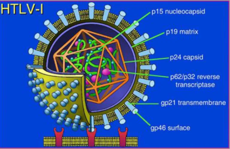
HTLVs are members of the delta-retrovirus family organized in enveloped virions of 80–100 nm in diameter, with two copies of a single stranded positive sense RNA genome (Figure 3). HTLV preferably infects CD4+ and CD8+ T cells, but presents a tropism for other cell types in vivo, including CD25+ lymphocytes, monocytes and B-lymphocytes, macrophages, dendritic cells, megakaryocytes as well as glial cells (astrocytes and microglial cells) (Koyanagi et al., 1993).
Figure 3. Schematic representation of the human T-cell leukemia virus
27
HTLV-1 spreads efficiently between T-cells via a tight organized cell-cell contact known as the virological synapse. Two types of cell-cell contacts have been identified to be critical for HTLV-1 transmission: tight cell-cell contacts and cellular conduits (Gross et al., 2016). In both cases, viral particles are transmitted in confined areas protected from the immune response of the host. The HTLV infection begins with a mature virion expressing the surface unit of the viral protein envelope (Env) and binding to a specific cellular receptor (Figure 4). HTLV-1 and HTLV-2 use the cellular receptors glucose transporter-1 (GLUT1) and neuropilin-1 (NRP-1). HTLV-1, differently to HTLV-2, also utilizes heparin sulfate proteoglycan (HSPG). When an infected cell comes into contact with an uninfected cell, a microtubule-organizing center (MTOC) is polarized at cell–cell junctions, termed virological synapses, impacting on the localization of HTLV Gag, Env and the genomic RNA. The formation of the virological synapses occurs when the intracellular adhesion molecule-1 (ICAM-1) is engaged by its ligand lymphocyte function-associated antigen 1 (LFA1) (Nejmeddine et al., 2009). When the viral RNA genome is delivered into the cytoplasm, it undergoes reverse transcription to convert the ssRNA molecule into dsDNA. The dsDNA translocates into the nucleus and integrates into the host cell genome, where the provirus is then transcribed by host RNA polymerase II. Once viral mRNA is transported into the cytoplasm the viral proteins are translated by the cell translation machinery. The structural proteins Gag, Pol and Env, together with the viral genomic RNA, assemble at a virus budding site along the plasma membrane forming an immature virus particle. When the budding particles are released from the plasma membrane the viral protease cleaves the viral polyproteins inducing a maturation of the virus particle (Martin et al., 2016). During primary infection, HTLV-1 has a period of active replication. The subsequent proliferation occurs mainly through clonal expansion of infected cells or by viral synapses. HTLV-1 integration into the host genome shows associations with specific transcription factor binding sites (Melamed et al., 2013), especially STAT1 and p53, and specific sites upstream of certain proto-oncogenes that are associated with ATL (Cook et al., 2014).
28
Figure 4. HTLV life cycle. Infectious HTLV-1 virion interacts with the target cell surface receptors GLUT1/HSPG/NRP-1 and fuses to the cell membrane (A). Following fusion, the viral genomic RNA is delivered into the cytoplasm ( B), and undergoes reverse transcription to convert the gRNA into dsDNA (C). The dsDNA is then integrated into the host genome (D, E, F). The provirus is then transcribed by cellular RNA polymerase II (G), as well as post-transcriptionally modified (H). Both full-length and spliced viral mRNAs are exported from the nucleus to the cytoplasm (I). The viral proteins are then translated (J), and the structural proteins transported to the plasma membrane along with of the gRNA genome (K) where they assemble to form an immature virus particle (L). The budding particle releases from the cell surface (M), and undergoes a maturation process (N) (Martin et al., 2017).
1.5 HTLV-1 and HTLV-2 Genomic Organization
HTLV-1 and HTLV-2 have a similar genomic structure (Figure 5) and share approximately 70% nucleotide sequence homology (Romanelli et al., 2013). HTLV proviral genome, like other retroviruses, encodes the gag (structural and core proteins), pro (protease), pol (reverse transcriptase), and env (envelope
29
glycoproteins) genes flanked by the long terminal repeat (LTR). The LTRs contain the U3, R and U5 regions at both the 5’- and 3’-ends, serving as promoters and regulators of viral gene expression. The translation of the polypeptide Gag is followed by cleavage into the 19 kDa matrix protein (p19), 24 kDa capsid protein (p24) and 15 kDa nucleocapsid protein (p15). The poly-protein precursor Env is cleaved into the 46 kDa surface glycoprotein (gp46) and 21 kDa transmembrane protein (p21). In addition, the HTLVs genome produces accessory and regulatory genes corresponding to the open reading frame (ORF) pX, which is located between env and the 3’-LTR.
1.5.1 The pX region
The HTLV positive strand of the pX region encodes the regulatory proteins Tax and Rex by the same doubly-spliced mRNA in two separate but overlapping reading frames, and the accessory proteins.
The HTLV negative strand encodes HTLV-1 basic leucine zipper factor (HBZ) protein, and HTLV-2 antisense protein (APH-2), for HTLV-1 and HTLV-2, respectively.
1.5.1.1 The accessory proteins
The HTLV accessory genes contribute to the regulation of viral gene expression and the evasion of the host’s immune response. The p30Tof/p28, p12/p10, and p21Rex/tRex proteins, in HTLV-1 and HTLV-2, respectively, are considered to be homologous based on their structure and functional properties. Among the other accessory proteins p13 and p8 are unique to HTLV-1, and p11 is peculiar to HTLV-2 (Ciminale et al., 2013). HTLV-1 p30Tof and HTLV-2 p28 play an important role in viral latency, sequestering the tax/rex mRNA in the nucleus. p12 also contributes to hinder lysis of HTLV-1-infected cells by CTL, reducing the expression of the β and γc chains of the interleukin-2 receptor (IL-2R), and of
MHC-I (Mulloy et al., 1996, Johnson et al., 2001). The p12 protein is cleaved in p8, which increases cell-to-cell viral transmission through the formation of
30
intercellular conduits (Prooyen et al., 2010a, b). In analogy to p12, HTLV-2 p10 binds the MHC heavy chain; however, p10 does not bind the IL2R β chain. HTLV-1 p13 influences both the turnover of infected cells and the balance between viral latency and productive infection (Silic-Benussi et al., 2010). The function of HTLV-2 p11 is still unclear; however, it has been demonstrated to bind the MHC heavy chain (Johnson et al., 2000).
1.5.1.2 Rex
Rex is the major factor involved in the post-transcriptional regulation; it binds viral mRNAs that contain cis acting sequences termed Rex response element (RxRE) at the R region of the viral LTR. The low expression of Rex during the early stages of the viral gene transcription results in the production of tax, rex,
p30, p12, p13 and hbz mRNAs. In the late stages of transcription, the expression
levels of Rex become higher and its splicing activity is reduced encoding the singly spliced (env) and unspliced (gag-pro-pol) mRNAs, which are then transported to the cytoplasm and translated into enzymes and structural proteins. Recent evidences demonstrate the presence of three alternatively spliced transcripts coding for novel Rex isoforms in primary samples from infected patients (Rende et al., 2015). These isoforms exhibit activities comparable to canonic Rex in terms of HTLV-1 protein expression regulation. However, from the analysis of kinetic expression of Rex, it has been observed that the early expression of dicistronic tax/rex mRNAs is followed by the expression of the monocistronic rex mRNAs, resulting in a prolonged duration of Rex function. The resulting pattern of viral gene expression is important to temporally restrain the expression of highly immunogenic viral epitopes (e.g. Tax, Gag, Env), thus favoring viral persistence and immune response escape (Rende et al., 2015).
31
Figure 5. Schematic representation of HTLV-1, HTLV-2, HTLV-3, and HTLV-4
genomic organization. Green colored boxes indicate ORF encoding regulatory proteins. Dark orange colored boxes indicate ORF encoding auxiliary proteins. Light orange colored boxes indicate putative ORF deduced by genomic sequence analyses (Romanelli et al., 2013).
32 1.6 HTLV Tax oncoproteins
1.6.1 Protein Structure of Tax-1 and Tax-2
The principal role of Tax during viral replication is to activate the LTR promoter transcription through a process that involves the recruitment of CREB/ATF complexes in the U3 region. Tax is recognized as the main viral oncoproteins necessary for the initial steps of T-cell transformation by HTLV. Tax-1 controls many cellular pathways implicated in cell survival and proliferation, directly interacting with components of the PI3K, AKT, MAPK, TGFβ, SRF and NF-κB pathways (Romanelli et al., 2013).
The tax gene is highly conserved between all four genotypes and different subtypes of the HTLV (Romanelli et al., 2013). The structural and functional domains of Tax-1, Tax-2, Tax-3, and Tax-4 are shown in Figure 6. The most characterized Tax-2 protein is the Tax-2B subtype that shares 75% nucleotide sequence homology and 85% amino acid sequence similarity with Tax-1. The N-terminal region of all Tax proteins contains an ATF/CREB-activating domain and two functional regions involved in CREB-binding protein (CBP)/p300 binding in the C-terminal region, which are required for HTLV transcription regulation, interaction with proteins involved in transcription, cell cycle progression, and cell signaling regulation (Rende et al., 2012). A nuclear localization signal (NLS) is present within the first 60 aa in Tax-1, Tax-3 and Tax-4, whereas Tax-2 possesses a nuclear localization determinant (NLD) within the first 42 aa (Romanelli et al., 2013). All Tax proteins contain a nuclear export sequence (NES) that is located at aa position 189–202 in Tax-1 and Tax-2 (Alefantis et al., 2003; Chevalier et al., 2005). The main characteristic that distinguishes the Tax-1 and Tax-2 is that only Tax-1 presents two leucine zipper-like motif regions (LZRs) at the 116-145 aa and 213-248 aa positions, whereas 2 does not. These regions are required for Tax-1 to activate the non-canonical NF-κB pathway through interaction with the pTax-100 factor (Shoji et al., 2009). Tax-1 and Tax-3, in contrast to Tax-2 and Tax-4 are characterized by the presence of a PDZ-binding motif (PBM) at the C-terminal region (Chevalier et al., 2006).
33
Figure 6. Structural and functional domains of the Tax proteins (Romanelli et al.,
2013).
1.6.2 Cellular Localization of Tax-1 and Tax-2 Proteins
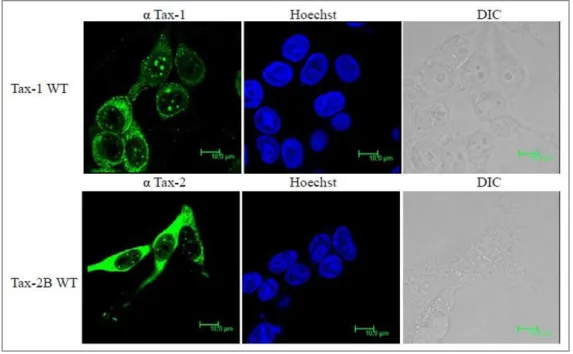
Tax-1 prevalently localizes into the nucleus in speckled structure or nuclear bodies (NBs) that are site of transcriptional activity (Figure 7). Promyelocytic leukemia (PML) protein–containing nuclear bodies also have been functionally linked to transcription, of viral genes and other nuclear processes such as RNA processing, transport, and RNP assembly (Mikecz et al., 2000). These unique nuclear structures containing Tax, also include NF-κB p50 and p65 and participate in the Tax-mediated activation of gene expression via the NF-κB pathway (Bertazzoni et al., 2011). In the cytoplasm Tax colocalizes with several host factors, among them we have observed that, Tax relocalizes p65 to cytoplasmic dotted structures which include the NF-κB factors TAB2, NEMO and the calreticulin (Avesani et al., 2010; Turci et al., 2012).
Differently from Tax-1, Tax-2 localizes predominantly in the cytoplasm of the HTLV-2 immortalized or transformed infected T-cells (Figure 7) (Marteens et al.,
34
2004) and in nuclear bodies (Turci et al., 2009). The domain between 89-113 aa confers to Tax-2 the property to accumulate in the cytoplasm.
Comparative analyses of Tax-1 and Tax-2 post-translational modifications highlighted the functional contribution of ubiquitination and sumoylation to the intracellular localization of Tax and its ability to activate NF-κB (Romanelli et al., 2013). Specific lysines are targeted of sumoylation and ubiquitination in Tax-1 and Tax-2 (lysines from K1 to K10 - Figure 6). Lysines K6 and K8, which are highly conserved in all Tax proteins, are critical for NF-κB activation.
Figure 7. Comparison of Tax-1 and Tax-2 subcellular localization (Bertazzoni et al., 2011).
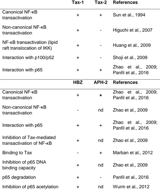
The main structural differences between Tax-1 and Tax-2 reflect different protein interactions, intracellular localization, but also transformation abilities, as described in Table 2. Tax-2, compared to Tax-1, is less efficient in the induction of micronuclei formation (Semmes et al., 1996b) and do not alter the expression of the cdk inhibitors p21 and p27 involved in the control of the cell cycle (Tripp et al., 2005).
35
Table 2. Summary of main functional and structural differences between Tax-1 and
Tax-2 (Romanelli et al., 2013).
Tax-1 Tax-2a References
Transactivating activity Higherb Lowerb [Semmes et al. 1996a]
Transformation capacity Higher Lower [Endo et al. 2002] Micronuclei formation + − [Semmes et al. 1996b]
Cell cycle arrest + − [Tripp et al. 2005]
Hematopoiesis suppression + − [Kubota et al. 1994] Reduction of histone gene
expression + − [Ego et al. 2002; Harrod et al. 2000] Inhibition of p53 functions Higher Lower [Mahieux et al. 2000;
Meertens et al. 2004; Jeong et al. 2005; Calattini et al. 2006] Total viral mRNA expression Higher Lower [Li and Green 2007] Proinflammatory cytokine
expression Higher Lower [Banerjee et al. 2007] Presence of PDZ motif + − [Feuer and Green
2005] Interaction with PDZ binding
proteins +
− [Higuchi and Fujii 2009]
Interaction with p100 + − [Shoji et al. 2009] Preferential cellular localization Nucleus Cytoplasm [Turci et al. 2009] NF-ĸB transactivation + + [Chevalier et al. 2012] NF-ĸB transactivation (lipid raft
translocation of IKK) + − [Huang et al. 2009]
In vitro CK2 phosphorylation + − [Bidoia et al. 2010]
Oligo-sumoylation + − [Turci et al. 2009]
Nuclear bodies Larger Smaller [Turci et al. 2009] Ubiquitination and sumoylation + + [Zane et al. 2012] Nuclear localization + + [Calattini et al. 2006] T-cell immortalization + + [Imai et al. 2013;
Chevalier et al. 2006]
a The properties of Tax-2 include both the results of Tax-2A and Tax-2B reported in
the literature.
b Higher and lower are relative to Tax-1 and Tax-2 comparison.
1.6.3 The Transforming Activity of Tax
The expression of Tax is crucial for promoting human T lymphocyte survival, proliferation and initiating HTLV-1-mediated oncogenesis (Ren et al., 2013). The transforming activity of Tax was first demonstrated using HTLV-1 infectious molecular clone in which the tax gene was disrupted. The wild type molecular clone easily transformed human primary T cells, while the tax mutant molecular clone failed to do so (Akagi et al., 1997). In Tax transgenic mice model, Tax-1
36
induces ATL-like leukemia (Grossman et al., 1995). The transforming activity of Tax is dependent on its ability to activate NF-κB, in fact Tax mutant defective in activating NF-κB fails to transform cells in vitro. In addition, Tax activates the transcription of cellular genes by activating the activating protein-1 (AP-1) transcription. This action is thought to contribute to the deregulated phenotypes and leukemogenesis of T cells infected with HTLV-1 (Gazon et al., 2017). AP-1 is composed of 18 dimeric complexes which included members of four families of DNA-binding proteins. Among these factors, c-Fos, Fra-1, c-Jun, JunB, and JunD genes have been shown to be activated by Tax at the transcriptional level (Fujii et al., 2000; Iwai et al., 2001). Studies conducted on Tax transgenic mouse model highlighted that, functional inactivation of p53 by HTLV-1 Tax is not critical for the initial tumor formation, but contributes to late-stage tumor progression (Portis et al., 2001).
Recent studies have demonstrated that, in addition to Tax, the antisense protein HBZ plays an essential role in oncogenesis by regulating viral transcription and modulating cellular signaling pathways (Satou and Matsuoka, 2012).
1.7 HBZ and APH-2 antisense proteins from HTLV-1 and HTLV-2 1.7.1 Protein Structure of HBZ and APH-2
HBZ and APH-2 are encoded by the minus strand from the 3’ LTR of HTLV-1 and HTLV-2 proviral genome, respectively. HBZ was first characterized for its ability to inhibit Tax-mediated viral transcription (Zhao et al., 2016). The inhibition is carried out by: the interaction between HBZ and CREB-2/CREB, and/or the interaction with CBP/p300, resulting in the inhibition of Tax-mediated recruitment of these transcription factors to the HTLV-1 promoter (Zhao et al., 2016). APH-2 can suppress Tax2-mediated viral transcription by a similar mechanism (Halin et al., 2009).
Two transcripts have been reported to be encoded by HBZ gene: a 206 amino acid protein spliced (sHBZ) and a 209 aa protein unspliced (usHBZ). Evidences demonstrated that the spliced form of HBZ gene is expressed in all ATL cells (Satou et al., 2006). HBZ protein is characterized by three distinct domains: 1) an
37
activation domain which contains two LXXLL-like motifs important for protein-protein interactions at the N-terminus and with transactivating potential; 2) a central domain which contains NLSs; 3) a basic leucine zipper (bZIP) domain for DNA binding. The bZIP domain enables HBZ to hetero-dimerize with cellular bZIP proteins such as CREB2, c-Jun, JunB, JunD, CREB, and ATF3 (Barbeau et al., 2013) (Figure 8). Concerning APH-2, only a spliced mRNA has been identified (Halin et al., 2009). APH-2 is a 183 aa protein, which exhibits less than 30% homology to HBZ. APH-2 contains a non-canonical bZIP region responsible for its interactions with c-Jun and JunB and a C-terminal CREB-binding motif responsible for its interaction with CREB (Halin et al., 2009; Marban et al., 2012). Unlike HBZ, APH-2 is not able to bind p300. The region from aa 102 to 183 is required to form complex between Tax-2B and APH-2 (Marban et al., 2012).
Figure 8. Schematic representations of HBZ and APH-2 functional domains (modified from
Fochi et al., 2018).
1.7.2 Cellular Localization of Antisense Proteins
HBZ exhibits a speckled distribution into the nucleus in infected cells and ATL cells, colocalizing with nucleolar structures (Raval et al., 2015). Three regions distributed in the central domain of the protein are associated with nuclear localization, including a DNA binding domain. HBZ contains a functional NES
38
site in its N-terminal region and it is exported from the nucleus via a CRM1-dependent pathway. Nuclear export of HBZ is essential for the induction mTOR pathway, resulting in the inhibition of the autophagic machinery (Mukai et al., 2014). In a recent study, it has been demonstrated that, in contrast with the HBZ nuclear localization in ATL, HAM/TSP patients show a HBZ localization confined to the cytoplasm, proposing this feature as a possible biomarker for HTLV-1-associated HAM/TSP (Baratella et al., 2017). The HTLV type 2 counterpart, APH-2, localizes in the nucleus, and its distribution in the cytoplasm is more evident compared to HBZ (Halin et al., 2009).
1.7.3 The Oncogenic Potential of HBZ
Approximately 60% of ATL cases lack Tax expression because of genetic and epigenetic changes in the proviral genome of HTLV-1, whereas HBZ is consistently expressed in all ATL cases (Zhao, 2016). The constitutive expression of HBZ promotes the spread of infection (Matsuoka and Green, 2009; Zhao and Matsuoka, 2012). HBZ is not essential for in vitro immortalization and but it is required for efficient infection, T-cell survival, and persistence of the virus in vivo (Arnold et al., 2006; Arnold et al., 2008). Several studies have provided evidences for HBZ role in HTLV-1 oncogenesis, which may be summarized in five actions: a) suppression of viral transcription mediated by Tax; b) promotion of T-cell proliferation; c) suppression of cellular apoptosis and senescence; d) induction of regulatory T-cell differentiation; e) impairment of cell-mediated immunity (Zhao et al., 2016).
A limited number of studies have compared the effects of APH-2 and HBZ on cellular pathways. The main results are summarized in Table 3. The comparative studies between HBZ and APH-2, have indicated that APH-2, unlike HBZ, does not promote T cell proliferation and lymphocytosis (Douceron et al., 2012). These different functions represent interesting aspect to investigate in order to clarify the distinct pathobiologies belonging to HTLV-1 and HTLV-2. Like HBZ, the APH-2 expression has been found to positively correlate with the proviral load in HTLV-2-infected carriers (Douceron et al., 2012). Interestingly, studies conducted in rabbits infected with HTLV-2 have demonstrated that the absence of APH-2
39
results in a high antibody response to the viral antigens and in an increased proviral load (Yin et al., 2012), suggesting that HTLV-2 replicates better when APH-2 is not expressed. HBZ and APH-2 show opposite effect on the modulation of cellular pathways, such as TGF-β and IRF-1 cell signaling and in the activation of AP-1 transcription.
Table 3. Overview of HBZ and APH-2 regulatory functions (Panfil et al., 2016).
Function Finding for:
HBZ APH-2
Expression in HTLV-infected cells Yes (100%) Yes (most but not all cases)
Required for in vitro immortalization No No Required for efficient in vivo infection and
persistence Yes No
HTLV 5′ LTRa transcription Inhibits Inhibits Promotion of T-cell proliferation Yes No
AP-1 transcription Inhibits Activates
JunD transcription Activates Activates TGF-β signaling Activates Inhibits (if
overexpressed) Modulation of classical NF-κB pathway Inhibits Inhibits Modulation of IRF-1 pathway Inhibits Activates aLTR, long terminal repeat.
1.8 Effect of Tax, HBZ and APH-2 on NF-κB Signaling Transduction
As stated above, one of the mayor effects derived by of Tax and antisense protein expression is the deregulation of cellular pathways. The deregulation of the NF-κB pathway has been investigated during the study presented in this PhD thesis. In the following paragraphs, it will be summarized the current knowledge of the involvement of Tax, HBZ and APH-2 on NF-κB signaling.
40 1.8.1 NF-κB Pathways
NF-κB, nuclear factor kappa-light-chain-enhancer of activated B cells, is a family of transcriptional factors that control a large number of cellular processes, such as immune and inflammatory responses, developmental processes, cell proliferation, apoptosis and oncogenesis (Masoumi et al., 2011). It has been demonstrated that aberrant NF-κB plays a crucial role in the initiation and progression of cancer. Many viruses have developed strategies to manipulate NF-κB signaling through the use of viral proteins. NF-κB family proteins consist of five transcriptional factors, p65 (termed also RelA), RelB, c-Rel, κB1 (known also p50) and NF-κB2 (alias p52). The different NF-κB members form various homodimers and heterodimers and once located in the nucleus, transactivate target genes bearing a κB enhancer sequence.
The activation of the NF-κB pathway occurs through two distinct pathways, known as the canonical/classical pathway and the non-canonical/alternative pathway, which involve different upstream, intermediated and effector factors (Figure 9). The classical NF-κB pathway is triggered by pro-inflammatory cytokines, which bind relevant cell surface receptor such as tumor necrosis factor receptor 1 (TNFR1) or Toll-like receptor. The ligand-receptor binding leads to the recruitment of the adaptor proteins as TNF receptor associated factors (TRAFs), tumor necrosis factor receptor type 1-associated DEATH domain protein (TRADD), or receptor interacting protein (RIP) to the cytoplasmic domain of cell membrane receptors. These adaptor proteins assemble a platform to recruit and activate the IKK complex (IκB kinase), composed of two catalytic kinase subunits, IKKα and IKKβ, and the regulatory non-enzymatic scaffold protein NEMO (termed also IKKγ). The activation of the IKK complex lead to the phosphorylation and degradation of the IκB inhibitor, that in physiological conditions, sequester the NF-κB proteins in the cytoplasm. This mechanism results in the nuclear translocation of the p50/RelA transcriptional factors.
In contrast, the non-canonical NF-κB pathway is induced by lymphotoxin B (LT-β), B-cell activating factor (BAF) or CD40 and involves an IKK complex formed by two subunits of IKKα kinases activated by the NF-κB-inducing kinase (NIK). The upstream ubiquitin ligase TRAF3, together with TRAF2 and cIAPs proteins,
41
acts as a negative regulator of the alternative pathway targeting NIK for constant ubiquitination and proteasomal degradation, in unstimulated cells. Upon stimulation, TRAF3 is degraded, allowing NIK to activate the IKKα complex, resulting in the processement of the precursor p100 to yield p52 and the final release of p52/RelB active heterodimer in the nucleus (Durand et al., 2017).
Figure 9. The NF-κB pathway (Masoumi et al., 2011).
1.8.2 Tax and NF-κB Signaling
It is well established that Tax-1 is a potent transactivator of the NF-κB cell signaling and its activity is critical for immortalization and transformation of HTLV-1-infected T cells (Giam et al., 2016). The activation of NF-κB, results in the transcriptional activation of many cellular genes that govern normal growth-signal transduction, such as cytokines and growth factors, including IL-2, IL-6, IL-15, TNF, GM-CSF, proto-oncogenes (c-Myc), and antiapoptotic proteins (bcl-xl) (Hiscott et al., 2001). Tax-mediated activation of the NF-κB transcription
42
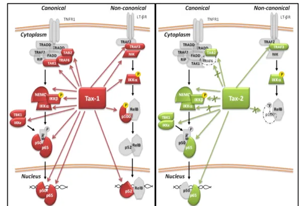
factors, involves two principal molecular mechanisms: the recruitment of Tax in cellular protein complexes (Qu and Xiao, 2011; Bertazzoni et al., 2011) and post-translational protein modifications (Lavorgna and Harhaj, 2014). Comparative studies between Tax-1 and Tax-2 highlighted relevant differences in the activation of NF-κB because of their divergent ability to interact with NF-κB members (Figure 10). Tax-1, unlike Tax-2, triggers the activation of the non-canonical pathway, directly interacting with p100 and promoting its processment to yield p52 (Higuchi et al., 2007; Shoji et al., 2009). Both Tax proteins interact with TAB2 and NEMO/IKKγ stimulating the translocation of the p50/p65 heterodimers into the nucleus, but only Tax-1 interacts with TRAF6, an E3 ligase that triggers the ubiquitination and activation of the downstream NF-κB signaling cascade (Avesani et al., 2010; Journo et al., 2013). We also contributed in a recent study to the identification of complex formation between Tax and two non-canonical IκB kinases, IKKε and TBK1, which act as regulatory factors in IFNα, STAT3 and NF-κB signaling (Shen and Hahn, 2011; Diani et al., 2015). The presence of Tax and TBK1 in lipid raft microdomains along with canonical IκB, supports the role of Tax as molecular crosstalker between the canonical IKKs and TBK1/IKKε, through NF-κB activity (Zhang et al., 2016). It is well known that Tax-1 forms complexes with the ubiquitin-conjugating enzyme Ubc13, NEMO, Tax1 binding protein1 (TAX1BP) and NRP/Optineurin in the membrane lipid rafts microdomain. Interestingly, in these complexes, the cell adhesion molecule 1 (CADM1) acts as a molecular scaffold recruiting Tax-1 and this interaction results in the activation of IKK complex and inactivation of the NF-κB negative regulator A20 enzyme (Pujari et al., 2015). An additional consequence of the Tax reorganization of the component of the lipid raft is the autophagy deregulation. Tax, in fact, acts as molecular crosstalker connecting the IKK complex to the autophagic complexes by interacting directly with Beclin1 and PI3KC3, participating in the assembly of authophagosomes (Ren et al., 2015; Chen et al. 2015). Tax induction of NF-κB also increases the expression of inhibitors of apoptosis, such as the anti-apoptotic c-Flip gene, and genes involved in cell cycle progression (Bangham and Matsuoka, 2017).
43
Figure 10. Schematic representation of Tax-1 (in red) and Tax-2 (in green)
interactions with factors of NF-κB pathway (Romanelli et al., 2013).
Currently, few host factors have been defined to exert a suppressive role on Tax– activation of NF-κB. Among them, the transcriptional regulator of the major histocompatibility complex class II (CIITA), which inhibits the persistent activation of NF-κB in two different manners. In the cytoplasm CIITA retains Tax impairing the nuclear translocation of p65, while in the nucleus, CIITA directly interacts with Tax-1/p65 in nuclear bodies, preventing Tax-1 mediated activation of NF-κB-responsive promoters (Forlani et al., 2013; Forlani et al., 2016). In addition to CIITA, the apoptotic regulator Bcl-3 inhibits p65 nuclear translocation and its DNA binding activity, resulting again in a downregulation of Tax-induced NF-κB activation (Wang et al., 2013). The reduction of Tax-NF-κB activation may also derive by Tax proteosomal degradation induced by host factor interaction (Lavorgna and Harhaj, 2014).
A second mechanism required for Tax-1 and Tax-2 mediated NF-κB activation occurs by post-translational modification, which includes ubiquitination, SUMOylation and phosphorylation. It is well known that Tax phosphorylation is
44
necessary for Tax nuclear translocation in the nuclear bodies where co-localize with p65 (Bex et al., 1997; Turci et al., 2006). The requirement of ubiquitination and SUMOylation is intensively investigated, but not fully clarified. A recent study proposed Tax as a ubiquitin E3 ligase protein, which catalyzes the assembly of mixed polyUb chains, in association with ubiquitin-conjugating enzyme (E2) (Wang et al., 2016). Afterwards, Shibata et al. demonstrated that Tax does not possess E3 ligase activity and they proposed a model in which Tax acts as a scaffold protein recruiting ubiquitin ligases, generating K63- and M1-linked hybrid polyubiquitin chains. In this interacting platform termed by the authors Taxisome, NEMO proteins and Ub-chains lead to the activation of the IKK complex (Shibata et al., 2017).
An additional mechanism that operates in the cells to maintain the NF-κB activation induced by Tax is the positive feedback loop derived by NF-κB target genes. Recently, it has been described that both NF-κB activation and Tax-induced the early growth response protein 1 (EGR1) stability upregulated EGR1, which in turn enhanced constitutive NF-κB activation (Huang et al., 2017). A similar positive loop has been defined by the overexpression of the interleukin receptor IL-17RB. Tax-1 promotes the expression of IL-17RB by NF-κB activation and establishes an IL-17RB-NF-κB feed-forward autocrine loop that maintains persistent NF-κB activation (Lavorgna et al., 2014).
1.8.3 HBZ, APH-2 and NF-κB Signaling
HBZ suppresses the classical NF-κB pathway activation, mediated by both the viral protein Tax and the host transcription factor p65 (Zhao et al., 2009). This inhibition derives by four properties of HBZ: a) the interaction with p65; b) the inhibition of p65 DNA binding; c) the enhanced degradation of p65 through PDLIM2 E3 ubiquitin ligase; d) the reduction of p65 acetylation (Figure 11). All these processes consequently result in the reduction of the expression of several NF-κB target genes (Zhao et al., 2009; Wurm et al., 2012). In this vein, Ma et al. demonstrated in a recent study that HBZ-mediated NF-κB inhibition contributes to the suppression of cyclin D1 gene, an essential regulator of the G1/S phase