ALMA MATER STUDIORUM – UNIVERSITÀ DI BOLOGNA
DOTTORATO DI RICERCA IN BIOINGEGNERIA
Ciclo XXVII
Settore concorsuale di afferenza: 09/G2
Settore scientifico disciplinare: ING-INF/06
Computational Modelling
of Cardiac Electrophysiology:
from Cell to Bedside
Presentata da
Elisa Passini
Coordinatore Dottorato:
Prof. Elisa Magosso
Supervisore:
Stefano Severi, PhD
Revisori:
Prof. Mauro Ursino
Prof. Ronald Wilders
Prof. Esther Pueyo
ALMA MATER STUDIORUM – UNIVERSITÀ DI BOLOGNA
DOTTORATO DI RICERCA IN BIOINGEGNERIA
Ciclo XXVII
Settore concorsuale di afferenza: 09/G2
Settore scientifico disciplinare: ING-INF/06
Computational Modelling
of Cardiac Electrophysiology:
from Cell to Bedside
Presented by
Elisa Passini
PhD Coordinator:
Prof. Elisa Magosso
Supervisor:
Stefano Severi, PhD
Reviewers:
Prof. Mauro Ursino
Prof. Ronald Wilders
Prof. Esther Pueyo
“It is a truth universally acknowledged,
that a single cell in possession of a good membrane,
must be in want of a model…”
Adapted from Pride & Prejudice, by Jane Austen
Table of Contents 9
TABLE OF CONTENTS
TABLE OF CONTENTS ... 9
SUMMARY ... 13
SOMMARIO ... 19
INTRODUCTION... 21
CHAPTER 1 Basic Concepts of the Electrical Activity of the Heart and its Mathematical Modelling ... 23THE CARDIAC ELECTRICAL ACTIVITY ... 25
MODELLING THE CARDIAC ELECTRICAL ACTIVITY... 31
CHAPTER 2 Human Ventricular Action Potential Models: a Literature Review ... 35
MODELS ON THE MARKET ... 37
MODELS COMPARISON ... 40
References ... 47
SECTION I
Extracellular Calcium and Action Potential Duration:
the Fine Balance between L-Type Calcium Current
10 Table of Contents
Elisa Passini
CHAPTER 3
A Novel Markov Model of L-Type Calcium Current to
Explore Inactivation Mechanisms ... 53
Abstract... 55
INTRODUCTION ... 55
METHODS ... 58
RESULTS ... 61
DISCUSSION AND CONCLUSIONS ... 67
CHAPTER 4 Extracellular Electrolyte Changes During Head-Down Bed-Rest: Effects on Action Potential Duration ... 69
Abstract... 71
INTRODUCTION ... 71
METHODS ... 72
RESULTS ... 75
DISCUSSION AND CONCLUSIONS ... 80
References ... 83
SECTION II
Haemodialysis
Therapy
Impact
on
Cardiac
Electrophysiology
87
CHAPTER 5 Human Atrial Cell Models to Analyse Haemodialysis-related Effects on Cardiac Electrophysiology: Work in Progress ... 89Abstract... 91
INTRODUCTION ... 91
CARDIAC CELL MODELLING AND HAEMODIALYSIS ... 93
ATRIAL CELL MODELLING: MATERIALS AND METHODS ... 94
ATRIAL CELL MODELLING: EFFECTS OF HD-RELATED CHANGES ... 101
Table of Contents 11
DISCUSSION AND CONCLUSIONS ... 114
CHAPTER 6 Recurrent Intradialytic Paroxysmal Atrial Fibrillation: Hypotheses on Onset Mechanisms Based on Clinical Data and Computational Analysis... 119
Abstract ... 121
INTRODUCTION ... 121
CASE REPORT ... 122
COMPUTATIONAL ANALYSIS ... 125
INSIGHTS INTO THE MECHANISMS OF INTRADIALYTIC ATRIAL FIBRILLATION ... 128
DISCUSSION AND CONCLUSIONS ... 134
References ... 137
SECTION III
Computational Modelling of Human Hypertrophic
Cardiomyopathy ... 149
CHAPTER 7 Pro-Arrhythmic Mechanisms and Potential Therapeutic Targets in Human Hypertrophic Cardiomyopathy ... 151
Abstract ... 153 INTRODUCTION ... 154 METHODS ... 158 RESULTS ... 166 CONCLUSIONS ... 195 References ... 197
GENERAL CONCLUSIONS ... 199
APPENDIX A ... 207
12 Table of Contents Elisa Passini
APPENDIX B ... 223
LIST OF PUBLICATIONS ... 245
RINGRAZIAMENTI ... 247
ACKNOWLEDGEMENTS ... 249
Summary 13
SUMMARY
Introduction
Heart diseases are the leading cause of death worldwide, both for men and women. However, the ionic mechanisms underlying many cardiac arrhythmias and genetic disorders are not completely understood, thus leading to a limited efficacy of the current available therapies and leaving many open questions for cardiac electrophysiologists.
On the other hand, experimental data availability is still a great issue in this field: most of the experiments are performed in vitro and/or using animal models (e.g. rabbit, dog and mouse), even when the final aim is to better understand the electrical behaviour of in vivo human heart either in physiological or pathological conditions.
Computational modelling constitutes a primary tool in cardiac electrophysiology:
in silico simulations, based on the available experimental data, may help to
understand the electrical properties of the heart and the ionic mechanisms underlying a specific phenomenon. Once validated, mathematical models can be used for making predictions and testing hypotheses, thus suggesting potential therapeutic targets.
Aims
This PhD thesis aims to apply computational cardiac modelling of human single cell action potential (AP) to three clinical scenarios, in order to gain new insights into the ionic mechanisms involved in the electrophysiological changes observed in
vitro and/or in vivo:
The first context is blood electrolyte variations, which may occur in patients due to different pathologies and/or therapies. In particular, we focused on extracellular Ca2+ and its effect on the AP duration (APD).
14 Summary
Elisa Passini
The second context is haemodialysis (HD) therapy: in addition to blood electrolyte variations, patients undergo a lot of other different changes during HD, e.g. heart rate, cell volume, pH, and sympatho-vagal balance.
The third context is human hypertrophic cardiomyopathy (HCM), a genetic disorder characterised by an increased arrhythmic risk, and still lacking a specific pharmacological treatment.
The general aim of this PhD thesis can therefore be referred to as “From Cell to Bedside”, meaning to correlate single cell electrophysiology with some specific clinical patient phenotypes, by using in silico techniques to highlight the mechanisms more likely contributing to them at the ionic level.
Methods
Many computational AP models published in literature, both atrial and ventricular, have been considered during this thesis. These models have been modified when needed, to improve their suitability to specific conditions, not originally taken into account during their development/validation. In particular, a new hybrid ventricular model has been developed, by using an existing one as basis and changing part of its original formulation.
All models provide a full description of the ionic currents and intracellular dynamics underlying the cardiac cell AP, represented by means of ordinary differential equations solved by using a variable order solver, based on numerical differentiation formulas. The models have been implemented mostly in Matlab (Mathworks Inc.) and CHASTE (Cancer, Heart and Soft Tissue Environment, University of Oxford), an open-source software specifically developed for cardiac modelling and based on C++.
In addition to the traditional single cell modelling techniques, the population of models (POMs) approach has been considered in the last Section of the thesis: instead of a single AP model, representative of the average cell behaviour, simulations have been run on thousands of models at the same time, hence representing the effect of biological variability.
Summary 15
Results Outline
Here below is a summary of the main results for the three different conditions investigated in this PhD thesis, each one illustrated in a different Section:
In Section I the effects of extracellular Ca2+ ([Ca2+]o) changes on the human
ventricular AP have been investigated: an increase of [Ca2+]o shortens AP and a
decrease of [Ca2+]o lengthens it. Both AP duration (APD) increase and decrease
are associated with a higher arrhythmic risk; therefore this dependence has to be considered in all the situations in which [Ca2+]o variations may occur. However,
most of the AP models currently available in literature do not reproduce properly the effect of [Ca2+]o changes on AP.
In Chapter 3 a new Markov model for the L-type Ca2+
current has been proposed and integrated into the O’Hara-Rudy human ventricular AP: this hybrid model well simulates the inverse APD-[Ca2+]o dependence, not
reproduced by the original one. Its development has been driven by the hypothesis that Ca2+-dependent inactivation is usually underestimated in AP models: our simulations confirmed the crucial role of this mechanism in determining the APD-[Ca2+]o relationship. Therefore, the hybrid model can be
applied to clinical conditions in which blood electrolyte concentrations change overtime, to evaluate the corresponding changes at the AP level and potential pro-arrhythmic effects.
In Chapter 4, the hybrid model described in Chapter 3 has been used to investigate the impact of blood electrolyte changes measured during bed-rest. Bed-rest is a ground-based experiment used to simulate on Earth the effect of microgravity on the human body, thus assessing the possibly increased arrhythmic risk for astronauts during space flights. Simulation results in single cells and 1D cable were compared with ECG data analysis, providing evidence of a biphasic trend in repolarisation: RT intervals decrease during bed-rest and increase afterwards. The electrolyte concentrations have been used as model inputs to simulate volunteer conditions before, during, and after bed-rest. Simulated AP and pseudo-ECG were both in agreement with the recorded ECG, suggesting that electrolyte variations occurring during bed-rest may be responsible for the repolarisation changes, and thus correlating the
16 Summary
Elisa Passini
electrophysiological phenotype with the modification at the cellular level. This project has been done in collaboration with Prof. Enrico G Caiani (Department of Electronics, Information and Bioengineering, Politecnico di Milano, Italy) and the experimental data have been acquired by the European Space Agency (ESA).
Section II investigates the impact of haemodialysis (HD) therapy on the electrical activity of the heart, focusing on the electrolyte variations occurring during a regular HD session and evaluating the corresponding changes at cellular level. Here, human atrial AP models have been considered, since atrial fibrillation (AF) incidence is high in end-stage renal disease (ESRD) patients. Chapter 5 presents a benchmarking of all the atrial AP models currently
available in literature, with respect to their suitability to the HD context. All models have been tested for variations in cell volume, extracellular electrolyte (K+, Ca+ and Na+) and acetylcholine concentration, computing a set of AP and Ca2+-transient biomarkers to compare simulation results with the expected behaviour, based on literature review. Some models proved to be more appropriate than others for single aspects, butall of them showed some drawbacks. Suggestions have been given for the potential development of a new atrial model, expected to reproduce properly all the HD-induced effects on human atrial AP.
Chapter 6 illustrates the case study of an ESRD patient showing recurring paroxysmal AF during HD therapy. Experimental data, i.e. blood electrolyte concentrations and heart rate, have been used to reproduce in silico the patient pre-HD and pre-AF conditions at cellular level, using a modified version of the Courtemanche atrial AP model, described in Chapter 5. By integrating simulation results and clinical observations, we formulated a new hypothesis about the mechanisms involved in AF onset during HD: AF episodes are induced by the presence of a trigger (ectopic beats) that acts upon an acute substrate induced by intra-dialytic electrolyte variations, especially K+ (increased AP depolarization time and shortened refractory period), on the background of autonomic nervous system changes. This project has been
Summary 17
done in collaborations with Simonetta Genovesi, MD and Antonio Vincenti, MD (Department of Health Sciences, University of Milano Bicocca, Italy). In Section III (Chapter 7) the population of models (POMs) approach has
been used to study the electrical remodelling occurring in human hypertrophic cardiomyopathy (HCM), in order to identify possible therapeutic targets for this disease. The POMs approach accounts for inter- and intra- subjects variability, which indeed seems to play an important role in HCM and which cannot be taken into account when considering a single AP model, representative of the average cellular behaviour.
As first, a control (CTRL) population of models has been built to reproduce an experimental dataset of AP and Ca2+ transient (CaT) biomarkers, acquired on human single cells from failing non-hypertrophic controls.
Then, a HCM population has been developed by applying to the CTRL population the electrophysiological changes measured in diseased cells, together with a few novel hypotheses based on literature review. The simulated HCM biomarkers resulted to be in agreement with the experimental ones, and the contribution of each single electrophysiological change to the global HCM phenotype has been evaluated.
The occurrence of repolarisation abnormalities, e.g. early after-depolarisations (EADs) and repolarisation failure (RF), has been investigated in the HCM population and the ionic mechanisms more likely to be responsible for them have been identified.
Since specific compounds are already available, the Late Na+
current (INaL)
and the Na+/Ca2+ exchanger (INCX) have been considered as potential
therapeutic targets. Both INaL and INCX selective blocks showed an
anti-arrhythmic effect, partially reversing the HCM phenotype and suppressing repolarisation abnormalities. The combination of both proved to be even more effective, suggesting the simultaneous block of INaL and INCX as a
18 Summary
Elisa Passini
This project has been done as a visiting student in the Department of Computer Science, University of Oxford (UK), under the supervision of Prof. Blanca Rodriguez, Alfonso Bueno-Orovio, PhD and Ana Mincholé, PhD, and in collaboration with Raffaele Coppini, MD and Elisabetta Cerbai, MD (NeuroFarBa Department, University of Florence, Italy). To summarize, the results presented in this thesis have improved the understanding of the ionic mechanisms underlying electrophysiological properties related to arrhythmic risk in specific clinical contexts, thus confirming computational modelling as a valuable tool in cardiac electrophysiology, especially when fully integrated with experimental data.
Sommario 19
SOMMARIO
Le malattie cardiache e cardiovascolari sono ad oggi la causa principale di morte nel mondo. Tuttavia, i meccanismi ionici responsabili di aritmie o di altre malattie cardiache non sono ancora del tutto conosciuti: questo spesso porta a una minore o mancata efficacia delle terapie attualmente disponibili, e lascia numerose domande aperte per gli elettrofisiologi. Inoltre, la difficoltà di acquisizione dei dati sperimentali rimane ancora uno dei problemi più grandi in questo campo. Infatti la maggior parte dei dati vengono raccolti in vitro e/o utilizzando modelli animali come coniglio, ratto o cane, sebbene l’obiettivo ultimo sia quello di una più completa comprensione del comportamento elettrico del cuore in vivo e nell’uomo, in condizioni sia fisiologiche sia patologiche.
In questo contesto, la modellistica computazionale costituisceuno strumento indispensabile: infatti, le simulazioni in silico permettono di superare, almeno in parte, i limiti sperimentali, e di investigare i meccanismi ionici alla base di specifici fenomeni a diversi livelli (singola cellula, tessuto, intero cuore). Una volta validati sui dati sperimentali, i modelli matematici possono essere dunque utilizzati per fare predizioni, testare ipotesi e valutare l’efficacia di eventuali interventi farmacologici. Lo scopo di questa tesi di dottorato è stato quello di applicare tecniche di modellistica matematica a problemi di elettrofisiologia cardiaca, in particolare utilizzando modelli di potenziale d’azione (PA) umano in tre diversi contesti:
Variazioni del livello di elettroliti (Na+, K+ e Ca2+) nel sangue, che possono verificarsi nei pazienti a causa di diverse patologie e/o terapie, con possibili conseguenze pro-aritmiche. Sono state considerate in particolare variazioni di Ca2+ e il loro effetto sulla durata del PA ventricolare, aspetto solitamente trascurato nei modelli a oggi disponibili. È stato sviluppato un nuovo modello di PA, integrando una nuova formulazione per la corrente di Ca2+ in un modello già esistente: il modello ibrido così ottenuto costituisce uno strumento importante per esplorare i contesti clinici in cui le variazioni elettrolitiche possono verificarsi. Come esempio applicativo, sono stati analizzati dati sperimentali
20 Sommario
Elisa Passini
raccolti dall’Agenzia Spaziale Europea (ESA) per valutare l’eventuale rischio aritmico per gli astronauti durante i voli nello spazio. Questo studio è stato svolto in collaborazione con il Prof. Enrico Caiani (Dipartimento di Elettronica, Informazione e Bioingegneria, Politecnico di Milano, Italia).
Variazioni elettrofisiologiche che avvengono durante la terapia dialitica. In questo contesto non si modificano soltanto le concentrazioni elettrolitiche ma anche la frequenza cardiaca, il volume cellulare e l’attività simpato-vagale. Dal momento che la fibrillazione atriale (FA) ha un’incidenza elevata nei pazienti in dialisi, sono stati considerati modelli di PA atriale, confrontando le loro caratteristiche e la loro applicabilità in questo contesto. Come esempio, è stato analizzato il caso di una paziente che presentava FA parossistica in ogni seduta dialitica. Questo studio è stato svolto in collaborazione con la Dott.ssa Simonetta Genovesi e il Dott. Antonio Vincenti, (Dipartimento di Scienze della Salute, Università degli Studi di Milano-Bicocca, Italia).
Cardiomiopatia ipertrofica (HCM), una malattia genetica caratterizzata da un alto rischio aritmico e causa principale di morte cardiaca improvvisa nei giovani adulti (<35 anni). Per tener conto della variabilità biologica, che sembra avere un ruolo determinante in questa patologia, soprattutto nella risposta individuale a un possibile trattamento farmacologico, è stato utilizzato un nuovo approccio computazionale: le popolazioni di modelli. Questo studio è stato svolto durante un periodo di ricerca all’estero presso il Dipartimento di Computer Science dell’Università di Oxford, sotto la supervisione della Prof. Blanca Rodriguez, il Dott. Alfonso Bueno-Orovio, e la Dott.ssa Ana Mincholé.
Il filo conduttore di questa tesi può quindi essere riassunto dall’espressione “Dalla Cellula al Paziente”. Non a caso, in tutti gli scenari analizzati, lo scopo principale è stato quello di correlare i cambiamenti elettrofisiologici a livello cellulare con il fenotipo osservato a livello macroscopico nel paziente, per identificare i meccanismi ionici che vi contribuiscono e suggerire di conseguenza possibili approcci farmacologici. I risultati ottenuti hanno confermato l’importanza dei modelli matematici come supporto all’elettrofisiologia cardiaca, specialmente quando l’approccio in silico viene utilizzato in sinergia con quello in vitro.
INTRODUCTION
___________________________________________________________________
CHAPTER 1
___________________________________________________________________Basic Concepts of the Electrical Activity of the
Heart and its Mathematical Modelling
Introduction – Chapter 1 25
THE CARDIAC ELECTRICAL ACTIVITY
The heart is situated slightly to the left of the middle of the thorax, underneath the sternum, between the lungs. It is supported inside a structure known as the pericardial sac, a double membrane structure containing a serous fluid to reduce friction during heart contractions.
There are four major chambers in the heart: the larger, lower, thicker walled chambers are the ventricles, while the smaller, upper, thinner chambers are the atria. The bottom of the ventricles is called the apex and their top part is known as the base. Both the atria and the ventricles are separated into independent left and right halves by the septal wall. The function of the right atrium is to collect deoxygenated blood from the body. After contraction of the atria, this blood is passed to the right ventricle and pumped into the lungs (pulmonary circulation) to produce the gas exchange between carbon dioxide and oxygen. The re-oxygenated blood from the lungs is then collected in the left atrium, from where it moves to the left ventricle which pumps it out to the body. Since the right ventricle only pumps blood through the pulmonary circulation system of the lungs, whilst the left ventricle pumps blood to the rest of the body, the left ventricle is considerably thicker than the right.
Mechanical contraction of the heart is caused by the electrical activation of myocardial cells. The electrical activation sequence (Figure 1.1, left side) of the human heart starts at the sinoatrial node, located in the right atrium at the superior vena cava. This node consists of specialized muscle cells which are self-excitatory, pacemaker cells, able to generate an electrical impulse at a rate of about 70 per minute. From the sinoatrial node, the wave of electrical activation propagates throughout the atria, but cannot propagate directly across the annulus of separation between the atria and the ventricles. The atrioventricular node, located at the boundary between the atria and ventricles, is the only conducting path from the atria to the ventricles in a normal heart. Conduction velocity through the atrioventricular node is considerably delayed in order to supply enough time to the atria to fill the ventricles with blood before the beginning of their contraction. Propagation then proceeds through a specialized conduction system, called the bundle of His. After a short distance, it separates into two bundle branches propagating along each side of
26 Introduction – Chapter 1
Elisa Passini
the septum, constituting the left and right bundle brunches. Both branches then continue to subdivide into a complex network of fibres called the Purkinje fibre network, which spreads across the endocardial surface and into the sub-endocardial region of both ventricles. The fast conduction through the bundle branches and Purkinje fibres causes the entire endocardium to be excited almost simultaneously, although apical regions contract first and the basal regions are usually the latest to be excited.
Figure 1.1: Electrophysiology of the heart. The different action potentials for each of the specialized
cells found in the heart, and their contribution to the total electrocardiogram waveform are shown (modified from [1]).
Cardiac Action Potential and ECG
Cardiac muscle cells or myocytes are approximately flattened tubes, about 80-100 μm long in human ventricular tissue, with elliptic cross sections with a major axis of 10-20 μm. They are arranged in discrete layers of fibres called sheets, roughly parallel to the heart surfaces (epicardium and endocardium), with the fibre axis continuously rotating counter clockwise from epicardium to endocardium in a range of 100°-120° as viewed from the top of epicardium. Each cardiac muscle cell
Introduction – Chapter 1 27
is bounded by a thin (5-7 nm) phospholipid membrane or sarcolemma. This membrane encapsulates a small volume that is known as the intracellular space, whereas the extracellular or interstitial space is therefore defined as the space that lies outside the sarcolemma. The membrane is heterogeneous, with numerous large, complex proteins embedded within it, combined to form small pores in the cell membrane. Under most circumstances these pores are selectively permeable, allowing the pass of only specific ions through the membrane and only under certain conditions, reason why they are commonly called ion channels.
The main ions that are of interest in cardiac electrophysiology are Na+, K+, Ca2+ and Cl−. At resting, the intracellular and extracellular concentrations of each ion are substantially different. In principle, this difference on concentrations would produce a chemical force that would make ions to flow down their concentration gradient to create a uniform distribution at both sides of the membrane. Nevertheless, different ionic concentrations also imply a net electrical charge difference between both sides of the membrane, what causes the establishment of an electrical gradient that acts to oppose the chemical gradient, thus allowing intra- and extracellular concentrations to be different. Consequently, at rest the cell membrane maintains a net membrane potential, which for cardiac muscle cells generally is between -90 and -80 mV, and the cell membrane is said to be in a polarized state.
However, under electrical excitation of the cell this electrochemical equilibrium is broken: this allows ions to flow through those ion channels to which they are permeable, if opened. Any positive increase of the transmembrane potential towards zero is therefore known as depolarization, while the term repolarization refers to the returning of the cell to its negative resting state.
Small perturbations in the potential difference across the cell membrane produce only a passive, linear response of the cardiac cell, followed by the returning of the transmembrane potential towards its resting state. On the contrary, when a sufficiently large stimulus is applied (i.e. able to rise the transmembrane potential above the threshold potential), an active, non-linear response, known as the action potential (AP) will be elicited.
Depending on the region of the heart, the cardiac AP may have different shapes and properties (Figure 1.1, right side): all these differences, together with the
28 Introduction – Chapter 1
Elisa Passini
particular activation sequence described above, are responsible for the macroscopical electrical activity of the heart, as measured in the electrocardiogram (ECG). In a conventional 12 lead ECG, ten electrodes are placed on the patient's limbs and on the surface of the chest. The overall magnitude of the heart's electrical potential is then measured from twelve different angles and recorded over a period of time. In this way, the overall magnitude and direction of the heart's electrical depolarization is captured at each moment throughout the cardiac cycle. The graph of voltage versus time produced by this non-invasive medical procedure, and referred to as ECG, is characterised mainly by 3 waves: a P wave (atrial depolarization), a QRS complex (ventricular depolarization) and a T wave (ventricular repolarization).
Despite these differences in shape and properties, the cardiac AP it is mainly characterised by 5 different phases, related to the opening/closing of the different ion channels (mainly Na+, Ca2+ and K+), as shown in Figure 1.2:
Phase 0 (upstroke) when the threshold is reached, there is a rapid influx of Na+ through the Na+ channels, creating the fast Na+ current (INa) who rise the
membrane potential up to positive values.
Phase 1: the Na+ channels close, while K+ channels open. Throughout the whole action potential duration there are different K+ currents that tend to bring the transmembrane potential back to its resting value. In this phase, the main contribution is the one of the transient outward K+ current, which causes a small deflection in the membrane voltage, called “notch”.
Phase 2: the outward K+ currents are counteracted by the opening of Ca2+ channels, responsible for the “plateau phase”, in which the membrane potential decreases very slightly. The duration of this phase may vary from one cell to the other, e.g. it is very short in atrial cells and longer in ventricular ones.
Phase 3: when the Ca2+ channels close, the “rapid” and “slow” delayed rectifier K+ currents (IKr and IKs respectively) play the major role, bringing the
transmembrane potential back to its resting value.
Phase 4: the cell is in its resting state; the resting membrane potential is depending mostly on the inward rectifying K+ current (IK1).
Introduction – Chapter 1 29
Figure 1.2: Representative action potential trace of a human ventricular endocardial cell.
Different cell types may have different ionic currents and formulations, causing the differences in shapes, e.g. ventricular vs atrial cells. An example is given in Figure 1.3, by comparing atrial and ventricular cells.
Figure 1.3: Main differences between atrial (left) and ventricular (right) action potentials, with the
30 Introduction – Chapter 1
Elisa Passini
Arrhythmias and pro-arrhythmic mechanisms
Sudden Cardiac Death (SCD) is a sudden, unexpected loss of heart function: the heart stops beating and blood stops flowing to the brain and other vital organs, causing death if not treated within minutes. Most SCD are caused by abnormal heart rhythms, called arrhythmias, in which heart beat is too fast, too slow or irregular: they are due to problems with the electrical conduction of the heart.
There are different types of arrhythmias: extra beats, either atrial or ventricular, supraventricular tachycardia, which include atrial flutter and atrial fibrillation, ventricular arrhythmias, i.e. ventricular tachycardia or fibrillation, and brady-arrhythmias. Most arrhythmias can be effectively treated, by medications or medical procedures, such as a pacemaker and surgery.
There are many pro-arrhythmic mechanisms which make the heart more vulnerable to arrhythmias. As an example, in Figure 1.4 and Figure 1.5 APD alternans and after-depolarisations (early, EADs and delayed DADs) are shown.
Figure 1.4: Cardiac action potential traces showing APD alternans, i.e. a beat to beat variability in
the AP duration (modified from [3]).
Introduction – Chapter 1 31
MODELLING THE CARDIAC ELECTRICAL
ACTIVITY
Equivalent Electric Circuit of the Membrane
The action potential (AP) represents a transient change of the transmembrane voltage of the cell, and it is the result of all the ionic currents (mostly Na+, K+ and Ca2+) flowing across the membrane. Considering the equivalent electric circuit, the membrane itself can be represented by a dielectric, with a capacitance of about 1µF/cm2, and each ionic current can be represented by a resistor (Figure 1.6).
Figure 1.6: Equivalent electric circuit of cell membrane.
The action potential of a single cell can be then reconstructed by solving the following differential equation, where Vm represents the transmembrane voltage, Cm
the capacitance of the cell and Itot the total current flowing, consisting of the sum of all the different ionic currents (Iion) and the stimuls current (IStim), required from the cell to reach the voltage threshold, thus developing a full action potential.
𝐼
𝑡𝑜𝑡= 𝐼
𝑖𝑜𝑛+ 𝐼
𝑆𝑡𝑖𝑚= 𝐶
𝑚∙
𝑑𝑉𝑚 𝑑𝑡32 Introduction – Chapter 1
Elisa Passini
The Hodgkin and Huxley formalism
The first AP mathematical model was developed by Alan Lloyd Hodgkin and Andrew Huxley, in 1952 [5], to explain the ionic mechanisms underlying the initiation and propagation of neural APs in the squid giant axon. In 1963, they received the Nobel Prize in Physiology or Medicine.
The Hodgkin-Huxley model was developed by performing a series of voltage clamp experiments on giant squid axons, i.e. holding the membrane to a constant voltage value, and measuring the corresponding current flow.
They identified three different contributions to the total current: - Na+ current
- K+ current
- Leakage current (carried by unspecified ions)
The leakage current was formulated simply as a maximal conductance times the corresponding driving force, while Na+ and K+ current formulations included also a voltage-dependent gating mechanism, regulating the channel opening/closing.
Here, gNa = 120, gK = 36 and gL = 0.3 (mS/cm2) are the maximal conductances associated with the Na+, K+ and leakage currents, respectively. ENa = 115, EK = −12
and EL = 10.613 (mV) are the reversal potentials of each ion, according to the
Nernst equation and relative to the resting membrane potential. Gating variables m,
h and n are voltage dependent and their values (always between 0 and 1) describe
the probability for the channel to be in an open state. When their value is 0 the gate is completely close and no current will flow, whereas when the value is 1 the gate is completely open. The leakage current does not have any gating mechanisms, the K+ current has only a gating variable, while the Na+ current has two of them: therefore,
𝐼
𝑁𝑎= 𝑔
𝑁𝑎∙ 𝑚
3∙ ℎ ∙ (𝑉
𝑚− 𝑉
𝑁𝑎)
𝐼
𝐾= 𝑔
𝐾∙ 𝑛
4∙ (𝑉
𝑚− 𝑉
𝐾)
Introduction – Chapter 1 33
the state of the Na+ gate relies on the product of both gating variable, and only when they are both equal to 1 the gate is completely open.
Each gating variable is described by a differential equation:
Here, αn and βn are known as rates and are usually voltage-dependent. Figure 1.7
shows the action potential generated by the Hodgkin-Huxley model, again relative to the resting membrane potential, and the three gating variable traces over time.
Figure 1.7: Transmembrane potential generated by the Hodgkin and Huxley model
(left panel) and the corresponding gating variables over time (right panel).
From Hodgkin-Huxley to Cardiac Models
Starting from Hodgkin and Huxley, AP mathematical models have gained a relevant role in the investigation of cellular electrophysiology. Their possible application to the heart has been soon realized and right from the beginning, cardiac cell modelling allowed to gain insights by predicting phenomena which have been later confirmed experimentally.
The earliest example consists in the pioneering work by Noble, who modified the Hodgking-Huxley model to simulate Purkinje fibres in mammals, identifying the energy-saving properties of the inward rectifier potassium current [6].
Due to the limited availability of human cardiomyocytes for experimental research, most electrophysiological models had been formulated for animals (mouse, guinea pig, rabbit, dog, etc.). However, animal and human cardiomyocytes
𝑑𝑛
34 Introduction – Chapter 1
Elisa Passini
differ in major aspects, such as action potential shape and duration, range of normal heart rates, action potential restitution and relative importance of ionic currents in the action potential generation. As all these factors may influence the mechanism of arrhythmias initiation and dynamics, simulation results obtained with animal models may prove inadequate to represent phenomena observed in human.
In recent years, more and more data on human ionic currents have been gathered from human cardiomyocytes. In addition, by cloning techniques voltage-clamp measurements of human ion channels have been acquired in heterologous cells. As a consequence, new several models have been developed to describe the origins of the human cardiac action potential, an important step towards a wider application in clinical practice.
In 2012, two comprehensive reviews of human atrial models have been published, [7, 8], comparing the different structure and ionic current formulations, and discussing the differences in AP and Ca2+ transient biomarkers.
As for ventricular cells, there is not any comprehensive review which includes the most recently published AP models. Therefore, in the next chapter we present a literature review of the current state of the art in human ventricular AP models.
___________________________________________________________________
CHAPTER 2
___________________________________________________________________Human Ventricular Action Potential Models:
a Literature Review
Introduction – Chapter 2 37
MODELS ON THE MARKET
Human ventricular cells modelling has begun in 1998 with a study by Priebe and Beuckelmann [9] (PB98), aimed at understanding the effects of electrophysiological alterations in heart failure. They used the Luo–Rudy model of guinea pig ventricular myocytes [10] as basis, parameterized anew with available human data, measured in normal and diseased myocytes.
The PB98 model has been the only one available until 2004, when two further models of human ventricular cells were introduced by Iyer et al. [11] (IW04) and Ten Tusscher et al. [12] (TP04). Both these models provide a more detailed description of ionic currents and fluxes, which reflects new insights in channel function understanding as well as the availability of new measurements from human cells and channels. The IW04 model describes the electrophysiology of sub-epicardial cells (Epi), applying Markovian models for most channels: however, this choice lead to a significant increase in complexity and hence computational time required for simulation. In contrast, the TP04 model uses the common Hodgkin-Huxley formulation for all currents, and in addition to Epi also considers sub-endocardial (Endo) and Midwall (M) cells. A comprehensive comparison of PB98, TP04 and IW04 models, including ion currents, action potential morphology and duration, rate adaptation and other properties, has been performed by Ten Tusscher et al. [13]. A revisited version of TP04 has been published by the same group in 2006 [14] (TP06), including more details on intracellular Ca2+-handling and cell compartmentalisation.
Two years later, Bueno-Orovio et al. [15] performed a new comparison of these models, proposing at the same time a minimal ventricular human model, specifically designed to reproduce tissue-level characteristics. An additional comparison of the IW04 and TP04 models as been performed by Niederer et al. [16], highlighting their significant differences in terms of voltage, ionic currents and concentrations during an action potential, although both models aim to represent the same physiological system. These differences can be partially explained by the different experimental data which have been used to characterise them.
38 Introduction – Chapter 2
Elisa Passini
More recently (2010), a model of human ventricular AP has been proposed by Grandi et al. [17] (GB10), using the rabbit model proposed by Shannon et al. [18] as basis, and including new formulations of ionic current densities and kinetics, according to novel human experimental data. With respect to the TP06 model, the GB10 shows a better steady-state AP response to frequency changes and to potassium current blockades. However, the GB10 model does not properly reproduce S1S2 restitution properties nor APD rate adaptation dynamics, as reported
by Carro et al. [19]. Those drawbacks probably have been acquired from the rabbit model used as basis, since S1S2 restitution and APD rate adaptation are notably
different in rabbit with respect to human. Indeed, in a recent review the GB10 model has been even referred to as a rearrangement of a rabbit model rather than a real new human model [20]. In 2011, Carro et al. [19] developed a refinement of the GB10 model (CP11), to rectify these drawbacks. In particular, they reformulated the L-type calcium current dynamics, in order to accurately reproduce S1S2 restitution and APD rate adaptation.
The most recent model of human ventricular cell has been proposed by O’Hara et al. [21] (OR11) in 2011. This model was developed and validated by using an extensive dataset, including many previously unpublished experimental data, from more than 100 undiseased human hearts. Due to the extensive validation on these new data, the authors claimed to have substantially increased human specific model accuracy: in fact, the model was shown to reproduce several physiological behaviours and drug blocks. Moreover, the effects of Ca2+/Calmodulin-dependent protein kinase II (CaMKII) were incorporated as well.
In Table 2.1 the published models of human ventricular electrophysiology available in literature are listed, together with a non-exhaustive reference to their extensions and/or refinements. Indeed, cardiac computational modelling has reached the stage in which many of the more recent works are focused on ‘fixing’ problems in previous models, as soon as new and better data become available and modellers discover possible applications for which the published models are not well-suited [20]. We chose to neglect minimal/reduced models, in which a single mathematical process represents multiple channel properties, since they have been developed mainly for multicellular simulations, while in this thesis we are mostly using single cells.
Introduction – Chapter 2 39
Table 2.1: Computational models of human ventricular cell electrophysiology.
Model Model extensions Cell types Citations Scopus 2014 Comments Ref # Priebe & Beuckelmann (PB98) 1998 n.s. 244
First human model, but largely based on animal data. Formulations for normal and failing hearts.
[9] Seemann et al. 2003 Endo, M, Epi 19 Focus on regional heterogeneity [22] Iyer et al.
(IW04) 2004 Epi 130 Joint first human models
prevalently based on human data. [11] Ten Tusscher et al. (TP04) 2004 Endo, M, Epi 477 [12] Ten Tusscher et al. (TP06) 2006 Endo, M, Epi 217 More detail on intracellular calcium handling [14] Fink et al. 2008 Epi 40 From TP06, new formulations for IK1 and
HERG, [23] Grandi et al. 2009 Endo, M, Epi 17
From TNNP04, new ICaL formulation to reproduce
APD shortening with increased [Ca]o
[24]
Grandi et al. (GB10) 2010
Endo,
Epi 90 From Shannon et al. 2004. [17]
Carro et al. 2011
Endo,
Epi 11
From Grandi et al. 2010
to study arrhythmias [19] O’Hara et al. (OR11) 2011 Endo, M, Epi 96 Substantially increased human-specific model accuracy from human data
40 Introduction – Chapter 2
Elisa Passini
Upon consideration of all these models, a legitimate question could be: “Why so many different models of the same human ventricular cell? Which one is the best?”. Unfortunately, the answer is not a simple one. In fact, each model has to be evaluated in its specific context, i.e. the experimental data which have been considered for its parameters identification and validation. Therefore, all the listed models (even the oldest ones) may have their advantages as well as their limitations, depending on the applications taken into account; i.e. a particular model may reproduce correctly the effects of a specific currents blockade, but it may be unsuitable for rate dependence analysis.
As an example, it is worth noting that the model with more citations in literature (based on Scopus Data, updated 31/12/2014) is TT04: in fact, even if quite old, this model is still widely used, especially for multidimensional simulations, because it is relatively good in variety of context and computationally much more efficient than its updated version (TT06) or the most recent models, as OR11 or GB10, which includes a very detailed description of intracellular processes and compartments.
MODELS COMPARISON
Among all the models included in Table 2.1, we chose to compare the 6 ones which have been more widely used: PB98, IW04, TT04, TT06, GB10 and OR11.
Action Potential Properties
All the considered models have been implemented in Matlab (Mathworks, Inc) and paced at 1 Hz until steady state (500 s), i.e. intracellular concentrations (Na+, Ca2+ and K+) stable over time. The current stimulus has been set to 2 ms of duration, with amplitude equal to twice the AP threshold for each model. Since PB89 and IW04 don’t reproduce different cell types, and their AP shape is similar to epicardial cells, we considered Epi cells only for all the models.
Simulated AP traces are shown in Figure 2.1Errore. L'origine riferimento non è stata trovata.. Each model has been represented using a different colour, but all the other model traces are shown in grey, to facilitate comparison. The high variability of AP shapes and duration among the models is mostly dependent on the different data used to construct them. In fact, after fitting the model parameters on
Introduction – Chapter 2 41
voltage-clamp data, current conductances are often “manually” adjusted to fit the AP data available.
For each model a set of AP biomarkers has been evaluated: the results are compared in Table 2.2 together with the time required for the simulations.
Figure 2.1: Simulated AP traces for the six considered models.
Table 2.2: AP biomarkers comparison for the six considered models
PB98 IW04 TP04 TP06 GB10 OR11 RMP (mV) -89.8 -90.7 -86.3 -86.0 -81.4 -87.9 AP peak (mV) 61.0 31.7 42.1 44.1 42.6 33.6 dV/dtMAX (V/s) 422 210 355 365 384 205 APD50 (ms) 298 278 232 268 232 184 APD90 (ms) 418 319 266 299 287 229 nODEs - 22 67 17 19 39 55 tSIM (n.u.) 1.18 4.36 1.00 1.04 1.83 2.42
RMP: resting membrane potential; AP peak: max AP voltage; dV/dtMAX: max upstroke velocity;
APD50 and APD90: AP duration, computed at 50% and 90% of repolarization; nODEs: number of
ordinary differential equations in the model; tSIM: time required to compute 500 s, normalised
42 Introduction – Chapter 2
Elisa Passini
As expected, the computational time is highly dependent on the number of differential equations: it is therefore very easy to understand why TT04 is still one of the most used human ventricular AP model, even if not very recent. At the same time, it is obvious how the large number of Markovian current models in IW04 affect its computational performances.
Intracellular Compartments
Intracellular compartments and Ca2+ release from the sarcoplasmic reticulum (SR) were first introduced in cardiac models by DiFrancesco and Noble [25]: indeed, their Purkinje cell model described intracellular Ca2+ dynamics in details, by including separate pools for cytosolic, non-junctional SR (NSR) and junctional SR (JSR) Ca2+ concentrations. Later on, it has been acknowledged that both the cellular and sub-cellular structure considerably shape the temporal evolution of Ca2+ concentration profiles.
However, model design in terms cellular compartmentalisation may be very different from one model to the other. Within human ventricular models, structure ranges from the simplest approach, as in TP04 (cytosol and SR only), to the most complex models with many different sub-compartments (cytosol, junctional space, sub-sarcolemma, NSR and JSR). In addition, most of the models also include different Ca2+ buffers for each compartment, especially the most recent ones, i.e. GB10 and OR11.
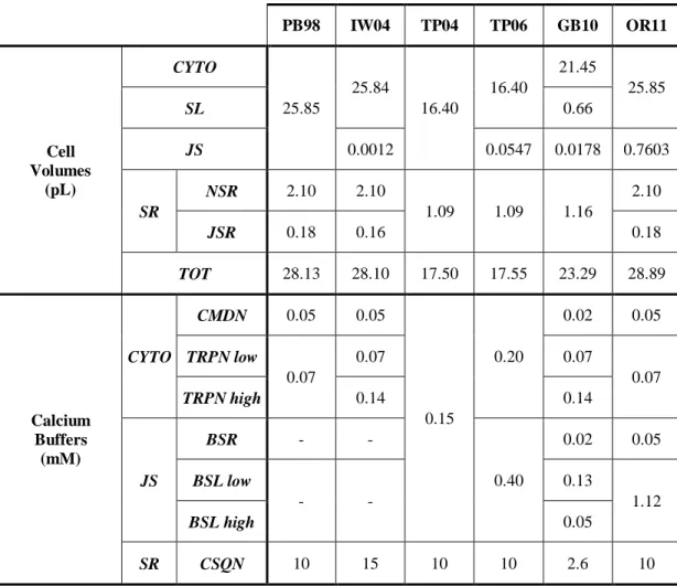
Table 2.3 shows the main structural properties of the considered human ventricular models, e.g. the number and size of intracellular compartments and the corresponding Ca2+ buffers.
The main challenge of the compartmental approach is the lack of corresponding distinct anatomical structures inside the cell. Thus, there is not a straightforward way of choosing the appropriate compartmentalization. This also explains the great variability among the different models.
Introduction – Chapter 2 43
Table 2.3: Main properties of the considered human ventricular AP models.
PB98 IW04 TP04 TP06 GB10 OR11 Cell Volumes (pL) CYTO 25.85 25.84 16.40 16.40 21.45 25.85 SL 0.66 JS 0.0012 0.0547 0.0178 0.7603 SR NSR 2.10 2.10 1.09 1.09 1.16 2.10 JSR 0.18 0.16 0.18 TOT 28.13 28.10 17.50 17.55 23.29 28.89 Calcium Buffers (mM) CYTO CMDN 0.05 0.05 0.15 0.20 0.02 0.05 TRPN low 0.07 0.07 0.07 0.07 TRPN high 0.14 0.14 JS BSR - - 0.40 0.02 0.05 BSL low - - 0.13 1.12 BSL high 0.05 SR CSQN 10 15 10 10 2.6 10
The six compartments refer to: bulk myoplasm (cytosol, CYTO); network SR (NSR); junctional SR (JSR); junctional space (JS); sub-sarcolemmal space (SSL). The Ca2+ buffers are: Calmodulin (CMDN), Troponin (TRPN), Calsequestrin (CSQN), SR Ca2+ buffers (BSR); junctional and sarcolemmal Ca2+ buffers (BSL).
Extracellular and Intracellular Concentrations
Extracellular ionic concentrations are quite similar in all the considered models, since they are the ones used for experimental recordings in single cells, often done in standard conditions. As for intracellular ionic concentrations, the differences between models are higher, especially when considering Ca2+ concentrations in the different compartments. A summary of the extra- and intra- cellular concentrations for every model is shown in Table 2.4: . Extracellular concentrations are constant in the models, while for intracellular concentrations the reported values are the diastolic ones, after pacing the models for 500 s at 1 Hz.
44 Introduction – Chapter 2
Elisa Passini
It is worth noticing that the GB10 model is the only one including Cl -concentrations, together with the Ca2+ activated Cl- current and a background Cl -current. We ran a few simulations blocking this current, and actually its effect on the AP is quite negligible, at least in physiological conditions. In addition, in GB10 both intracellular K+ and Cl- are clamped to constant values.
Table 2.4: Extracellular and intracellular ionic concentrations for the considered models.
PB98 IW04 TP04 TP06 GB10 OR11 Extracellular Concentrations [Na+]o (mM) 138 138 140 140 140 140 [K+]o (mM) 4.0 4.0 5.4 5.4 5.4 5.4 [Ca2+]o (mM) 2.0 2.0 2.0 2.0 1.8 1.8 [Cl-]o (mM) - - - - 150 - Intracellular Concentrations [Na+]i (mM) 10.6 9.8 11.6 10.4 8.4 7 [K+]i (mM) 140 125 140 140 120* 144 [Ca2+]i (nM) 200 86 200 130 87 85 [Ca2+]SL (nM) 100 [Ca2+]JS (nM) 140 360 200 85 [Ca2+]SR (mM) 2.5 0.3 0.2 3.7 0.6 1.6 [Cl-]i (mM) - - - - 15* -
[Y]o: extracellular concentration for Y; [Y]i: intracellular concentration for Y; SL:
sarcolemmal space; JS: junctional space; SR: sarcoplasmic reticulum.
* in GB10, [K+]i and [Cl
-] i are clamped to constant values.
Membrane Ionic Currents and Ca
2+subsystem
The main ionic currents are presents in all the considered models: INa, ICaL, Ito,
IK1, IKr, IKs, Na+/K+ pump (INaK) and Na+/Ca2+ exchanger (INCX). However, there are
a few differences in the small currents (e.g. background ones), and in Ca2+ related channels/pumps distribution. To avoid unnecessary complexity, we will focus on the differences between the three most recent models: TP06, GB10 and OR11.
A summary of ionic currents distribution is shown in Table 2.5, together with the corresponding intracellular compartments in which they are distributed. As for ionic fluxes, the two connected compartments for each of them are shown.
Introduction – Chapter 2 45
Table 2.5: Ionic currents and fluxes with the corresponding intracellular compartments for the six considered human ventricular AP models.
PB90 IW04 TT04 TT06 GB10 OR11
Na+ currents
INa CYTO CYTO CYTO CYTO
11% JS 89% SL CYTO INaL - - - CYTO ICaNa - - - - 90% JS 10% SL JS INaB CYTO CYTO CYTO CYTO
11% JS
89% SL CYTO Ca2+ currents ICaL
CYTO CYTO CYTO JS 90% JS
10% SL JS ICab CYTO CYTO CYTO CYTO 89% SL 11% JS CYTO
K+ currents
Ito CYTO CYTO CYTO CYTO CYTO CYTO
IK1 CYTO CYTO CYTO CYTO CYTO CYTO
IKr CYTO CYTO CYTO CYTO CYTO CYTO
IKs CYTO CYTO CYTO CYTO CYTO CYTO
ICaK - CYTO - - 90% JS 10% SL JS IKp - - CYTO CYTO 11% JS 89% SL CYTO Cl- currents IClB - - - - CYTO - IClCa - - - - 11% JS 89% SL - Pumps and Exchangers
INaK CYTO CYTO CYTO CYTO
11% JS
89% SL CYTO INCX CYTO CYTO CYTO CYTO 89% SL 11% JS
20% JS 89% CYTO IpCa - CYTO CYTO CYTO
11% JS 89% SL CYTO SR Fluxes Jrel JSR <-> CYTO JSR <-> JS SR -> CYTO SR <-> JS JSR <-> JS JSR -> JS Jleak NSR -> CYTO - SR <-> CYTO SR <-> CYTO JSR <-> JS NSR -> CYTO Jup -> NSR CYTO <-> NSR CYTO CYTO -> SR CYTO -> SR <-> SR CYTO -> NSR CYTO
Jtr NSR <-> JSR NSR <-> JSR - - - NSR <-> JSR Intracellular Fluxes JdiffCa - CYTO ˂-˃ JS - CYTO ˂-˃ JS CYTO/SL SL/JS CYTO ˂-˃ JS JdiffNa - - - - CYTO/SL SL/JS CYTO ˂-˃ JS JdiffK - - - CYTO ˂-˃ JS
As an example, in TP06 and OR11, all the ICaL actually flows into the JS. Even if
it is well know that most of the L-type Ca channels are located in the junctional portion of the membrane, this seems to be a rather extreme choice. More realistically in GB10, based on rat data [26], 90% of the channels are located in the JS membrane and the remaining 10% in the cytoplasm.
Another difference is in the INCX distribution. It is still not completely clear if
46 Introduction – Chapter 2
Elisa Passini
more uniform distribution in the ventricular sarcolemma [28]. In GB10 the INCX
channels are distributed evenly throughout the cell membrane (89% in the SL, 11% in the JS), while in OR11 the fraction located in the JS is slightly higher (20%). O’Hara et al. indicated this choice as necessary in order to correctly reproduce the rate dependence of intracellular Ca2+ peak [21]. It is worth noticing that all the ionic currents were considered uniformly distributed throughout the cell membrane in the GB10 model (89% in the SL, 11% in the JS), as suggested e.g. for INaK in
mammalian cardiomyocytes [29]. On the contrary, they were considered absent from JS membrane in TP06 and OR11.
A visual comparison of TT06, GB10 and OR11, showing all the ionic currents, fluxes and compartments for this three models, is given in Figure 2.2.
Figure 2.2: Visual comparison of the three most recent human ventricular model properties, in
particular showing the distribution of each ionic current on the cell, and the ions diffusion (credit: Caterina Passini).
It is worth noticing that, after this literature review has been completed (2013), a new mathematical model have been published for human ventricular cells by Asakura-Noma [30], as well as a new paper comparing the behaviour of some of the human ventricular models considered here in tissue, by Elshrif and Cherry [31].
Introduction – References 47
References
[1] J. Malmivuo and R. Plonsey, Bioelectromagnetism: Principles and Applications of
Bioelectric and Biomagnetic Fields. 1995.
[2] U. Ravens and E. Cerbai, “Role of potassium currents in cardiac arrhythmias” Europace, vol. 10, no. 10, pp. 1133–7, Oct. 2008.
[3] Z. Qu, Y. Xie, A. Garfinkel, and J. N. Weiss, “T-wave alternans and arrhythmogenesis in cardiac diseases” Front Physiol, vol. 1, p. 154, Jan. 2010.
[4] Longo, Fauci, Kasper, Hauser, Jameson, and LoScalzo, Harrison’s Principles of Internal
Medicine, 18th ed. 2012.
[5] A. L. Hodgkin and A. F. Huxley, “A quantitative description of membrane current and its application to conduction and excitation in nerve” J Physiol, vol. 117, no. 4, pp. 500–44, Aug. 1952.
[6] D. Noble, “A modification of the Hodgkin--Huxley equations applicable to Purkinje fibre action and pace-maker potentials” J Physiol, vol. 160, pp. 317–52, Feb. 1962.
[7] M. Wilhelms, H. Hettmann, M. M. Maleckar, J. T. Koivumäki, O. Dössel, and G. Seemann, “Benchmarking electrophysiological models of human atrial myocytes” Front Physiol, vol. 3, p. 487, Jan. 2012.
[8] O. Dössel, M. W. Krueger, F. M. Weber, M. Wilhelms, and G. Seemann, “Computational modeling of the human atrial anatomy and electrophysiology” Med Biol Eng Comput, vol. 50, no. 8, pp. 773–99, Aug. 2012.
[9] L. Priebe and D. J. Beuckelmann, “Simulation study of cellular electric properties in heart failure” Circ Res, vol. 82, no. 11, pp. 1206–23, Jun. 1998.
[10] C. H. Luo and Y. Rudy, “A dynamic model of the cardiac ventricular action potential. I. Simulations of ionic currents and concentration changes” Circ Res, vol. 74, no. 6, pp. 1071– 96, Jun. 1994.
[11] V. Iyer, R. Mazhari, and R. L. Winslow, “A computational model of the human left-ventricular epicardial myocyte” Biophys J, vol. 87, no. 3, pp. 1507–25, Sep. 2004.
[12] K. H. W. J. ten Tusscher, D. Noble, P. J. Noble, and A. V Panfilov, “A model for human ventricular tissue” Am J Physiol Heart Circ Physiol, vol. 286, no. 4, pp. H1573–89, Apr. 2004.
[13] K. H. W. J. Ten Tusscher, O. Bernus, R. Hren, and A. V Panfilov, “Comparison of electrophysiological models for human ventricular cells and tissues” Prog Biophys Mol Biol, vol. 90, no. 1–3, pp. 326–45, 2006.
[14] K. H. W. J. ten Tusscher and a V Panfilov, “Alternans and spiral breakup in a human ventricular tissue model” Am J Physiol Heart Circ Physiol, vol. 291, no. 3, pp. H1088–100, Sep. 2006.
[15] A. Bueno-Orovio, E. M. Cherry, and F. H. Fenton, “Minimal model for human ventricular action potentials in tissue” J Theor Biol, vol. 253, no. 3, pp. 544–60, Aug. 2008.
48 Introduction – References
Elisa Passini
[16] S. a Niederer, M. Fink, D. Noble, and N. P. Smith, “A meta-analysis of cardiac electrophysiology computational models” Exp Physiol, vol. 94, no. 5, pp. 486–95, May 2009.
[17] E. Grandi, F. S. Pasqualini, and D. M. Bers, “A novel computational model of the human ventricular action potential and Ca transient” J Mol Cell Cardiol, vol. 48, no. 1, pp. 112–21, Jan. 2010.
[18] T. R. Shannon, F. Wang, J. Puglisi, C. Weber, and D. M. Bers, “A mathematical treatment of integrated Ca dynamics within the ventricular myocyte” Biophys J, vol. 87, no. 5, pp. 3351– 71, Nov. 2004.
[19] J. Carro, J. F. Rodríguez, P. Laguna, and E. Pueyo, “A human ventricular cell model for investigation of cardiac arrhythmias under hyperkalaemic conditions” Philos Trans A Math
Phys Eng Sci, vol. 369, no. 1954, pp. 4205–32, Nov. 2011.
[20] D. Noble, A. Garny, and P. J. Noble, “How the Hodgkin-Huxley equations inspired the Cardiac Physiome Project,” J Physiol, vol. 590, no. Pt 11, pp. 2613–2628, 2012.
[21] T. O’Hara, L. Virág, A. Varró, and Y. Rudy, “Simulation of the undiseased human cardiac ventricular action potential: model formulation and experimental validation” PLoS Comput
Biol, vol. 7, no. 5, p. e1002061, May 2011.
[22] G. Seemann, F. B. Sachse, D. L. Weiss, and O. Dössel, “Quantitative reconstruction of cardiac electromechanics in human myocardium: regional heterogeneity” J Cardiovasc
Electrophysiol, vol. 14, no. 10 Suppl, pp. S219–28, Oct. 2003.
[23] M. Fink, D. Noble, L. Virag, A. Varro, and W. R. Giles, “Contributions of HERG K+ current to repolarization of the human ventricular action potential” Prog Biophys Mol Biol, vol. 96, no. 1–3, pp. 357–76, Jan. 2008.
[24] E. Grandi, F. S. Pasqualini, C. Pes, C. Corsi, A. Zaza, and S. Severi, “Theoretical investigation of action potential duration dependence on extracellular Ca2+ in human cardiomyocytes” J Mol Cell Cardiol, vol. 46, no. 3, pp. 332–42, Mar. 2009.
[25] D. DiFrancesco and D. Noble, “A model of cardiac electrical activity incorporating ionic pumps and concentration changes” Philos Trans R Soc Lond B Biol Sci, vol. 307, no. 1133, pp. 353–98, Jan. 1985.
[26] D. R. Scriven, P. Dan, and E. D. Moore, “Distribution of proteins implicated in excitation-contraction coupling in rat ventricular myocytes” Biophys J, vol. 79, no. 5, pp. 2682–91, Nov. 2000.
[27] J. S. Frank, G. Mottino, D. Reid, R. S. Molday, and K. D. Philipson, “Distribution of the Na(+)-Ca2+ exchange protein in mammalian cardiac myocytes: an immunofluorescence and immunocolloidal gold-labeling study” J Cell Biol, vol. 117, no. 2, pp. 337–45, Apr. 1992.
[28] W. J. Lederer, S. He, S. Luo, W. duBell, P. Kofuji, R. Kieval, C. F. Neubauer, A. Ruknudin, H. Cheng, M. B. Cannell, T. B. Rogers, and D. H. Schulze, “The molecular biology of the Na(+)-Ca2+ exchanger and its functional roles in heart, smooth muscle cells, neurons, glia, lymphocytes, and nonexcitable cells” Ann N Y Acad Sci, vol. 779, pp. 7–17, Apr. 1996. [29] A. A. McDonough, Y. Zhang, V. Shin, and J. S. Frank, “Subcellular distribution of sodium
pump isoform subunits in mammalian cardiac myocytes” Am J Physiol, vol. 270, no. 4 Pt 1, pp. C1221–7, Apr. 1996.
Introduction – References 49
[30] K. Asakura, C. Y. Cha, H. Yamaoka, Y. Horikawa, H. Memida, T. Powell, A. Amano, and A. Noma, “EAD and DAD mechanisms analyzed by developing a new human ventricular cell model” Prog Biophys Mol Biol, vol. 116, no. 1, pp. 11–24, Sep. 2014.
[31] M. M. Elshrif and E. M. Cherry, “A quantitative comparison of the behavior of human ventricular cardiac electrophysiology models in tissue” PLoS One, vol. 9, no. 1, p. e84401, Jan. 2014.
50 Introduction – References
SECTION I
___________________________________________________________________
Extracellular Calcium and Action Potential
Duration: the Fine Balance between L-Type
Calcium Current Inactivation Mechanisms
___________________________________________________________________
CHAPTER 3
___________________________________________________________________A Novel Markov Model of L-Type
Calcium Current to Explore
Inactivation Mechanisms
___________________________________________________________________
The content of this chapter has been published in:
Passini E, Severi S.
Extracellular Calcium and L-Type Calcium Current Inactivation Mechanisms: a Computational Study.
Computing in Cardiology, Vol. 40, 2013
Passini E, Severi S.
Computational Analysis of Extracellular Calcium Effect on Action Potential Duration
![Figure 3.1: Experimental Data from literature showing the inverse dependence of APD vs [Ca 2+ ] o : A) guinea pig ventricular cells (modified from [2]); B) human atrial cells (modified from [6]); C) human atrial cells (modified from [7]); D)](https://thumb-eu.123doks.com/thumbv2/123dokorg/8153058.126487/56.893.123.717.109.496/figure-experimental-literature-dependence-ventricular-modified-modified-modified.webp)
![Figure 3.3: APD-[Ca 2+ ] o dependence in two of the most recent human ventricular AP models:](https://thumb-eu.123doks.com/thumbv2/123dokorg/8153058.126487/57.893.235.746.345.820/figure-apd-dependence-most-recent-human-ventricular-models.webp)

![Figure 3.8: Comparison of I CaL steady state activation curves: original ORd model (blue line), modified ORd model (pink line) and experimental data from [18] (black squares)](https://thumb-eu.123doks.com/thumbv2/123dokorg/8153058.126487/61.893.184.757.600.959/figure-comparison-steady-activation-original-modified-experimental-squares.webp)
![Figure 3.9: Comparison of I CaL steady state inactivation curves: original ORd model (blue line), modified ORd model (pink line) and experimental data from [18] (black squares)](https://thumb-eu.123doks.com/thumbv2/123dokorg/8153058.126487/62.893.121.701.97.473/figure-comparison-steady-inactivation-original-modified-experimental-squares.webp)
![Figure 3.12: A) Experimental recording of Ca 2+ and Ba 2+ currents [10]: the latter, in absence of CDI mechanism, shows a much slower inactivation; B) Effects of CDI block on the L-Type Ca 2+ current for the original and modified ORd mode](https://thumb-eu.123doks.com/thumbv2/123dokorg/8153058.126487/63.893.219.761.713.1028/experimental-recording-currents-mechanism-inactivation-effects-original-modified.webp)
![Figure 3.14: Simulation results for the original and modified ORd models, showing APs (top panels) and I CaL (bottom panels) for different [Ca 2+ ] o concentrations](https://thumb-eu.123doks.com/thumbv2/123dokorg/8153058.126487/65.893.187.777.118.672/figure-simulation-results-original-modified-showing-different-concentrations.webp)

![Figure 5.5: AP traces corresponding to different [K + ] o for the NG model. When decreasing [K + ] o , the RMP becomes lower and the APD 90 increases, both as expected](https://thumb-eu.123doks.com/thumbv2/123dokorg/8153058.126487/104.893.117.687.126.663/figure-traces-corresponding-different-decreasing-lower-increases-expected.webp)
