1
UNIVERSITY OF ROME
"TOR VERGATA"
FACULTY OF SCIENCE
DOCTORATE DEGREE IN CHEMICAL SCIENCES
XXI CYCLE
NEW PHYSIOLOGICAL ROLES OF
GLUTATHIONE TRANSFERASES
Kutayba Farhan Dawood
2008/2009
Supervisor: Prof. Giorgio Ricci
Coordinator: Prof. Bruno Crociani
2
ACKNOWLEDGMENTS
First, all praise and thanks to God Almighty – ALLAH. I would like to
thank the Italian Ministry of Foreign Affairs.
I would like to record my gratitude to Prof. Giorgio Ricci for his
supervision, advice, and guidance from the very early stage of this research
as well as giving me extraordinary experiences throughout the work. Above
all and the most needed, he provided me unflinching encouragement and
support in various ways.
Special thank to my friend Dr. Raffaele Fabrini for all his assistances, as
a brother.
Lastly, and most importantly, I wish to thank my parents. They bore me,
raised me, supported me, taught me, and loved me. To them I dedicate this
thesis.
To my wife, and our daughter, all I can say is it would take another
thesis to express my deep love for you.
3
TABLE OF CONTENTS
ABSTRACT
5
ABBREVIATIONS
6
1. GLUTATHIONE TRANSFERASE
General Introduction
8
1.1 Glutathione Transferases (GSTs)
9
1.2 Classification of Glutathione Transferases
9
1.3 GST of the malarial parasites
10
1.4 GST enzymatic functions
11
1.5 Ligandin properties
13
1.6 Apoptosis regulation
14
1.7 GSTs structures
14
1.7.1 Mammalian cytosolic GST
14
1.7.2 GST of human malarial parasites
(Plasmodium falciparum)
17
1.8 GSTs and Nitric Oxide
19
1.8.1 Nitric oxide (Nitrogen monoxide)
19
1.8.2 Dinitrosyl iron complex
20
1.9 References
23
AIM OF THESIS
32
2. STUDY OF THE INTERACTION BETWEEN GSTs
AND DNDGIC IN INTACT CELLS AND TISSUES
33
2.1 Introduction
34
2.2 Experimental Procedures
36
2.3 Results
40
2.4 Discussion
50
4
3. STUDY OF GST LOCALIZATION IN NUCLEI
AND SUBCELLULAR FRACTIONS OF RAT
55
3.1 Introduction
56
3.2 Experimental Procedures
57
3.3 Results
61
3.4 Discussion
72
3.5 References
76
4. STUDY OF GST OF MALARIAL PARASITE
(Plasmodium falciparum)
79
4.1 Introduction
80
4.2 Experimental Procedures
82
4.3 Results
85
4.4 Discussion
95
4.5 References
97
CONCLUDING REMARKS
98
5
ABSTRACT
Glutathione transferases (GSTs) are enzymes able to conjugate GSH to a lot of toxic compounds thereby favoring their excretion. Recently, other protective roles of these enzymes have been discovered. In particular, it has been observed that a peculiar and strong interaction exists between some mammalian GSTs and an endogenous carrier of nitric oxide, the dinitrosyl-diglutathionyl iron complex (DNDGIC). This iron complex is a paramagnetic molecule with a characteristic EPR spectrum centered at g = 2.03, that is spontaneously formed when NO enters the cell. This complex is a strong irreversible inhibitor of glutathione reductase. The present work explores the possible role of GSTs like a protection system against DNDGIC. Actually, mammalian GSTs bind DNDGIC with extraordinary affinity (KD = 10-9-10-10 M). When
rat hepatocytes are incubated in the presence of GSNO, a natural source of NO, a rapid formation of 0.1 - 0.2 mM intracellular DNDGIC has been observed. This concentration would be lethal for glutathione reductase. However the complex does not appear like a free species but completely bound to GSTs, that are present at the cytosolic level of 0.8 mM. In this form the complex is completely harmless for glutathione reductase.
Surprisingly, electron paramagnetic data, reveal that DNGIC-GST is partially associated to subcellular fractions and in particular to nuclei. Our data indicate that about 10% of the cytosolic pool GST is electrostatically associated with the outer nuclear membrane, and a similar quantity is compartmentalized inside the nucleus. Mainly Alpha class GSTs, in particular GSTA1-1, GSTA2-2 and GSTA3-3, are involved in this double modality of interaction. Confocal microscopy and immunofluorescence experiments have been used to detail the electrostatic association in hepatocytes. A quantitative analysis of the membrane-bound Alpha GSTs suggests the existence of a multilayer
6
assembly of these enzymes at the outer nuclear envelope that could represent a potent protection shell for the nucleus and an amazing novelty in cell physiology.
A second target of this study is represented by the particular GST isoenzyme expressed by the Plasmodium falciparum (PfGST), the parasite causative of malaria. This enzyme is characterized by a peculiar dimer/tetramer transition that occurs in the absence of GSH and that causes a total loss of its enzymatic activity. Moreover PfGST binds hemin with high affinity and this interaction is finalized to the protection of the parasite against this toxic compound. Binding of hemin is regulated by a cooperative mechanism and does not occur in the tetrameric enzyme. Side directed mutagenesis, steady-state kinetic experiments, fluorescence anisotropy and X-ray crystallography were used to verify the involvement of some protein segment in the tetramerization process and in the cooperative phenomenon. Actually the loop 113-118 represents one the most prominent structural difference between PfGST and other GSTs. Our results demonstrate that truncation, increased rigidity or even a simple point mutation of this loop cause a dramatic change of the tetramerization kinetics that becomes hundred times slower than that observed in the native enzyme. Furthermore all mutants loose the positive cooperativity for hemin binding found in the native structure suggesting that the integrity of this peculiar loop is essential for intersubunit communication. Interestingly, the tetramerization process, that is very fast in the absence of GSH in the native enzyme, is prevented not only by GSH but even by GSSG. This result indicate that the protection of the parasite against free hemin is independent of the redox status of the cell.
7
ABBREVIATIONS
CDNB 1-Chloro-2,4-dinitrobenzene
CEM Human acute lymphoblastic T-cell leukemia Cell
DNDGIC Dinitrosyl-diglutathionyl iron complex EPR Electron Paramagnetic Resonance
GSH Glutathione (γ-glutamyl-cysteinyl-glycine)
GSNO S-Nitrosoglutathione
GSSG Glutathione Reductase
GST Glutathione Transferase
JNK Jun N-terminal kinase
MAPEG Membrane Associated Proteins in Eicosanoid and Glutathione
metabolism
NBD-Cl 4-chloro-7-nitro-2,1,3-benzoxadiazole
NBDHEX 6-(7-Nitro-2,1,3-benzoxadiazol-4-ylthio)hexanol
NOS Nitric Oxide Synthase
PfGST GST of Plasmodium falciparum parasite
SDS-PAGE Sodium Dodecyl Sulphate - Poly Acrylamide Gel Electrophoresis
8
1. GLUTATHIONE TRANSFERASE
General Introduction
9
1.1 Glutathione Transferases (GSTs)
Glutathione transferases are a superfamily of multifunctional enzymes that catalyze the addition of the nucleophilic thiol GSH (the tripeptide γ-glutamyl-cysteinyl-glycine) to xenobiotic and endogenous compounds which have electrophilic centers. Their substrates include alkyl and aryl halides, carboxylates, sulphate and phosphate esters, epoxides, organic nitrates, lactones, quinones, ozonides, thiocyanates and hydroperoxides (1-5). GSTs are involved in phase II of the mechanism of cellular detoxification. These proteins are found in all eukaryotic and prokaryotic systems, in the cytoplasm, in the microsomes and in mitochondria (6, 7). In human cells GSTs are present in high concentrations. For example in hepathocytes they represent about 3 - 5% of all cytosolic proteins. The GSTs are ubiquitous, especially abundant in the liver, lungs, in the placenta and skin (8, 9).
1.2 Classification of Glutathione Transferases
Over 100 different isoenzymes of GST have been described in species ranging from microorganisms to humans. Currently cytosolic GSTs are grouped into 12 isoenzymatic classes based on amino acid sequences, immunological properties and substrate and inhibitor specificity (10, 11).
A particular class is represented by the microsomal GST (MAPEG). This isoenzyme is associated to the microsomal membrane, and displays a peculiar trimeric structure (12). The cytosolic and mitochondrial GSTs are involved in the metabolism of xenobiotics, as well as in the detoxification against endogenous toxic compounds. In contrast, MAPEG is not involved in detoxification processes, but is active in the synthesis of prostaglandins and leukotrienes (5, 12).
In mammals 8 families of cytosolic GSTs are expressed, termed Alpha, Mu, Omega, Pi, Sigma, Theta and Zeta. Several other soluble GST classes have been reported: Delta and Epsilon in insects (13); Phi, Tau, Lambda in plants (14); Beta (15) and Chi in bacteria (16). The first classes discovered in mammals, i.e. Alpha, Mu and Pi, (17), are also the most advanced from an evolutionary point of view. The Alpha and Mu GST class are particularly abundant in the liver. The Alpha class GST plays also an additional detoxification role showing a peroxidase activity with organic peroxides. The GSTA1-1 and GSTA2-2 isoforms are highly
10
substrate-promiscuous with catalytic activity toward many structurally unrelated toxins (18-22) and the GSTA3-3 is involved in steroid biosynthesis and the metabolism of some xenobiotics (23).
1.3 GST of the malarial parasites
GST isoenzymes play a crucial role in parasites as they represent the main detoxification system due to the lack of Cytochrome P450 activity. A curious exception is represented by some protozoans such as Trypanosoma cruzi,
Trypanosoma brucei, Plasmodium berghei and Leishmania donovani that replace
the redox couple GSH/GSH reductase with trypanothione/trypanothione reductase (24-26). Furthermore, knockout studies in Plasmodium falciparum support the importance of functional cytosolic GSTs in these organisms (27). These findings place the parasite GSTs as targets for the development of new antiparasitic drugs (28, 29).
Tropical malaria, which is caused by the protozoan parasite Plasmodium
falciparum, is responsible for about 515 million clinical cases (30) and one to three
million deaths annually (31). The emergence and spread of drug resistance to commonly used chemotherapeutics are major factors contributing to this increasing burden. Thus, the characterization of alternative drug targets is urgently required (32-34). GST activity has been reported in all Plasmodium species studied so far as well as in all intraerythrocytic stages of the parasite (27).
The malaria parasite gives rise to disease only during its blood stage. This part of the lifecycle occurs largely within the red blood cell of the human host (35), where it digests a major proportion of the red cell hemoglobin (36). It has been demonstrated that Plasmodium falciparum, the causative agent of almost all fatal cases of malaria, detoxifies host hemoglobin-derived ferriprotoporphyrin IX in an acidic digestive vacuole mainly by converting it to hemozoin (37). Hemozoin is now known to be a crystalline cyclic dimer of ferriprotoporphyrin IX in which the propionate group of one porphyrin moiety coordinates to the Fe(III) center of its partner and vice-versa, while the second propionic acid group of each ferriprotoporphyrin IX hydrogen bonds to a neighboring dimer in the crystal (38).
11
Glutathione transferase of Plasmodium falciparum (PfGST) is the sole GST isoenzyme expressed by the parasite and it represents >1% of the total cellular protein (39, 40). A role of GST from Plasmodium falciparum in the development of drug resistance in malarial parasites has been postulated but is still controversial (28, 39). The PfGST differs significantly from human GSTs, and PfGST cannot be assigned to any of the previously known GST classes, thus representing a novel GST isoform (27, 29, 41-50) that may exert a particular protective role in the parasite. In fact, beside the usual activity that promote the conjugation of GSH to electrophilic toxic compounds, this protein binds efficiently hemin and thus it could protect the parasite (that lives in the erythrocytes) from the oxidative stress caused by residual free hemin that did not polymerize into hemozoin.
1.4 GST Enzymatic functions
GSTs display multifunctional nature because they are involved in different types of processes and have different enzyme activities (51). The main function is the transferase activity i.e. the reaction of conjugation of GSH to a wide variety of hydrophobic compounds, endobiotics or xenobiotics (Fig. 1.1), that have an electrophilic centre (4, 52, 53).
These electrophilic substrates include epoxides, alkyl and aryl halides, esters, activated alkenes, quinones and -unsaturated carbonyls compounds (3, 41). With this conjugation the glutathione transferase plays the function of detoxification against of toxic compounds, both of endogenous nature, such as secondary metabolites of oxidative stress, both of exogenous nature, such as drugs, carcinogens, environmental pollutants, pesticides and herbicides. In addition to this
specific activity, some isoforms also show a glutathione peroxidase selenium-independent activity, allowing them to catalyze the reduction of lipid
12
FIGURE 1.1. Reactions catalyzed by GSTs. Examples of conjugation,
reduction, thiolysis, and isomerization reactions catalyzed by GST. The following substrates are shown: (a) chlorodinitrobenzene, (b) 4-nitrophenyl acetate, (c) sulforaphane (d) trinitroglycerin, (e) cumene hydroperoxide, (f) maleylacetoacetate, (g) prostaglandin PGH2.
13
Thanks to this activity of peroxidation, combined with that of conjugation, the GSTs play an important role in the tissue protection process against oxidative damage (5). Other isoenzymes show an additional isomerase activity to various
compounds such as unsaturated 5-3-chetosteroid, maleylacetoacetic acid and
maleylacetone (Fig. 1.1). GSTs are also able to bind endogenous compounds such as leukotrienes and prostaglandins acting both in their catabolism, through the classic reaction of conjugation with the GSH (Fig. 1.2), and in their process of biosynthesis (55).
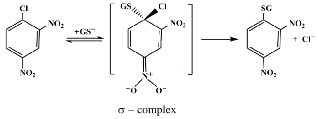
As an example of enzymatic mechanism is reported below the conjugation of GSH to the universal co-substrate 1-chloro-2,4-dinitrobenzene (CDNB):
complex
FIGURE 1.2. The conjugation of GSH to CDNB catalyzed by GSTs.
The catalysis of nucleophilic aromatic substitution reactions can be divided into steps involving binding of substrates to the enzyme active site, activation of GSH by deprotonation of the thiol to form the nucleophilic thiolate (56), and nucleophilic attack by the thiolate at the electrophilic center to form a σ-complex.
1.5 Ligandin properties
The panel of GSTs present in nature encompasses enzymes that catalyze conjugation, reduction and isomerase reactions, as well as proteins that act non-enzymatically as ligandines. Glutathione transferases are able to bind some hydrophobic compounds like bilirubin, heme, steroid and thyroid hormones. Some of these ligands neither bind to the G site nor to the H site, but to the interface of the two subunits, and the binding often inhibits glutathione transferases. This
14
interaction and compartmentalization prevents a possible cytotoxic accumulation in tissues of these lipophilic molecules (17).
1.6 Apoptosis regulation
Recently an important protein-protein interaction has been described involving GST and Jun N-terminal kinase (JNK), a protein involved in the cellular apoptotic process. Apoptosis is programmed cellular dead primed from free JNK and other enzymes in cascade reactions. As long as JNK protein remains bound to the GST it is not in position to trigger cellular death. This GST-JNK interaction is regulated by their cytosolic concentrations. When the cell is in the increasing phase JNK is bound to GSTs and is inactive. Interaction with inhibitors, UV irradiation and oxidative stress, induce GSTs modification making the GST-JNK complex unstable. Therefore JNK becomes active priming the apoptotic process (57).
1.7 GSTs structures
1.7.1 Mammalian cytosolic GSTs
Representative tridimensional structures of at least one member from each cytosolic GSTs have been solved by X-ray diffraction studies (Fig. 1.3). The crystals were obtained in complex with the GSH (or its analogues) or inhibitors. All soluble GSTs display similar dimeric structures assembled mainly in α-helix (48-59%) and in minor measure by β-sheets (8-10%), the rest is constituted by regions irregularly structured and loops. Each subunit is constituted by two domains linked together by one peptide of a few amino acids. The first domain in these enzymes is located in the N-terminal portion and is responsible for GSH binding, hence the name “G-site”. The domain is conserved for all classes with a thioredoxin-like fold comprised of three helixes and four sheets in a βαβαββα run. The binding of glutathione is done in an extended conformation at one end of the four strands of the G-site and it is anchored to the domain through electrostatic interactions.
15
FIGURE 1.3. Three-dimensional structure of the main glutathione transferase monomers (58).
16
The second domain contains the H-site, i.e., the site for binding of the second, often hydrophobic, substrate. The amino acid residues that are involved in the binding of the electrophilic substrate may also, if correctly positioned, contribute to the chemical steps on the reaction pathway. In any case, the structure of the H-site governs the substrate specificity of a GST. Interestingly, these enzymes have activity only as dimers (Fig. 1.4).
FIGURE 1.4. The homodimer of GSTP1-1 (59).
The argument used to explain this behavior has been the cooperativity (positive or negative) between subunits, recently demonstrated in Plasmodium falciparum and some subclasses of mammalians isoenzymes in which the subunits showed interactions which modulate the binding of different compounds (60-62). In general, GSTs monomers have molecular masses of 23–28 kDa with an average of 220 amino acids in their sequences. The dimer may have identical subunits (homodimer) or different subunits (heterodimer) of the same class.
The three-dimensional structures of members of mammalian GSTs from classes Alpha (63, 64), Mu (65-66) and Pi (67-69) and mutational studies have provided
17
important details about amino acid residues putatively involved in the catalytic mechanism. Activation of GSH occurs at the G-site by different amino acids according to the class. Tyrosine (Mu, Pi, Alpha, and Sigma classes), serine (Zeta class) or cysteine (Omega class) allow conjugation or the thiol transfer. The first two amino acids, tyrosine and serine, promote the formation and stabilization of the thiolate anion of GSH, lowing its pKa from 9.0 to 6.2. This is achieved through hydrogen bond donation of their hydroxyl group, which gets GSH ready for conjugation. When a Cys residue is used there is a thiol transfer, and it forms mixed disulphides with GSH. This kind of reaction is closely related to redox reactions.
The second domain, called the H-site, binds hydrophobic substrates, is located in the C-terminal region and is comprised exclusively of α-helixes. The number of the helixes varies from 4 to 7. This fact and residue variations in the H-site have been taken as arguments for the wide substrate diversity and preferences for detoxification among classes. For example, the Mu class has very efficient catalysis with molecules containing oxiranes and unsaturated carbonyl groups whereas class Alpha acts on 4-hydroxyalkenals and peroxides (4, 51, 70). However, the subclasses might also be distinguished by their substrate specificities.
1.7.2 GST of human malarial parasites (Plasmodium falciparum)
Structures for parasite cytosolic GSTs come from the protozoa Plasmodium
falciparum (PfGST), the nematode Onchocerca volvulus (OvGST2) and trematodes Schistosoma haematobium, Schistosoma japonicum, Schistosoma mansoni and Fasciola hepatica. In all cases, the differences with human cytosolic GSTs provide
opportunities to develop specific inhibitors against these parasites (71-76). The GST gene of Plasmodium falciparum (PfGST) was cloned and expressed in Escherichia
coli, yielding a homodimeric active enzyme. According to primary structure and
substrate specificity, the protein can be placed into the vicinity of the Mu or Pi subclass of GSTs (40). PfGST is the only enzyme of the GST family, which in the absence of GSH shows a tetrameric structure rather than a dimeric structure (77).
The structure of PfGST at 1.9 and 2.2Å resolution exhibits a shorter C-terminal section, with only five residues after the 8 helix, which implicates a more solvent-accessible H-site area and an amphiphilic character that is reflected in its
18
substrate spectrum. Amphiphilic compounds, including inhibitors, can access the H-site of PfGST but cannot enter the hydrophobic H-site of human isoforms. Alignments of the PfGST structures with crystals of human Alpha, Mu and Pi classes indicate a significantly high root mean-square (rms; relative position between two atoms) deviation of ~ 1.2 Å, compared to members within classes which show rms deviations of ~ 0.7 Å, as occurs between S. japonicum and F.
hepatica, which have an rms deviation of 0.85 Å (27, 72, 75). Again, a broader and
more solvent-exposed site is found and it is due to amino acid variation in the H-site of the parasite cytosolic GST in contrast to mammalian Pi class (74). X-ray study of this enzyme, showed by an atypic extra loop connecting helix 4 and helix 5 (residues 113-118) that could be involved in the dimer/dimer interaction. Actually, in the absence of ligands two biological dimers (AA1 and BB1) form a tetramer and these homodimers (Fig. 1.5) are interlocked with each other by the loop 113-118 of monomer B (B1), which occupies the H-site of monomer A (A1) (50).
FIGURE 1.5. Structure of the PfGST. (A), dimer structure, (B), tetramer
19
Upon binding of S-hexylglutathione, the H-site loop 113-118 rearranges, residues Asn-114, Leu-115, and Phe-116 form an additional coil in helix -4 and the side chains of Asn-111, Phe-116, and Tyr-211 flip into the H-site. The changed course of the residues 113-120 in the liganded enzyme prevents the interlocking of the dimers; as a consequence, the molecules are packed as dimers (78).
1.8 GSTs and Nitric Oxide
1.8.1 Nitric oxide (Nitrogen monoxide)
Nitric oxide (NO) is a paramagnetic inorganic gas with good solubility in water, weakly polar and thermodynamically unstable (79). NO is produced in bodies from endogenous and exogenous sources. In the second case, it is synthesized by the enzyme nitric oxide synthase (NOS) in which L-arginine is converted into NO and L-citrulline (80, 81). There are three NOS isoforms: neuronal NOS (nNOS, NOS-1), inducible NOS (iNOS, NOS-2), and endothelial NOS (eNOS, NOS-3) (81). All mammalian NOS are hemoproteins, require
NADPH and O2 for the production of NO, and use FAD, FMN, and
tetrahydrobiopterin as cofactors (81-83).
NO is an important molecule involved in a wide range of physiological functions as control of blood pressure, vasodilatation, inhibition of platelet aggregation, neurotransmission, memory formation, penile erection. Furthermore NO is involved in the operation of the immune system, iron metabolism and contributes to the cytotoxicity of activated macrophages against tumor cells and intracellular parasites (84-88). Nitric oxide is reported to induce apoptosis and initiate differentiation in certain types of cancer cells, suggesting that NO is a potential cancer therapeutic agent with novel mechanisms of action (89-91). Furthermore endogenously synthesized NO has been implicated as being responsible for the development of various diseases (92-94). Although the precise mechanisms of biological action of nitric oxide are not completely elucidated, the physiological effects of nitric oxide are dependent on its local concentration and duration of exposure (95, 96).
20
The most important biological reactions of NO are with oxygen, superoxide and metal ions (97). NO is an odd electron species with a half-life of only a few seconds in biological systems (98) and it reacts rapidly with superoxide to form the cytotoxic peroxynitrite (ONOO−) (99). It degrades rapidly to nitrite (NO2
−
) then nitrate (NO3−) (79, 100, 101). Putative intermediate metabolites include an array of low and high molecular weight compounds, including nitrosoglutathione, nitrosoalbumin, and S-nitrosohaemoglobin (102-104). The presence of a single electron in the NO molecule gives it a radical character and therefore, high reactivity. Between its reactions, NO can lose an electron forming nitrosylic compounds, of mostly covalent character, or to coordinate with variety of metals forming metal-nitrosyl compounds (105).
1.8.2 Dinitrosyl iron complex
All biological activities of nitric oxide can be affected not only by NO itself but
also by relatively stable physiological NO carriers or NO donors among which S-nitrosothiol (RSNO) and dinitrosyl iron complexes (DNICs) are included (79, 106). DNICs are stable paramagnetic molecules that exhibits a characteristic spectrum of electron paramagnetic resonance (EPR) (107-110). It is generated in cells and tissues from various sources following exposure to endogenous or exogenous NO and can be detected by EPR spectroscopy (111-119). Mononuclear dinitrosyl iron complexes are formed when ferrous iron and NO react with low molecular weight thiols (120) amino acids, peptides and various proteins (110). In mammalian tissues and cells, the first detection of DNIC derived EPR signals (g =2.03), has been reported more than 30 years ago by Vanin et al. (108).
In vivo, dinitrosyl-diglutathionyl iron complex (DNDGIC) and other low mass DNICs could be in equilibrium with several protein-bound forms after replacing one or both the free thiol ligands with protein residues like His, Cys and Ser (121). Both low mass and high mass DNICs seem to be more stable than NO and may possibly act as storage of nitric oxide (122-124) as well as intermediates in the iron-catalyzed formation and decomposition of S-nitrosothiols (125). DNICs can cross cell membranes to donate Fe to tissues (126) and can transnitrosylate acceptor targets in vitro and in vivo (125, 127, 128) demonstrating their bioavailability and
21
potential role as NO carrier molecules. DNICs also inhibit platelet aggregation (129), reduce blood pressure (130), relax vascular vessels (131), induce accumulation of heat shock protein Hsp70 (132, 133), and modulate ion channel activity (134).
Up to a few years ago no relation had been found between GSTs and the NO carriers like DNICs, but in 2001 Ricci and co-workers have described the peculiar interaction between of DNDGIC (Fig. 1.6) with GSTP1-1, a representative member of the human glutathione transferase superfamily (135).
FIGURE 1.6. Dinitrosyl-diglutathionyl iron complex (DNDGIC).
The study of the interaction of DNDGIC with GSTP1-1 demonstrated that this complex is a potent natural competitive inhibitor of this enzyme (135). EPR spectroscopy and molecular modeling indicated that DNDGIC is stabilized in the G-site through the usual polar and hydrophobic interactions of protein residues with one GSH molecule, coordination of the iron ion to the hydroxyl group of Tyr-7, and additional van der Waals interactions of NO moieties with Ile-104 and Tyr-108 (135). This was late confirmed by the crystal structure (136).
More recently Ricci and co-workers demonstrated that not only GSTP1-1 but representative members of all mammalian GSTs interact with DNDGIC showing similar binding mechanism and cooperativity (137). This complex binds with extraordinary affinity to the active site of all GSTs (dimeric enzymes); GSTA1-1 shows the strongest interaction (KD ≈ 10-10 M), whereas GSTM2-2 and GSTP1-1 display similar and slightly lower affinities (KD ≈ 10-9 M). Binding of the DNDGIC to GSTA1-1 triggers structural intersubunit communication, which lowers the affinity for DNDGIC in the vacant subunit and also causes a drastic loss of enzyme activity. Negative cooperativity is also found in GSTM2-2 and GSTP1-1, but it does
22
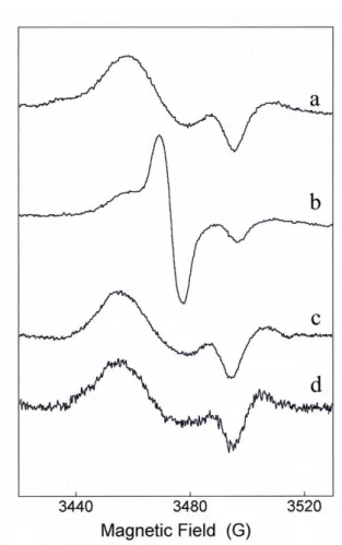
not affect the catalytic competence of the second subunit (137). The EPR spectrum of the GST-bound dinitrosyl iron complexes changes appreciably according to the GST isoform used (Fig. 1.7). The A1-1 and M2-2 classes of GSTs give essentially axial spectra, whereas P1-1 and T2-2 give strongly rhombic spectra, but there are minor differences in the spectra that in practice make it possible to identify the type of glutathione transferase involved directly from the EPR spectrum.
All these findings suggest a new and important role of GSTs in the metabolism of NO in the cell that may disclose interesting scenarios of research.
The dinitrosyl iron complex is firmly bound to these GST isoforms through the glutathione thiolate-iron-tyrosinate ligand arrangement; constant S-Fe-O bond angles and bond lengths are essential for the high affinity, whereas the positions of the NO groups, which do not seem to contribute much to binding, will be determined by the space available in the active site of the enzyme (137).
FIGURE 1.7. EPR spectra of DNDGIC bound to different GST isoforms (137).
23
1.9 References
1. Sheehan, D., Meade, G., Foley, V. M., and Dowd, C. A. (2001) Biochem. J.
360, 1-16
2. Keen, J. H., and Jakoby, W. B. (1978) J. Biol. Chem. 253, 5654-5657
3. Hayes, J. D., and Pulford, D. J. (1995) Crit. Rev. Biochem. Mol. Biol. 30, 445-600
4. Armstrong, R. N. (1997) Chem. Res. Toxicol. 10, 2-18
5. Hayes, J. D., and McLellan, L. I. (1999) Free Radic. Res. 31, 273-300
6. Laughlin, L. T., Bernat, B. A., and Armstrong, R. N. (1998) Chem. Biol.
Interact. 111-112, 41-50
7. Pemble, S. E., Wardle, A. F., and Taylor, J. B. (1996) Biochem. J. 319,
749-754
8. Awasthi, Y. C., Sharma, R., and Singhal, S. S. (1994) Int. J. Biochem. 26,
295-308
9. Sugimoto, M. (1995) Nippon Rinsho 53, 1253-1259
10. Ladner, J. E., Parsons, J. F., Rife, C. L., Gilliland, G. L., and Armstrong, R. N. (2004) Biochem. J. 43, 352-361
11. Robinson, A., Huttley, G. A., Booth, H. S., and Board, P. G. (2004).
Biochem. J. 379, 541-552
12. Jakobsson, P. J., Morgenstern, R., Mancini, J., Ford-Hutchinson, A., and Persson, B. (1999) Protein Sci. 8, 689-669
13. Alias, Z., and Clark, A. G. (2007) Proteomics 7, 3618-3628
14. Dixon, D. P., Davis, B. G., and Edwards, R. (2002) J. Biol. Chem. 277, 30859-30869
15. Allocati, N., Favaloro, B., Masulli, M., Alexeyev, M. F., and Di Ilio, C. (2002) Biochem. J. 373, 305-311
16. Wiktelius, E., and Stenberg, G. (2007) Biochem. J. 406, 115-123
17. Mannervik, B. (1985) Adv. Enzymol. Relat. Areas. Mol. Biol. 57, 357- 417 18. Dreij, K., Sundberg, K., Johansson, A. S., Seidel, E., Persson, B.,
24
19. Bruns, C. M., Hubatsch, I., Ridderstrom, M., Mannervik, B., and Tanner, J. A. (1999) J. Mol. Biol. 288, 427-439
20. Nilsson, L. O., Gustaffson, A., and Mannervik, B. (2000) Proc. Natl. Acad.
Sci. U. S. A. 97, 9408-9412
21. Hurst, R., Bao, Y., Jemth, P., Mannervik, B., and Williamson, G. (1998)
Biochem. J. 332, 97-100
22. Zhao, T., Singhal, S. S., Piper, J. T., Cheng, J., Pandya, U., Clark-Wronski, J., Awasthi, S., and Awasthi, Y. C. (1999) Arch. Biochem. Biophys. 367, 216-224
23. Tetlow, N., Coggan, M., Casarotto, M. G., Board P. G. (2004)
Pharmacogenetics 14, 657-663
24. Agosin, M., Naquira, C., Capdevila, J., and Pulin, J. (1976) Int. J.
Biochem. 7, 585-593
25. Precious, W., and Barret, J. (1989) Biochem. Biophys. Acta 992, 215-222 26. Tracy, J. W., and Vande Waa, E. A. (1995) Biochemistry and Molecular
Biology of Parasites. pp. 161-173, Academic Press, San Diego
27. Deponte, M., and Becker, K. (2005) Methods Enzymol. 401, 240-252 28. Barrett, J. (1998) Comp. Biochem. Physiol. 121, 181-183
29. Brophy, P. M., and Pritchard, D. I. (1994) Exp. Parasitol. 79, 89-99
30. Snow, R. W., Guerra, C. A., Noor, A. M., Myint, H. Y., and Hay, S. I. (2005) Nature 434, 214-217
31. Sachs, J., and Malaney, P. (2002) Nature 415, 680-685 32. Olliaro, P. (2001) Pharmacol. Ther. 89, 207-219
33. Greenwood, B., and Mutabingwa, T. (2002) Nature 415, 670-672 34. Ridley, R. G. (2002) Nature 415, 686-693
35. Bannister, L. H., Hopkins, J. M., Fowler, R. E., Krishna S., and Mitchell, G. H. (2000) Parasitol. Today 16, 427-433
36. Krugliak, M., Zhang F., and Ginsburg, H. (2002) Mol. Biochem. Parasitol.
25
37. Egan, T. J., Combrinck, J. M., Egan, J., Hearne, G. R., Marques, H. M., Ntenteni, S., Sewell, B. T., Smith, P. J., Taylor, D., van Schalkwyk, D. A., and Walden, J. C. (2002) Biochem. J. 365, 343-347
38. Pagola, S., Stephens, P. W., Bohle, D. S., Kosar A. D., and Madsen, S. K. (2000) Nature 404, 307-310
39. Harwaldt, P., Rahlfs, S., and Becker, K. (2002) Biol. Chem. 383, 821-830 40. Liebau, E., Bergmann, B., Campbell, A. M., Teesdale-Spittle, P., Brophy,
P. M., Luersen, K., and Walter, R. D. (2002) Mol. Biochem. Parasitol. 124, 85-90
41. Mannervik, B., and Danielson, U. H. (1988) CRC Crit. Rev. Biochem. 23, 283-337
42. Chemale, G., Morphew, R., Moxon, J. V., Morassuti, A. L., Lacourse, E. J., Barrett, J., Johnston, D. A., and Brophy, P. M. (2006) Proteomics 6, 6263-6273
43. Girardini, J., Amirante, A., Zemzoumi, K., and Serra, E. (2002) Eur. J.
Biochem. 269, 5512-5521
44. Hong, S., Lee, J., Lee, D., Sohn, W., and Cho, S. (2001) Mol. Biochem.
Parasitol. 115, 69-75
45. Kang, S., Ahn, I., Park, C., Chung, Y., Hong, S., Kong, Y., Cho, S., and Hong, S. (2001) Exp. Parasitol. 97, 186-195
46. Rathaur, S., Fisher, P., Domagalski, M., Walter, R. D., and Liebau, E. (2003) Exp. Parasitol. 103, 177-181
47. Salinas, G., Braun, G., and Taylor, D. W. (1994) Mol. Biochem. Parasitol.
66, 1-9
48. Salvatore, L., Wijffels, G., Sexton, J., Panaccio, M., Mailer, S., McCauley, I., and Spithill, T. (1995) Mol. Biochem. Parasitol. 69, 281-288
49. Vibanco-Pèrez, N., Jimènez, L., Mendoza-Hernàndez, G., and Landa, A., (2002) Parasitol. Res. 88, 398-404
50. Fritz-Wolf, K., Becker, A., Rahlfs, S., Harwaldt, P., Schirmer, R.H., Kabsch, W., and Becker, K. (2003) Proc. Natl. Acad. Sci. U. S. A. 100, 13821-13826
26
51. Hayes, J. D., Flanagan, J. U., and Jowsey, I. R. (2005) Annn. Rev.
Pharmacol. Toxicol. 45, 51-88
52. Wilce, M. C., and Parker, M. W. (1994) Biochim. Biophys. Acta 1205, 1-18
53. Pickett, C. B., and Lu, A. Y. (1989) Annu. Rev. Biochem. 58, 743-764
54. Ketterer, B., Fraser, G., and Meyer, D. J. (1990) Adv. Exp. Med. Biol. 264, 301-310
55. Meyer, D. J., Crease, D. J., and Ketterer, B. (1995) Biochem. J. 306, 565-569
56. Graminski, G. F., Kubo, Y., and Armstrong, R. N. (1989) Biochemistry 28, 3562-3568
57. Adler, V., Yin, Z., Fuchs, S. Y., Benezra, M., Rosario, L., Tew, K. D., Pincus, M. R., Ardana, M., Henderson, C. J., Wolf, C. R., Davis, R. J., and Ronai, Z. (1999) EMBO J. 18, 1321-1334
58. Thomas, D. D., Liu, X., Kantrow, S. P., and Lancaster, J. R. (2001) Proc.
Natl. Acad. Sci. U. S. A. 98, 355-360
59. Oakley, A. J., Lo Bello, M., Mazzetti, A. P., Federici, G., and Parker, M. W. (1997) FEBS Lett. 419, 32-36
60. Ricci, G., Turella, P., De Maria, F., Antonini, G., Nardocci, L., Board, P. G., Parker, M. W., Carbonelli, M. G., Federici, G., and Caccuria, A. M. (2004) J. Biol. Chem. 279, 33336-33342
61. Armstrong, R.N. (1994) Adv. Enzymol. 69, 1-44
62. Liebau, E., De Maria, F., Burmeister, C., Perbandt, M., Turella, P., Antonini, G., Federici, G., Giansanti, F., Stella, L., Lo Bello, M., Caccuri, A. M., and Ricci, G. (2005) J. Biol. Chem. 280, 26121-26128
63. Sinning, I., Kleywegt, G. J., Cowan, S. W., Reinemer, P., Dirr, H. W., Huber, R., Gilliland, G. L., Armstrong, R. N., Ji, X., Board, P. G., Olin, B., Mannervik, B., and Jones, T. A. (1993) J. Mol. Biol. 232, 192-212
64. Cameron A. D., Sinning, I., L'Hermite, G., Olin, B., Board, P. G., Mannervik, B., and Jones, T. A. (1995) Structure 3, 717-727
65. Ji, X., Zhang, P., Armstrong, R. N., and Gilliland, G. L. (1992)
27
66. Raghunathan, S., Chandross, R. J., Kretsinger, R. H, Allison, T. J., Penington, C. J., and Rule G. S. (1994) J. Mol. Biol. 238, 815-832
67. Reinemer, P., Dirr, H. W., Ladenstein, R., Schäffer, J., Gallay, O., and Huber, R. (1991) EMBO J. 10, 1997-2005
68. Reinemer, P., Dirr, HW., Ladenstein, R., Huber, R., Lo Bello, M., Federici, G., and Parker, M. W. (1992) J. Mol. Biol. 227, 4-26
69. Dirr, H., Reinemer, P., and Huber, R. (1994) J. Mol. Biol. 243, 72-92 70. Frova, C. (2006) Biomol. Eng. 23, 149-169
71. Baiocco, P., Gourlay, L. J., Angelucci, F., Fontaine, J., Herv´e, M., Miele, A. E., Trottein, F., Brunori, M., and Bellelli, A. (2006) J. Mol. Biol. 360, 678-689
72. Burmeister, C., Perbandt, M., Betzel, C., Walter, R. D., and Liebau, E. (2003) Acta Crystallogr. D Biol. Crystallogr. 59, 1469-1471
73. McTigue, M., Williams, D. R., and Tainer, J. A., (1995) J. Mol. Biol. 246, 21-27
74. Perbandt, M., Höppner, J., Betzel, C., Walter, R. D., and Liebau, E. (2005)
J. Biol. Chem. 280, 12630-12636
75. Rossjohn, J., Feil, S. C., Wilce, M. C. J., Sexton, J. L., Spithill, T. W., and Parker, M. W. (1997) J. Mol. Biol. 273, 857-872
76. Trottein, F., Vaney, M. C., Bachet, B., Pierce, R. J., Colloch, N., Lecocq, J. P., Capron, A., and Mornon, J. P. (1992) J. Mol. Biol. 224, 515-518
77. Tripathi, T., Rahlfs, S., Becker, K., and Bhakuni, V. (2007) BMC Struct
Biol. 7, 67
78. Hiller, N., Fritz-Wolf, K., Deponte, M., Wende, W., Zimmermann, H., and Becker, K. (2006) Protein 15, 281-289
79. Butler, A. R., and Williams, D. L. H. (1993) Chem. Soc. Rev. 22, 233-241 80. Stuehr, D. J., and Griffith, O. W. (1992) Advances in enzymology and
related areas of molecular biology. pp. 287-346, John Wiley & Sons, New
York
81. Morris, S. M. Jr. (2005) Vasc. Med. 10, 83-87
82. Knowles, R. G., and Moncada, S. (1994) Biochem. J. 298, 249-258 83. White, K. A., and Marletta, M. A. (1992) Biochem. J. 31, 6627-6631
28
84. Moncada, S. J. (1994) Hypertens. Suppl. 12, 35-39
85. Loscalzo, J., and Welch, G. (1995) Prog. Cardiovasc. Dis. 38, 87-104 86. Nathan, C. (1992) FASEB J. 6, 3051-3064
87. Stamler, J. S. (1994) Cell 78, 931-936
88. Thippeswamy, T., McKay, J. S., Quinn, J. P., and Morris, R. (2006) Histol.
Histopathol. 21, 445-458
89. Magrinat, G., Mason, S. N., Shami, P. J., and Weinberg, J. B. (1992) Blood
80, 1880-1884
90. Shami, P. J., Moore, J. O., Gockerman, J. P., Hathorn, J. W., Misukonis, M. A., and Weinberg, J. B. (1995) Leukemia Res. 19, 527-533
91. Shami, P. J., Sauls, D. L., and Weinberg, J. B. (1998) Leukemia 12, 1461-1466
92. Moncada, S., Palmer, R. M., and Higgs, E. A.(1991) Pharmacol. Rev. 43, 109-142
93. Tamir, S., and Tannenbaum, S. R. (1996) Biochim. Biophys. Acta 1288, 31-36
94. Mey, C. (1998) Curr. Med. Res. Opin. 14, 187-202
95. Wink, D. A., and Mitchell, J. B. (2003) Free Rad. Biol. Med. 34, 951-954 96. Wink, D. A., Vodovotz, Y., Laval, J., Laval, F., Dewhirst, M. W., and
Mitchell, J. B. (1998) Carcinogenesis 19, 711-721
97. Stamler, J. S., Singel, D. J., and Loscalzo, J. (1992) Science 258, 1898-1902 98. Thomas, D. D., Liu, X., Kantrow, S. P., and Lancaster, J. R., Jr. (2001)
Proc. Natl. Acad. Sci. U. S. A. 98, 355-360
99. Beckman, J. S., Beckman, T. W., Chen, J., Marshall, P. A., and Freeman, B. A. (1990) Proc. Natl. Acad. Sci. U. S. A. 87, 1620-1624
100. Lam, A. A., Hyland, K., and Heales, S. J. (2007) J. Inherit. Metab. Dis. 30, 256-622
101. Lancaster, J. R., (1994) Proc. Natl. Acad. Sci. U. S. A. 91, 8137-8141
102. Kukovetz, W. R., Holzmann, S., and Schmidt, K. (1991). Eur. Heart J. 12, 1-24
103. Marks, D. S., Vita, J. A., Folts, J. D., Keaney, J. F., Welch, G. N., and Loscalzo, J. (1995) J. Clin. Invest. 96, 2630-2638
29
104. Jia, L., Bonaventura, C., Bonaventura, J., and Stamler, J. S. (1996) Nature
380, 221-226
105. Xu, W. M. and Liu, L. Z. (1998) Cell Res. 8, 251-258
106. Henry, Y., Lepoivre, M., Ducrocq, C., Boucher J. L., and Guissani A. (1993) FASEB J. 7, 1124-1134
107. Vithayathil, A. J., Ternberg, J. L., and Commoner, B. (1965) Nature 207, 1246-1249
108. Vanin, A. F., and Nalbandian R. M. (1965) Biofizika 10, 167-168
109. Vanin, A. F., Bliumenfel'd, L. A., and Chetverikov, A.G. (1967) Biofizika
12, 828-848
110. Woolum, J. C., Tiezzi, E., and Commoner, B. (1968) Biochim. Biophys.
Acta 160, 311-320
111. Woolum, J. C., and Commoner, B. (1970) Biochim. Biophys. Acta 201, 131-135
112. Chiang, R. W., Woolum, J. C., and Commoner, B. (1972) Biochim.
Biophys. Acta 257, 452-460
113. Reddy, D., Lancaster, J. R., and Cornforth, D. P. (1983) Science 221, 769-770
114. Drapier, J. C., Pellat, C., and Henry, Y. (1991) J. Biol. Chem. 266, 10162-10167
115. Lancaster, J. R., Langrehr, J. M., Bergonia, H. A., Murase, N., Simmons, R. L., and Hoffman, R. A. (1992) J. Biol. Chem. 267, 10994-10998
116. Stadler, J., Bergonia, H. A., Di Silvio, M., Sweetland, M. A., Billiar, T. R., Simmons, R. L., and Lancaster, J. R. (1993) Arch. Biochem. Biophys. 302, 4-11
117. Vanin, A. F., Mordvintcev, P. I., Hauschildt, S., and Mülsch, A. (1993)
Biochim. Biophys. Acta 1177, 37-42
118. Bastian, N. R., Yim, C. Y., Hibbs, J. B., and Samlowski, W. E. (1994) J.
Biol. Chem. 269, 5127-5131.
119. Lancaster, J. R., Werner-Felmayer, G., and Wachter, H. (1994) Free Radic.
30
120. McDonald, C. C., Phillips, W. D., and Mower, H. F. (1965) J. Am. Chem.
Soc. 87, 3319-3326
121. Ueno, T., and Yoshimura, T. (2000) Jpn J. Pharmacol. 82, 95-101
122. Mülsch, A., Mordvintcev, P., Vanin, A. F., and Büsse, R. (1991) FEBS Lett.
294, 252-256
123. Mülsch, A., Mordvintcev, P. I., Vanin, A. F., Busse, R. (1993) Biochem.
Biophys. Res. Commun. 196, 1303-1308
124. Muller, B., Kleschyov, A. L., and Stoclet, J. C. (1996) Br. J. Pharmacol.
119, 1281-1285
125. Vanin, A. F., Malenkova, I. V., and Serezhenkov, V. A. (1997) Nitric Oxide
1, 191-203
126. Ueno, T., Suzuki, Y., Fujii, S., Vanin, A. F., and Yoshimura, T. (1999) Free
Radic. Res. 31, 525-534
127. Ueno, T., Suzuki, Y., Fujii, S., Vanin, A. F., and Yoshimura, T. (2002)
Biochem. Pharmacol. 63, 485-493
128. Boese, M., Mordvintcev, P. I., Vanin, A. F., Büsse, R., and Mülsch, A. (1995) J. Biol. Chem. 270, 29244-29249
129. Vanin, A. F. (1991) FEBS Lett. 289, 1-3
130. Manukhina, E. B., Malyshev, I. Y., Malenyuk, E. B., Zenina, T. A., Pokidyshev, D. A., Mikojan, V. D., Kubrina, L. N., and Vanin, A. F. (1998)
Biull. Eksp. Biol. Med. 125, 30-33
131. Mülsch, A. (1994) Arzneim. Forsch. 44, 408-411
132. Malyshev, I. Y., Malugin, A. V., Golubeva, L. Y., Zenina, T. A., Manukhina, E. B., Mikojan, V. D., and Vanin, A. F. (1996) FEBS Lett. 391, 21-23
133. Malyshev, I. Y., Zenina, T. A., Golubeva, L. Y., Saltykova, V. A., Manukhina, E. B., Mikojan, V. D., Kubrina, L. N., and Vanin, A. F. (1999)
Nitric Oxide 3, 105-113
134. Giannone, G., Takeda, K., and Kleyshov, A. L. (2000) J. Physiol. (Lond.)
31
135. Lo Bello, M., Nuccetelli, M., Caccuri, A. M., Stella, L., Parker, M. W., Rossjohn, J., McKinstry, W. J., Mozzi, A. F., Federici, G., Polizio, F., Pedersen, J. Z., and Ricci, G. (2001) J. Biol. Chem. 276, 42138-42145
136. De Maria, F., Pedersen, J. Z., Caccuri, A. M., Antonini, G., Turella, P., Stella, L., Lo Bello, M., Federici, G., and Ricci, G. (2003) J. Biol. Chem.
278, 42283-42293
137. Cesareo, E., Parker, L. J., Pedersen J. Z., Nuccetelli, M., Mazzetti, A. P., Pastore, A., Federici, G., Caccuri, A. M., Ricci, G., Julian J., Adams, J. J., Parker, M. W., and Lo Bello, M. (2005) J. Biol. Chem. 280, 42172-42180
32
AIM OF THESIS
Targets of the present study may be summarized as follows:
1. Study on the interaction of rat liver GSTs with DNDGIC in living cells and tissues. Investigation on the possible physiological roles of this interaction.
2. Study of the subcellular localization of GST in rat hepatocytes. Role of Alpha GSTs as protection enzymes localized near the nuclear envelope.
3. Identification of protein segments in the glutathione transferase from
Plasmodium falciparum that modulate the peculiar dimer - tetramer transition and
33
2. STUDY OF THE INTERACTION
BETWEEN GSTs AND DNDGIC IN
INTACT CELLS AND TISSUES
34
2.1 Introduction
Dinitrosyl iron complexes (DNICs) are paramagnetic compounds observed in isolated cells or tissues incubated or perfused with NO or NO-generating systems (1-5). Traces are also present in tissues under physiological conditions (4). These complexes, in which ferrous ion coordinates two nitric oxide molecules together with two other ligands, show characteristic EPR spectra centered at about g = 2.03 that made possible their discovery in cells or tissues. Although the occurrence of DNICs has been demonstrated unequivocally, their chemical identity in vivo is still ambiguous. In fact, they may exist as free low molecular mass complexes of the general formula (NO)2(RS)2Fe, e.g. the dinitrosyl-diglutathionyl iron complex (DNDGIC) and dinitrosyl-dicysteinyl iron complex but the existence of such free complexes in vivo has never been demonstrated; they always appear bound to unknown proteins (1). The binding to proteins is possible after replacing one thiol ligand of the free complex with a protein serine, tyrosine, or cysteine to complete the coordination shell of the iron. All these paramagnetic species show very similar EPR spectra centered around g = 2.03, thus this technique is unable to define their precise chemical composition (6). Also the physiological role of DNICs is controversial; it has been suggested that they function as more stable natural NO carriers, but they are also known to have toxic effects in biological systems (1). In particular, DNDGIC at micromolar concentrations is a potent and irreversible inhibitor of glutathione reductase (7, 8).
It has been proposed that glutathione transferases (GSTs) could be involved in the DNIC binding, storage, and detoxification in living systems (9-11). In fact, we recently demonstrated that Alpha, Pi, and Mu class GSTs, which represent 90-95% of all mammalian GSTs, bind the dinitrosyl-diglutathionyl iron complex with extraordinary high affinity, showing KD values of 10-10 - 10-9 M (9-11). The
association of DNDGIC to GSTs has been thoroughly investigated, revealing that one of the glutathiones in the iron complex binds to the enzyme G-site, whereas the other GSH molecule is lost and is replaced by a tyrosine phenolate in the coordination of the ferrous ion (11). Thus, the bound complex is a monoglutathionyl species (DNGIC). The X-ray crystallographic structure of DNGIC bound to
35
GSTP1-1 has been solved recently, confirming the structure proposed on the basis of molecular modeling studies (12). Binding of DNGIC to the first subunit of the dimeric Alpha, Pi, and Mu GSTs also triggers a peculiar intersubunit communication, which lowers the affinity of the second subunit (11). We suggested that in crude liver homogenates one target of DNICs could be the pool of GSTs (10), which thus could represent a significant part of the "unknown" proteins that apparently bind DNICs. Furthermore, the intracellular iron source for DNIC formation has never been determined. This study demonstrates that DNDGIC is formed spontaneously in intact rat hepatocytes after exposure to GSNO; this complex is never detected as free species but always bound to GSTs. The preferential binding proteins in rat hepatocytes are the Alpha class GSTs, which stabilize the complex for many hours. Ferritin is the iron source for DNDGIC, but the amount of complex formed never exceeds the buffer capacity of the endogenous pool of GSTs. Evidence is also given that this highly specific interaction is essential to protect glutathione reductase against irreversible inactivation by DNDGIC.
36
2.2 Experimental Procedures
Materials - Human GSTA1-1, GSTM2-2, GSTP1-1 were expressed in Escherichia
coli and purified as described previously (13-15). MGST1, the microsomal GST,
was a generous gift of Prof. R. Morgenstern. The enzyme concentrations reported in the text for all GSTs refer to the single subunit. Horse spleen ferritin (16% iron) was a Fluka product (Buchs, Switzerland).
Preparation of GSNO - GSNO was prepared as described previously (9). Briefly, a few drops of HCl were added to a solution containing equimolar amounts of GSH and sodium nitrite until pH 1.5 was reached. After standing for 5 min at room temperature, the red GSNO was neutralized with NaOH. GSNO displays an
absorption maximum of 750 M-1 cm-1 at 332 nm and appears to be stable for a few
days at room temperature. Appropriate aliquots of freshly synthesized compound were stored at 80°C and used when necessary, after checking their absorbance at 332 nm.
Synthesis of DNDGIC - Dinitrosyl-diglutathionyl-iron complex was synthesized according to the following procedure. Suitable amounts of ferrous ions (FeSO4, ranging from 10 to 50 µM) were added to a mixture containing 20 mM GSH and 2 mM GSNO in 0.1 M phosphate buffer, pH 7.4, and 25°C. The synthesis of the complex was completed in the first 15-20 min and gives an extinction coefficient of 3000 M-1 cm-1 at 403 nm (9).
Preparation of rat liver homogenate - Rat liver homogenate was prepared starting from 10 g of Sprague-Dawley male rat liver washed twice with 200 ml of phosphate-buffered saline. The tissue was homogenized in 100 ml of 0.25 M sucrose and centrifuged at 1000 × g to remove the nuclear fraction. The estimated concentration of the GST pool was 18 µM. Alternatively, the rat liver was homogenized in 30 ml of 0.25 M sucrose to obtain a more concentrated GST medium 56 µM). Hepatocytes were isolated from male Wistar rats (2 months old, 100-120 g) as reported previously (16). Rats were anesthetized by pentobarbital
37
(50 mg/kg body weight, injected intraperitoneally) before rapid killing by cervical dislocation and subsequent liver dissection. Experiments were carried out in accordance with the ethical guidelines for animal research (Italian Ministry of Health).
Preparation of subcellular fractions - After perfusion with 0.25 M sucrose and heparin to remove blood, livers from male rats (about 10 g) were excised, minced, and homogenized in a Potter-Elvehjem in 0.25 M sucrose and 10 mM potassium phosphate buffer, pH 7.4 (50 ml per 5 g of liver). After a brief centrifugation to remove unbroken cells, the homogenate was incubated with 1 mM GSNO for 2 h and then centrifuged at 1000 × g for 10 min to isolate the nuclear fraction. The nuclear pellet was washed three times with 20 ml of 0.25 M sucrose and 10 mM potassium phosphate buffer, pH 7.4. The collected supernatants were centrifuged at 3,300 × g for 10 min to isolate the mitochondrial fraction. With similar procedures the lysosomal fraction (16,300 × g for 20 min) and the microsomal pellet (105,000 × g for 30 min) were isolated. Each fraction was washed three times with 10 volumes of 0.25 M sucrose in 10 mM potassium phosphate buffer, pH 7.4. Each fraction was tested for purity through measurement of the activities of several marker enzymes, typically located in separate cellular compartments as follows: glucose-6-phosphate dehydrogenase for the cytosol, cytochrome oxidase for mitochondria, acid lipase for lysosomes, and glucose-6-phosphatase for microsomes. In addition, the quality of isolated nuclei was examined using electron microscopy (not shown). Cross-contamination in each fraction was below 10%. The nuclear fraction showed less than 2% of cytosol contamination; the mitochondrial fraction contained less than 1% of nuclei as judged by DNA content.
GST activity - GST activity was assayed in 0.1 M potassium phosphate buffer, pH 6.5, in the presence of 10 mM GSH and 1 mM of 1-chloro-2,4-dinitrobenzene at 25°C. The reaction was followed spectrophotometrically at 340 nm where the GSH-2,4-dinitrobenzene adduct absorbs (ɛ = 9,600 M-1 cm-1).
38
Glutathione reductase activity - Glutathione reductase activity was assayed at 25°C using a solution of 1 mM GSSG and 0.1 mM NADPH in 1 ml (final volume) of 0.1 M potassium phosphate buffer, pH 7.4. The activity was followed spectrophotometrically at 340 nm.
EPR analysis - Samples for EPR experiments were usually prepared using hepatocytes in phosphate-buffered saline or rat liver homogenate in 0.25 M sucrose with DNDGIC added from a freshly made stock solution. EPR measurements were carried out at room temperature with a Bruker ESP300 X-band instrument
(Bruker, Karlsruhe, Germany) equipped with a high sensitivity TM110-mode cavity.
To optimize instrument sensitivity, spectra were recorded using samples of 80 µl contained in flat glass capillaries (inner cross-section 5 × 0.3 mm) (17). Unless otherwise stated, spectra were measured over a 200-G range using 20 milliwatts power, 2.0 G modulation, and a scan time of 42 s; typically 4-40 single scans were accumulated to improve the signal to noise ratio. The EPR signal was quantified by comparison with standard samples containing known concentrations of DNDGIC and GST, as described previously (11). The limit of detection was ~2 µM, and the range was linear up to at least 50 µM DNGIC-GST.
Glutathione reductase activity - Glutathione reductase activity was assayed at 25°C using a solution of 1 mM GSSG and 0.1 mM NADPH in 1 ml (final volume) of 0.1 M potassium phosphate buffer, pH 7.4. The activity was followed spectrophotometrically at 340 nm.
Calculation of intracellular DNIC concentrations - DNDGIC and DNGIC-GST were determined on the basis of EPR spectra. Calculations of the cytosolic concentration of both DNGIC-GST and GSTs in rat hepatocytes and in rat liver homogenates were made assuming a hepatocyte volume of 8 × 10-12 liters and a cytosol volume corresponding to 56% of the cell volume. The volume of the cytosol is 0.28 ml per g of fresh liver (18). The concentration of the cytosolic GSTs was 0.7 mM.
39
Theoretical inhibition of the cytosolic GSTs because of DNDGIC binding - An inhibition simulation algorithm has been developed based on the following assumptions. (a) In the male rat liver, Alpha and Mu GSTs are 43 and 56%, respectively (19, 20). These values were confirmed for our male rat liver preparations by means of high pressure liquid chromatography. (b) Specific activities of Alpha and Mu GSTs are 16 and 22 units/mg, respectively. These values are the weighted average of the specific activities of the three major Alpha isoenzymes, i.e. GSTA1-1 (18 units/ mg), GSTA2-2 (18 units/mg), and GSTA3-3 (14 units/mg), and of the two major Mu isoenzymes, i.e. GSTM1-1 (29 units/mg) and GSTM2-2 (15 units/mg) (21). (c) KD values for the high and low affinity
binding sites of Alpha and Mu GSTs were reported previously (11). (d) Half-site inhibition is operative for the Alpha GSTs, i.e. 95% inhibition when the enzyme is half-saturated (11).
40
2.3 Results
2.3.1 Interaction of GSTs with DNDGIC in rat liver homogenate
Kinetics and EPR experiments were used to verify if in rat liver GST represents the prime protein target for DNDGIC among all the cytosolic proteins.
Incubation of variable amounts of DNDGIC in a liver homogenate (56 µM total GSTs) caused instantaneous and concentration-dependent loss of GST
activity. By considering the relative levels of Alpha and Mu GSTs, their different affinities for the complex (KD = 10-10 for Alpha class and 10-9 M for Mu class) of GSTs (11), and their different specific activities, (see “Experimental Procedures”), it is possible to calculate the extent of this inhibition in case DNDGIC binds stoichiometrically and exclusively to GSTs, assuming that the isoenzyme with higher affinity (Alpha GST) is involved first. As shown in Fig. 2.1, the inhibition calculated corresponds well to that found experimentally.
FIGURE 2.1. Inhibition of rat liver GSTs by substoichiometric DNDGIC. ▲, DNDGIC added to a rat liver homogenate (diluted 1:3 in 0.25 M sucrose). Final concentration of GSTs is 28 µM; ■, DNDGIC added to the purified pool of rat liver GSTs (28 µM final concentration); ●, theoretical inhibition curve for exclusive binding of DNDGIC to GSTs, calculated as reported under “Experimental Procedures.”
41
The inhibition pattern of the purified pool of liver GSTs is also very similar (Fig. 2.1). As expected, the EPR analysis of the homogenate after reaction with substoichiometric DNDGIC confirmed that all complex is bound to proteins (Fig. 2.2). It should be remembered that in rat liver homogenate the GST-DNGIC signal is stable for many hours, whereas DNDGIC in a GST-depleted homogenate appears as a free species and is highly unstable, with a t1⁄2 of 10 min (10).
2.3.2 Spontaneous formation of DNDGIC by GSNO in rat liver homogenate
When a rat liver homogenate (56 µM GSTs) depleted only of the nuclear fraction is incubate with 1 mM GSNO, a time-dependent accumulation of DNIC has been observed. The complex reaches an apparent plateau of ~18 µM after two hours of incubation (Fig. 2.3). This is followed by a second phase with a very slow increase that ends only after 14-16 hours, at a concentration of ~26 µM DNIC (not shown).
FIGURE 2.2. EPR spectra of DNDGIC and DNGIC-GST.
Spectrum a, authentic DNDGIC
(5 µM) in 0.1 M potassium phosphate buffer, pH 7.4.
Spectrum b, DNDGIC (9 µM)
added to a rat liver homogenate containing 18 µM GSTs.
Spectrum c, DNDGIC (9 µM)
added to the purified pool of rat liver GSTs (18 µM) at pH 7.4.
Spectrum d, DNDGIC (10 µM)
added to purified GSTA1-1 (20 µM). Spectrum e, rat liver homogenate as a control.
42
FIGURE 2.3. DNDGIC formation in rat liver homogenate. Rat liver homogenate
(diluted 1:3 in 0.25 M sucrose) incubated with 1 mM GSNO at 25°C; ■, DNGIC-GST measured by EPR. ▲, GST activity; ●, theoretical inhibition for an exclusive binding of DNDGIC to GSTs.
The EPR spectra suggest that the iron complex is entirely bound to proteins and that it does not exist as a free species (Fig. 2.4). The EPR spectrum is very similar to that obtained after addition of authentic DNDGIC to the homogenate. The identity of DNGIC-GST confirmed by the GST inhibition pattern that is close to that expected assuming GSTs to be the sole target of this complex (Fig. 2.3). Increasing the final concentration of GSH in the homogenate up to the physiological levels in rat hepatocytes (10 mM) results in faster kinetics of the first phase for DNDGIC formation, but the final amount of complex formed is the same (not shown). The kinetics of DNDGIC formation also depends on GSNO concentration (in the range from 0.2 to 5 mM), but the final concentration of DNGIC-GST does not change appreciably (Fig. 2.5). Thus it appears that iron availability is the limiting factor for the final level of the complex. In our experimental conditions, DNDGIC never exceeds the amount of the endogenous GST pool, which is 56 µM. Only by adding 50 µM of exogenous ferrous ions to the homogenate can the typical EPR signal of unbound DNICs be seen, superimposed on a large GST-DNGIC signal (Fig. 3.4). In that case, the GST activity almost disappears, and the amount of the bound DNIC corresponds to the concentration of the entire pool of cytosolic GSTs.
43
2.3.3 Formation of DNDGIC in intact rat hepatocytes
Incubation of rat hepatocytes with 1 mM GSNO causes a time-dependent intracellular accumulation of a paramagnetic species with an EPR spectrum centered at g = 2.03 very similar to that obtained in the crude homogenate after incubation with GSNO and reasonably due to a DNGIC-GST complex (Fig. 2.4). Also in this case, the kinetics of DNIC formation is proportional to the GSNO concentration (within 0.5 mM and 2 mM) while the final level of the complex is
FIGURE 2.4. EPR spectra of DNGIC-GST formed by GSNO. Spectrum a,
homogenate (56 µM GSTs) after 1 h of incubation with 1mM GSNO. Spectrum b, as in a with 50 µM Fe(II) added before incubation; spectrum is shown at half the actual size. Spectrum c, hepatocytes (4×10⁷cells) after 1 h of incubation with 1mM GSNO. Spectrum d, membrane fraction isolated from sample c; spectrum was amplified twice.
FIGURE 2.5. Dependence of
DNGIC-GST formation on GSNO concentration. Variable amounts of
GSNO were incubated at 25°C with rat liver homogenate (diluted 1:3 in 0.25 M sucrose). ▲, 0.2 mM GSNO; ■, 1mM GSNO; ●, 5 mM GSNO.