Mitochondria are highly dynamic organelles, the location, size and distribution of which are controlled by a family of proteins that modulate mitochondrial fusion and fission. Recent evidence indicates that mitochondrial morphology is crucial for cell physio logy, as changes in mitochondrial shape have been linked to neuro degeneration, calcium signalling, lifespan and cell death. Because immune cells contain few mitochondria, these organelles have been considered to have only a marginal role in this physio logical context—which is conversely well characterized from the point of view of signalling. Nevertheless, accumulating evidence shows that mitochondrial dynamics have an impact on the migration and activation of immune cells and on the innate immune response. Here, we discuss the roles of mitochondrial dynamics in cell patho physiology and consider how studying dynamics in the context of the immune system could increase our knowledge about the role of dynamics in key signalling cascades.
Keywords: mitochondria; fusion/fission; t cell; migration; adaptive/innate immunity
EMBO reports advance online publication 20 August 2010; doi:10.1038/embor.2010.115 See glossary for abbreviations used in this article.
Introduction
Mitochondria are dynamic and versatile organelles that produce most of the cellular atp and are essential components of several sig nalling pathways crucial for the life and death of the cell (Ernster & Schatz, 1981; rizzuto et al, 2000; green & Kroemer, 2004). their functional versatility is paralleled by their morphological com plexity (Frey & Mannella, 2000), which is heterogeneous in living cells as a result of the balance between fusion and fission of mito chondrial membranes (BereiterHahn & Voth, 1994). Mitochondrial morphology is controlled by a set of ‘mitochondriashaping’ pro teins that either share structural homology with the large gtpases— dynamins (okamoto & Shaw, 2005)—or are members of a cohort of ‘nonconventional’ proteins, the molecular function of which is less characterized. growing evidence indicates that morphology and the
+regulation of mitochondrial shape are crucial for cellular physio logy (cereghetti & Scorrano, 2006) and new mitochondriashaping pro teins continue to be discovered. However, the field is young and many questions remain open (Sidebar a). in this review, we provide an overview of mitochondrial fusion and fission in mammals and consider recent findings on the role that morphological changes of mitochondria have in the immune system—a research area that shows great promise for understanding the multifaceted role of mitochondrial shape in cell signalling.
The fusion and fission machineries
the two dynaminrelated gtpases MFn1 and MFn2 are the princi pal regulators of mitochondrial outer membrane fusion in mammals (Santel & Fuller, 2001). although the two are highly homologous to one another, they have different roles in cell physio logy. MFn2 is the more versatile and participates in cell metabolism — tethering the Er to mitochondria—and cell proliferation (Fig 1; de Brito & Scorrano, 2008a). Moreover, mutations in the MFN2 gene are associated with the peripheral neuropathy charcot–Marie–tooth disease type 2a (zuchner et al, 2004).
another dynaminlike gtpase, opa1, is a key profusion player (Fig 1; cipolat et al, 2004) that is anchored to the mitochondrial inner membrane and is mutated in the disease aDoa (alexander et al, 2000), which results in the loss of some or most of the fibres of the optic nerve. Both opa1 and the activities of other mitochondria shaping proteins are tightly regulated by complex posttranscriptional mechanisms that include proteolytic processing (Ehses et al, 2009; liesa et al, 2009). interestingly, opa1 has other functions indepen dent of its fusogenic activity: heterocomplexes of different opa1 forms regulate apoptosis by controlling the structure of cristae and the release of cytochrome c (germain et al, 2005; cipolat et al, 2006; Frezza et al, 2006; yamaguchi et al, 2008). the list of pro teins involved in the control of mitochondrial fusion recently grew to include lEtM1 (Dimmer et al, 2008); the phospholipase plD, which is associated with the outer membrane (choi et al, 2006); and prohibitins that act as scaffolds for opa1 processing or assem bly (Merkwirth et al, 2008).
cytosolic Drp1 (Smirnova et al, 2001) and outermembrane associated FiS1 (Fig 1; James et al, 2003) have important roles in regu lating mitochondrial division in mammalian cells. Drp1 needs to be activated and recruited to mitochondria to induce mito chondrial fission (yoon et al, 2003) and its activity is highly regulated by post translational modifications (Fig 1; Harder et al, 2004; yonashiro et al,
Mitochondrial shape changes: orchestrating cell
pathophysiology
Silvia Campello
1& Luca Scorrano
1,2+1
University of Geneva, Genève, Switzerland, and
2Venetian Institute of Molecular Medicine, Padova, Italy
1Department of Cell Physiology and Metabolism, University of Geneva, 1 Rue M. Servet,
1206 Genève, Switzerland
2Dulbecco–Telethon Institute, Venetian Institute of Molecular Medicine, Via Orus 2,
35129 Padova, Italy
+Corresponding author. Tel: +41 (0)22 3795235; Fax: +41 (0)22 3795338;
E-mail: [email protected]
2006; Han et al, 2008). there is some evidence that FiS1 is the recep tor on the outer membrane for Drp1 (yoon et al, 2003), although evi dence of its absolute requirement remains contro versial. in addition to these two players, other molecules that were later identified as fission regulators are endophilin B1 (Karbowski et al, 2004), Mtp18 (tondera et al, 2005), MiB (Eura et al, 2006) and gDap1, which is an outer membrane protein mutated in charcot–Marie–tooth disease type 4a (pedrola et al, 2005).
Physiological roles for mitochondrial morphology
in recent years we have learnt that changes in mitochondrial shape influence crucial cellular functions, from ca2+ signalling (Szabadkai et al, 2004) to the generation of reactive oxygen species (yu et al, 2006), and from neuronal plasticity (li et al, 2004) to muscle atro phy (romanello et al, 2010) and even lifespan (Scheckhuber et al, 2007). Why is mitochondrial morphology and movement so impor tant for cellular physiology? We doubt that there is a unique deter minant for this; rather, we think a few key aspects should be taken into account: spatial compartmentalization achieved by specific mitochondrial localization, the effects of shape changes on mito chondrial function, and the downstream effects of mitochondrial shape on cellular viability.
the cytosolic localization of mitochondria is not random: these organelles accumulate where high amounts of atp are required, or where ca2+ signalling needs to be regulated. in this respect, one can regard mitochondria as movable tuners of signalling events that the cell can deploy where they are most needed. not surprising ly, mitochondrial movement is highly coordinated with changes in organelle shape in order to produce mitochondria whose size is compatible with their movement (de Vos et al, 2005). For exam ple, the expression of profusion shaping proteins decreases mito chondrial movement along axons and dendrites and consequently reduces the number of dendritic spines and synapses (li et al, 2004), the formation or maintenance of which require local levels of high mitochondrial atp production. in fact, mitochondria cluster at many sites of high atp demand in different cell types. as such, a possible direct, functional interaction between mito chondria and atpconsuming cellular structures has been suggested in Drosophila neuro muscular junctions: synaptic mitochondria are required to fuel the myosin atpase that mobilizes a reserve pool of vesicles (Verstreken et al, 2005).
Mitochondriashaping proteins could alternatively influence morpho genesis by having an impact on organellar function. in the 1960s, Hackebrock described the classical transition of mitochondria
from the orthodox morphology of quiescent “state 4” to the condensed one of respiring “state 3” (Hackenbrock, 1966, 1972), demonstrating a relationship between mitochondrial structure and function. However, these studies were performed with isolated organelles and in non physiological, sugarbased incubation media. recently, an associa tion between different phosphorylation abilities and mitochondrial morphology has been unveiled; however, an unequivocal relation ship between p/o ratios (the number of atp molecules produced per oxygen molecule oxidized) and mitochondrial morphology is lack ing, raising the question of whether the two are linked (Benard et al, 2007; Benard & rossignol, 2008; rossignol & Karbowski, 2009).
Mitochondriashaping proteins have been reported to have a cru cial role in apoptosis. there is intense debate about whether and how they modulate the complete release of cytochrome c and other pro apoptotic cofactors (Wasilewski & Scorrano, 2009). at an early stage during apoptosis, mitochondria undergo marked structural changes, including fragmentation and socalled cristae remodelling that ulti mately mobilizes the cristae pool of cytochrome c (Frank et al, 2001; Scorrano et al, 2002). the mechanism of the massive and reversible fragmentation is still unclear, but Drp1 is involved (Martinou et al,
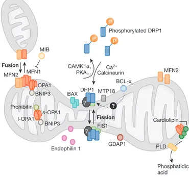
? P P P Phosphorylated DRP1 CAMK1α, PKA... MFN1 Fusion Fission MFN2 MFN2 PLD MIB OPA1 BNIP3 BNIP3 s-OPA1 Prohibitin I-OPA1 GDAP1 Endophilin 1 Phosphatidic acid Cardiolipin FIS1 BAX ? ? MTP18 BCL-xL DRP1 CalcineurinCa 2+
Fig 1 | The dynamics of mitochondrial fission and fusion. The localization, as
well as some interaction and modification of the principal proteins involved in the two processes are shown. Once dephosphorylated, DRP1 is recruited to the outer membrane by FIS1 or by another, unknown, component. The oligomerization of DRP1 is followed by constriction of the membrane and mitochondrial fission. The pro-fusion proteins (MFNs on the outer membrane and OPA1 on the inner membrane) oligomerize to induce fusion of the membranes. Other additional components of the machinery are shown. BAX, BCL2-associated X protein; BNIP3, BCL2/E1B 19 kDa-interacting protein 3; CAMK1a, calcium/calmodulin-dependent protein kinase 1a; DRP1, dynamin-related protein 1; FIS1, fission protein 1; GDAP1, ganglioside-induced differentiation-associated protein 1; l-OPA1, long form of OPA1; MFN, mitofusin; MIB, mitofusin-binding protein; MTP18, mitochondrial protein 18 kDa; OPA1, optic atrophy 1; PKA, protein kinase A; PLD, phospholipase D; s-OPA1: short form of OPA1.
Sidebar A | In need of answers
(i) Which is the exact mechanism of fusion of both the outer and inner mitochondria membranes?
(ii) What happens to the inner membrane during fusion?
(iii) Which proteases are responsible for cleavage and processing of OPA1? (iv) Is FIS1 the only partner of DRP1 in the fission machinery? How does
recruited DRP1 induce fragmentation of mitochondria? (v) Are mitochondria-shaping proteins crucial in the regulation/
amplification of programmed cell death? Do they influence a physiological form of cell death?
(vi) How do mitochondria fragment during apoptosis? Do mitochondria-shaping proteins interact directly with members of the BCL2 family? (vii) Do mitochondrial dynamics have a crucial role in key T-cell processes
1999; Frank et al, 2001). When the proapoptotic Bcl2 family mem ber BaX translocates to mitochondria, it colocalizes with Drp1 and MFn2 on the induction of stressdependent apoptosis (Karbowski et al, 2002). thus, BaX might be targeting subdomains of mitochondria that are most prone to dynamic membrane rearrangements. Similarly, the consequences of fragmentation are unclear: most laboratories have reported a role for fragmentation in the progression of apopto sis (lee et al, 2004) and in the survival of cells in clonogenic assays (cereghetti et al, 2010), but others have shown that fragmentation has no role in cell death (autret & Martin, 2009).
cristae remodelling is a process by which cristae junctions are widened, leading to the mobilization of the cytochrome c pool from the cristae to the intermembrane space (iMS) for its complete release (Scorrano et al, 2002). a complex comprising a longer membrane bound fraction and a shorter soluble iMS fraction of opa1 gener ated by the rhomboid protease parl controls the cristae junction size (Frezza et al, 2006; cipolat et al, 2006). During apoptosis this complex is disrupted—even before the activation of multidomain proapoptotic Bcl2 proteins (yamaguchi et al, 2008)—leading to complete cytochrome c release. changes in cristae structure have been reported to be caspasedependent (Sun et al, 2007) or very subtle (yamaguchi et al, 2008). it is beyond the scope of this review to address the detailed experimental differences leading to these results. Here, it suffices to mention that (i) the disassembly of the opa1 complex has been found in all the apoptotic paradigms in which it has been tested; (ii) cristae remodelling was originally identified (and is still studied) by using an in vitro system in which caspases are absent, ruling out a role for any postmitochondrial pathway; and (iii) differences in the experimental conditions and in the way that measurements in electron tomograms are performed could blunt changes in the cristae junction. Mounting evidence sub stantiates a model in which changes in mitochondrial ultrastructure occur during apoptosis, are regulated by the opa1 complex, and are required for complete cytochrome c release. Whether or not the opa1 complex is a direct target for proapoptotic members of the Bcl2 family remains unclear. the BH3only member of the Bcl2 family, Bnip3, was recently found to interact with opa1, inhibit ing its fusogenic function and inducing opa1 deoligo merization in a BaX/BaK and BH3domaindependent manner (landes et al, 2010). interestingly, Bnip3 is positioned at the crossroad between apoptosis and autophagy and is believed to participate in the selective autophagy of mitochondria, called mitophagy.
autophagy is a form of recycling that reprocesses cytosolic constituents, from bulk cytoplasm to entire organelles (glick et al, 2010). the importance of this mechanism is emerging in the context of crucial physiological as well as pathological processes (cecconi & levine, 2008). Hypoxiainduced autophagy depends on both Bnip3 and niX (Bellot et al, 2009; zhang & ney, 2009; novak et al, 2010) and the fragmentation of mitochondria occurs before being engulfed by autophagosomes (twig et al, 2008). the disruption of the mitochondrial network similarly constitutes an amplificatory loop during autophagy in muscular atrophy (romanello et al, 2010). During atrophy, conserved atrophic factors, such as FoXo3, trig ger Bnip3dependent mitochondrial fragmentation, which causes mitochondrial dysfunction and is required for organelle removal. interestingly, mitochondrial fission associated with dysfunction is able to induce muscle atrophy, elucidating a feedforward loop that stems from mitochondria to activate the conserved autophagic path ways (romanello et al, 2010).
Mitochondrial dynamics in the immune system
as we have outlined above, mitochondrial dynamics have a role in the regulation of several key cellular processes. one of the emerg ing areas in which mitochondria and mitochondrial dynamics are increasingly recognized as crucial is the immune system. the immune response can be broadly divided into innate and adaptive immunity: the first is thought to constitute an evolutionarily older defence strategy and provides immediate and nonspecific defence against infection (by recognizing components that are conserved among broad groups of microorganisms), whereas the second is com posed of highly specialized, systemic cells that provide the vertebrate immune system with the ability to recognize and remember specific pathogens, and to mount stronger attacks each time the pathogen is encountered. the signalling pathways involved are fairly well charac terized in both innate and adaptive immunity, making these systems useful to elucidate whether mitochondrial shape has any role.
Mitochondrial dynamics in innate immunity
When an organism senses a viral infection, it directly activates immune cells through a complex signalling pathway. the recogni tion of viral material is performed mainly by two groups of receptor:
Glossary
ADOA autosomal dominant optic atrophy AP1 activator protein 1
APC antigen-presenting cell BAX BCL2-associated X protein BAK BCL2-antagonist/killer BCL2 B-cell lymphoma protein 2 BH3 Bcl-2 homology domain 3
BNIP3 Bcl2/E1B 19 kDa-interacting protein 3 CXCL12 CXC (chemokine) ligand 12 CRAC Ca2+ release-activated Ca2+ channel
DRP1 dynamin-related protein 1 ER endoplasmic reticulum FIS1 fission protein 1 FOXO3 forkhead box O3
GDAP1 ganglioside-induced differentiation-associated protein 1 IKK IκB kinase
LETM1 leucine zipper-EF-hand-containing transmembrane protein 1 MAVS mitochondrial antiviral signalling
MFN1/2 mitofusin 1/2
MHC major histocompatibility complex MIB mitofusin-binding protein MTP18 mitochondrial protein 18 kDa
MyD88 myeloid differentiation primary response gene 88 NIX Nip3-like protein X
NFAT nuclear factor of activated T cells OPA1 optic atrophy 1
ORAI1 calcium-release-activated calcium modulator 1 PARL presenilin-associated rhimboid-like protease PLD phospholipase D
RLH retinoic acid-inducible gene I-like helicase SRC abbreviation of Sarcoma
STIM1 stromal interacting molecule 1 STING stimulator of interferon genes TBK1 TANK-binding kinase 1 TCR T-cell receptor TLR Toll-like receptor
vMIA viral mitochondria-localized inhibitor of apoptosis ZAP70 zeta-chain-associated protein kinase 70
the tlrs and the rlH family. these specific receptors interact with cytoplasmic adaptor molecules such as MyD88 (loiarro et al, 2009) and MaVS (Seth et al, 2005), which in turn activate two cytosolic protein kinase complexes, tBK1 and iKK, leading to the production of type i interferons and proinflammatory cytokines (Moore & ting, 2008). Mitochondria were thought to have a role when the specific mitochondrial localization of MaVS was discovered (Seth et al, 2005). More recently, mitochondrial morphology has been found to have a crucial role in modulating downstream signalling of MaVS (castanier et al, 2010). after specific rlH activation, mitochondria elongate and enhance antiviral signalling. accordingly, MaVS and the promitochondrial fusion protein MFn1 interact under normal condi
tions. Mechanistically, this could be explained by the fact that mito chondrial elongation regulates the association of MaVS with Sting, an Er molecule crucial for the antiviral cell response (ishikawa & Barber, 2008). the activation of rlH induces the degradation of one MaVS isoform with the consequent disruption of a MaVS–MFn1– Sting complex located at sites of Er–mitochondrial tethering. this frees MFn1 to orchestrate mitochondrial fusion. Fusion is required to enhance the Sting–MaVS—that is, the remaining isoform—inter action and the downstream signalling propagation. the expression of the viral protein vMia—used by viruses to enhance infectiv ity—fragments mitochondria, reduces Sting–MaVS association and delays the degradation of the MaVS isoform.
Similarly, MFn2 inhibits mitochondrial antiviral immunity: MFn2 overexpression blocks antiviral signalling induction and its silencing decreases viral replication (yasukawa et al, 2009). interestingly, MFn2 is a physical tether of Er to mitochondria (de Brito & Scorrano, 2008b), pointing to a role of Er–mitochondria tethering in the modulation of the innate immune response.
A link between mitochondrial dynamics and signalling?
lymphocytes are key cells of the immune system that are involved mainly in adaptive immunity and in the effective clearance of infec tions from the organism. they circulate in the blood until they are activated in response to an infection, at which point they sense and respond to chemoattractant gradients that recruit them to the site of infection with asymmetrical changes in cell morphology (polari zation) and mobility (chemotaxis). to achieve directed movement, cells organize and maintain spatial and functional asymmetry with a defined anterior (leading edge) and posterior (uropod; lauffenburger & Horwitz, 1996). thus, tcell migration or chemotaxis is required to maintain homeostasis and to achieve appropriate immuno logical reactions. once at the site of infection, t cells deploy an array of responses that lead to the activation and proliferation of specific effec tor t cells and an effective immune response. the activation of t cells occurs through the simultaneous engagement of the tcr and the co stimulatory receptor cD28 on the t cell by the MHc peptide and B7 family members on the apc, respectively (Krammer et al, 2007). one of the earliest events in tcell activation is the induction of the tyro sine kinase activity of members of the Src and zap70 kinase families. Downstream from these, activation of the transcription factors nFκB and ap1, as well as ca2+/calcineurindependent activation of the tran scription factor nFat, all induce the transcription of a pleiotropic set of genes that promote the longterm proliferation of activated t cells. any of these steps requires energy or depends on ca2+, and is there fore likely to involve some degree of control by mitochondria.cells of the immune system have few mitochondria and are usually regarded as glycolytic; however, emerging evidence indi cates a role for mitochondria in the response of t cells to exter nal cues. thus, t cells could be a good and ‘simplified’ system with which to characterize the interplay between mitochondrial morphology and signalling cascades, the latter of which are well characterized in t cells.
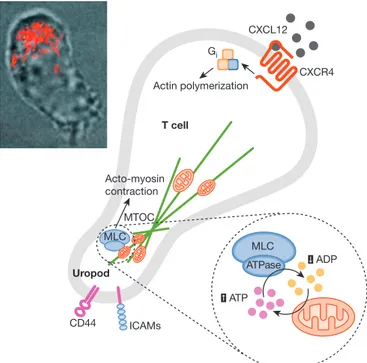
Mitochondria and mitochondrial dynamics have thus far been linked to at least two aspects of the function of t cells: chemotaxis and the regulation of the formation and activity of the immuno logical synapse. We recently demonstrated that mitochondria specifically redistribute to and accumulate at the uropod during directed leukocyte migration to provide atp, thus regulating the cell motor of migrating lymphocytes (Fig 2; campello et al, 2006). Gi CXCL12 CXCR4 Actin polymerization Acto-myosin contraction Uropod MTOC CD44 ICAMs ATP ADP MLC MLC ATPase T cell
Fig 2 | A model for mitochondrial dynamics in the orchestration of T-cell
chemotaxis. To achieve directed movement, lymphoid cells organize and maintain spatial and functional asymmetry, with a leading edge containing the machinery for actin polymerization and gradient sensing and a uropod containing the adhesion molecules and the MTOC. At the uropod, myosin filaments contract actin filaments to provide the tension required for cell movement. Myosin II activity is controlled mainly through the phosphorylation of MLC, which induces a conformational change allowing actin–myosin interactions and activating its ATPase activity. Myosin II ATPase depends on availability of the substrate and clearance of the product. Even in the presence of optimal ATP concentration, accumulation of ADP near the enzyme will slow down its activity and block migration. However, during lymphocyte migration, mitochondria are transported to the uropod along microtubules, in a process requiring Gi protein signalling and mitochondrial fission. The position of mitochondria is strategic for myosin ATPase activity: mitochondria not only supply ATP, but also withdraw ADP, thus creating optimal conditions for MLC. Inset: reconstruction of a confocal z-stack of images of a polarized Jurkat cell expressing mitochondrially targeted dsRED merged with a bright field image of the same cell showing polarization of mitochondria at the uropod. CXCL12, CXC ligand 12; CXCR4, CXC chemokine receptor 4; ICAM, intercellular adhesion molecule 1; MLC, myosin light chain; MTOC, microtubule organizing centre.
More interestingly, the accumulation of mitochondria at the uro pod requires their fission. genetic manoeuvres that elongate mito chondria were found to block mitochondrial and cell polarization in response to chemokines and, more importantly, prevented the migration of t lymphocytes towards cXcl12 gradients. this inhi bition of mitochondrial transport could reflect an inability of the cytoskeleton to transport organelles that are too large. alternatively, altered mitochondriashaping proteins could be unable to interact with the components of the mitochondria transport machinery, such as the kinesin motors or the adaptor Miro–Milton complex (liu & Hajnoczky, 2009). Kinesin and dynein motors control the move ment of mitochondria along microtubules, which in turn regulates the distribution of the organelles in the cell. to move, the extensive mitochondrial network must be divided into smaller organelles that can be moved readily by the motors (Hollenbeck & Saxton, 2005). to this end, the machinery that transports mitochondria is probably coordinated with mitochondriashaping proteins, as substantiated by the finding that disruption of the dynein complex results in mito chondrial elongation caused by Drp1 blockage (Varadi et al, 2004). it remains to be seen whether changes in mitochondrial transport can reproduce the effects of unopposed fusion, in vitro and in vivo. irrespective of the mechanism, when mitochondria do not relocate properly, tcell polarization and migration are impeded, revealing a previously unexpected role in the immune system for morpho logical adaptations of mitochondria.
the second key physiological process in which mitochondrial shape has been implicated is the regulation of gene expression after the activation of t cells. the role of global ca2+ signals for gene expression regulation (Feske et al, 2001), cell activation and pro liferation (Schwarz et al, 2007) in t cells is already well established. an in dispensable step during tcell activation is ca2+ entry across the plasma membrane through the opening of crac/orai1 channels, which are in turn opened by StiM1 as a consequence of the deple tion of intracellular ca2+ stores (cahalan & chandy, 2009). to prevent ca2+dependent inactivation of crac/orai1 channels, mitochon dria act efficiently as inflowing ca2+ buffers (parekh & putney, 2005). it has been shown recently that mitochondria translocate towards the plasma membrane and the immunological synapse, which is the contact site between activator and effector cells, during activation of at least certain tcell subpopulations (abarcarojano et al, 2009; Kummerow et al, 2009). Mitochondrial fragmentation is implicated in this relocation, therefore adding another role for the fission machin ery in immunological synapse formation and maintenance. However, mitochondrial accumulation at the immunological synapse seems to be more a consequence of cellular, rather than organellar, shape changes (Quintana et al, 2009). it therefore remains to be investigated whether the modulation of mitochondrial fusion, fission and transport influences the formation and stability of the immunological synapse, as well as the ability of mitochondria to regulate ca2+ influx through storeoperated channels.
Conclusions
the machinery controlling mitochondria shape and dynamics are becoming well established and defined. Emerging evidence increas ingly suggests that these processes are crucial for the physiology of the cell, as well as for the pathology of the organism. conversely, our knowledge of the role of mitochondria dynamics in the immune system is still rather scarce. nevertheless, appealing yet preliminary data have shed light on the potential importance of mitochondrial
dynamics in tcell physiology. in addition, the signalling cascades that lead to immune cell activation are reasonably well understood, and genetic models make it possible to isolate the key steps of these cascades. thus, t cells are an appealing system in which to place mitochondrial dynamics in the context of known elements of sig nalling. in the long term, mitochondria and mitochondrial dynamics could be unveiled as an important therapeutic target to modulate tcell function in common autoimmune diseases, graftversushost diseases and infections, as well as to modulate viral infections in innate immunity.
acKnoWlEDgEMEntS
l.S. is a senior telethon scientist of the Dulbeccotelethon institute and is supported by Swiss national Foundation grant 31118171, oncoSuisse and telethon italy. S.c. was supported by a roche Foundation postdoctoral Fellowship and is supported by a Fp7pEoplEiEF2008 grant (grant agreement 235595).
conFlict oF intErESt
the authors declare that they have no conflict of interest.
rEFErEncES
abarcarojano E, MunizHernandez S, Morenoaltamirano MM, MondragonFlores r, Enriquezrincon F, Sanchezgarcia FJ (2009) reorganization of mitochondria at the nK cell immune synapse. Immunol Lett 122: 18–25
alexander c et al (2000) opa1, encoding a dynaminrelated gtpase, is mutated in autosomal dominant optic atrophy linked to chromosome 3q28. Nat Genet 26: 211–215
autret a, Martin SJ (2009) Emerging role for members of the Bcl2 family in mitochondrial morphogenesis. Mol Cell 36: 355–363
Bellot g, garciaMedina r, gounon p, chiche J, roux D, pouyssegur J, Mazure nM (2009) Hypoxiainduced autophagy is mediated through hypoxiainducible factor induction of Bnip3 and Bnip3l via their BH3 domains. Mol Cell Biol 29: 2570–2581
Benard g, rossignol r (2008) Mitochondrial fluidity matters. Focus on ‘inherited complex i deficiency is associated with faster protein diffusion in the matrix of moving mitochondria’. Am J Physiol Cell Physiol 294: c1123 Benard g, Bellance n, James D, parrone p, Fernandez H, letellier t,
rossignol r (2007) Mitochondrial bioenergetics and structural network organization. J Cell Sci 120: 838–848
BereiterHahn J, Voth M (1994) Dynamics of mitochondria in living cells: shape changes, dislocations, fusion, and fission of mitochondria. Microsc Res Tech 27: 198–219
cahalan MD, chandy Kg (2009) the functional network of ion channels in t lymphocytes. Immunol Rev 231: 59–87
campello S, lacalle ra, Bettella M, Manes S, Scorrano l, Viola a (2006) orchestration of lymphocyte chemotaxis by mitochondrial dynamics. J Exp Med 203: 2879–2886
castanier c, garcin D, Vazquez a, arnoult D (2010) Mitochondrial dynamics regulate the rigilike receptor antiviral pathway. EMBO Rep 11: 133–138 cecconi F, levine B (2008) the role of autophagy in mammalian
development: cell makeover rather than cell death. Dev Cell 15: 344–357 cereghetti gM, Scorrano l (2006) the many shapes of mitochondrial death.
Oncogene 25: 4717–4724
cereghetti gM, costa V, Scorrano l (2010) inhibition of Drp1dependent mitochondrial fragmentation and apoptosis by a polypeptide antagonist of calcineurin. Cell Death Differ [Epub 21 May] doi:10.1038/cdd.2010.61 choi Sy, Huang p, Jenkins gM, chan Dc, Schiller J, Frohman Ma (2006)
a common lipid links Mfnmediated mitochondrial fusion and SnarE regulated exocytosis. Nat Cell Biol 8: 1255–1262
cipolat S, Martins de Brito o, Dal zilio B, Scorrano l (2004) opa1 requires mitofusin 1 to promote mitochondrial fusion. Proc Natl Acad Sci USA 101: 15927–15932
cipolat S et al (2006) Mitochondrial rhomboid parl regulates cytochrome c release during apoptosis via opa1dependent cristae remodeling. Cell 126: 163–175
de Brito oM, Scorrano l (2008a) Mitofusin 2: a mitochondriashaping protein with signaling roles beyond fusion. Antioxid Redox Signal 10: 621–633
de Brito oM, Scorrano l (2008b) Mitofusin 2 tethers endoplasmic reticulum to mitochondria. Nature 456: 605–610
de Vos KJ, allan VJ, grierson aJ, Sheetz Mp (2005) Mitochondrial function and actin regulate dynaminrelated protein 1dependent mitochondrial fission. Curr Biol 15: 678–683
Dimmer KS, navoni F, casarin a, trevisson E, Endele S, Winterpacht a, Salviati l, Scorrano l (2008) LETM1, deleted in Wolf–Hirschhorn syndrome is required for normal mitochondrial morphology and cellular viability. Hum Mol Genet 17: 201–214
Ehses S, raschke i, Mancuso g, Bernacchia a, geimer S, tondera D, Martinou Jc, Westermann B, rugarli Ei, langer t (2009) regulation of opa1 processing and mitochondrial fusion by maaa protease isoenzymes and oMa1. J Cell Biol 187: 1023–1036
Ernster l, Schatz g (1981) Mitochondria: a historical review. J Cell Biol 91: 227s–255s
Eura y, ishihara n, oka t, Mihara K (2006) identification of a novel protein that regulates mitochondrial fusion by modulating mitofusin (Mfn) protein function. J Cell Sci 119: 4913–4925
Feske S, giltnane J, Dolmetsch r, Staudt lM, rao a (2001) gene regulation mediated by calcium signals in t lymphocytes. Nat Immunol 2: 316–324 Frank S, gaume B, Bergmannleitner ES, leitner WW, robert Eg, catez F,
Smith cl, youle rJ (2001) the role of dynaminrelated protein 1, a mediator of mitochondrial fission, in apoptosis. Dev Cell 1: 515–525
Frey tg, Mannella ca (2000) the internal structure of mitochondria. Trends Biochem Sci 25: 319–324
Frezza c et al (2006) opa1 controls apoptotic cristae remodeling independently from mitochondrial fusion. Cell 126: 177–189
germain M, Mathai Jp, McBride HM, Shore gc (2005) Endoplasmic reticulum BiK initiates Drp1regulated remodelling of mitochondrial cristae during apoptosis. EMBO J 24: 1546–1556
glick D, Barth S, Macleod KF (2010) autophagy: cellular and molecular mechanisms. Pathol J 221: 3–12
green Dr, Kroemer g (2004) the pathophysiology of mitochondrial cell death. Science 305: 626–629
Hackenbrock cr (1966) ultrastructural bases for metabolically linked mechanical activity in mitochondria. i. reversible ultrastructural changes with change in metabolic steady state in isolated liver mitochondria. J Cell Biol 30: 269–297
Hackenbrock cr (1972) Energylinked ultrastructural transformations in isolated liver mitochondria and mitoplasts. preservation of configurations by freezecleaving compared to chemical fixation. J Cell Biol 53: 450–465 Han XJ, lu yF, li Sa, Kaitsuka t, Sato y, tomizawa K, nairn ac, takei K,
Matsui H, Matsushita M (2008) caM kinase i alphainduced
phosphorylation of Drp1 regulates mitochondrial morphology. J Cell Biol 182: 573–585
Harder z, zunino r, McBride H (2004) Sumo1 conjugates mitochondrial substrates and participates in mitochondrial fission. Curr Biol 14: 340–345
Hollenbeck pJ, Saxton WM (2005) the axonal transport of mitochondria. J Cell Sci 118: 5411–5419
ishikawa H, Barber gn (2008) Sting is an endoplasmic reticulum adaptor that facilitates innate immune signalling. Nature 455: 674–678
James Di, parone pa, Mattenberger y, Martinou Jc (2003) hFis1, a novel component of the mammalian mitochondrial fission machinery. J Biol Chem 278: 36373–36379
Karbowski M, lee yJ, gaume B, Jeong Sy, Frank S, nechushtan a, Santel a, Fuller M, Smith cl, youle rJ (2002) Spatial and temporal association of Bax with mitochondrial fission sites, Drp1, and Mfn2 during apoptosis. J Cell Biol 159: 931–938
Karbowski M, Jeong Sy, youle rJ (2004) Endophilin B1 is required for the maintenance of mitochondrial morphology. J Cell Biol 166: 1027–1039 Krammer pH, arnold r, lavrik in (2007) life and death in peripheral t cells.
Nat Rev Immunol 7: 532–542
Kummerow c, Junker c, Kruse K, rieger H, Quintana a, Hoth M (2009) the immunological synapse controls local and global calcium signals in t lymphocytes. Immunol Rev 231: 132–147
landes t, Emorine lJ, courilleau D, rojo M, Belenguer p, arnaunepelloquin l (2010) the BH3only Bnip3 binds to the dynamin opa1 to promote mitochondrial fragmentation and apoptosis by distinct mechanisms. EMBO Rep 11: 459–465
lauffenburger Da, Horwitz aF (1996) cell migration: a physically integrated molecular process. Cell 84: 359–369
lee yJ, Jeong Sy, Karbowski M, Smith cl, youle rJ (2004) roles of the mammalian mitochondrial fission and fusion mediators Fis1, Drp1, and opa1 in apoptosis. Mol Biol Cell 15: 5001–5011
li z, okamoto K, Hayashi y, Sheng M (2004) the importance of dendritic mitochondria in the morphogenesis and plasticity of spines and synapses. Cell 119: 873–887
liesa M, palacin M, zorzano a (2009) Mitochondrial dynamics in mammalian health and disease. Physiol Rev 89: 799–845
liu X, Hajnoczky g (2009) ca2+dependent regulation of mitochondrial
dynamics by the Miro–Milton complex. Int J Biochem Cell Biol 41: 1972–1976
loiarro M, gallo g, Fanto n, de Santis r, carminati p, ruggiero V, Sette c (2009) identification of critical residues of the MyD88 death domain involved in the recruitment of downstream kinases. J Biol Chem 284: 28093–28103 Martinou i, Desagher S, Eskes r, antonsson B, andre E, Fakan S, Martinou Jc
(1999) the release of cytochrome c from mitochondria during apoptosis of ngFdeprived sympathetic neurons is a reversible event. J Cell Biol 144: 883–889
Merkwirth c, Dargazanli S, tatsuta t, geimer S, lower B, Wunderlich Ft, von Kleistretzow Jc, Waisman a, Westermann B, langer t (2008) prohibitins control cell proliferation and apoptosis by regulating
opa1dependent cristae morphogenesis in mitochondria. Genes Dev 22: 476–488
Moore cB, ting Jp (2008) regulation of mitochondrial antiviral signaling pathways. Immunity 28: 735–739
novak i et al (2010) nix is a selective autophagy receptor for mitochondrial clearance. EMBO Rep 11: 45–51
okamoto K, Shaw JM (2005) Mitochondrial morphology and dynamics in yeast and multicellular eukaryotes. Annu Rev Genet 39: 503–536
parekh aB, putney JWJ (2005) Storeoperated calcium channels. Physiol Rev 85: 757–810
pedrola l, Espert a, Wu X, claramunt r, Shy ME, palau F (2005) gDap1, the protein causing charcot–Marie–tooth disease type 4a, is expressed in neurons and is associated with mitochondria. Hum Mol Genet 14: 1087– 1094
Quintana a, Kummerow c, Junker c, Becherer u, Hoth M (2009)
Morphological changes of t cells following formation of the immunological synapse modulate intracellular calcium signals. Cell Calcium 45: 109–122 rizzuto r, Bernardi p, pozzan t (2000) Mitochondria as allround players of the
calcium game. J Physiol 529: 37–47
romanello V et al (2010) Mitochondrial fission and remodelling contributes to muscle atrophy. EMBO J 29: 1774–1785
rossignol r, Karbowski M (2009) Editorial of the directed issue on
mitochondrial dynamics in biology and medicine. Int J Biochem Cell Biol 41: 1748–1749
Santel a, Fuller Mt (2001) control of mitochondrial morphology by a human mitofusin. J Cell Sci 114: 867–874
Scheckhuber cQ, Erjavec n, tinazli a, Hamann a, nystrom t, osiewacz HD (2007) reducing mitochondrial fission results in increased life span and fitness of two fungal ageing models. Nat Cell Biol 9: 99–105
Schwarz Ec et al (2007) calcium dependence of t cell proliferation following focal stimulation. Eur J Immunol 37: 2723–2733
Scorrano l, ashiya M, Buttle K, Weiler S, oakes Sa, Mannella ca, Korsmeyer SJ (2002) a distinct pathway remodels mitochondrial cristae and mobilizes cytochrome c during apoptosis. Dev Cell 2: 55–67 Seth rB, Sun l, Ea cK, chen zJ (2005) identification and characterization of
MaVS, a mitochondrial antiviral signaling protein that activates nFκB and irF 3. Cell 122: 669–682
Smirnova E, griparic l, Shurland, Dl, van der Bliek aM (2001) Dynamin related protein Drp1 is required for mitochondrial division in mammalian cells. Mol Biol Cell 12: 2245–2256
Sun Mg, Williams J, Munozpinedo c, perkins ga, Brown JM, Ellisman MH, green Dr, Frey tg (2007) correlated threedimensional light and electron microscopy reveals transformation of mitochondria during apoptosis. Nat Cell Biol 9: 1057–1065
Szabadkai g, Simoni aM, chami M, Wieckowski Mr, youle rJ, rizzuto r (2004) Drp1dependent division of the mitochondrial network blocks intraorganellar ca2+ waves and protects against ca2+mediated apoptosis.
Mol Cell 16: 59–68
tondera D, czauderna F, paulick K, Schwarzer r, Kaufmann J, Santel a (2005) the mitochondrial protein Mtp18 contributes to mitochondrial fission in mammalian cells. J Cell Sci 118: 3049–3059
twig g et al (2008) Fission and selective fusion govern mitochondrial segregation and elimination by autophagy. EMBO J 27: 433–446 Varadi a, Johnsoncadwell li, cirulli V, yoon y, allan VJ, rutter ga (2004)
cytoplasmic dynein regulates the subcellular distribution of mitochondria by controlling the recruitment of the fission factor dynaminrelated protein 1. J Cell Sci 117: 4389–4400
Verstreken p, ly cV, Venken KJ, Koh tW, zhou y, Bellen HJ (2005) Synaptic mitochondria are critical for mobilization of reserve pool vesicles at Drosophila neuromuscular junctions. Neuron 47: 365–378 Wasilewski M, Scorrano l (2009) the changing shape of mitochondrial
apoptosis. Trends Endocrinol Metab 20: 287–294
yamaguchi r, lartigue l, perkins g, Scott rt, Dixit a, Kushnareva y, Kuwana t, Ellisman MH, newmeyer DD (2008) opa1mediated cristae opening is Bax/Bak and BH3 dependent, required for apoptosis, and independent of Bak oligomerization. Mol Cell 31: 557–569
yasukawa K, oshiumi H, takeda M, ishihara n, yanagi y, Seya t, Kawabata S, Koshiba t (2009) Mitofusin 2 inhibits mitochondrial antiviral signaling. Sci Signal 2: ra47
yonashiro r et al (2006) a novel mitochondrial ubiquitin ligase plays a critical role in mitochondrial dynamics. EMBO J 25: 3618–3626 yoon y, Krueger EW, oswald BJ, Mcniven Ma (2003) the mitochondrial
protein hFis1 regulates mitochondrial fission in mammalian cells through
an interaction with the dynaminlike protein Dlp1. Mol Cell Biol 23: 5409–5420
yu t, robotham Jl, yoon y (2006) increased production of reactive oxygen species in hyperglycemic conditions requires dynamic change of mitochondrial morphology. Proc Natl Acad Sci USA 103: 2653–2658 zhang J, ney pa (2009) role of Bnip3 and niX in cell death, autophagy,
and mitophagy. Cell Death Differ 16: 939–946
zuchner S et al (2004) Mutations in the mitochondrial gtpase mitofusin 2 cause charcot–Marie–tooth neuropathy type 2a. Nat Genet 36: 449–451