Letter to the Editor
Percutaneous coronary intervention driven by combined use of
intracoronary anatomy and physiology
Towards a tailored therapy for coronary artery disease
Giampiero Vizzari
a, Alessandro Di Giorgio
a, Francesco Saporito
a, Olimpia Trio
a,
Francesco Versaci
b,c, Giuseppe Andò
a,⁎
a
Cardiology Section, Department of Medicine and Pharmacology, University Hospital of Messina, Italy b
Department of Cardiovascular Diseases, Ospedale“A. Cardarelli”, Campobasso, Italy c
Department of Cardiovascular Diseases, Ospedale“F. Veneziale”, Isernia, Italy
a r t i c l e i n f o
Article history: Received 31 March 2015 Accepted 1 April 2015 Available online 2 April 2015 Keywords:
PCI
Percutaneous coronary revascularization Optical coherence tomography (OCT) Fractionalflow reserve (FFR) Multimodality coronary imaging
Coronary angiography classically allows a bidimensional evaluation of the vascular lumen, however with many limitations in the case of ec-centric lesions, irregular contour or tortuosity of the vessel. Moreover, it does not enable to assess neither the features of the vessel wall, nor the functional significance of a lesion[1]. Newer technologies are available to overcome these limitations. We present a case of percutaneous coro-nary revascularization optimized by combined use of two of the most widely used techniques.
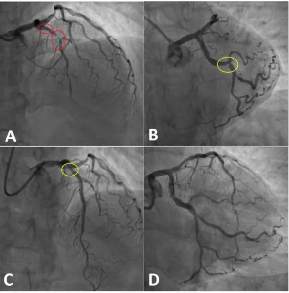
A 59-year-old woman, with a history of psoriasis, primary biliary cirrhosis and untreated hypercholesterolemia, presented to the Emer-gency Room for sudden onset of chest pain at rest, radiating to the left arm and the jaw. The electrocardiogram showed symmetrical inverted T-waves in the anterior leads; the patient was then admitted to the Cardiac Care Unit to undergo further treatment of the underlying acute coronary syndrome (ACS). Cardiac troponin was normal and GRACE score was low (74); nonetheless, because of dynamic T-wave changes (primary high-risk criterion) with positivization during admission, the patient was referred to an invasive evaluation. Coronary angiogram showed an ambiguous ostial lesion of the Left Anterior
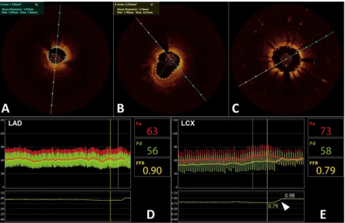
Descending (LAD) artery, followed by a 99% stenosis in the proximal-mid tract, involving the bifurcation with thefirst diagonal branch (Fig. 1-A). In addition, a focal, eccentric intermediate stenosis was present in the middle tract of circumflex artery (Cx) (Fig. 1-B). The angiogram of the right coronary artery was unremarkable. After predilatation of both the LAD and thefirst diagonal, the decision was made to run an optical coherence tomography (OCT) scan, in order to get additional information. OCT was performed to primarily obtain an accurate sizing of the culprit lesion on proximal-mid LAD, aimed to the appropriate choice of the stent; furthermore, it was hypothesized that it might quantify the severity of the ostial LAD stenosis. OCT revealed the existence of a long atheromatous tract, involving the whole middle segment of the LAD, mainly consisting of lipid pool
(signal-poor regions) and deep calcific (signal-rich) components
(Fig. 2-A). Moreover, analysis of the proximal LAD showed a circumfer-ential lipid plaque, resulting in a minimal lumen area (MLA) at the osti-um of about 3.5 mm2(Fig. 2-B), thus lower than the severity cut-off of
4 mm2, which is considered the threshold for a significant
flow-limiting stenosis in large (N3 mm diameter) vessels[2,3]. We proceeded by treating the culprit lesion with two overlapped drug eluting stents for a total length of 46 mm (Fig. 1-C); the diagonal branch was left un-stented (provisional side branch stenting). OCT scan, performed after stent deployment, confirmed an optimal expansion with complete struts apposition also in the overlapped segment and no residual dissec-tions at the two edges (Fig. 2-C). Further evaluation of both ostial LAD and middle Cx intermediate lesions was deferred to a subsequent procedure to avoid contrast volume overload[4,5]. The need of a func-tional assessment of the LAD lesion was based on both the ostial location (a poorly evaluable position) and the ambiguous MLA value at OCT imaging; in addition, the borderline severity of the Cx lesion supported this decision.
One month later, the patient was readmitted to undergo a second
procedure with possible fractionalflow reserve (FFR)-guided PCI.
Central intravenous infusion of Adenosine (140μg/kg/min) was admin-istered to induce maximal hyperemia. FFR calculated in the LAD, distally to both the ostial lesion and the two previously implanted stents, was 0.90, therefore not indicating ischemia (Fig. 2-D). On the contrary, the focal stenosis in the mid Cx demonstrated a 0.79 FFR, resulting eligible
International Journal of Cardiology 187 (2015) 562–564
⁎ Corresponding author at: Via Santa Cecilia, 98, 98123 Messina, Italy. E-mail address:[email protected](G. Andò).
http://dx.doi.org/10.1016/j.ijcard.2015.04.010
0167-5273/© 2015 Elsevier Ireland Ltd. All rights reserved.
Contents lists available atScienceDirect
International Journal of Cardiology
for revascularization. Pressure-wire pullback confirmed the significant coronaryflow reduction induced by this lesion at maximal hyperemia, showing a direct step from 0.79 to the normal value of about 1.0 at the retrograde crossing of the stenosis (Fig. 2-E). Drug eluting stent implantation was then performed and thefinal post-PCI FFR was 0.97, indicating a good result of the intervention withflow improvement (Fig. 1-D).
OCT is a safe, effective and reproducible technique, routinely used for an assessment of plaque morphology and lesion severity. OCT allows optimal identification of thrombus, intimal rupture, lipid plaques, as well as measurement offibrous cap thickness. In addition, it provides accurate quantitative measurements, in terms of lumen areas and diam-eters, able to detect significant coronary lesions requiring an
interven-tional approach[6,7]. MLA values measured by OCT are currently
compared with cut-off values defined by intravascular ultrasound
(IVUS), despite the definition of the lumen–plaque border is significant-ly improved by OCT as compared to IVUS. Different observational studies validated IVUS for the assessment of lesion severity, mainly for
left main coronary artery (LMCA) disease [8,9]. An absolute MLA
lower than 4 mm2was classically correlated with a FFR lower than
0.80 in non-LMCA lesions[2,3,10]. More recent studies have questioned thesefindings[11]and especially the concept of using absolute MLA for the assessment of lesion severity[12]. Our case is a further evidence of
the low reliability of these quantitative criteria, as well of their limita-tions in guiding PCI[13–15].
FFR provides on the contrary a cut-off value for significant ischemia which is independent from the vessel size. It allows reproducible functional assessment of complex anatomy, poorly visible or over-lapped lesions, multivessel disease or multiple stenoses within one artery, long and ostial lesions and LMCA disease. In several randomized trials, FFR-guided PCI showed the improvement of the outcome of patients with functionally significant stable coronary artery disease (FFR of 0.80 or less), compared with medical therapy alone[16,17]. Moreover, in multivessel disease patients, it allows to detect lesions that really need revascularization, hence reducing overtreatment[18].
In conclusion, this case shows the importance of a multimodality approach for both functional and anatomic evaluation of different coronary lesions in multivessel disease. Although a combined use of these techniques might not be feasible in all patients, it allows improv-ing success and durability of percutaneous coronary interventions in specific complex cases.
Conflict of interest
The authors report no relationships that could be construed as a con-flict of interest.
Fig. 1. Coronary angiograms. Diagnostic angiogram of the LAD (A) and Cx (B). (C) Final angiogram after deployment of 2 overlapped drug eluting stent in the proximal-mid LAD. (D) Final angiogram after deployment of 1 drug eluting stent in the Cx. Colored ovals indicate the segments evaluated with OCT (red) and FFR (yellow). (For interpretation of the references to color in thisfigure legend, the reader is referred to the web version of this article.)
563 G. Vizzari et al. / International Journal of Cardiology 187 (2015) 562–564
References
[1] I.D. Kilic, N. Konstantinidis, G. Caiazzo, E. Fabris, S.S. Sherif, C. Di Mario, Influence of multimodality coronary imaging on revascularization strategy, Int. J. Cardiol. 177 (2014) 515–516.
[2] F. Prati, E. Regar, G.S. Mintz, E. Arbustini, C. Di Mario, I.K. Jang, et al., Expert review document on methodology, terminology, and clinical applications of optical coher-ence tomography: physical principles, methodology of image acquisition, and clini-cal application for assessment of coronary arteries and atherosclerosis, Eur. Heart J. 31 (2010) 401–415.
[3] C. Briguori, A. Anzuini, F. Airoldi, G. Gimelli, T. Nishida, M. Adamian, et al., Intravas-cular ultrasound criteria for the assessment of the functional significance of inter-mediate coronary artery stenoses and comparison with fractionalflow reserve, Am. J. Cardiol. 87 (2001) 136–141.
[4] G. Ando, G. Morabito, C. de Gregorio, O. Trio, F. Saporito, G. Oreto, Age, glomerular filtration rate, ejection fraction, and the AGEF score predict contrast-induced ne-phropathy in patients with acute myocardial infarction undergoing primary percu-taneous coronary intervention, Catheter. Cardiovasc. Interv. 82 (2013) 878–885.
[5] G. Ando, C. de Gregorio, G. Morabito, O. Trio, F. Saporito, G. Oreto, Renal function-adjusted contrast volume redefines the baseline estimation of contrast-induced acute kidney injury risk in patients undergoing primary percutaneous coronary in-tervention, Circ. Cardiovasc. Interv. 7 (2014) 465–472.
[6] F. Prati, G. Guagliumi, G.S. Mintz, M. Costa, E. Regar, T. Akasaka, et al., Expert review document part 2: methodology, terminology and clinical applications of optical co-herence tomography for the assessment of interventional procedures, Eur. Heart J. 33 (2012) 2513–2520.
[7] A. Belkacemi, P.R. Stella, D.S. Ali, P.W. Novianti, P.A. Doevendans, E. van Belle, et al., Diagnostic accuracy of optical coherence tomography parameters in predicting in-stent hemodynamic severe coronary lesions: validation against fractionalflow re-serve, Int. J. Cardiol. 168 (2013) 4209–4213.
[8] J.M. de la Torre Hernandez, F. Hernandez Hernandez, F. Alfonso, J.R. Rumoroso, R. Lopez-Palop, M. Sadaba, et al., Prospective application of pre-defined intravascular ultrasound criteria for assessment of intermediate left main coronary artery lesions results from the multicenter LITRO study, J. Am. Coll. Cardiol. 58 (2011) 351–358.
[9] S.L. Chen, B. Xu, J.B. Chen, T. Xu, F. Ye, J.J. Zhang, et al., Diagnostic accuracy of quan-titative angiographic and intravascular ultrasound parameters predicting the func-tional significance of single de novo lesions, Int. J. Cardiol. 168 (2013) 1364–1369.
[10] S.J. Park, J.M. Ahn, S.J. Kang, S.H. Yoon, B.K. Koo, J.Y. Lee, et al., Intravascular ultrasound-derived minimal lumen area criteria for functionally significant left main coronary artery stenosis, J. Am. Coll. Cardiol. Intv. 7 (2014) 868–874.
[11] T. Pawlowski, F. Prati, T. Kulawik, E. Ficarra, J. Bil, R. Gil, Optical coherence tomogra-phy criteria for defining functional severity of intermediate lesions: a comparative study with FFR, Int. J. Cardiovasc. Imaging 29 (2013) 1685–1691.
[12] R. Waksman, J. Legutko, J. Singh, Q. Orlando, S. Marso, T. Schloss, et al., FIRST: Frac-tional Flow Reserve and Intravascular Ultrasound Relationship Study, J. Am. Coll. Cardiol. 61 (2013) 917–923.
[13] B.K. Koo, H.M. Yang, J.H. Doh, H. Choe, S.Y. Lee, C.H. Yoon, et al., Optimal intravascu-lar ultrasound criteria and their accuracy for defining the functional significance of intermediate coronary stenoses of different locations, J. Am. Coll. Cardiol. Intv. 4 (2011) 803–811.
[14] X.J. Jin, S.J. Tahk, H.M. Yang, H.S. Lim, M.H. Yoon, S.Y. Choi, et al., The relationship be-tween intravascular ultrasound-derived percent total atheroma volume and frac-tionalflow reserve in the intermediate stenosis of proximal or middle left anterior descending coronary artery, Int. J. Cardiol. 185 (2015) 56–61.
[15] S. Sen, J. Davies, Can anatomy be used as a surrogate for physiology? The IVUS co-nundrum, Int. J. Cardiol. 168 (2013) 631–632.
[16] B.C. Zhang, Z.W. Zhou, C. Wang, Y.F. Ma, W.H. Li, D.Y. Li, Fractionalflow reserve im-proves long-term clinical outcomes in patients receiving drug-eluting stent implan-tation: insights from a meta-analysis of 14,327 patients, Int. J. Cardiol. 177 (2014) 1044–1048.
[17] H.S. Lim, P.A. Tonino, B. De Bruyne, A.S. Yong, B.K. Lee, N.H. Pijls, et al., The impact of age on fractionalflow reserve-guided percutaneous coronary intervention: a FAME (Fractional Flow Reserve versus Angiography for Multivessel Evaluation) trial substudy, Int. J. Cardiol. 177 (2014) 66–70.
[18]B. Xu, R. Whitbourn, A. Wilson, A.T. Burns, P.D. Williams, C. Judkins, et al., Clinical impact of fractionalflow reserve in a real-world cohort of patients, Int. J. Cardiol. 172 (2014) 251–252.
Fig. 2. Intracoronary imaging of the LAD (A–C) and functional assessment of both LAD and Cx arteries (D–E). (A) Severe stenosis in the middle tract of the LAD (culprit lesion), investigated by OCT scan. (B) Circumferential lipid plaque in the ostial–proximal LAD, with minimal lumen area lower than 4 mm2
. (C) Optimal stent expansion in the middle LAD, with complete struts apposition and no residual dissections. (D) FFR interrogation performed in the LAD to evaluate the ostial–proximal stenosis resulted 0.90, demonstrating non significant flow-reduction. (E) Significant FFR value in the mid Cx (0.79), indicating critical stenosis. Pullback of the pressure-wire back through the lesion showed a direct step from 0.79 to the basal value of about 1 (arrowhead), further confirming the severity of the lesion.