Ph.D. Thesis
C-tactile fibers mediate Affective Touch:
from childhood to individual differences to neural correlates
Pietro Zingaretti
Department of Psychology
Medicine and Psychology
Sapienza University of Rome
Rome, Italy
Dottorando: Tutor:
Pietro Zingaretti Grazia Fernanda Spitoni
Table of contents
Table of contents 2
Introduction 4
Section I 7
Chapter 1. Touch and CT System 7
1.1. The principal features of CT fibers 9
1.2 Behavioral output of CT fibers: affective touch and methodological issues 14
Chapter 2. Experiencing affective touch 18
2.1. Affective touch in the lifespan 18
2.2. The social touch perspective and individual differences in experiencing Affective Touch 20
Chapter 3. The neural correlates of affective touch 26
Section II 34
Study I: Tactile sensitivity, tactile acuity and affective touch: from childhood to early adolescence 36
3.2 Material and Methods 38
3.3 Results 44
3.4 Discussion 48
Study II: Altered perception of affective touch in disorganized attachment 52
4.1 Introduction 52
4.2 Methods 54
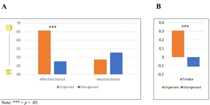
4.3 Results 58
4.4 Discussion 60
Study III: Brain mechanism for processing affective touch 63
in disorganized attachment: preliminary results 63
5.1 Introduction 63 5.2 Methods 64 5.3 Results 69 5.4 Discussion 75 General Discussion 80 Conclusion 85 References 86
Introduction
In the past 3 years, throughout my doctoral project, I had the chance to carry out a series of
researches ranging from a behavioral to neuroimaging techniques.
The main issue that has interested me since the very first steps of my journey, was the tactile
system and in particular a specific function of this system that is the affective touch. The
affective touch is a construct that, in the last years, has gained much interest from the
international scientific community. This is because the correlates of this construct appear to be
linked to different domains of our lives, to the body of course, but also to the human mind, thus
both on a physical and on a physiological level. Basically, as the reader will understand moving
forward trhough the current thesis, the affective touch is nothing but a simple and powerful
behavioral output, it is a feeling of pleasure perceived by stimulating the skin at a specific
frequency. The feeling of pleasure is given by a specific group of fibers - C fibers - which have
peculiar characteristics such as slow conduction. Up to a few years ago, these fibers were
mainly studied for their role on conduction of pain perception. Actually, the step forward that
has interested the researchers, is that this class of unmyelinated fibers, if stimulated to a
determined frequency, produces a sensation of pleasure that is associated to the human caress.
Thus, the fact that stimulation of these tactile receptors evokes a feeling of shared and
measurable pleasantness has opened a wide new perspective. My thesis work lays right within
this beautiful paradigm and my goal is to study this phenomenon through three experiments. In
order to address the contents of the current work more clearly, I decided to divide this
manuscript into two main sections. The first, is an introductive and reviewing part in which I
will describe the basics that will be useful to the reader to understand the affective touch, the C
tactile fibers and the literature directly linked to consecutive experiments.
A second section is dedicated to the experiments that I conducted in my doctoral project. I
distinct studies that, however, follow a temporal progression that reflects both the way they
have been carried out and the logical sequence of the rationale underpinning my doctoral
project.
The first study aimed to verify something very important for me, that is: is the affective
tactile system mature also in children as shown in adults? More specifically, if I stimulate a
child with the same frequency with which an adult perceives affective touch, will a child
perceive that stimulation as pleasant? In other words, is the sensation of affective touch already
present in children and from what age? So, the first study wanted to verify whether the
stimulations of C-fibers in children had pleasantness as a behavioral output. In the first
experiment, I committed myself to demonstrating it.
As for the second study, I tried to apply affective touch in another neuroscientific perspective
that has to do with individual differences. In particular, a growing body of literature on affective
touch has shown how affective touch perception can be altered in people with
psychopathological characteristics such as eating disorders or even in autism. In other words,
what is clear is that people with psychiatric and neuropsychological diseases have different
responses to affective touch. Now the aim of the second study was precisely to verify whether
a particular group of people, i.e. those with a particular pattern of attachment, could respond to
the affective touch in a different way. The reasons why I chose the attachment are essentially
two. On one hand, because attachment is a psychological pattern linked to cure received, such
as human caress that recalls on mind the perception of the affective touch. On the other hand,
because the few studies in question have shown that there is a strong correlation between the
attachment pattern and the affective touch, but no one had ever implemented it using the only
true measure of adult attachment. So, an important aspect of the second study is that we
administered the Adult Attachment Interview, which is the gold standard for defining the state
with a specific attachment pattern, such as the Disorganized one that is the pattern that has been
linked to higher maternal neglect, perceive the affective touch in the same way as individuals
with attachment pattern characterized by less difficulties in relationships such as the Organized
pattern.
Finally, as a physiological development of this second experiment, we wanted to verify if
the interesting results obtained in the previous study, could also have implications at the neural
level. We wondered if it was possible to verify if subjects previously classified as Disorganized,
and therefore with a behavioral alteration of affective touch perception, could also have specific
functional characteristics at the cortical level. So, in the last experiment we conducted a
functional magnetic resonance imaging study administering the procedure of affective touch in
subjects classified as Disorganized and Organized with respect to their attachment pattern,
involving a series of comparative analyses of both the resting-state and the functional activation
in response to affective touch stimulation.
It is clear that the three experiments have engaged me in these three years and that while I
am writing this thesis the last part of the fMRI study is still ongoing because we would like to
confirm the results on a large sample for a wider understanding. Currently these three
experiments seem to have clarified that the structure of affective touch is already present in
childhood, that the affective touch is perceived differently depending on the type of attachment
and that even the cortical circuits that process the response and processing of affective touch in
Section I
Chapter 1. Touch and CT System
In humans, touch is the first sensory system to develop (Bremner & Spence, 2017; McGrath,
2004). The earliest sensations we experienced are tactile. Already at 12 weeks of gestation, the
cutaneous receptors and the somatosensory functions are matured (Humphrey, 1964) and the
fetus is able to make movements when lips are touched (Hooker, 1952); dissimilarly, other
sensory modalities, such as hearing and vision, develop later. The early need of tactile functions
in fetal growth, implies that the initial tactile experiences are of crucial importance in the
development and the maturation of an organism. This issue has been demonstrated in several
developmental pathways, from biological growth (Bremner & Spence, 2017) to psychological
maturity (Ardiel & Rankin, 2010), to social skills achievements (Cascio et al., 2018).
The sense of touch is processed by mechanosensory neurons that are embedded in the skin
and that transmit signals from the periphery to the central nervous system. In essence, the
sensation of touch occurs when a specialized afferent mechanoreceptor in the skin is activated
by a contact stimulus. This can be as little as a gentle breeze over the arms, to the high force
exerted from trapping a finger in the door. A single mechanoreceptive afferent can encode many
aspects of the stimulus (e.g. force, speed, direction and roughness), and when activated together
with other mechano and somatosensory afferents, specific percepts are generated (e.g. wetness
and oiliness; see Bentley 1900).
Most of the research on the human somatosensory touch system has been devoted to
myelinated (Aβ) low threshold mechanoreceptive (LTMR) afferents. This system consists of
large diameter fibers with rapid conduction velocities (approximately 50m s-1) optimized for signaling immediate detection of and discriminative information about a touch stimulus. Aβ
afferents are present throughout the skin, i.e. both in hairy and in glabrous skin, with the highest
resolution during explorative tactile behavior. This is in contrast to the hairy skin, where the
density of myelinated mechanoreceptive afferents is much lower. These fibers are thought to
provide information about tactile discrimination and tactile sensitivity, where the sensitivity
relates to whether something can be felt or not and discrimination relates to spatial tactile acuity
(e.g. differentiating between two points—you could feel a fly that landed on your back but you
would not know how many legs it had!). The whole skin is sensitive to a tactile event; for
example, the upper half of the body is generally more sensitive than the lower half, where the
lips, cheeks and nose are maximally sensitive to pressure (Weinstein, 1968).
Not all types of touch are the same, and specific tactile afferents convey different properties
of touch such as discriminative, thermal, painful, pruritic (itch), or affective information to the
central nervous system. These input channels can be further classified as sub-serving sensory
functions, such as spatial and temporal discrimination, and the provision of essential
information for controlling and guiding exploratory manual behaviours, or affective functions
that include the provision of the subjective experience of affective or emotional pleasurable
touch. As seen above, signaling in fast-conducting myelinated peripheral nerve fibers (Aβ
afferents) is important for the discriminative properties of tactile sensations. On the other hand,
another class of tactile fibers, namely C-tactile (CT) afferents seems to be important for the
rewarding, emotional properties of touch. As a full description of the properties of the tactile
system overcome the aims of this dissertation, I will focus on a selective class of tactile fiber,
named CT.
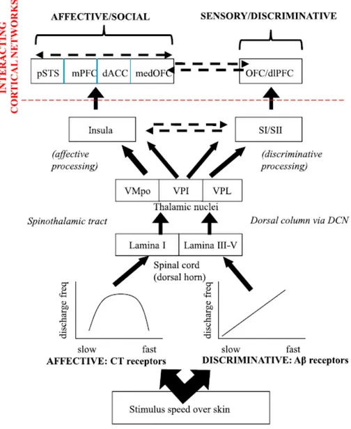
It is worth noting that Aβ and CT fibers underpin different properties. As stated above, Aβ
fibers encode sensory-discriminative aspects of touch with high rapidity and acuity, with
pathways projecting ultimately to primary and secondary sensory cortical areas (see Figure 1
Instead, CT fibers may ‘‘pick out’’ a range of tactile stimuli likely, for the purposes of further
hedonic, rewarding processing in affect-related brain areas such as the insula. Importantly, these
two systems are not separate, despite being at least partly dissociable; it is likely that
sensory-discriminative and motivational-affective pathways for touch interact. CT afferents may
operate as selectors, activated in parallel with Aβ afferents and their cooperation may provide
a complete elaboration of a tactile percept. Still, both from a physiological and functional
perspective, the CT system is characterized by very peculiar features that I shall describe in the
following paragraphs.
1.1. The principal features of CT fibers
The existence of a slow tactile system (C Low Threshold Mechanosensitive Receptors;
CLTMR) was first presented almost 80 years ago in animals. In 1939, the Swedish physiologist
Zotterman proposed that light touch activates not only large afferents but also small
unmyelinated afferents (Zotterman, 1939). Recording from thin strands of the cat saphenous
nerve, he noticed that touching the skin on the lower leg produced impulses of three different
sizes, designated A beta (β), A delta (δ), and C in accordance with the Erlanger−Gasser scheme
(Erlanger and Gasser, 1924). Zotterman emphasized a unique and striking response feature of
the low threshold C mechanoreceptive afferents, that is, a prominent and long-lasting
after-discharge which was not seen in large diameter tactile afferents. On the basis of this finding, he
suggested that unmyelinated tactile afferents might account for the sensation of tickle: “The
itching after-sensation to light touch”- Zotterman said- “is most probably due to fibers
conducting at C rates” (Zotterman, 1939). An important step was taken about 20 years later
when Douglas and Ritchie (1957) demonstrated a number of fundamental properties of the slow
tactile system. Using a cat saphenous nerve preparation with intact connection to the skin,
repetitive electrical stimuli. They showed beyond doubt, that the slow tactile afferents
conducted impulses at a speed of about 1 m s−1 indicating unmyelinated axons. In addition, their study demonstrated that these fibers are abundant in nerves innervating hairy skin of the
cat.
Single unit analysis of CLTMR was pioneered by Iggo and coworkers, who presented
detailed descriptions of response properties to innocuous touch, for example, high sensitivity
to skin deformation, large response to hair movements, intermediate adaptation, and
pronounced post-activation fatigue effect of the sense organ, which may last up to 30 min (Iggo,
1960; Iggo and Kornhuber, 1977). In 1971, an important publication from Perl’s group (Bessou
et al., 1971) emphasized a difference between CLTMR and Aβ tactile afferents with regard to
their dynamic response properties. They wrote that as the velocity of a glass rod “stroked across
the receptive field ... is progressively decreased, the frequency (of the discharge) ... first
increases and then declines”. In myelinated tactile afferents, on the other hand, impulse rate
increases monotonously with velocity of touch movement.
In the last three decades, the investigation of physiological properties of slow-conducting
fibers was shifted to human models, highlighting specific neurophysiological, neural and
behavioral properties. The CT fibers has specific properties in terms of impulse rate,
distribution, density, receptive field, conduction velocity, impulse frequency and fatigue effect.
Impulse Rate: From the literature (Cole et al., 2006; Vallbo et al., 1999), it has been seen
that CT afferents respond to very low indentation forces in the range 0.3–2.5 mM and with
high-frequency responses (50–100 impulses s-1) to innocuous stimuli, such as gentle stroking with a soft brush. This impulse rate is close to the maximum reported for other C afferents
(Kumazawa and Perl, 1977). Despite a very large heterogeneity in CT responses, CTs are
nonetheless a electrophysiologically constrained population of cutaneous afferents, the abiding
Distribution: The distribution of the slow tactile system has been first verified in the skin of
face, forearm, and leg. More specifically, afferent impulses in unmyelinated CT fibers have
been recorded in the small supra- and infraorbital nerves innervating facial skin, in lateral and
dorsal ante-brachial cutaneous nerves innervating the hairy skin of the forearm and hand
dorsum, and in the lateral cutaneous femoral and peroneal nerves innervating the thigh, lower
leg, and the foot (Johansson et al., 1988; Nordin, 1990; Vallbo et al., 1993, 1999; Edin, 2001;
Wessberg et al., 2003; Löken et al., 2007, 2009). On the other hand, nerves innervating the skin
of the trunk have not been exploited so far. Although these findings strongly suggest that CT
innervation of human skin is ubiquitous, a distinct exception is indicated by the fact that CT
has never been encountered in recordings from the glabrous skin of the hand in spite of
extensive analyses of tactile afferents in this skin area. Moreover, the difference between hairy
and glabrous skin in man is consistent with findings in cats, rodents, and nonhuman primates
where the slow tactile system, that is, CLTMR afferents have never been found in nerves
supplying foot pads or monkey glabrous skin.
Density: Information about density of CT afferents comes from microneurography studies.
A monkey study indicates a proximo-distal gradient with fewer CLTMR in the distal parts of
the extremities (Kumazawa and Perl, 1977). A similar gradient is suggested in a human study
focused on distal hairy skin (Löken et al., 2007). Only a few CT afferents were found on hand
dorsum and in lower leg in recordings from the radial and peroneal nerves. In general, present
data suggest that CTs are abundant in the hairy skin of the human body, scarcer in the distal
parts of the extremities and seem to be lacking altogether in the glabrous skin.
Receptive field: Receptive field of CTs mechanoreceptive afferents, defined as the skin area
where adequate stimuli are effective to produce afferent impulses, vary considerably in size and
complexity. It may be a single spot, about 0.25 mm2 in size or it may include up to nine hotspots distributed over an area of 35 mm2; mean field size was 7 mm2 (s.d. 8 mm2 ). Fields are roughly
oval in shape with no preferred orientation. No dependence on location along the forearm
emerged. This field analysis indicates that the stem fiber of a CT afferent commonly branches
to terminate with a varying number of clusters of sensory terminals irregularly distributed
within a relatively small area, rather than providing a continuous mesh of responsive terminals
as suggested by many previous studies based on handheld field exploration (Olausson et al.,
2016).
Conduction velocity: Moving to another feature of the CT fibers, namely the conduction
velocity, it is known that it is characterized by a long latency from stimulus to impulse response
occurring much later the onset of stimulus due to a 30–50 times difference in propagation
velocity. Conduction velocity of CT afferents is about 1 m s−1 (0.6–1.2 m s−1) as assessed from the unit’s response to mechanical stimuli. Neither significant correlation has been found between conduction velocity of individual afferents and location of receptive field along the
extremity, nor with other functional properties of CT units (McGlone et al., 2007).
Impulse frequency: In order to investigate the impulse frequencies, the study of Iggo (1960)
showed that stroking with a soft brush across CT receptive fields evokes peak impulse rates
between 50 and 100 s−1 in a majority of the afferents. Although these rates are not very impressive compared to firing of Aβ afferents, they are, in fact, relatively speaking very high
considering that maximal rate of C mechanoreceptive afferents as found in animal experiments
is 100 impulses s−1 (Iggo, 1960).
Fatigue effect. One of the most distinctive features of CT fibers is the fatigue effect: a
specific response feature of CT-units is that sensory endings of CTs exhibit a pronounced
postactive depression. This characteristic shows a marked decrease of the response after a single
stimulus. When a series of successive indentations are delivered, the response usually decreases
with the first 2–4 stimuli to settle around a submaximal level which is dependent on
resting period of 300 s is substantially larger in terms of impulse rate, number of impulses, as
well as duration of discharge than that after 60 s. There are indications that full recovery may
take several minutes. However, systematic analyses regarding development of fatigue and time
course of recovery remain to be pursued in man (Iggo, 1960; Iggo & Kornhuber, 1977).
Before moving to the behavioral features of the CT system, is of importance to briefly describe
the spinal and cortical processing of CT fibers. Data on the spinal pathways enrolled in the CT
processing, came from human studies after surgical sectioning of the anterolateral
spinothalamic tract for treatment of chronic intractable pain. The earliest observation that
cutting this tract impacts on aspects of CT signaling was made by a German neurologist and
neurosurgeon, Otfrid Foerster (Foerster et al., 1932):
‘Except for the pain and temperature sensations, also other sensory qualities were spoiled after the anterolateral transection. First of all, the feelings of tickle and itch were included, but so were all other feelings of pleasure and displeasure as well’. (p. 43, translated from the
original German).
This lack of ‘‘pleasure’’ after transection of the spinothalamic tract provides at least
circumstantial evidence that CTs ascend in the same tract as C-nociceptors. Lahuerta et al.
(1994) made similar observations in a cohort of anterolateral cordotomized patients, reporting
that they do not experience cutaneous erotic sensation when receiving low-intensity tactile
stimulation. These clinical observations support the existence of a spinothlamic pathway for
signaling CT-mediated pleasant properties of touch. Through the caudal part of the
posterolateral-ventral and the lateral central nuclei of the thalamus, the sensorial percept reaches
primary and higher cortical areas where it is elaborated and decoded (Dum et al., 2009). In
healthy humans, a number of studies have found different cortical areas and signatures that
sake of reading, I will fully address the issue of the neural correlate of the Affective Touch in
the Chapter 3, of my dissertation.
1.2 Behavioral output of CT fibers: affective touch and methodological issues
The CT system has been associated to a pleasant, positive hedonic sensation since it has been
systematically studied in humans for the first time. Following the studies by Olausson and
colleagues (2002, 2008; see previous section), the relation between CT fibers and pleasantness
has been further explored by Lӧken and colleagues (2009); using a microneurography technique
for recording single afferent activity in awake humans and a robotic device to deliver moving
tactile stimuli, authors stimulated CT units in the hairy skin of the subjects’ forearm with a soft
brush moving at different speeds. The relationship between brush stroking velocity and units
firing rate was distinctly different between CT and myelinated afferents: CT fibers showed an
inverted U-shaped relationship between brushing velocity and mean firing rate, with highest
responses at 1, 3 and 10 cm s-1. In contrast, mean firing increased monotonically with brushing velocity in all myelinated afferents. When asked to rate on a visual-analog scale (VAS), the
hedonic quality of the brush stroking, subjects rated 1, 3 and 10 cm s-1 as being the most pleasant velocities, with a peak of pleasantness around 3 cm s-1. Authors found a significant linear correlation between mean firing rates and mean ratings of pleasantness for CT but not for
myelinated units. These results are the first demonstration of a relationship between positive
hedonic sensation and coding at level of peripheral afferent nerve, suggesting that CT fibers
contribute critically to pleasant touch. This perspective has been further documented by several
imaging studies demonstrating the crucial function of the posterior insula in the process of the
and recognition of pleasant touch (for a full description of the studies, see Chapter 3)
These evidences sustained the hypothesis that the activation of the CT system is strongly
correlated to a pleasant, hedonic sensation that we experience in everyday life when interacting
the posterior insula, an area that is known for being involved in processing visceral inputs and
in emotion regulation. Implications for the discovery of a segregated system that is specifically
devoted to signaling and processing affective, emotional and social information will be the
focus of the following sections.
The growing number of studies involving affective touch and its relationship with the CT
system highlighted the necessity to develop controlled and valid paradigms to explore this new
dimension of touch. Even in recent years very approximate stimulus control has been accepted
for the study of emotional touch, with hand application of soft cosmetic or artist’s brushes being
commonly used as a prototypically pleasant stimulus (Cascio et al., 2008; Olausson et al., 2002,
2008). This is acceptable in the sense that a pleasant stimulus, such as a soft cosmetic brush,
remains pleasant almost regardless of how it is moved across the skin, even if it is not delivered
with the optimal stimulus parameters. Nevertheless, it was necessary to set precise parameters
and create standard, endorsed and reproducible paradigms allowing for a scientific approach
for the exploration of affective touch.
A primary issue with any psychophysics is how to adequately control the parameters of
stimulation. One of the early attempts to provide improved stimulus control was via a brushing
stimulator that allowed different materials to be moved across the skin with controlled velocity
(Essick et al., 1999). The development of this robotic device was a primary step in CT-related
work, not only to provide hitherto unavailable stimulus control, but also to control for
experimenter-induced effects. For example, the physical attractiveness of the experimenter can
influence the responses he/she obtains from participants (Donley and Allen, 1977; Hartnett et
al., 1976). To overcome these limits, Essick and colleagues (2010) implemented an automatic
device, termed the ‘Rotary Tactile Stimulator’ (RTS). The RTS allows stimuli to be brushed
onto, across, and then off the skin with control of brushing direction, speed, and force of
delivery. The main psychophysical study that has used the RTS was relatively complex,
assessing pleasantness responses to multiple fabric materials, at multiple body sites, for both
sexes (Essick et al., 2010); they demonstrated the curvilinear nature of the pleasantness
response with stimulus speed. What Essick and colleagues have left unexplored is whether the
use of RTS could actually control for the potential influence of the humanity of the
experimenters or not. A more recent study from Triscoli and colleagues (2013) compared
pleasantness ratings in response to caress-like brush strokes on the hairy skin of the forearm
either produced by the RTS or by hand by an experimenter with three different velocities (0.3,
3, and 30 cm s-1). Results showed that pleasantness ratings were very similar in both conditions. This was found across stimulus velocities and regardless of whether the subjects were informed
about the source of the on-going stroke or not. As robot and human touch are highly comparable
in terms of perceived pleasantness, handheld stimulation may be used in studies on AT,
allowing for a more ecologic and affordable to examine this dimension of touch.
Another methodological issue concerns the specific activation of CT fibers. According to
the physiological properties of CT afferents, many studies (e.g. Olausson et al., 2002, 2008;
Lӧken et al., 2009; Morrison et al., 2011a, 2011b) have used different velocities to stimulate
this population of fibers; specifically, velocities between 1 and 10 cm s-1 are considered optimal to selectively activate CT afferents (Lӧken et al., 2009). Other studies used the site of
stimulation as control for the selective activation of these fibers. In fact, CT afferents are not
present in the glabrous but only on the hairy skin of mammals (Johansson et al., 1988; Nordin,
1990; Vallbo et al., 1993, 1999); behavioral studies have shown that CT optimal stimulation on
the forearm are rated as being more pleasant than when delivered on the palm (Essick et al.,
2010; Triscoli et al., 2013). Similar findings are reported in a fMRI study by Perini and
colleagues (2015); authors delivered brush stroking on the participants’ palm and forearm at
actively chose whether the caress they would receive in the next trial would be the same speed
or different. Since preferred stroking speed should be sought with greater frequency than
non-preferred speeds, this paradigm provided a measure of such preferences in the form of active
choices. Results showed a preference for stimulations delivered at 1, 3 and 10 cm s-1 on the forearm and only at 3 cm s-1 when delivered on the palm. Referring to different control methods, Ackerley and colleagues (2014) assessed tactile pleasantness using five velocities (0.3, 1, 3, 10,
30 cm s-1), over five skin sites: forehead, arm, palm, thigh and shin. The assessment of tactile pleasantness over the skin resulted in a preference for the middle velocities (1 - 10 cm s-1); this preference was found across all the skin sites, apart from the palm, where no decrease in
pleasantness for the faster stroking velocities was seen.
Before moving to the next chapter, it is important to clarify a terminological issue.
Certainly, the reader is now more than convinced that the tactile stimulation of a hairy body
part at a rate of 1 - 10 cm s-1, produces a pleasant sensation, similar to a human caress; and maybe, the reader will be tempted to associate the pleasantness of a caress with the affective
aspect of touch. This possible scenario has been evoked by several authors, who coined the
term “Affective Touch”, just to labelled the behavioral and perceptual output of the CT-fibers
stimulation at 1 - 10 cm s-1. For this reason, I will refer to Affective Touch (rather than CT-fibers stimulation, CT optimal stroking velocity or CT targeted stimulation), throughout the rest
Chapter 2. Experiencing affective touch
In the second chapter, I will address two mains aspects related to affective touch: the
development and the individual differences. As mentioned in the introduction, in the first
paragraph, I will examine the literature on affective touch in the lifespan that is directly linked
to my first study. Subsequently in the second paragraph, I will describe how the individual
differences and the social relationships, may affect directly and/or un-directly the perception of
the Affective Touch.
2.1. Affective touch in the lifespan
Touch serving as a “sensory scaffold on which we come to perceive our own bodies and our
sense of self (Bremner and Spence, 2017)”. In the first few months of postnatal life, touch is a
key “active ingredient” in the development of secure attachment (Duhn, 2010) and the
formation of family bonds (Gordon et al., 2010). In the last years, researches on touch have
pointed out the essential role of CT fibers in conveying emotional and rewarding features of
touch, proposing these afferents as a strong candidate for the biological substrate of affective
touch. Recent studies showed that the CT system responds to a pleasant, affective touch in an
adult-like manner in infants at 7 (Miguel et al., 2017) and 2 months (Jӧnsson et al., 2017) and
at 11-36 days (Tuulari et al., 2017) after birth, suggesting that AT processing already exists in
childhood and evokes specific neural (Bjӧrnsdotter et al., 2014; Kida & Shinohara, 2013; May
et al., 2014) and autonomic responses (Fairhurst, Lӧken, & Grossmann, 2014).
A growing number of studies examined affective touch at different stages of life, aiming to
provide new evidence on how the neural and behavioral responses to pleasant touch emerge
and develop throughout our lifetime. For example, Bjӧrnsdotter and colleagues (2014) used
functional magnetic resonance imaging (fMRI) to study brain responses to soft brush stroking
(14–17 years), and adults (25–35 years). Results showed a significant activation in the primary
and secondary somatosensory cortices, the insular cortex and right posterior superior temporal
sulcus, in all groups of age, suggesting that brain mechanisms associated with both
sensory-discriminative ad affective-motivational aspects of touch are established in school-aged
children. A similar study by May and colleagues (2014) examined behavioral and neural
processing as a function of age during stimulation of A-beta (Aβ) and CT afferents using a soft
brush stroke task. 16 adolescents (ages15–17), 22 young adults (ages20–28), and 20 mature
adults (ages 29–55) were stroked whether on their forearm or palm at 2 cm s-1, during fMRI. Results showed that adolescents displayed greater bilateral posterior insula activation than
young and mature adults across all conditions. Despite this, no behavioral differences were
found between groups when asking to participants to rate pleasantness in response to forearm
and palm stimulations. Behavioral differences as a function of age were found by Sehlstedt and
colleagues (2016) in a study examining affective touch responses in a sample of healthy
subjects from 13 to 82 years of age. Keeping the intensity of touch controlled by using the RTS,
stimulations were delivered on the participants’ left forearm at six different velocities (0.1, 0.3,
1, 3, 10, e 30 cm s-1) in a pseudo-randomized order asking to rate subjective pleasantness on a VAS. Results showed that pleasantness ratings for all velocities grew as a function of age and,
specifically, the intermediate speeds (1, 3 and 10 cm s-1) were those considered as the most pleasant. Conversely, intensity perception was negatively correlated to age. Authors suggest
that the perception of tactile intensity and the perception of hedonic properties of touch follow
dissociated developmental pathways: in fact, despite touch is perceived as less intense as age
grows, its hedonic values enhances as a function of age. In a recent study (Croy et al., 2017),
the behavioral response to affective touch was measured also in a sample of children from 5 to
this age. Interestingly, the preference for CT-optimal speeds is positively correlated with age,
suggesting that affective touch is a dynamic facet that changes over time.
Taken together, these results show that the brain differently processes affective touch and
non-affective touch since early stages of life. Human newborns are extremely dependent on
their caregivers and early formation of an attachment is critical for survival. Infant brain
possesses a specialized system which enables them to distinguish affective from non-affective
tactile cues already few weeks after birth (Tuulari et al., 2017; Jonsson et al., 2017). This,
highlights the importance of affective touch early in life and could add important implications
for the care of newborn babies under both normal and more special circumstances such as
preterm care and care in cases of mothers suffering post-partum depression where interaction
with the newborn is sometimes compromised (Feldman and Eidelman, 2007). Moreover, this
kind of touch is associated to a pleasant, rewarding sensation from childhood to old age
(Sehlstedt et al., 2016; Croy et al., 2017). Affective touch has been linked to functional roles in
the social touch perspective, including affiliative behavior and communication (Morrison et al.,
2010; McGlone et al., 2014). The important social roles of the affective aspect of touch can be
regarded from the perspective of social neuroscience and will be the core of the following
section.
The first experimental study of my Phd experience, has been totally devoted to the
investigation over the presence/absence of an effective response to tactile stimulation already
in young and preadolescent children.
2.2. The social touch perspective and individual differences in experiencing Affective Touch
Research in social neuroscience tends to focus on visual and auditory channels as routes for
way we think and feel about, and interact with the other one, touch can mediate social
perceptions in various ways (Morrison et al., 2010). Anyway, the fact that tactile stimulation
may evoke pleasure is mentioned in the early papers on cutaneous psychophysics. Müller
(1838) listed Kitzel (meaning tickle/titillation) and Wollust (meaning lust/pleasure) among the
cutaneous, sensory qualities. His brief comment on the subject was that feelings of Kitzel and
the closely associated Wollust could be evoked from all parts of the body. Von Frey (1926) was
more explicit. He noted that Kitzel was a fickle sensation, which could not be captured unless
the stimulus was moving and that this sensory quality required stimulation characteristics,
which were similar to those of the tactile sensibility.
Hedonically positive touch in human social interactions is ubiquitous despite cultural
differences in its regulation, with roles ranging from the casual to the sexual. Sexual and parent–
infant interactions are undeniably vital arenas of social touch. For example, the erotic dimension
of human touch affects everyday interactions even among people who are not sexually
involved, by introducing a culturally influenced ‘‘erotic barrier’’ which precludes certain types
of casual touch (Heslin and Alper, 1983; Olausson et al., 2016). Touch also influences
developmental pathways: maternal licking of rat pups can influence the behavior of the adult
rat (Menard et al., 2004), and monkey infants deprived of tactile contact with a mother or
mother surrogate become stressed and even ill-nourished (e.g., Harlow, 1958). Here, however,
we focus on primarily nonsexual, positively hedonic forms of interaction between adult
humans, while acknowledging that these may have sources in and links with sexual and
maternal touch behavior.
The most salient nonsexual, positively hedonic forms of social touch can be tentatively
divided into categories. ‘‘Simple’’ touch involves brief, intentional contact to a relatively
restricted location on the body surface of the receiver during a social interaction; the person
making a request is engaging in ‘‘simple’’ touch. ‘‘Protracted’’ touch involves longer and often
mutual skin-to-skin contact between individuals, and usually includes a component of pressure,
for example embracing, holding hands, and cuddling. Finally, ‘‘dynamic’’ touch involves
continuous movement over the skin from one point to another, and can often be repetitious, as
in stroking, rubbing, and caressing. What is the role of ‘‘pleasantness’’—the positive hedonic
facet—in these categories of human social touch? First, pleasant touch may serve as a
foundation for affiliative behavior. For example, holding a loved one’s hand can reduce the
anxiety posed by an impending threat (Coan et al., 2006) and stroking an infant can not only
give rise to positive emotions in the baby, but can also modulate negative ones, compared to
other forms of touch (Pelaez-Nogueras et al., 1996). Second, it may provide a mechanism for
the formation and maintenance of social bonds. For example, in romantic partnerships,
relationship satisfaction, previous experience of familial affection, and trust were positively
correlated with self-reports of mutual grooming (Nelson and Geher, 2007). The same study
showed that individuals who scored higher on anxiety subscales of an attachment questionnaire
also reported more frequent grooming behavior, suggesting that an anxious attachment style
may be accompanied by behavior likely to lead to more secure bonds. Third, it is a nonverbal
means for the communication of emotions (Morrison et al., 2010) that it can be used to convey
thoughts and feelings, to regulate them in others, or both. Tactile communication need not
always involve mutual touching, but the giver’s touch may affect participants’ emotions and
consequent signals without answering touches (Hertenstein et al., 2006).
In light of what has been said, touch is a fundamental channel for interactions and emotions.
Specifically, CT fibers and the affective touch for their properties seem to play a primary role
in signaling emotional and hedonic information through caress like touch, mediating the
relationship between touch and the intra – and interpersonal life. For example, recent animal
of physical contact in nurturing and social interactions. In humans, affective touch has
important functions in social interactions and beneficial implications in the modulation of pain
(Krahé, Drabek, Paloyelis, & Fotopoulou, 2016; McGlone, Wessberg, & Olausson, 2014; von
Mohr, Kirsch, & Fotopoulou, 2017); moreover, it has been suggested that CT afferent
stimulation mediates the release of oxytocin during affiliative tactile interactions (Walker,
Trotter, Swaney, Marshall, & McGlone, 2017).
Furthermore, the relation between these social aspects and affective touch, seems to recall
the research of Harlow (1958, 1959) on the rhesus monkeys and Bowlby’s theory (1969, 1982)
on the human attachment. In his famous studies, Harlow demonstrated that infant rhesus
monkeys would rather cling to a surrogate wire mother covered in warm cloth, than to one that
provided milk but made up only of wires. Indeed, Harlow also observed that in case of a sudden
frightening stimulus the cloth model was again preferred to the wire one, with the monkeys
sought immediate physical contact with the cloth model after which their fear decreased.
Finally, from these findings, Harlow suggested that the absence of comforting touch led to
psychological stress in the monkeys. Probably, these studies provided the seminal evidence of
the influential role of bodily contact in the development of the infant monkey’s attachment.
On the other hand, in the attachment theory, Bowlby suggested that children come into the
world biologically pre-programmed to form attachments with others, because this will help
them to survive. According to Bowlby’s theory (1969, 1982), the attachment behaviors are
instinctive and are activated by any conditions that seem to threaten the achievement of
proximity, such as separation, insecurity, and fear. In respect to the purpose of our work, what
seems of interest is that after the nineties’, several authors used the sense of touch to further
explore the theory of attachment. For example, Reite (1990) suggested that in human, touch is
fundamental because it allows the formation of an affective relationship with the caregiver
regulation as well as social interactions. On the basis of similar premises, Anisfeld et al. (1990)
found strong evidence that in infants at 13 months of age, increased physical contact would
promote more secure attachment. In another study on touch and attachment, Weiss et al. (2000)
explored aspects of maternal touch and its relation to a low-birth-weight infant’s security of
attachment at 1 year of age. Results of this observational study showed that at 1 year, nurturing
touch was associated with more secure attachment; authors also found that children whose
mothers felt more secure about their own childhood experiences of touch were more likely to
develop secure attachments. More recently, Krahé and colleagues (2016) have used affective
touch to study the influence of the attachment styles in the perception of physical pain.
Specifically, the authors investigated whether different properties of touch may modulate
subjective and neural responses to pain in respect to individual attachment style. Interestingly,
results showed that pleasant touch reduces the perception of pain in individuals with higher
attachment anxiety and conversely it increases pain in individuals with higher attachment
avoidance. Finally, in a more recent study always on pleasantness perception of affective touch
and attachment (Krahé et al., 2018), authors found that insecure and anxiety attachment was
associated with reduced pleasantness discrimination between affective vs. non-affective,
neutral touch.
Taken together the results suggest that there is a strong link between social dimensions and
touch. In addition to specific aspects of development, the intrinsic bonding between affective
touch and psychological dimension may imply an alteration of the former in case of
psychological vulnerabilities. These circumstances have been investigated in several studies,
among which the majority focused on autism traits. Probably, the first research that explored
such a relationship, was that of Voos and collaborators (2013), who found that autistic traits of
healthy participants, were associated with diminished neural response to affective touch. These
healthy control group, children and adolescents with Autism Spectrum Disorder, exhibited
reduced activity in response to Affective Touch stimuli on the forearm CT- versus non-
Affective Touch stimuli on the palm, in a network of brain regions typically involved in social–
emotional information processing. Furthermore, the link between affective touch and
psychopathology has been investigated also from the perspective of psychiatric disorders. For
example, Crucianelli (2016), demonstrated that patients affected by Anorexia Nervosa
perceived affective touch less pleasant than healthy controls. In a descriptive study, Croy et al.
(2016a) tested the modulation of affective touch in a large sample of outpatients’ psychotherapy
affected by a broad range of mental disorders (mood and affective disorders, disorders of
personality, post-traumatic stress disorder and anxiety disorders). The authors found that
patients rated touch generally less pleasant than controls but interestingly this effect was
stronger in patients with disorders of personality. Interestingly to note, the autistic spectrum,
anorexia nervosa and personality disorders share impaired skills in the social domain.
There is growing circumstantial and neurobiological evidence that touch is more than a
sensory input for discrimination of what is on the skin, or control of movement, and that the
rewarding value of physical contact in nurturing and social interactions reflects the presence of
an evolutionary mechanism—mediated via CT/CLTMs—that promotes physical contact in
specific contexts. From the proposed perspective, touch may be viewed as a biologically
necessary form of stimulation, not just a sentimental and romantic human indulgence (Casler,
1965; Korner and Grobstein, 1966; Thayer, 1986) and its alteration may be associated to
atypical patterns of development characterized by social and behavioral abnormalities.
Having said that, the second study of my dissertation deals with the possibility that diverse
Chapter 3. The neural correlates of affective touch
In the following and last chapter of the first part of this dissertation, I will expose the
empirical literature on neural correlates and brain mechanism involved in processing
information driven by the CT system and affective touch perception. The review of the
researches published until now has guided and inspired the third study of the current thesis.
Specifically, I will address the role of the principal areas of a complex brain network involved
in the processing of affective touch and the abnormality expression in psychopathology.
CTs system neural projections have been studied for the first time on a patient who suffered
permanent specific loss of large-diameter myelinated afferents, including Aβ fiber (Olausson
et al., 2002). By studying her brain responses to gentle touch stimulations, researchers found
out that the somatosensory cortices were not activated. Instead the insula was found activated
like in healthy controls: consistent activations were found in the posterior Ig2 (granular) region
of the insular cortex in the hemisphere contralateral to the stimulated limb. Similar results came
from another study from the same group (Olausson et al., 2008) who observed the same pattern
of insular activation in another patient lacking large-diameter myelinated afferents. Interesting,
for both patients, who were unable to detect any touch stimuli applied on their skin surface, a
soft stroking on the skin was reported as a faint pressure that was clearly pleasant, while failing
to provide a percept of intensity. Complementary studies came from Morrison and colleagues
(2011a, 2011b) that further explored the relation between CT fibers and insular cortices.
Authors found that in healthy adults within this region a slow stimulation, optimal to activate
CTs (3 cm/s), elicits a larger brain response than a faster stimulation (30 cm/s) (Morrison et al.,
2011a). Moreover, a study conducted on patients with selective loss of CT afferents (HSAN
type V) without affecting Aβ afferents showed that they perceive slow arm stroking but rate it
a lack of insular cortex activation following CT-optimal stroking velocity (3 cm/s), showing no
differences with non-optimal stroking velocity (30 cm/s) (Morrison et al., 2011b). Taken
together, these results agree on the role that the insular cortex plays as the primary neural target
of the CT fibers.
The insula is associated with an astonishing array of functions, ranging from basic
processing of sensory and visceral information (Augustine, 1985) to complex processing of
emotion and self-awareness (Craig, 2009). Insular cortex is a region of great interest in relation
to affective mechanisms and is considered as a gateway from sensory systems to the emotional
systems of the frontal lobe (Augustine, 1996; Craig, 2008). It responds to a wide number of
visceral and noxious stimuli (Segerdahl et al., 2015) and is anatomically connected to the
somatosensory cortices (Dum et al., 2009; Cerliani et al., 2012). At the same time, it is
important to consider the insula as a part of a wider brain network that co-works in processing
this kind of stimuli. According to Craig’s model (2009), the insula plays an important role in
the awareness of the physiological condition of the body. From this perspective, the posterior
insula is the basis for the sense of the physiological condition of the entire body. These
conditions are then re-represented in the mid-insula and again in the anterior insular cortices
(on the left or right side or both, depending on the source of the activity). The mid-insula
integrates these homeostatic re-representations with activity that is associated with emotionally
salient environmental stimuli of many sensory modalities, probably by way of input from
higher-order sensory regions, the temporal pole and the amygdala. Thus, this
posterior-to--anterior progression provides a substrate for the sequential integration of homeostatic
conditions with the sensory environment and with motivational, hedonic and social conditions
represented in other parts of the brain, and this substrate is constructed on the foundation
Other cortical areas seem to be involved in the processing of affective touch, although they
probably receive less input from the CTs pathway compared to posterior insula. In fact, primary
and secondary somatosensory cortices activation have been observed during affective
stimulations. It is important to remember that these two systems are not separate, despite being
at least partly dissociable; it is likely that sensory-discriminative and motivational-affective
pathways for touch interact (McGlone et al., 2014). CT afferents may operate as selectors,
activated in parallel with Aβ afferents and their cooperation may provide a complete elaboration
of a tactile percept. For example, secondary somatosensory cortex (S2) is associated with
intensity perception and salience of stimuli (Case et al., 2017); although it is not associated with
perception of touch pleasantness, a recent meta-analysis showed that S2 is likely activated by
both affective and discriminative aspects of touch. Probably, S2 is tied only indirectly to
processing of tactile pleasantness (Morrison, 2016a). Moreover, posterior insula and sensory
cortices are activated by tactile stimulations delivered on palms (where CTs are absent) and
arms (where CTs are abundant); however, preferred arm stroking engaged only the posterior
insula whereas preferred palm stroking involved parietal, primary and secondary
somatosensory areas as well. This finding corroborates the hypothesis that different skin types
involve different, yet related, processing on the cortical level (Perini et al., 2015).
Beyond somatosensory cortices, particular importance has the “social brain”, which refers
to the neuronal networks enabling our interactions with the social world: for example, our
interest in others, our sensitivity to their emotions and thoughts, and our ability to interact with
them (Brauer et al., 2016). Furthermore, affective touch is considered to be the scaffolding
through which the social brain is shaped (Crucianelli & Filippetti, 2018). There are different
areas and functions that interact in this network. For instance, the network involved in
processing CT afferents seems to interact with reward and decision-making networks; in fact,
1999; Disbrow et al., 2000; Rolls et al., 2003; McCabe et al., 2008; McGlone et al., 2014) which
is a fundamental area involved in sensibility to reward and in problem solving. Different areas
of the OFC are activated by gentle touch or painful stimuli and pleasantness derived from CT
stimulations appears to be related to the activation of the medial/mid-orbitofrontal cortex
(Rolls, 2016). Thus, the mid-anterior OFC, an area that encodes subjective pleasure
(Kringelbach and Rolls, 2004), is activated by CT-optimal stimulations, while Aβ mediated
touch does not produce such activation (McGlone et al., 2012). Affective touch elicits the
activation of medial Prefrontal Cortex (mPFC) as well. This region is associated with
mentalizing abilities, social-cognitive processes such as self-referential (Gusnard et al., 2001)
and other-inferential (Mitchell et al., 2005) tasks. Therefore, this result may be the consequence
of a self-reflection on one’s own feelings induced by affective touch, or alternatively it may
represent the personal reflection on the brusher’s mental state (Voos et al., 2013).
Another evidence is that during an affective tactile stimulation an increased functional
connectivity between the mPFC/dorsoanterior cingulate cortex (dACC), the insula and
amygdala can be seen (Gordon et al., 2013). The amygdala is greatly involved with social
processing, emotion, reward learning and assessment of hedonic value of stimuli; its activation
in association with affective touch may be a signal of social relevance (Sander et al., 2003).
The activation seen between amygdala, mPFC/dACC and insula may represent a circuit devoted
to coding the social relevance and social reward of affective tactile stimulations (Gordon et al.,
2013). Little is known about how the amygdala may process touch; however, it is known that
there are touch-sensitive neurons in the primate amygdala which may contribute to extract
positive or negative valence of tactile stimuli similarly to other neurons in this area (Mosher et
al., 2016).
Moreover, a meta-analysis reported that parietal opercular regions (PO) are more likely to
selective since parietal operculum seems to be activated by both affective and discriminative
touch. Parietal opercular somatosensory areas may be minor cortical targets of the
CT-spinothalamic pathway since this area is adjacent and interconnected with the posterior insula.
One possibility is that PO regions are able to process certain aspects of affective touch which
are integrated with more selective information from insula through cortico-cortical connections
(Morrison, 2016a).
From the study by Vrticka and Vuilleumier (2012) has been seen that temporal lobes are
essential for social cognition and for a healthy development of attachment. The posterior
superior temporal sulcus responds strongly to a wide range of social stimuli in various sensory
domains; its involvement in the neural processing of affective touch is confirmed by fMRI
studies (Voos et al., 2013) and this result has been replicated with the use of fNIRS (Bennett et
al., 2013). Interestingly, posterior Superior Temporal Sulcus (pSTS) responses correlated
significantly only with participants’ subjective pleasantness of affective touch.
To sum up, besides insular cortex, CTs seem to have minor connections with other brain
areas such as the OFC, PO and STS suggesting the presence of a wide network involved in
Figure 1. Schematic model of affective and sensory-discriminative pathways for dynamic touch in hairy skin.
Another researches have focused on whether this network may show abnormalities related
to psychopathology. For example, a study conducted by Davidovic and colleagues (2018)
proved the presence of abnormalities in cortical processing of affective touch in patients
suffering from anorexia nervosa (AN). Interestingly, they did not found any differences
between subjects with AN and healthy controls regarding neural response to AT in the insular
cortex. However, the AN group showed significantly less activity in areas including caudate
nucleus and lateral occipital cortex (LOC). LOC is not involved in the tactile domain, but it has
activity may be due to their disturbed body image perception. On the other hand,
Bischoff-Grethe and colleagues (2018) found that women remitted from AN had a lower brain response
relative to controls during anticipation of touch, but a greater response when experiencing touch
in the right ventral mid-insula.
Another finding comes from a research on adults with autistic traits. Voos and colleagues
(2013) showed that in healthy individuals CT-optimal stimulations produce an activation of the
medial prefrontal cortex, insula, amygdala, superior temporal sulcus and orbitofrontal cortex
while in adults with more autistic traits such stimulations produce less activation in the latter
two areas (Voos et al., 2013). Similarly, results from another study (Cascio et al., 2012) showed
that gentle stroking of the forearm’s skin (5 cm/s), conducted with 3 different textures (a plastic
mesh material, a soft cosmetic brush and a burlap fabric) produces different patterns of
activation in healthy subjects and in adults with autism spectrum disorders. The former group
showed significant increases in BOLD response to all three textures relative to the latter group’s
responses. Interestingly, subjects with autism exhibited greater BOLD response compared to
healthy subjects in areas such as the posterior cingulate cortex and the insula when the
stimulation was delivered with the most unpleasant texture. These results show that autism is
associated with brain’s over-reactivity to unpleasant and under-reactivity to pleasant textures,
which may stand for autism’s typical tactile defensiveness. Moreover, it was found out that
children and adolescents with autism (with ages ranging from 6 to 20 years) appear to have an
atypical social brain hypoactivation since they showed a hypo- reactivity - following CT
optimal touch versus CT suboptimal stimulation - in a network of brain regions which is
involved in social–emotional information processing. The network included: the bilateral insula
and insular operculum, the right posterior superior temporal sulcus, bilateral temporoparietal
bilateral ventrolateral prefrontal cortex including the inferior frontal and precentral gyri (Kaiser
Section II
As seen in the previous chapters, the affective touch and the C tactile fibers are crucial in
different dimensions of our biological and social lives. Despite the growing interest displayed
by the scientific community towards this new and thriving research field, many interesting
aspects have been left unexplored. Section II will address and discuss the results coming from
three empirical studies I have conducted throughout my doctoral project. These researches
focused on three specific issues concerning different declinations of affective touch:
development, individual differences and neural correlates. As briefly reported in the
introduction, I decided to report the three studies as they have been submitted to the scientific
community, leaving a final discussion as an overall exposition of the strengths and limits of my
works. In the first study, I will explore and discuss how the perception of discriminative and
affective features of touch change as a function of age from childhood to early adolescence; In
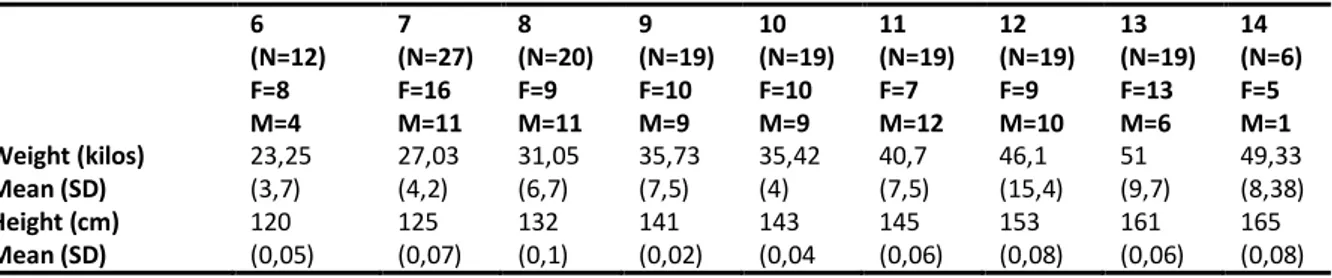
order to do so, I recruited a sample of 160 subjects across different schools of Rome. In the
second study, I will show and discuss how the affective touch perception changes in relation to
different attachment patterns in a sample of healthy adults. For the evaluation of adult
attachment, it was decided to use the Adult Attachment Interview. Despite it requires a long
work both in the administration and in the transcription of the interview, the Adult Attachment
Interview is the gold standard for the evaluation of the state of mind with respect to the
attachment. It is easy to imagine that the recruitment procedure was prolonged and demanding
in order to achieve a sample of over 60 subjects, where three days of work were required to
correctly code the state of mind for each subject. Considering results from this study, I chose
to implement a new research to investigate whether the behavioral differences observed
between groups, would have been reflected also in differences on the cortical activations of the
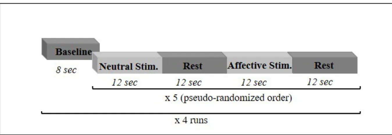
participants of the two groups. More specifically, we reproduced the affective touch stimulation
differences in subjects with different attachment patterns. Until now this study is yet to be
completed because data recollection started during my third and last year of doctoral project
and the entire procedure is taking some time. Despite this, I will present the preliminary results
Study I: Tactile sensitivity, tactile acuity and affective touch: from childhood to early adolescence
3.1 Introduction
The sense of touch is thought to be the first sense to develop and, perhaps, it continues to be
the most emotionally central throughout our lives. However, even though the maturation of the
tactile perception has been well characterized among adults, very little is known about how
tactile functions develop in childhood. If on one hand it has been demonstrated that tactile
abilities decline with age, on the other, the literature is not consistent on the essential question
of whether tactile functions improve, decline, or remain unchanged with age early in life
(Bleyenheuft et al., 2006; Güçlü & Oztek, 2007; Stevens & Choo, 1996).
The lack of data on the maturation of touch in childhood seems unexpected if we think to
the fundamental roles that the touch plays in the early stage of human development. For
example, it has been showed that in premature neonates deprived of normal sensory stimulation,
substitute stimulation facilitates growth and development (Ardiel & Rankin, 2010). Similarly,
the administration of 10 minutes of additional handling per day produced a significant reduction
in regurgitation (Hopper & Pinneau, 1957). In older children, Casler (1965) reported that
institutionalized infants receiving an additional 1000 minutes of extra tactile stimulation
administered impersonally for 10 weeks, had higher scores on developmental assessments. The
aforementioned studies, together with other similar reports, suggest that the physical and
cognitive deficits observed in deprived children could have been the effect of the lack of sensory
deprivation (namely mechanosensory stimulation) rather than merely the maternal care
withdrawal. Touch is also important for the development of affective and social interactions. In
a recent review on this topic, Cascio et al. (2018) revised an impressive number of studies, both
social touch that is based on stroking speed (Della Longa et al., 2017; Miguel et al., 2017;
Pirazzoli et al., 2018; Tuulari et al., 2017). It has been proposed that a specific class of tactile
fibers, known as C-Tactile (CT) afferents (Lӧken et al., 2009; McGlone & Spence, 2010),
respond optimally when the skin is stroked at a speed of about 1–10 cm/s (Morrison et al.,
2011a; Sailer & Ackerley, 2017). These fibers are found in the hairy, but not in the glabrous
skin and they are linked with the perception of pleasant touch similar to a caress. Several authors
proposed the term Affective Touch to label the pleasant tactile perception evoked by the
stimulation of the CT-system (Gordon et al., 2013; McGlone et al., 2014; Perini et al., 2015).
Coming back to the initial reflection, it seems that the literature on the development of the
basic functions of discriminative touch in normal childhood is lacking. Moreover, there have
been relatively few studies of tactile sensitivity and acuity on hairy skin that is the preferential
site for Affective Touch stimulation. As a matter of fact, studies on tactile sensitivity and tactile
acuity in the lifespan, focused on the glabrous skin (Peters & Goldreich, 2013; Stevens &
Patterson, 1995) and, with the exception of the study of Mancini and colleagues (2014) on
adults, no data are available on tactile sensitivity and acuity in the hairy skin of children and
early adolescents.
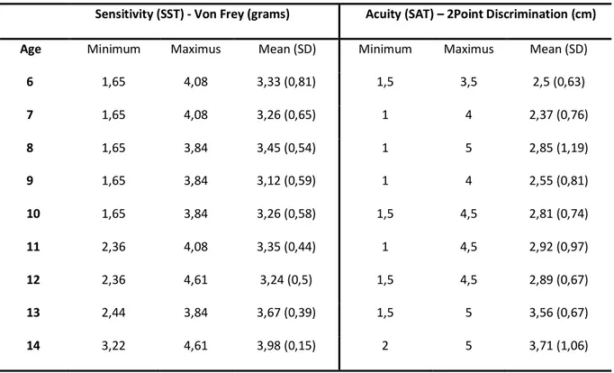
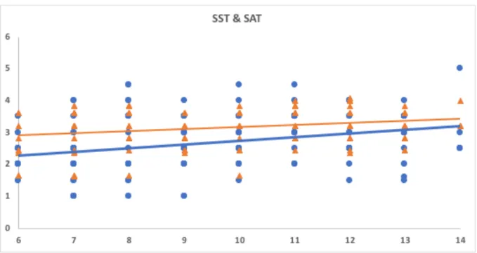
Under these circumstances, the first aim of this study is to explore the tactile sensitivity and
the tactile acuity, two dimension of basic somato-sensation, of hairy skin from early childhood
to early adolescence; we hypothesized that both tactile functions could be modified by age
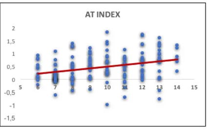
growth. A second aim of this study is to analyze whether tactile sensitivity and acuity are linked
to affective touch. Recent evidence points to orthogonal somatosensory subsystems for basic
discriminative functions of touch and affective touch in adult (McGlone et al., 2014), so we
hypothesized that, also in childhood and early adolescence, the two tactile systems should not
be connected. Lastly, a third aim is to analyze whether or not the perception of affective touch