Development/Plasticity/Repair
Synaptic Plasticity and PDGF Signaling Defects Underlie
Clinical Progression in Multiple Sclerosis
Francesco Mori,
1,2* Silvia Rossi,
1,2* Sonia Piccinin,
3,4Caterina Motta,
1,2Dalila Mango,
1,2Hajime Kusayanagi,
1,2Alessandra Bergami,
5Valeria Studer,
1,2Carolina G. Nicoletti,
1,2Fabio Buttari,
1,2Francesca Barbieri,
1,2Nicola B. Mercuri,
1,2Gianvito Martino,
5Roberto Furlan,
5Robert Nistico`,
1,3and Diego Centonze
1,21Istituto Di Ricovero e Cura a Carattere Scientifico Fondazione Santa Lucia, 00143 Rome, Italy,2Clinica Neurologica, Dipartimento di Medicina dei Sistemi, Universita` Tor Vergata, 00133 Rome, Italy,3Dipartimento di Fisiologia e Farmacologia, Universita` di Roma La Sapienza, 00185, Rome, Italy,4Laboratorio di Farmacologia della Plasticita` Sinaptica, European Brain Research Institute, 00143 Rome, Italy, and5Neuroimmunology Unit, Institute of Experimental Neurology, Division of Neuroscience, San Raffaele Scientific Institute, 20132 Milan, Italy
Neuroplasticity is essential to prevent clinical worsening despite continuing neuronal loss in several brain diseases, including multiple
sclerosis (MS). The precise nature of the adaptation mechanisms taking place in MS brains, ensuring protection from disability
appear-ance and accumulation, is however unknown. Here, we explored the hypothesis that long-term synaptic potentiation (LTP), potentially
able to minimize the effects of neuronal loss by providing extra excitation of denervated neurons, is the most relevant form of adaptive
plasticity in stable MS patients, and it is disrupted in progressing MS patients. We found that LTP, explored by means of transcranial
magnetic theta burst stimulation over the primary motor cortex, was still possible, and even favored, in stable relapsing-remitting
(RR-MS) patients, whereas it was absent in individuals with primary progressive MS (PP-MS). We also provided evidence that
platelet-derived growth factor (PDGF) plays a substantial role in favoring both LTP and brain reserve in MS patients, as this molecule: (1) was
reduced in the CSF of PP-MS patients, (2) enhanced LTP emergence in hippocampal mouse brain slices, (3) was associated with more
pronounced LTP in RR-MS patients, and (4) was associated with the clinical compensation of new brain lesion formation in RR-MS. Our
results show that brain plasticity reserve, in the form of LTP, is crucial to contrast clinical deterioration in MS. Enhancing PDGF signaling
might represent a valuable treatment option to maintain brain reserve and to attenuate the clinical consequences of neuronal damage in
the progressive phases of MS and in other neurodegenerative disorders.
Introduction
Neuronal loss and gray matter atrophy progress since the early
stages of multiple sclerosis (MS) (
Tiberio et al., 2005
;
Compston
and Coles, 2008
), but their clinical consequences are initially well
compensated, likely because of plastic adaptations of surviving
neurons (
Schirmer et al., 2013
). The precise nature of the
adap-tation mechanisms taking place in MS brains, ensuring
protec-tion from disability appearance and accumulaprotec-tion, is however
unknown, and its identification could be of great relevance for
future treatments of primary and secondary progressive MS.
LTP of excitatory transmission is the most studied form of
synaptic plasticity, and its occurrence in spared neurons might
well compensate for neuronal loss occurring in acute and chronic
neurological diseases. LTP, indeed, consists of the strengthening
of synaptic communication between two connected neurons
(
Bliss and Lomo, 1973
) and is virtually able therefore to restore
membrane excitability of neurons that have lost part of their
synaptic inputs (
Singer et al., 2011
;
Zepeda et al., 2013
). It can be
hypothesized, therefore, that a certain degree of white and gray
matter damage is tolerated in relapsing-remitting MS (RR-MS)
as a consequence of LTP occurrence in unaffected neurons,
al-though reversible or irreversible, clinical disability appears when
the adaptive abilities of the brain fail. Instead, in primary
progres-sive MS (PP-MS), accumulating disability might reflect the
pro-gression of neuronal damage without any compensation by
adaptive LTP mechanisms exhausted during clinically silent
in-flammatory episodes.
LTP can be explored noninvasively in humans by means of
transcranial magnetic stimulation (TMS) (
Mariorenzi et al.,
1991
;
Stefan et al., 2000
), which offers therefore the possibility of
addressing the hypothesis of synaptic plasticity involvement in
the attenuation of MS clinical deficits. The synaptic plasticity
hypothesis of clinical recovery in MS predicts that LTP is possible
in RR-MS but not in PP-MS and that molecular factors regulating
LTP induction also impact on MS disease clinical manifestation.
The present investigation was therefore specifically designed
at investigating whether LTP is differentially expressed in
non-progressing and in non-progressing MS patients, and to try to uncover
the role of inflammatory molecules in the regulation of LTP
in-Received June 15, 2013; revised Sept. 12, 2013; accepted Oct. 14, 2013.
Author contributions: N.B.M., G.M., R.F., R.N., and D.C. designed research; F.M., S.R., S.P., C.M., D.M., H.K., A.B., V.S., C.G.N., F. Buttari, F. Barbieri, R.F., and R.N. performed research; F.M., S.R., S.P., C.M., D.M., and V.S. analyzed data; D.C. wrote the paper.
This work was supported by Fondazione Italiana Sclerosi Multipla Special Project Grant to D.C. The authors declare no competing financial interests.
*F.M. and S.R. contributed equally to this work as first authors.
Correspondence should be addressed to Dr. Diego Centonze, Clinica Neurologica, Dipartimento di Medicina dei Sistemi, Universita` Tor Vergata, Via Montpellier 1, 00133 Rome, Italy. E-mail: [email protected].
DOI:10.1523/JNEUROSCI.2536-13.2013
duction. According to the idea that synaptic plasticity is crucial to
counterbalance clinical progression in MS subjects, we found
that LTP was still possible, and even favored, in stable RR-MS
patients, whereas it was absent in individuals with PP-MS. Recent
studies reported that the PDGF can induce LTP in vitro (
Peng et
al., 2010
), and that it may represent a key molecule for the
recov-ery phase of MS because of its neuroprotective action (
Vana et al.,
2007
). In the present study, we also identified PDGF as a crucial
inflammatory molecule able to facilitate LTP induction and to
promote the clinical compensation of brain damage associated
with MS.
Materials and Methods
The study involving human subjects was approved by the Ethics Com-mittee of the University Hospital Tor Vergata, Rome. All experiments in mice were performed in accordance with the Guide for the Care and Use of Laboratory Animals and the European Communities Council Direc-tive of 24 November, 1986 (86/609/EEC).
Human subjects and CSF withdrawal. A total of 194 central-southern
Italian subjects (118 females, 76 males) were included in this study (Table 1). MS subjects were admitted to the neurological clinic of the University Hospital Tor Vergata of Rome and later diagnosed as suffering from RR-MS (n⫽ 116), or PP-MS (n ⫽ 30). After their admittance, all patients underwent for diagnostic purposes, in sequence, brain (and in selected cases also spinal) MRI scan, and CSF withdrawal within 24 h. In all instances, patients underwent detection of oligoclonal banding in the CSF (positive in 88% of cases). Patients were drug-free before CSF with-drawal and neurophysiological assessment. Corticosteroids or other MS-specific immunoactive therapies were initiated later when appropriate.
The diagnosis of RR-MS or PP-MS was established by clinical, labora-tory, and MRI parameters, and matched published criteria (Polman et al., 2005,2011). Demographic and clinical information was derived from medical records. MS disease onset was defined as the first episode of focal neurological dysfunction indicative of MS. Disease duration was esti-mated as the number of years from onset to the last assessment of dis-ability. Disability was determined by a specially trained (Neurostatus training and documentation DVD for a standardized neurological exam-ination and assessment of Kurtzke’s functional systems and Expanded Disability Status Scale for MS patients. Basel, Switzerland: Neurostatus, 2006; available at http://www.neurostatus.net) and certified examining neurologist using Expanded Disability Status Scale (EDSS), a 10 point disease severity score derived from nine ratings for individual neurolog-ical domains (Kurtzke, 1983). Relapses were defined as the development of new or recurrent neurological symptoms not associated with fever or infection lasting at least 24 h. As controls, we used CSF from 48 age- and gender-matched healthy subjects (HSs) without inflammatory or degen-erative diseases of the central or peripheral nervous system. These sub-jects underwent lumbar puncture because of a clinical suspect of acute peripheral neuropathy, meningitis, or subarachnoidal hemorrhage, which were not confirmed.
All the subjects gave their written informed consent to the study. Clin-ical and demographic data are presented as the mean⫾ SD. Differences between two groups were compared by univariate analysis using Stu-dent’s t test or Mann–Whitney test for continuous variables and Fisher’s exact test for categorical variables. Multiple comparisons were analyzed
by performing ANOVA for independent measures followed by Tukey HSD.
MRI acquisition and analysis. Three Tesla MRI scan consisted of
dual-echo proton density, FLAIR, T2-weighted spin-dual-echo images and pre-contrast and postpre-contrast T1-weighted spin-echo images. All images were acquired in the axial orientation with 3-mm-thick contiguous slices. The presence of gadolinium-enhancing (Gd⫹; 0.2 ml/kg e.v.) lesions was assessed by a neuroradiologist who was unaware of the patient’s clinical details (Mori et al., 2011).
TMS. TMS protocols were performed in a subgroup of 75 MS patients
(63 RR-MS and 12 PP-MS), and in 13 healthy controls (Table 2). In a subgroup of 20 RR-MS patients, TMS measurements were performed during admission to the neurological clinic and within 24 h from CSF withdrawal. A subgroup of 62 MS subjects (50 RR-MS and 12 PP-MS) were evaluated at least 60 d since stabilization/resolution of a previous relapse in case of RR-MS (50 females, 12 males) (Table 2).
All subjects gave consent to the examination and were asymptomatic in the upper right limb. EMG traces were recorded from the right first dorsal interosseus muscle (FDI) with surface cup electrodes. The active electrode was placed over the muscle belly and the reference electrode over the metacarpophalangeal joint of the index finger. Responses were amplified with a Digitimer D360 amplifier (Digitimer) through filters set at 20 Hz and 2 kHz with a sampling rate of 5 kHz, then recorded by a computer with SIGNAL software (Cambridge Electronic Devices). Motor-evoked potentials (MEPs) were evoked through a figure-of-eight coil with external loop diameter of 70 mm connected to a Magstim 2002 magnetic stimulator (Magstim). Coil position was adjusted to find the optimal scalp site to evoke motor responses in the contralateral FDI, the motor “hot spot,” at the beginning of each experimental session and marked over the patients scalp with a pencil. The coil was held tangen-tially to the scalp surface with the handle pointing posteriorly and later-ally at⬃45° with respect to the mid-sagittal axis of the head.
Intermittent theta burst stimulation (iTBS) or continuous theta burst stimulation (cTBS) was delivered over the primary motor cortex (M1) “hot spot” of the right FDI through a Magstim Rapid2stimulator. The resting motor threshold (RMT) was defined as the minimum stimulation intensity required to evoke a liminal motor potential from the FDI at rest (⬃50V in 50% of 10 trials). The active motor threshold (AMT) was defined as the minimum stimulation intensity required to evoke a liminal motor potential from the FDI during voluntary contraction (⬃200V in 50% of 10 trials). Stimulation intensity was 80% of AMT. The iTBS protocol consisted of 10 bursts, each burst composed of three stimuli at 50 Hz, repeated at a theta frequency of 5 Hz every 10 s for a total of 600 stimuli (200 s). The cTBS protocol was delivered as a sequence of 200 bursts (600 stimuli) given at a rate of 5 Hz (total duration of 40 s).
The effect of iTBS or cTBS on corticospinal excitability was quantified by measuring the amplitude of MEPs evoked in the right FDI by a con-stant intensity TMS pulse given over the contralateral motor cortex. Twenty-five MEPs were collected before iTBS or cTBS (baseline) and at two different time points (0 and 15 min) after the end of stimulation procedure. Stimulation intensity was set to induce a stable MEP of⬃1 mV peak to peak amplitude in the relaxed right FDI at baseline and remained unchanged until end of recordings. MEP amplitudes were then averaged at each time point and normalized to the mean baseline amplitude.
Differences between groups for MEP latency, RMT, and AMT were evaluated through one-way ANOVA. For iTBS or cTBS aftereffects, we Table 1. Demographic and clinical characteristics of enrolled subjects
Total Controls RR-MS PP-MS p
Number 194 48 116 30
Sex (F/M) 118/76 28/20 72/44 18/12 NS
Age (years) 36.3⫾ 9.3 36.5 ⫾ 10.2 35.6 ⫾ 9.5 38.5 ⫾ 6.3 NS Disease duration (years) NA NA 3.2⫾ 5.1 4.7 ⫾ 5.2 NS
EDSS NA NA 1.3⫾ 1.1 3.5 ⫾ 0.9 ⬍0.01
EDSS range NA NA 0 – 6.0 2.5– 6.0
NS, Not significant; NA, not applicable.
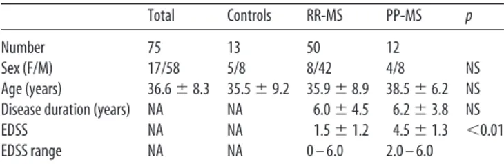
Table 2. Demographic and clinical characteristics of the subgroup of subjects enrolled in TMS experiments
Total Controls RR-MS PP-MS p
Number 75 13 50 12
Sex (F/M) 17/58 5/8 8/42 4/8 NS
Age (years) 36.6⫾ 8.3 35.5 ⫾ 9.2 35.9 ⫾ 8.9 38.5 ⫾ 6.2 NS Disease duration (years) NA NA 6.0⫾ 4.5 6.2⫾ 3.8 NS
EDSS NA NA 1.5⫾ 1.2 4.5⫾ 1.3 ⬍0.01
EDSS range NA NA 0 – 6.0 2.0 – 6.0
used a repeated-measures ANOVA with between-subjects GROUP (RR-MS, PP-(RR-MS, and controls) and within-subjects TIME (baseline, 0 and 15 min after iTBS or cTBS) main factors. Correlations between PDGF CSF levels and iTBS or cTBS effects on MEP size were analyzed through the Pearson correlation coefficient.
PDGF determination in the CSF. For measurements of PDGF
concen-tration, the CSF was centrifuged and immediately stored at⫺80°C until analyzed using Bio-Plex Multiplex Cytokine Assay (Bio-Rad), according to the manufacturer’s instructions. Concentrations of PDGF were calcu-lated according to a standard curve generated for the specific target and expressed as pictograms per milliliter. When the concentrations of PDGF were below the detection threshold, they were assumed to be 0 pg/ml. For data presented as the mean⫾ SEM, statistical analysis was performed using one-way ANOVA for independent measures followed by Tukey HSD. Dif-ferences among two groups were compared by univariate analysis using Student’s t test.
In vitro electrophysiology. Preparation of mouse brain slices was
per-formed in accordance with the European Communities Council Direc-tive (86/609/EEC). Parasagittal hippocampal slices (400 m) were prepared from 3- to 4-week-old male C57BL/6J mice as previously de-scribed (Molinaro et al., 2011;Nistico` et al., 2013).
Slices were incubated for 1 h and then transferred to a recording cham-ber submerged in a continuously flowing artificial CSF (ACSF) (30°C, 2–3 ml/min), gassed with 95% O2and 5% CO2containing 124 mMNaCl, 2.5 mMKCl, 1.25 mMNaH2PO4, 2.5 mMCaCl2, 1.3 mMMgSO4, 26 mM NaHCO3, and 10 mMglucose. Hippocampal CA1 field EPSP (fEPSP) was evoked by Schaffer collateral stimulation (0.2 ms current pulses) using a bipolar tungsten-stimulating electrode. Synaptic responses were re-corded with ACSF-filled microelectrodes (2– 4 M⍀) positioned in the stratum radiatum and were quantified as the initial slope of fEPSP in CA1.
For slices in which the presynaptic fiber volley was distinguishable, input-output relations were examined by plotting the initial slope of the fEPSP against the amplitude of the presynaptic fiber volley. LTP was induced by conventional TBS applied to the Schaffer collateral-CA1 syn-apses TBS (4 trains of 5 pulses at a frequency of 100 Hz, with an intertrain interval of 200 ms) (Errico et al., 2008).
All data are presented as mean⫾ SEM. normalized to the precondi-tioning baseline (at least 30 min of stable responses) and assessed for significance using the Student’s t test.
Results
Characteristics of enrolled subjects
The three groups (control, RR-MS, PP-MS) did not differ in
terms of the demographic characteristics, and the two MS groups
(RR-MS and PP-MS) did not differ in terms of the main clinical
characteristics (
Table 1
). Of note, disease duration, reported to be
inversely related to PDGF concentration in the CSF of MS
pa-tients (
Harirchian et al., 2012
), was similar in our samples. It was
therefore not taken into account as confounding factor during
subsequent analyses. EDSS was higher in PP-MS patients,
ac-cording to the different grade of disability progression here
investigated.
TMS was well tolerated from all subjects, and no adverse effect
was recorded. One-way ANOVA revealed that MEP latency and
AMT differed in the three groups. Post hoc comparisons revealed
that MEP latency was significantly different between the three
groups (PP-MS, 23.83
⫾ 3.2 ms; RR-MS, 22.02 ⫾ 2.25 ms; HSs,
21.1
⫾ 0.9 ms; all p ⬍ 0.05). RMT differed significantly between
the three groups (PP-MS, 41.5
⫾ 13.0%; RR-MS, 34.1 ⫾ 7.2; HSs,
30.7
⫾ 4.96; all p ⬍ 0.05). AMT mean values were higher in the
PP-MS (41.5
⫾ 13.0%) than in the RR-MS (34.1 ⫾ 7.2) and HSs
(30.7
⫾ 4.96) groups, but not statistically different between the
three groups.
LTP induction in the motor cortex of RR-MS and of PP-MS
To assess plasticity reserve in nonprogressing RR-MS and in
PP-MS, we delivered the iTBS and cTBS protocols over the M1 in 50
stable RR-MS patients, in 12 PP-MS subjects, and in 13 HSs
(
Table 2
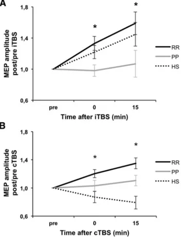
). Repeated-measures ANOVA showed a significant
TIME per GROUP interaction (F
⫽ 3.80; p ⬍ 0.05). According to
the idea that LTP reserve is greater in RR-MS than in PP-MS
subjects, post hoc contrasts showed that iTBS-induced LTP was
more pronounced in RR-MS than in PP-MS at 0 min (F
⫽ 3.50,
p
⬍ 0.05) and 15 min (F ⫽ 3.60, p ⬍ 0.05) after the stimulation
procedure. No differences emerged between RR-MS and healthy
individuals, as iTBS caused the expected LTP-like phenomenon
(
Huang et al., 2005
) in both groups (
Fig. 1
A).
We then explored the effects of the cTBS protocol, which also
induces plasticity effects (LTP or LTD) in healthy individuals
(
Gentner et al., 2008
;
Iezzi et al., 2008
;
Huang et al., 2011
).
Repeated-measures ANOVA showed a significant effect of
GROUP (F
⫽ 3.83, p ⬍ 0.05) and a significant TIME per GROUP
interaction (F
⫽ 4.15; p ⬍ 0.05). Post hoc contrasts showed that,
15 min after cTBS, MEP amplitude was significantly higher in
RR-MS subjects compared with PP-MS (F
⫽ 3.26, p ⬍ 0.05) and
compared with HSs (F
⫽ 4.89, p ⬍ 0.05) at 0 min (F ⫽ 3.84, p ⬍
0.05) and 15 min (F
⫽ 4.15, p ⬍ 0.05) and significantly lower in
HSs compared with PPMS at 15 min (F
⫽ 3.31, p ⬍ 0.05),
reveal-ing that this alternative stimulation protocol caused a measurable
LTP in RR-MS patients, although it failed to induce synaptic
plasticity effects in PP-MS. In healthy individuals, cTBS caused a
LTD-like phenomenon in the majority of the subjects (9 of 13), as
already described (
Huang et al., 2011
) (
Fig. 1
B).
Figure 1. Cortical excitability changes induced by (A) iTBS and (B) cTBS in RR-MS, PP-MS, and HSs. *p⬍ 0.05.
Together, these results indicate that LTP reserve is preserved
in stable RR-MS and lost in PP-MS patients, and that LTP
induction is favored in RR-MS patients compared with
healthy individuals.
PDGF levels in the CSF of RR-MS and of PP-MS
Some inflammatory molecules released in the CSF of MS patients
can alter excitatory synaptic transmission (
Rossi et al., 2012
) and
could be therefore potentially implicated in the differential
syn-aptic plasticity expression in RR-MS and in PP-MS. Because of its
ability to enhance LTP induction and expression in vitro (
Peng et
al., 2010
) and of its proposed role in limiting MS disease
progres-sion (
Harirchian et al., 2012
), we measured PDGF CSF levels in
RR-MS, in PP-MS, and in a sample of control individuals.
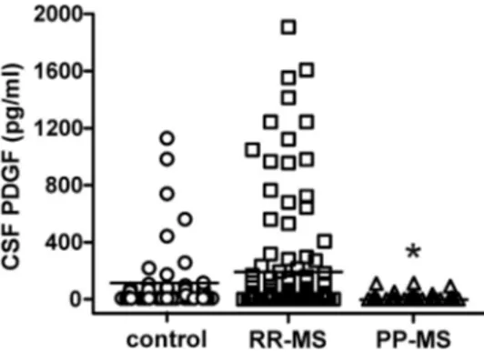
PDGF levels were significantly affected by MS disease (F
⫽
3.65, p
⫽ 0.03). Post hoc analysis showed that PDGF was
signifi-cantly lower in PP-MS patients than in RR-MS, suggesting a
contribution of this molecule in the differential expression of
LTP seen in RR-MS and in PP-MS. PDGF levels were also lower
in PP-MS patients than in controls, without reaching statistical
significance. Of note, PDGF levels were higher, although not
sig-nificantly, in the CSF of RR-MS subjects, in line with the TMS
data showing a favored LTP induction compared with healthy
individuals (
Fig. 2
).
Role of PDGF in in vitro LTP
Having determined that PDGF CSF levels positively correlate to
LTP induction in stable RR-MS, we next sought to test the effect
of PDGF on synaptic transmission and LTP in mouse
hippocam-pal slices. A scatter plot relating the initial slope of the fEPSP to
the size of the presynaptic fiber volley showed that slices
incu-bated with PDGF (20 ng/ml for 1 h) displayed normal
input-output characteristics in the CA1 area (
Fig. 3
A). Next, to
determine whether synaptic plasticity is modulated by PDGF,
we studied hippocampal LTP induced by TBS. Similarly to
previous results (
Peng et al., 2010
), CA1-LTP was always
fa-cilitated when slices were preincubated with PDGF (
Fig. 3
B).
Indeed, the magnitude of potentiation measured between 50
and 60 min after TBS was significantly higher in PDGF-treated
(162
⫾ 9%, n ⫽ 6) compared with control slices (134 ⫾ 9%,
n
⫽ 6) (p ⬍ 0.05;
Fig. 3
B).
Thus, our in vitro data indicate that PDGF modulates LTP
induction at excitatory synapses and are consistent with the idea
that this molecule could contribute to the maintenance of
plas-ticity reserve seen in nonprogressing MS patients.
Association between PDGF levels and clinical relapses in
RR-MS patients
Based on the assumption that PDGF-favored plasticity reserve
could be relevant for the compensation of evolving brain damage
in RR-MS, we also examined the impact of PDGF on the clinical
correlates of disease evolution in RR-MS subjects. To this aim,
RR-MS subjects with active MS lesions (n
⫽ 49), as evidenced by
Gd enhancement at the MRI, were divided into two groups
(re-lapsing, n
⫽ 26 vs silent, n ⫽ 23), based on the presence or
absence of concomitant clinical symptoms. Gd
⫹-relapsing
pa-tients had significantly lower PDGF CSF concentrations than
Gd
⫹clinically silent patients ( p
⫽ 0.01), a finding that is in
agreement with the idea that PDGF plays a role in plasticity
re-serve and clinical compensation of brain damage in MS, by
pre-venting symptom appearance despite new lesion formation (
Fig.
4
A). In line with this, relapsing patients were significantly more
frequent among subjects with undetectable PDGF at the time of
MRI activation (undetectable PDGF, n
⫽ 23; detectable PDGF,
n
⫽ 26; 78% vs 30%, p ⫽ 0.001) (
Fig. 4
B).
Correlation between PDGF levels and LTP amplitude in
RR-MS patients
To further address the proposed involvement of PDGF in LTP
reserve in RR-MS patients, the correlation between CSF
concen-trations of this molecule and the amplitude of this form of
syn-aptic plasticity was explored in a subgroup of 20 RR-MS patients
(5 males and 15 females, aged 17– 47 years). The results of this
investigation were in agreement with the conclusion that PDGF
has a role in the maintenance of brain plasticity potential in MS
subjects because those individuals with higher CSF PDGF levels
showed larger LTPs in response to cTBS than those with low
PDGF levels (r
⫽ 0.57, p ⬍ 0.05) (
Fig. 5
). Conversely, no
signif-icant correlation emerged between CSF PDGF levels and MEP
latency, AMT, or RMT (data not shown).
Discussion
LTP is a form of use-dependent synaptic plasticity that
strength-ens the communication between two connected neurons, able to
minimize the effects of neuronal loss in a network by providing
extra excitation of denervated neurons (
Bliss and Lomo, 1973
).
The present study showed defective LTP in patients with
pro-gressive MS course, supporting the idea that altered synaptic
plas-ticity reserve plays a substantial role in the manifestation and
progression of clinical deficits in MS. In stable RR-MS patients, in
contrast, LTP was still possible and even favored, as evidenced by
the finding that not only iTBS but also cTBS resulted in
signifi-cant and long-lasting enhancement of cortical excitability. In
healthy individuals, conversely, cTBS generally results in LTD,
but LTP has been found to emerge in response to cTBS under
specific circumstances altering the excitability of the motor
cor-tex (
Gentner et al., 2008
;
Iezzi et al., 2008
).
NMDA receptor-dependent LTP induction is greatly
facili-tated in response to ischemic brain damage in rodents (
Ben-veniste et al., 1984
;
Picconi et al., 2006
), and its occurrence in the
peri-infarct area is associated with better clinical outcome (
Cen-tonze et al., 2007
). Also, in humans, LTP reserve explored with
iTBS after focal brain ischemia is associated with better
func-tional recovery (
Di Lazzaro et al., 2010
), a finding that is in good
agreement with our results, and extends to other acute and
chronic pathological conditions the concept that this form of
synaptic plasticity is indeed able to attenuate the clinical
conse-quences of brain tissue damage.
Figure 2. PDGF levels in the CSF of MS subjects. The graph shows that PDGF was significantly lower in PP-MS patients than in RR-MS. *p⬍ 0.05, versus RR-MS.
Immune cells infiltrating the CNS and
causing tissue damage contribute to
neu-ronal and oligodendroglial cell survival
and tissue repair by secreting different
growth factors (
Schwartz et al., 1999
;
Ker-schensteiner et al., 2003
) and, among
these, PDGF acts as a key molecule for the
recovery phase. PDGF, indeed, promotes
neuronal differentiation (
Williams et al.,
1997
;
Erlandsson et al., 2001
), improves
significantly remyelination and
oligoden-drocyte density during acute
demyelina-tion, and reduces apoptosis during the
recovery period after chronic
demyelina-tion (
Vana et al., 2007
). Furthermore,
strong upregulation of PDGF occurs in
peripheral lymphocytes of experimental
MS, with the highest expression after the
disease maximum (
Koehler et al., 2008
).
PDGF has also been implicated in
neuro-protection against energy deprivation and
oxidative injury (
Cheng and Mattson,
1995
), against human immunodeficiency
virus protein toxicity (
Peng et al., 2008
),
and after injurious events, such as focal
brain ischemia (
Egawa-Tsuzuki et al.,
2004
). Importantly, PDGF concentration
in the CSF of MS patients decreases with
disease duration, and its serum and CSF
levels have been proposed as markers of
disease severity (
Harirchian et al., 2012
).
Here, we also provided evidence that
PDGF plays a substantial role in favoring
both LTP and brain reserve in MS
pa-tients, as this molecule: (1) enhanced LTP
emergence in hippocampal slices, (2) was
associated with more pronounced LTP in
RR-MS patients, (3) was associated with
clinically stable disease course, and (4)
was associated with the clinical
compen-sation of new brain lesion formation in
RR-MS. The ability of PDGF to favor LTP,
and its association with clinically stable MS course, is indeed
consistent with its prosurvival effects because a striking
conver-gence between the intracellular signaling pathway mediating LTP
and neuronal survival exists (
Bartlett and Wang, 2013
).
As expected, PP-MS patients had higher EDSS values and
more pronounced MEP alterations (higher MEP latency and
RMT). PP-MS patients may thus have lost the ability to express
LTP resulting from increased axonal damage and thus to a lower
number of synaptic connections. It must be considered, however,
that activity-dependent LTP is essential for synaptogenesis (
Col-lin et al., 1997
) and that axonal damage occurs also in RR-MS
patients since the early stages (
Schirmer et al., 2013
). However,
LTP was higher in RR-MS than in HSs in our study. At the present
stage, this point remains controversial and needs to be elucidated
in further studies.
The mechanism by which PDGF enhances LTP is only
mar-ginally understood, but animal studies showed that PDGF and its
receptors are widely expressed in the CNS (
Sasahara et al., 1991
;
Gozal et al., 2000
), where they modulate the expression of Arc/
Arg3.1 and give rise to LTP in hippocampal slices (
Peng et al.,
2010
; present study). While reduced PDGF activity seems to
con-tribute to exhausted LTP in PP-MS patients, LTP facilitation in
RR-MS, as evidenced by the induction of this form of synaptic
plasticity not only after iTBS but also after cTBS, does not seem to
be directly dependent upon PDGF. Although higher than in
PP-Figure 3. Effect of PDGF on hippocampal synaptic function. A, Input/output curves are shown as plots of the fEPSP slopes against the corresponding presynaptic fiber volley amplitudes. Each treatment group is the average (⫾SEM) of at least six recordings performed on separate slices. No significant differences between treatment groups were detected. B, Time plot of fEPSP responses showing that pretreatment with PDGF enhances the magnitude of CA1-LTP. Representative fEPSP recordings from time points 1 and 2 are shown for each condition. Calibration: 0.5 mV, 10 ms.Figure 4. Association between PDGF levels and clinical compensation in RR-MS. A, The histogram shows that PDGF CSF levels were lower in Gd⫹relapsing patients than in Gd⫹clinically silent patients. B, Relapsing patients were more frequent among RR-MS subjects with undetectable PDGF levels at the time of MRI activation. *p⬍ 0.05.
Figure 5. PDGF favors LTP over LTD in RR-MS patients. Correlation plot shows that PDGF levels in the CSF correlate with the magnitude of LTP-like changes induced 15 min after cTBS in RR-MS patients.
MS, indeed, PDGF CSF levels were similar in RR-MS and in
control subjects, who showed LTD rather than LTP in response to
cTBS.
Facilitated LTP is likely to be important for prompt
compen-sation of brain damage in RR-MS and could instead be favored by
inflammatory molecules released during MS but not in
nondis-eased brains. Among them, IL-1 has already been found to
lower the threshold for LTP induction and to favor LTD to LTP
switch in MS brains, possibly through the inhibition of GABA
synapses (
Nistico` et al., 2013
). cTBS, indeed, induces a mixture of
excitatory and inhibitory long-lasting effects (
Stagg et al., 2009
),
and the direction of the post-cTBS synaptic plasticity is
deter-mined in physiological conditions by the excitation/inhibition
balance (
Huang et al., 2011
). Accordingly, LTP is attenuated by
enhancing GABA transmission (
Wigstro¨m and Gustafsson, 1983
;
Chapman et al., 1998
;
Grover and Yan, 1999
;
Levkovitz et al.,
1999
;
Lu et al., 2000
;
Yoshiike et al., 2008
;
Martin et al., 2010
) and
potentiated by reducing it (
Hess and Donoghue, 1996
;
Sta¨ubli et
al., 1999
).
During a relapse, MS patients show decreased short interval
intracortical inhibition (
Caramia et al., 2004
). These findings
could reflect changes of both NMDA receptors (
Schwenkreis et
al., 1999
) or GABAA receptor activities (
Ziemann et al., 1996
).
Thus, the increased LTP responses observed in RR-MS patients
may possibly be secondary to alterations in both NMDA or
GABA receptor activity. In this respect, reduction of cortical
in-hibition has been proposed as a mechanism to augment plastic
properties (
Baroncelli et al., 2011
;
Imbrosci and Mittmann,
2011
), as synaptic inhibition limits the plastic properties of the
cortex (
Teo et al., 2009
).
In our study, the excitability of the motor cortex was evaluated
indirectly, through the amplitude of the MEP, which is
consid-ered reflective of corticospinal excitability. This may represent a
limitation, as the plastic modulation of the MEP amplitude may
have occurred at noncortical sites. Indeed, plastic changes in the
spinal H reflex after 5 consecutive days of iTBS over the motor
cortex have been reported in MS patients with lower limb
spas-ticity (
Mori et al., 2010
). However, H reflex remained unchanged
after only one single session of iTBS (
Huang et al., 2005
;
Mori et
al., 2010
;
Conte et al., 2012
) or cTBS (
Huang et al., 2005
;
Zapal-low et al., 2012
).
Also, spinal recordings through epidural electrodes at the
cer-vical site showed that, after a TMS pulse at intensities 3% below
the AMT over the motor cortex, no corticospinal volleys could be
recorded (
Di Lazzaro et al., 1998
). The lack of change in the H
reflex after TBS and the low intensity used by TBS suggest that
there is no effect of one single session of motor TBS on excitability
of spinal motor neurons and the inhibitory circuits around
these neurons. Moreover, EEG (
Vernet et al., 2013
), MEG
(
McAllister et al., 2013
), and cervical epidural (
Di Lazzaro et
al., 2005
) recordings showed that TBS induces excitability
changes at the cortical level. Investigating the relationship
be-tween cortical plasticity through measures of corticocortical
connectivity (i.e., by means of paired pulse TMS, EEG, MRI),
and clinical and biochemical variables in MS may provide
further and more direct evidence of the role of neuronal
plas-ticity and its regulators in MS.
LTP can be influenced by a number of different factors,
among these, proinflammatory cytokines, with detrimental
ef-fects on MS progression (
Pickering et al., 2005
;
Haji et al., 2012
;
Rossi et al., 2012
;
Mori et al., 2013
). It thus appears reasonable
that the net effect on plasticity is the result of a very complex
interaction between proinflammatory and anti-inflammatory
cy-tokines and genetic factors. Future studies designed to evaluate
the impact of multiple variables and of their interaction on MS
clinical severity are thus warranted.
In conclusion, despite brain damage progresses overtime,
maintenance of LTP-like, brain plasticity reserve is crucial to
contrast clinical deterioration in MS, and possibly in other acute
or chronic neurological diseases. Enhancing PDGF signaling
might represent a valuable treatment option to preserve brain
reserve and to attenuate the clinical consequences of neuronal
damage in the progressive phases of MS. This possibility should
be carefully considered as plasticity can also lead to maladaptive
clinical manifestations as reported in dystonia (
Quartarone et al.,
2003
), dyskinesias (
Cenci and Konradi, 2010
) or spasticity itself
(
Tan et al., 2012
).
References
Baroncelli L, Braschi C, Spolidoro M, Begenisic T, Maffei L, Sale A (2011) Brain plasticity and disease: a matter of inhibition. Neural Plast 2011: 286073.CrossRef Medline
Bartlett TE, Wang YT (2013) The intersections of NMDAR-dependent syn-aptic plasticity and cell survival. Neuropharmacology 74:59 – 68.CrossRef Medline
Benveniste H, Drejer J, Schousboe A, Diemer NH (1984) Elevation of the extracellular concentrations of glutamate and aspartate in rat hippocam-pus during transient cerebral ischemia monitored by intracerebral micro-dialysis. J Neurochem 43:1369 –1374.CrossRef Medline
Bliss TV, Lomo T (1973) Long-lasting potentiation of synaptic transmission in the dentate area of the anaesthetized rabbit following stimulation of the perforant path. J Physiol 232:331–356.Medline
Caramia MD, Palmieri MG, Desiato MT, Boffa L, Galizia P, Rossini PM, Centonze D, Bernardi G (2004) Brain excitability changes in the relaps-ing and remittrelaps-ing phases of multiple sclerosis: a study with transcranial magnetic stimulation. Clin Neurophysiol 115:956 –965.CrossRef Medline
Cenci MA, Konradi C (2010) Maladaptive striatal plasticity inL -DOPA-induced dyskinesia. Prog Brain Res 183:209 –233.CrossRef Medline
Centonze D, Rossi S, Tortiglione A, Picconi B, Prosperetti C, De Chiara V, Bernardi G, Calabresi P (2007) Synaptic plasticity during recovery from permanent occlusion of the middle cerebral artery. Neurobiol Dis 27:44 – 53.CrossRef Medline
Chapman CA, Perez Y, Lacaille JC (1998) Effects of GABA(A) inhibition on the expression of long-term potentiation in CA1 pyramidal cells are de-pendent on tetanization parameters. Hippocampus 8:289 –298.CrossRef Medline
Cheng B, Mattson MP (1995) PDGFs protect hippocampal neurons against energy deprivation and oxidative injury: evidence for induction of anti-oxidant pathways. J Neurosci 15:7095–7104.Medline
Collin C, Miyaguchi K, Segal M (1997) Dendritic spine density and LTP induction in cultured hippocampal slices. J Neurophysiol 77:1614 –1623.
Medline
Compston A, Coles A (2008) Multiple sclerosis. Lancet 372:1502–1517.
CrossRef Medline
Conte A, Belvisi D, Bologna M, Ottaviani D, Fabbrini G, Colosimo C, Wil-liams DR, Berardelli A (2012) Abnormal cortical synaptic plasticity in primary motor area in progressive supranuclear palsy. Cereb Cortex 22: 693–700.CrossRef Medline
Di Lazzaro V, Restuccia D, Oliviero A, Profice P, Ferrara L, Insola A, Mazzone P, Tonali P, Rothwell JC (1998) Effects of voluntary contraction on de-scending volleys evoked by transcranial stimulation in conscious humans. J Physiol 508:625– 633.CrossRef Medline
Di Lazzaro V, Pilato F, Saturno E, Oliviero A, Dileone M, Mazzone P, Insola A, Tonali PA, Ranieri F, Huang YZ, Rothwell JC (2005) Theta-burst repetitive transcranial magnetic stimulation suppresses specific excitatory circuits in the human motor cortex. J Physiol 565:945–950.CrossRef Medline
Di Lazzaro V, Profice P, Pilato F, Capone F, Ranieri F, Pasqualetti P, Colosimo C, Pravata` E, Cianfoni A, Dileone M (2010) Motor cortex plasticity predicits recovery in acute stroke. Cereb Cortex 20:1523–1528.CrossRef Medline
Egawa-Tsuzuki T, Ohno M, Tanaka N, Takeuchi Y, Uramoto H, Faigle R, Funa K, Ishii Y, Sasahara M (2004) The PDGF B-chain is involved in the
ontogenic susceptibility of the developing rat brain to NMDA toxicity. Exp Neurol 186:89 –98.CrossRef Medline
Erlandsson A, Enarsson M, Forsberg-Nilsson K (2001) Immature neurons from CNS stem cells proliferate in response to platelet-derived growth factor. J Neurosci 21:3483–3491.Medline
Errico F, Nistico` R, Palma G, Federici M, Affuso A, Brilli E, Topo E, Centonze D, Bernardi G, Bozzi Y, D’Aniello A, Di Lauro R, Mercuri NB, Usiello A (2008) Increased levels of D-aspartate in the hippocampus enhance LTP but do not facilitate cognitive flexibility. Mol Cell Neurosci 37:236 –246.
CrossRef Medline
Gentner R, Wankerl K, Reinsberger C, Zeller D, Classen J (2008) Depression of human corticospinal excitability induced by magnetic theta-burst stimulation: evidence of rapid polarity-reversing metaplasticity. Cereb Cortex 18:2046 –2053.CrossRef Medline
Gozal D, Simakajornboon N, Czapla MA, Xue YD, Gozal E, Vlasic V, Lasky JA, Liu JY (2000) Brainstem activation of platelet-derived growth factor-beta receptor modulates the late phase of the hypoxic ventilatory response. J Neurochem 74:310 –319.CrossRef Medline
Grover LM, Yan C (1999) Blockade of GABAA receptors facilitates induc-tion of NMDA receptor-independent long-term potentiainduc-tion. J Neuro-physiol 81:2814 –2822.Medline
Haji N, Mandolesi G, Gentile A, Sacchetti L, Fresegna D, Rossi S, Musella A, Sepman H, Motta C, Studer V, De Chiara V, Bernardi G, Strata P, Cen-tonze D (2012) TNF-␣-mediated anxiety in a mouse model of multiple sclerosis. Exp Neurol 237:296 –303.CrossRef Medline
Harirchian MH, Tekieh AH, Modabbernia A, Aghamollaii V, Tafakhori A, Ghaffarpour M, Sahraian MA, Naji M, Yazdanbakhsh M (2012) Serum and CSF PDGF-AA and FGF-2 in relapsing-remitting multiple sclerosis: a case-control study. Eur J Neurol 19:241–247.CrossRef Medline
Hess G, Donoghue JP (1996) Long-term depression of horizontal connec-tions in rat motor cortex. Eur J Neurosci 8:658 – 665.CrossRef Medline
Huang YZ, Edwards MJ, Rounis E, Bhatia KP, Rothwell JC (2005) Theta burst stimulation of the human motor cortex. Neuron 45:201–206.
CrossRef Medline
Huang YZ, Rothwell JC, Chen RS, Lu CS, Chuang WL (2011) The theoret-ical model of theta burst form of repetitive transcranial magnetic stimu-lation. Clin Neurophysiol 122:1011–1018.CrossRef Medline
Iezzi E, Conte A, Suppa A, Agostino R, Dinapoli L, Scontrini A, Berardelli A (2008) Phasic voluntary movements reverse the aftereffects of subse-quent theta-burst stimulation in humans. J Neurophysiol 100:2070 – 2076.CrossRef Medline
Imbrosci B, Mittmann T (2011) Functional consequences of the distur-bances in the GABA-mediated inhibition induced by injuries in the cere-bral cortex. Neural Plast 2011:614329.CrossRef Medline
Kerschensteiner M, Stadelmann C, Dechant G, Wekerle H, Hohlfeld R (2003) Neurotrophic cross-talk between the nervous and immune sys-tems: implications for neurological diseases. Ann Neurol 53:292–304.
CrossRef Medline
Koehler NK, Roebbert M, Dehghani K, Ballmaier M, Claus P, von Hoersten S, Shing M, Odin P, Strehlau J, Heidenreich F (2008) Up-regulation of platelet-derived growth factor by peripheral-blood leukocytes during ex-perimental allergic encephalomyelitis. J Neurosci Res 86:392– 402.
CrossRef Medline
Kurtzke JF (1983) Rating neurologic impairment in multiple sclerosis: an expanded disability status scale (EDSS). Neurology 33:1444 –1452.
CrossRef Medline
Levkovitz Y, Avignone E, Groner Y, Segal M (1999) Upregulation of GABA neurotransmission suppresses hippocampal excitability and prevents long-term potentiation in transgenic superoxide dismutase-overexpressing mice. J Neurosci 19:10977–10984.Medline
Lu YM, Mansuy IM, Kandel ER, Roder J (2000) Calcineurin-mediated LTD of GABAergic inhibition underlies the increased excitability of CA1 neu-rons associated with LTP. Neuron 26:197–205.CrossRef Medline
Mariorenzi R, Zarola F, Caramia MD, Paradiso C, Rossini PM (1991) Non-invasive evaluation of central motor tract excitability changes following peripheral nerve stimulation in healthy humans. Electroencephalogr Clin Neurophysiol 81:90 –101.CrossRef Medline
Martin LJ, Zurek AA, MacDonald JF, Roder JC, Jackson MF, Orser BA (2010) Alpha5GABAA receptor activity sets the threshold for long-term potentiation and constrains hippocampus-dependent memory. J Neuro-sci 30:5269 –5282.CrossRef Medline
McAllister CJ, Ro¨nnqvist KC, Stanford IM, Woodhall GL, Furlong PL, Hall
SD (2013) Oscillatory beta activity mediates neuroplastic effects of mo-tor cortex stimulation in humans. J Neurosci 33:7919 –7927.CrossRef Medline
Molinaro P, Viggiano D, Nistico` R, Sirabella R, Secondo A, Boscia F, Pannac-cione A, Scorziello A, Mehdawy B, Sokolow S, Herchuelz A, Di Renzo GF, Annunziato L (2011) Na⫹-Ca2⫹exchanger (NCX3) knock-out mice display an impairment in hippocampal long-term potentiation and spa-tial learning and memory. J Neurosci 31:7312–7321.CrossRef Medline
Mori F, Codeca` C, Kusayanagi H, Monteleone F, Boffa L, Rimano A, Bernardi G, Koch G, Centonze D (2010) Effects of intermittent theta burst stim-ulation on spasticity in patients with multiple sclerosis. Eur J Neurol 17:295–300.CrossRef Medline
Mori F, Rossi S, Sancesario G, Codeca` C, Mataluni G, Monteleone F, Buttari F, Kusayanagi H, Castelli M, Motta C, Studer V, Bernardi G, Koch G, Bernardini S, Centonze D (2011) Cognitive and cortical plasticity defi-cits correlate with altered amyloid- CSF levels in multiple sclerosis. Neu-ropsychopharmacology 36:559 –568.CrossRef Medline
Mori F, Nistico` R, Mandolesi G, Piccinin S, Mango D, Kusayanagi H, Berretta N, Bergami A, Gentile A, Musella A, Nicoletti CG, Nicoletti F, Buttari F, Mercuri NB, Martino G, Furlan R, Centonze D (2013) Interleukin-1 promotes long-term potentiation in patients with multiple sclerosis. Neu-romolecular Med. Advance online publication. Retrieved July 28, 2013. doi: 10.1007/s12017– 013-8249 –7.CrossRef Medline
Nistico` R, Mango D, Mandolesi G, Piccinin S, Berretta N, Pignatelli M, Feli-gioni M, Musella A, Gentile A, Mori F, Bernardi G, Nicoletti F, Mercuri NB, Centonze D (2013) Inflammation subverts hippocampal synaptic plasticity in experimental multiple sclerosis. PLoS One 8:e54666.
CrossRef Medline
Peng F, Dhillon N, Callen S, Yao H, Bokhari S, Zhu X, Baydoun HH, Buch S (2008) Platelet-derived growth factor protects neurons against gp120-mediated toxicity. J Neurovirol 14:62–72.CrossRef Medline
Peng F, Yao H, Bai X, Zhu X, Reiner BC, Beazely M, Funa K, Xiong H, Buch S (2010) Platelet-derived growth factor-mediated induction of the synap-tic plassynap-ticity gene Arc/Arg3.1. J Biol Chem 285:21615–21624.CrossRef Medline
Picconi B, Tortiglione A, Barone I, Centonze D, Gardoni F, Gubellini P, Bonsi P, Pisani A, Bernardi G, Di Luca M, Calabresi P (2006) NR2B subunit exerts a critical role in post ischemic synaptic plasticity. Stroke 37:1895– 1901.CrossRef Medline
Pickering M, Cumiskey D, O’Connor JJ (2005) Actions of TNF-alpha on glutamatergic synaptic transmission in the central nervous system. Exp Physiol 90:663– 670.CrossRef Medline
Polman CH, Reingold SC, Edan G, Filippi M, Hartung HP, Kappos L, Lublin FD, Metz LM, McFarland HF, O’Connor PW, Sandberg-Wollheim M, Thompson AJ, Weinshenker BG, Wolinsky JS (2005) Diagnostic criteria for multiple sclerosis: 2005 revisions to the “McDonald Criteria.” Ann Neurol 58:840 – 846.CrossRef Medline
Polman CH, Reingold SC, Banwell B, Clanet M, Cohen JA, Filippi M, Fujihara K, Havrdova E, Hutchinson M, Kappos L, Lublin FD, Montalban X, O’Connor P, Sandberg-Wollheim M, Thompson AJ, Waubant E, Wein-shenker B, Wolinsky JS (2011) Diagnostic criteria for multiple sclerosis: 2010 revisions to the McDonald criteria. Ann Neurol 69:292–302.
CrossRef Medline
Quartarone A, Bagnato S, Rizzo V, Siebner HR, Dattola V, Scalfari A, Mor-gante F, Battaglia F, Romano M, Girlanda P (2003) Abnormal associa-tive plasticity of the human motor cortex in writer’s cramp. Brain 126: 2586 –2596.CrossRef Medline
Rossi S, Furlan R, De Chiara V, Motta C, Studer V, Mori F, Musella A, Bergami A, Muzio L, Bernardi G, Battistini L, Martino G, Centonze D (2012) Interleukin-1 causes synaptic hyperexcitability in multiple scle-rosis. Ann Neurol 71:76 – 83.CrossRef Medline
Sasahara M, Fries JW, Raines EW, Gown AM, Westrum LE, Frosch MP, Bonthron DT, Ross R, Collins T (1991) PDGF B-chain in neurons of the central nervous system, posterior pituitary, and in a transgenic model. Cell 64:217–227.CrossRef Medline
Schirmer L, Merkler D, Ko¨nig FB, Bru¨ck W, Stadelmann C (2013) Neuroax-onal regeneration is more pronounced in early multiple sclerosis than in traumatic brain injury lesions. Brain Pathol 23:2–12.CrossRef Medline
Schwartz M, Cohen I, Lazarov-Spiegler O, Moalem G, Yoles E (1999) The remedy may lie in ourselves: prospects for immune cell therapy in central nervous system protection and repair. J Mol Med 77:713–717.CrossRef Medline
Schwenkreis P, Witscher K, Janssen F, Addo A, Dertwinkel R, Zenz M, Malin JP, Tegenthoff M (1999) Influence of the N-methyl-D-aspartate antago-nist memantine on human motor cortex excitability. Neurosci Lett 270: 137–140.CrossRef Medline
Singer BH, Gamelli AE, Fuller CL, Temme SJ, Parent JM, Murphy GG (2011) Compensatory network changes in the dentate gyrus restore long-term potentiation following ablation of neurogenesis in young-adult mice. Proc Natl Acad Sci U S A 108:5437–5442.CrossRef Medline
Stagg CJ, Wylezinska M, Matthews PM, Johansen-Berg H, Jezzard P, Rothwell JC, Bestmann S (2009) Neurochemical effects of theta burst stimulation as assessed by magnetic resonance spectroscopy. J Neurophysiol 101: 2872–2877.CrossRef Medline
Sta¨ubli U, Scafidi J, Chun D (1999) GABAB receptor antagonism: facili-tatory effects on memory parallel those on LTP induced by TBS but not HFS. J Neurosci 19:4609 – 4615.Medline
Stefan K, Kunesch E, Cohen LG, Benecke R, Classen J (2000) Induction of plasticity in the human motor cortex by paired associative stimulation. Brain 123:572–584.CrossRef Medline
Tan AM, Chakrabarty S, Kimura H, Martin JH (2012) Selective corticospi-nal tract injury in the rat induces primary afferent fiber sprouting in the spinal cord and hyperreflexia. J Neurosci 32:12896 –12908.CrossRef Medline
Teo JT, Terranova C, Swayne O, Greenwood RJ, Rothwell JC (2009) Differ-ing effects of intracortical circuits on plasticity. Exp Brain Res 193:555– 563.CrossRef Medline
Tiberio M, Chard DT, Altmann DR, Davies G, Griffin CM, Rashid W, Sastre-Garriga J, Thompson AJ, Miller DH (2005) Gray and white matter vol-ume changes in early RRMS: a 2-year longitudinal study. Neurology 64: 1001–1007.CrossRef Medline
Vana AC, Flint NC, Harwood NE, Le TQ, Fruttiger M, Armstrong RC (2007)
Platelet-derived growth factor promotes repair of chronically demyeli-nated white matter. J Neuropathol Exp Neurol 66:975–988.CrossRef Medline
Vernet M, Bashir S, Yoo WK, Perez JM, Najib U, Pascual-Leone A (2013) Insights on the neural basis of motor plasticity induced by theta burst stimulation from TMS-EEG. Eur J Neurosci 37:598 – 606. CrossRef Medline
Wigstro¨m H, Gustafsson B (1983) Facilitated induction of hippocampal long-lasting potentiation during blockade of inhibition. Nature 301:603– 604.CrossRef Medline
Williams BP, Park JK, Alberta JA, Muhlebach SG, Hwang GY, Roberts TM, Stiles CD (1997) A PDGF-regulated immediate early gene response ini-tiates neuronal differentiation in ventricular zone progenitor cells. Neu-ron 18:553–562.CrossRef Medline
Yoshiike Y, Kimura T, Yamashita S, Furudate H, Mizoroki T, Murayama M, Takashima A (2008) GABA(A) receptor-mediated acceleration of aging-associated memory decline in APP/PS1 mice and its pharmacolog-ical treatment by picrotoxin. PLoS One 3:e3029.CrossRef Medline
Zapallow CM, Asmussen MJ, Bolton DA, Lee KG, Jacobs MF, Nelson AJ (2012) Theta burst repetitive transcranial magnetic stimulation attenu-ates somatosensory evoked potentials from the lower limb. BMC Neuro-sci 13:133.CrossRef Medline
Zepeda A, Aguilar-Arredondo A, Michel G, Ramos-Languren LE, Escobar ML, Arias C (2013) Functional recovery of the dentate gyrus after a focal lesion is accompanied by structural reorganization in the adult rat. Brain Struct Funct 218:437– 453.CrossRef Medline
Ziemann U, Lo¨nnecker S, Steinhoff BJ, Paulus W (1996) The effect of loraz-epam on the motor cortical excitability in man. Exp Brain Res 109:127– 135.Medline