Scuola Dottorale in Biologia
Sezione “Biologia Cellulare e Molecolare”
Ciclo di Dottorato XXV
“Development of strategies to find inhibitors
of HIV-1 Nef cellular interaction partners”
“Sviluppo di strategie per la ricerca di
inibitori dei partner cellulari di interazione
della proteina Nef di HIV-1”
A.A. 2012/2013
Coordinatore:
Prof. Paolo Mariottini
Candidato:
Dott.ssa Valentina Gallo
Docente Guida:
Prof.ssa Elisabetta Affabris
Co-tutor:
ABSTRACT
This Ph.D. project has been focused on the study of HIV-1 Nef protein. Nef is a regulatory phosphoprotein and studies on animal models and seropositive patients have demonstrated its importance as virulence factor. Nef, in fact, plays a pivotal role in maintaining high viral load and in progression to AIDS, and is already object of interest of the HIV scientific community in the search of new targets to prevent or block HIV disease. Nef exerts three main functions in HIV-1 target cells (i.e.CD4+ T-helper lymhpocytes, monocytes/macrophages, dendritic cells, microglial cells): 1) the modulation of cellular signalling pathways; 2) the enhancement of viral infectivity; 3) the regulation of expression of cell surface receptors, including the well-studied down-regulation of CD4 and certain class I major histocompatibility complex (MCH-I) antigens.
Altogether these effects increase viral infectivity and spreading contributing to render the virus able to escape the immune system response.
Nef presents a two-domains structure: a structurally flexible N-terminal membrane anchor domain and a well conserved and folded C-terminal core domain. In addition it is post-translationally modified by phosphorylation and by myristoylation which is important for the Nef signalling activity. Nef exerts its functions working as a molecular adaptor, interacting and influencing the activity of more than 30 intracellular partners and several Nef interaction sites have been identified. As a consequence of Nef ability to bind multiple targets certain Nef effects dominate over others in time- or cell type- dependent manner.
Macrophages are the first cells to be infected by HIV and represent the main reservoir of the virus. In these cells Nef induces the synthesis and the release of a specific sub-set of chemokines and cytokines able to recruit T-cells on the infectious site rendering them susceptible to HIV infection. Moreover, recent studies, carried out by the research group where it has been performed this Ph.D. project, have demonstrated that Nef is efficiently internalized by human non infected monocytes derived macrophages (MDMs) in in vitro cultures and mediates the NF-kB-mediated synthesis of specific cytokines and chemokines that in turn lead to the activation by phosphorylation of STAT (Signal Transducer and Activator of Transcription)-1, -2 and -3. Importantly, it has been demonstrated that the Nef interaction site designated as acidic cluster (A60QEEEE65) is required for this process. In addiction, modelling analysis and silencing experiments indicate that this region represents a putative binding motif for specific TNF receptor associated factor (TRAF) adaptor family members. It has been
proposed, in fact, that in macrophages Nef intersects the CD40 signalling pathway and TRAF are involved in the signalling events downstream this receptor. Therefore, the study of the Nef interactome, of the molecular basis underlying the Nef acidic cluster/TRAFs interactions, and the finding of molecules able to block the main Nef interaction sites is certainly of great interest both to clarify the so far enigmatic biology of Nef and to open new perspectives in the field of HIV-AIDS therapy.
In detail, this project has been centred on the study of Nef anchor domain and of its acidic cluster with the aim to further investigate on the role of these regions in the Nef-mediated signalling.
To achieve these goals, the first part of the project has been focused on the production of a Nef target region designated as N-Term76-Nef that has been used first to carry out experiments on cellular systems, and secondly as target for phage display experiments in order to identify Nef binding peptides. Thus, an efficient protocol for the expression and the purification of the N-Term76-Nef has been setup and as result the production of a high level of purity of the target protein has been obtained.
During the second part of the work N-Term76-Nef has been used for the treatment of THP-1 cells in order to evaluate the role of the Nef anchor domain in the Nef signalling effects. It has been demonstrated that N-Term76-Nef region alone is able to affect the macrophages cell signalling as the Nef full-length protein does. This finding highlights on the importance of the Nef anchor domain in the Nef functions and opens new intriguing perspectives regarding the biology of Nef.
Finally, phage display of random peptide experiments have been performed to identify peptide to be used both for the characterization of the Nef interactome and for the search of potential Nef binding inhibitors.
To this purpose, a 50 amino acid long phage random peptide library has been designed and produced and N-Term76-Nef has been used as target for the affinity selection cycles that has been performed through bio-panning methodology. The selection cycles have been carried out until the obtainment of a final phage sub-library enriched for the interested phage. Sequencing analysis of the selected phage genomes have been performed while modelling analysis and pull-down experiments are currently under planning and design with the final goal to isolate peptides displaying the best binding affinity for Nef. Once found, the more suitable peptides will be tested in cell culture systems to verify their potential Nef inhibitory activity.
RIASSUNTO
La presente tesi di dottorato ha come argomento di studio la proteina retro virale Nef del virus dell’immunodeficienza umana (HIV)-1, agente eziologico della sindrome dell’immunodeficienza acquisita (AIDS).
Nef è un importante fattore di virulenza fortemente coinvolto nel mantenimento di un’elevata carica virale e nella progressione verso la fase conclamata della malattia, ed è attualmente oggetto di intenso studio da parte della comunità scientifica nella ricerca di nuovi target terapeutici per la prevenzione ed il contenimento della malattia da HIV.
Nef esercita sulle cellule suscettibili all’infezione (linfociti T CD4 positivi, cellule della linea monocito-macrofagica, cellule dendritiche e microgliali), tre principali funzioni: 1) regolazione di vie di trasduzione del segnale; 2) aumento dell’infettività del virus; 3) regolazione dell’espressione di alcuni recettori sulla membrana plasmatica, tra cui tra cui down-regolazione del recettore CD4 e di alcuni antigeni del complesso maggiore di istocompatibilità di classe I (MCH-I). Complessivamente gli effetti si traducono nell’incremento dell’infettività e della diffusione virale e contribuiscono all’evasione del virus dal sistema immune.
La struttura di Nef è caratterizzata dalla presenza di due domini principali: una regione N-terminale strutturalmente e geneticamente flessibile (anchor
domain) seguita da una regione C-terminale altamente conservata e
strutturalmente definita (core domain). Entrambi questi domini presentano numerosi siti funzionali coinvolti nell’interazione tra Nef e i suoi effettori molecolari. Nef è, infatti, un adattatore molecolare i cui effetti sono mediati dalle numerose interazioni tra la proteina e i suoi diversi partner cellulari. In questo contesto è di grande interesse, sia sotto un profilo di ricerca di base che applicata, lo studio dell’interattoma di Nef e l’identificazione di peptidi di binding ad azione inibitoria sui suoi siti di interazione. In particolare, il presente studio si è avvalso di precedenti risultati sperimentali, condotti su cellule umane non infette della linea monocito-macrofagica, ottenuti dal gruppo di ricerca dove è stato svolto il dottorato. Questi studi, hanno dimostrato che il trattamento esogeno di macrofagi con la proteina Nef miristoilata determina la produzione, NF-kB mediata, di uno specifico
sub-set di citochine e chemochine pro-infiammatorie (MIP1α, MIP1β, IL-6,
TNFα, IFNβ) che mediano l’attivazione dei trasduttori del segnale ed attivatori della trascrizione STAT(Signal Transducer and Activator of Transcription)-1, -2 e -3. In particolare, è stato dimostrato il coinvolgimento del cluster acidico (AC) presente nel braccio N terminale di Nef (A60QEEEE65) in questo processo e la possibile interazione della proteina
con alcuni membri delle proteine TRAF (adattatori molecolari coinvolti nella trasduzione del segnale, che parte da membri della famiglia dei recettori per il TNF).
Nel presente progetto la ricerca è stata incentrata sul anchor domain di Nef con particolare riferimento al cluster acidico nel tentativo di chiarire alcuni meccanismi alla base della complessa biologia di Nef e il significato biologico delle sue multiple interazioni.
Per la realizzazione di questi obiettivi la prima parte del lavoro ha richiesto la produzione di una specifica regione della proteina definita come N-Term76-Nef comprendente l’anchor domain e la regione del cluster acidico. A tale scopo, è stato messo a punto un efficiente protocollo di espressione/purificazione per l’ottenimento della regione target altamente purificata. La regione N-Term76-Nef così ottenuta è stata quindi utilizzata sia per esperimenti su colture cellulari che come target per esperimenti di
display fagico. A questo scopo N-Term76-Nef è stata utilizzata per il
trattamento di cellule THP-1 ed è stato dimostrato che questa regione è in grado di indurre la fosforilazione in tirosina di STAT-1 nei macrofagi così come la proteina full-length, suggerendo che N-Term76-Nef attivi un processo di trasduzione del segnale identico a quello innescato dalla proteina intera. Questi risultati evidenziano l’importanza del anchor domain in questo processo e aprono nuove interessanti prospettive sull’ancora enigmatica biologia di Nef.
L’ultima parte del progetto è stata focalizzata sull’identificazione di peptidi, tramite esperimenti di display fagico di librerie peptidiche casuali, per la caratterizzazione dell’interattoma di Nef e la ricerca di potenziali inibitori di interazione. A tale proposito, è stata prodotta una libreria fagica di peptidi casuali utilizzata per cicli di selezione diretti contro N-Term76-Nef realizzati tramite la metodologia del biopanning. I cicli di selezione sono stati portati avanti fino all’ottenimento di un’ultima sotto-libreria fagica arricchita per i fagi esibenti le caratteristiche attese. Il processamento di tale libreria, che è attualmente in corso, prevede l’isolamento di peptidi che presentano alta affinità di legame per i principali siti di interazione di Nef tra cui il cluster acidico, tramite analisi di modelling ed esperimenti di
pull-down. Una volta isolati, questi peptidi saranno testati in sistemi cellulari per
INDEX
1. INTRODUCTION 1
1.1. Human immunodeficiency virus-1 (HIV-1) and its
involvement in AIDS. 1
1.1.1. HIV-1 structure and genomic organization. 1
1.1.2. HIV-1 life cycle. 4
1.1.3. HIV-1 tropism, pathogenesis and progression to
acquired immunodeficiency syndrome (AIDS). 6 1.2. Nef protein and its role in HIV pathogenesis. 9
1.2.1. HIV-1 Nef: an overview. 9
1.2.2. HIV-1 Nef structure and the “Nef cycle”. 10
1.2.3. HIV-1 Nef functions. 15
1.2.4. Nef and macrophages. 17
1.2.5. The Nef anchor domain and its acidic cluster motif as potential targets to block Nef functions. 19
1.3. Phage display technology. 23
1.3.1. Biology of filamentous bacteriophage. 23 1.3.2. Phagemid vector construction and phage display
principles. 26
1.3.3. Phage display of random peptides. 26
2. AIM OF THE WORK 28
3. RESULTS AND DISCUSSION 29
3.1. Expression and purification of HIV-1 Nef protein
N-terminal anchor domain. 29
3.1.1. N-Term76-nef amplification and plasmids
3.1.2. Expression of the recombinant N-Term76-Nef. 32 3.1.3. N-Term76-Nef purification procedures. 35 3.2. HIV-1 Nef anchor domain: its involvement in cell
signalling and strategies to block its activities. 41 3.2.1. Treatment of MDMs cells with exogenous
recombinant HIV-1 Nef full-length protein: state-of-the-art.
41
3.2.2. Treatment of THP-1 cells with HIV-1 recombinant
N-Term76-Nef. 43
3.2.3. Assuming a novel mechanism of action of Nef: a possible role of HIV-1 protease in regulating Nef activity.
48
3.3. Searching for Nef peptide inhibitors: Phage display
experiments. 49
3.3.1. Design and production of synthetic oligonucleotide
random library. 49
3.3.2. Construction of phagemid vector and phagemid
library. 49
3.3.3. Production of phage library. 51 3.3.4. Phage display of random peptides: selection cycles to
find N-Term76-Nef binding peptides. 55
4. CONCLUSIONS AND PERSPECTIVES 57
1. INTRODUCTION
1.1. Human immunodeficiency virus-1 (HIV-1) and its involvement in AIDS.
1.1.1. HIV-1 structure and genomic organization.
Human immunodeficiency virus-1 (HIV-1) is a primate complex retrovirus (Goff, 2001) belonging to Lentivirus genus (Ratner et al., 1985; Wain-Hobson et al., 1985) and, together with HIV-2, is the etiological agent of the acquired immunodeficiency syndrome (AIDS) (Barrè-Sinoussi et al., 1983; Popovic et al., 1984; Sarngadharan et al., 1984).
HIVs enveloped mature virions are about 100-120 nm in size and present a cone-shaped cylindrical core (capsid) (Fig. 1.1).
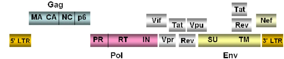
HIV-1 genome consists of two copies of linear single stranded RNA molecules with positive sense that are linked at the 5’ end through a self-complementary sequence called dimer linkage structure (DLS). The genomic organization shares gag, pro, pol, and env structural genes with the other retrovirus members while presents, in addition, six overlapping open reading frames (ORFs) of regulatory and accessory genes such as vif, vpr,
vpu, tat, rev and nef. The products of the structural genes (gag, pro, pol and env) are initially translated as polyprotein precursors. The gag gene leads
the expression of the 55-Kda Gag precursor that, after cleavage, generates matrix (MA), capsid (CA), nucleocapsid (NC) and p6 proteins; Gag-Pol precursor is a 160-Kda polyprotein and after processing generates protease (PR), reverse transcriptase (RT) and integrase (IN); the Env precursor (gp160) gives rise to the envelope proteins of surface (SU) gp120 and trans membrane (TM) gp41 (Fig. 1.2).
The products of accessory genes are generated through mRNA alternative splicing (Freed and Martin, 2001) and are classified in structural (present in the virion) and non-structural proteins (founded exclusively in the infected cells). Below their known functions will be described, even if their role is still partially solved. However it is known that these accessory proteins manipulate host biology to promote the viral life cycle.
The main regulatory proteins that are indispensable for virus replication are Tat and Rev. Tat (from Trans Activator of transcription) is a 14 KDa trans activator of proviral genome transcription while Rev is a phosphoprotein of 19 KDa involved in m-RNA maturation and transport and gets its name from Regulator of Virion Expression.
Viral Infectivity Factor, Vif, is about 23 KDa and plays a central role in the
APOBEC cellular enzyme ubiquitination and degradation, directly disrupting its antiviral activity (Stanley et al., 2008).
Viral protein R, Vpr, is a structural virion protein, is 14 KDa in size
involved in regulation of nuclear import of pre-integration complex (PIC). In addition, Vpr has an important role in apoptosis regulation and in G2 cell
cycle phase arrest (Bukrinsky and Adzhubei, 1999; Muthumani et al., 2006).
Virus Protein U, Vpu, is the distinguishing character of HIV-1, the protein
is in fact not present in HIV-2 and SIV. Vpu is a multimeric integral membrane phosphoprotein of 81 amino acid (Wray et al., 1999). The protein is involved in increasing virus release and in CD4 degradation (Bour and Strebel, 2003).
Finally, the virulence factor Nef which, being the topic of this work, will be in depth treated during the following sections.
Figure 1.1. Human Immunodeficiency Virus (HIV): virion anatomy. (www.con-tatto.org).
Figure 1.2. HIV-1 genomic organization. HIV-1 has three structural genes (gag, pol and env)
codifying for structural proteins and enzymes. HIV-1 genome also contains six overlapping open reading frames codifying for the regulatory and accessory proteins Tat, Rev, Nef, Vif, Vpr and Vpu.
1.1.2. HIV-1 life cycle.
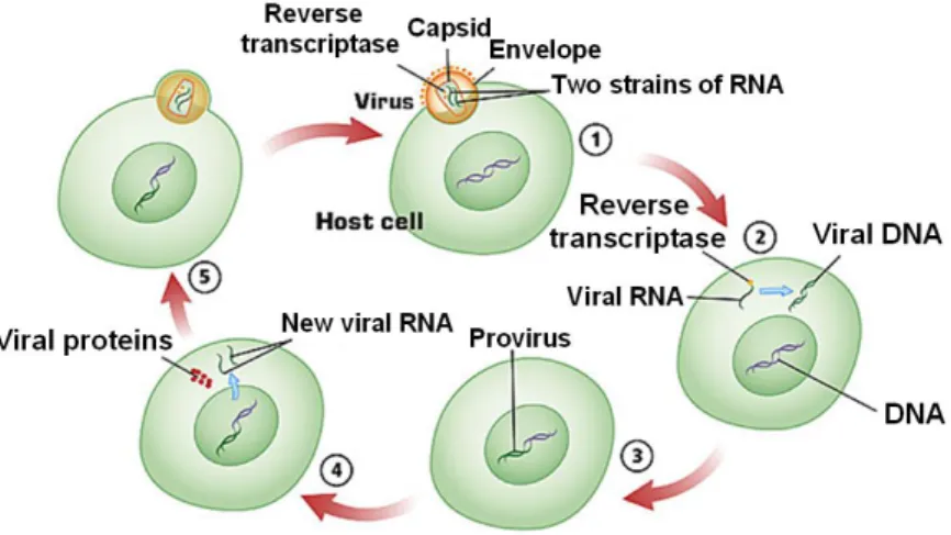
HIV-1 life cycle, as that of the others retroviruses, consists in a multi stage process and each step is crucial for successful replication (Fig. 1.3).
Figure 1.3. Schematic representation of HIV-1 life cycle. The figure shows the principal
stages of the viral cycle: 1) host cell penetration; 2) uncoating and reverse transcription of HIV genome; 3) Integration of the retroviral DNA; 4) transcription of the proviral DNA; 5) Virus budding. (Modified from Manninen, 2001).
The first step of HIV-1 infection, is characterized by the interaction between the surface subunit gp120, of the trimeric viral envelope glycoprotein (Env) gp120/gp41, presents on the virion surface and the CD4+ receptor presents on suitable host cells (i.e. T helper lymphocytes, machrophages and dendritic cells) (Dalgleish et al., 1984). The binding of gp120 to CD4 receptor is necessary but not enough for the entry of HIV into the cells: for an efficient process also a co-receptor (i.e. CCR5 or CXCR4 chemokines receptors) is required (Edinger et al., 1998; Simmons et al., 1998). The binding of gp120 with the CD4 receptor, in fact, promotes its further binding to the co-receptor. This event leads to a conformational change in gp120 that results in viral fusion mediated by gp41 trans membrane subunit (Blumenthal et al., 2012). After membrane fusion, penetration and
uncoating steps occur. During this process, who’s molecular mechanisms
have not yet been completely elucidated, viral capsid enters the cytoplasm and is digested (uncoating) inside the host cell releasing RNA genome and the three essential replication enzymes integrase, reverse transcriptase and protease to allow the formation of the reverse transcription complex (Arhel, 2010). Therefore, reverse transcriptase begins the reverse transcription of viral RNA. This enzyme presents two catalytic domains: an aminoterminal DNA polymerase active site and a carboxyterminal RNase H active site (Skalka and Goff, 1993; Tanese and Goff, 1988).
Reverse transcription is an extremely complex process comprising three main stages that are hereafter briefly schematized (an exhaustive description of the process lies outside the aim of this work): 1) Single stranded viral RNA is transcribed by DNA polymerase domain in a RNA/DNA double helix; 2) the RNA/DNA duplex is then processed by RNase H that degrades RNA strand; 3) the single stranded DNA is used as template by DNA polymerase to be converted in a full-length linear double stranded DNA bordered at each end by the long terminal repeat (LTR) containing all the genetic information for viral gene expression.
Once reverse transcription is concluded the integration of the viral DNA in the genome of the host cell takes place. Integration is crucial for retroviral life cycle and also accounts for the ability of viruses to persist in the infected cells as provirus and to permanently enter the germline (Goff, 2001).
The integration of retroviral DNA is mediated by the viral integrase protein. Integrase cleaves, at a highly conserved CA sequence, a dinucleotide from each 3’ end of DNA creating two sticky ends with protruding 5’ ends. Integrase then transfers the DNA into the cell nucleus and facilitates its integration into the host cell genome (Gallay et al., 1997). The mechanisms of integration consists in a Sn2-type reaction in which the 3’ OH ends generated by integrase are used to attack the phosphodiester bonds of the host DNA (Fujiwara and Mizuuchi, 1988). Thus, the protruding 5’ end of the proviral DNA is joined to the host DNA in a process that doesn’t require the viral integrase.
The integration of the viral DNA signs the end of the early phase of HIV-1 replication cycle and the beginning of the late phase mediated mostly by host enzymes. The late phase is characterized by the synthesis of viral RNA and proteins and progeny virions assembly (Freed and Martin, 2001). Transcription of proviral DNA into mRNA mediated by cellular Pol II polymerase starts at U3 part of 5’ LTR sequences which are the major determinants regulating virus replication. This process is controlled and
regulated by the viral regulatory protein Tat that stimulates transcriptional elongation of the full length viral mRNA. Viral mRNA migrates from the nucleus to the cytoplasm in a process involving Rev regulatory viral protein (Karn and Staltzfus, 2012). Into the cytoplasm a portion of the transcripts that remain unspliced is utilized as the viral RNA genome and also serves as Gag and Gag Pol mRNAs. Another portion is partially spliced for Env, Vif, Vpr and Vpu mRNAs and completely spliced to generate Tat, Rev, and Nef (Freed and Martin, 2001). Some of these proteins are translated as protein precursors and then processed by the viral protease during and after virion assembly to produce viral mature proteins; this step is critical for the creation of an infective virus. After synthesis, viral proteins and RNA genome came together to form the capsid. This immature viral particle buds out the cell and acquires the envelope containing both host and viral proteins. After this process the virus matures, becomes infective and equipped to infect other cells.
1.1.3. HIV-1 tropism, pathogenesis and progression to acquired immunodeficiency syndrome (AIDS).
How emerges from the modalities of virus entry, HIV infection is confined mainly to the specific sub set of immune system cells that presents CD4 receptor on its surface, such as CD4+ T-helper lymphocytes, macrophages and dendritic cells, and carrying the co-receptors CCR5 or CXCR4. On the basis of the type of co-receptor utilized in in vitro fusion assays, a classification of HIV-1 isolates has been proposed (Baba et al., 1999; Donzella et al., 1998).
HIV-1 strains that use CCR5 receptor are predominantly macrophage- or M-tropic (able to replicate in macrophages), nonsyncytium-inducing (NSI) and are defined slow/low because are characterized by slow replication and low production of viral progeny, these isolates are called R5.
HIV-1 strains that use CXCR4 co-receptor (X4 viruses) present a T-cell line tropism (TCL-tropic), are syncytium-inducing (SI) and, on the basis of their characteristics of replication and particles production, are defined
rapid/high and exhibit an enhanced cytopathicity. Although the in vivo
situation is obviously more complex, on the basis of this simplified classification, it has been observed that HIV-1 R5 strains are predominant during the asymptomatic phase of infection (Zhu et al., 1993) while X4 viruses emerge mostly during the acute symptomatic phase (Koot et al., 1993). During the asymptomatic phase, in fact, the major part of the
depleting CD4+ T lymphocytes are memory T-cells expressing CCR5 that are located principally in the gastrointestinal tract and in lung; only a low percentage of these R5-target cells are usually present in the peripheral blood (5-10%), so that no relevant T-lymphocytes depletion is yet observable in this tissue at this time of disease (Mehandru et al., 2004). The emerging of X4 strains during the late acute phase is accompanied by the substantial depletion of peripheral blood CD4+ T-lymphocytes (to < 200 cells/μl) that represents the salient clinical manifestation of HIV disease. The major part of the CD4+ T-cells circulating in peripheral blood (80% to 95%), in fact, are naïve T-cells expressing CXCR4 receptor and suitable to X4 virus infection. This massive T-lymphocytes depletion is associated with a rapid progression to AIDS (Connor et al., 1993).
How mentioned above, T-lymphocytes are not the only sub set of immune cells to be infected by HIV. A pivotal role in HIV dissemination and persistence in target tissues (i.e. lung, gastrointestinal tract, bone marrow, central nervous system, lymph nodes), in fact, is ascribed to macrophages and dendritic cells.
Macrophages are involved in mucosal transmission of the virus and represent the main viral reservoir. Macrophages are the first cells to be infected and are involved in viral spreading because able to directly infect CD4+ T-cells through a transient adhesive cell to cell contact (Groot et al., 2008). Further, macrophages exhibit a host/pathogen interaction response that significantly differs from that observed in T-lymphocytes and consisting in a great resistance to the cytopathic effects of the virus. More, in macrophages, HIV is able to grow inside endocytic compartments designated as multivescicular bodies (MVBs) that confer protection. Altogether these features counter for the long-term persistence of productive infection mediated by these cells (Carter and Ehrlich, 2008). Like to macrophages, dendritic cells (DC) are also infected by HIV and, being antigen presenting cells (APC), should be the first defence line during infection, but in the case of HIV-1 infection, DCs appear to promote viral spread. The interaction between DCs and HIV-1, in fact, differs from the mechanisms of infection observed in machrophages and CD4+ T-cells. In DCs the virus interaction is mediated mainly by a different kind of receptor known as DC-SIGN that binds and retains HIV on dendritic cell surface and mediates the infection of CD4+ T lymphocytes in a process called
trans-infection (Geijtenbeek et al., 2000).
To resume, the specific contribution made by diverse and specific immune system HIV suitable cells on the intricate dynamics of HIV-infection, converges in the massive depletion of CD4+ T-lymphocytes. This event
constitutes a strong insult to the immune system and results in the wide plethora of clinical manifestations, normally do not present in a health immune system, typical of AIDS. These manifestations include: generalized lymphadenopathy; a wide variety of severe opportunistic infections mainly caused by Pneumocystis carinii, Toxoplasma gondii and cytomegalovirus; the development of unusual neoplasms such as Kaposi’s sarcoms and non-Hodgking’s lymphoma, due principally to oncogenic viruses co-infections (i.e. Epstein-Barr virus, papilloma virus, herpesvirus).
Although after the discover that HIV is the etiological agent of AIDS several successes have been reached to contain the pandemy, AIDS is still a great menace for public health and represents also a huge worldwide socio-economic problem. The ability of the virus to mutate and to escape the immune system accounts for the difficulty to find a resolutive therapy. So far antiretrovirals that interfere with the crucial steps of HIV cell cycle have given the best results, while no efficacious vaccines have been still developed. However, also the antiretroviral therapies present several limitations in terms of costs and side effects. Hence, the necessity to find new targets to prevent or block HIV disease. Nef protein represents a good candidate to this purpose and is already object of intense study from a large part of the scientific community.
1.2. Nef protein and its role in HIV pathogenesis. 1.2.1. HIV-1 Nef: an overview.
Nef is a primate lentiviruses (HIV-1, HIV-2 and SIV) accessory protein encoded by the nef gene that is localized at the 3’ end of the viral genome (see figure 1.3).
HIV-1 Nef is a 27-35 Kda regulatory myristoyled phosphoprotein and despite to the originally belief that confined Nef functions to downregulation of virus replication (Nef, in fact, derived its name from
negative factor), is now clear that the protein has not negative effects on
virus replication but is strongly involved in maintenance of high-viral load
in vivo and plays a crucial role in progression to AIDS (Hanna et al., 1998;
Kestler et al., 1991). The central role of Nef in pathogenicity in vivo is supported by studies on animal models and seropositive patients that showed that nef defective viruses lead to an attenuated clinical phenotype with a reduced viral load. Are reported, in fact, a number of long term non-progressor (LTNP) individuals whose viruses presented marked depletion in the nef gene (Kirchhoff et al., 1995). Further, it has been demonstrated that
nef transgenic mice develop an AIDS-like disease (Hanna et al., 1998). Nef,
actually, is considered an important virulence factor that, lacking of any enzymatic activity, fulfils its functions working as a molecular adaptor. It has been reported, in fact, that Nef interacts and interferes with the activity of more than 30 intracellular partners (Fackler and Baur, 2002) mostly involved in membrane receptors trafficking (Doms and Trono, 2000) and in signal transduction pathways (Geyer et al., 2001).
Well ascribed Nef functions in vitro include downregulation of diverse cell-surface molecules (such as CD4, MHC-I, MHC-II, CD3 receptor complex, CD28) (Garcia and Miller, 1991; Schwarz et al., 1996), increase of virus infectivity (Miller et al., 1994), regulation of apoptosis (Fackler and Baur, 2002) and modulation of cell signalling such as T-cell activation pathways (Sawai et al., 1994). These functions will be analyzed in detail in the following sections after a preliminary description of the Nef structure.
1.2.2. HIV-1 Nef structure and the “Nef cycle”.
Nef is a small protein of about 200 amino acids that exists in diverse allelic forms varying slightly in length (Percario et al., 2011), however in this work we will refer mainly to SF2 Nef allele that is about 27 Kda in size in its full length myristoylated form or 25 Kda in the trunked form that is translated from a second start codon. Nef is expressed early and abundantly during the early stages of the viral cycle and is post-translational modified by phosphorilation and by N-terminus myristoylation that is critical for its functions.
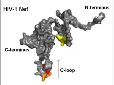
NMR spectroscopy and X-ray crystallography have been used to determine the three-dimensional structure of Nef protein (Grzesck et al., 1997; Arold et al., 1997). The structure of whole Nef protein has been determined trough the overlap of the single fragmented components separately analyzed, because the full-length structure is quite difficult to obtain due to problems in its crystallization. Nef presents a specific cleavage site for a viral protease (between Trp 57 and Leu 58 residues) that has been a good tool for the determination of its structure because splits the protein in its two principal domains: the N-terminal anchor domain that is globally unfolded and structurally flexible and the C-terminal core domain that is the only part of the protein to present a stable tertiary fold (respectively 2-61 and 62-210 residues), both involved in cellular signalling and trafficking (Geyer et al., 1999; Breuer et al., 2006; Geyer and Peterlin, 2001); further a flexible loop, also important for Nef functions, of about 30 amino acids, projects out of the core domain (Fig. 1.4).
Figure 1.4. HIV Nef structure. The principal Nef domain are shown: the unstructured
N-terminus anchor domain, the well conserved and folded N-terminus core domain and the C-loop that projects out of the core domain. (Modified from Götz et al., 2012).
The core domain has been studied both alone and in association with SH3 domains of Nef interaction partner proteins. It is a highly conserved region characterized by the presence of a PxxP motif that, in association with the SH3 (Src homology 3) domain, assumes a left-handed polyproline type II helix. Over PxxP motif, is also present an α-β motifinwhich a central parallel β sheet of four strands is flanked N-terminally by two long anti-parallel α-helices and C-terminally by two short α-helices (Arold et al., 1997; Lee et al., 1996). Diverse binding sites are present in the core domain. PxxP motif is one of the most important, allowing the binding of a great part of Nef molecular partners. Further, core domain plays a role in the Nef oligomerization that has been observed both in vitro and in vivo, even if its significance has still to be clarified (Arold et al., 2000; Arold and Baur, 2001).
The structure of the anchor domain has been characterized both in the presence and in the absence of N-terminus myristoylation. The anchor domain is a genetically varied unstructured region of about 60 amino acids.
Globally, in the not myristoylated anchor domain secondary folded elements have not been found except for a short two-turn α-helix (H2) between Arg35 and Gly41 and for another helical secondary structure element (H1) in the arginine-rich region (Arg17 to Arg22). The myristoylation, that involves the N-terminal glycine residue, confers stability to this secondary elements and renders it more defined (Geyer et al., 1999).
How mentioned above the anchor domain is a moderately conserved region and the only sequence highly conserved is the motif MGxxx(S/T) that is the
consensus sequence for N-myristoyl-transferase target proteins (Resh, 1999;
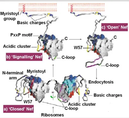
Geyer et al., 2001) and this accounts for the crucial role that myristoylation plays in the Nef functions specially in its signalling activity (see below in the chapter). Later in this chapter, will be described detailed examples relative to the importance of myristoylation in Nef biology. However, N-terminus myristoylation plays a role in the cellular localization of Nef that is determinant for Nef molecular interactions and signalling. How mentioned above, in fact, Nef is a molecular adaptor that alters cellular pathways via multiple protein-protein interactions and diverse Nef interaction motifs have been already identified (table 1.1). Indeed, Nef presents both cytoplasmic and membrane localization and the wide plethora of Nef-mediated effects are the results of the different set of effectors that interacts with Nef depending on its cellular localization and on the particular phase of the viral life cycle. In this regard it has been speculated on the existence of a Nef cycle that consents a time/space-dependent exposure of different Nef motifs (Arold and Baur, 2001). The great plasticity of Nef, in fact, is certainly due to its peculiar flexible structure that consents drastic conformational adjustments that are basic for Nef multiple interactions, and these conformational changes could occur during the hypothetic Nef cycle. The Nef cycle is a speculative model that follows the path of Nef immediately after its translation until its bind to cellular membrane. According to this model, after protein translation, Nef assumes a “closed conformation” in which presumably are shown just some of its interaction sites. After the bind with the membrane, mediated by myristic acids, Nef adopts a semi-open form that binds first with signalling molecules downstream the T-cell receptor (TCR). These interactions lead Nef to assume the open signalling conformation involved in the interaction with endocytotic machinery and in Nef trafficking and signalling (see next paragraph) (Fig. 1.5).
Figure 1.5. The Nef Cycle. On the basis of this speculative model, Nef assumes different
conformations depending on its cellular localization and on the viral cycle stages. a) the closed conformation that Nef could adopt immediately after translation, in this form Nef hides most of its interaction sites; b) after the bond with membrane, Nef adopts a semi-open signalling conformation mainly involved in the interaction with T-cell receptor signalling molecules; finally, Nef assumes an open conformation and exposes the C-loop. This conformation could be involved in the interaction with endocytic machinery. (Modified from Arold and Baur, 2001).
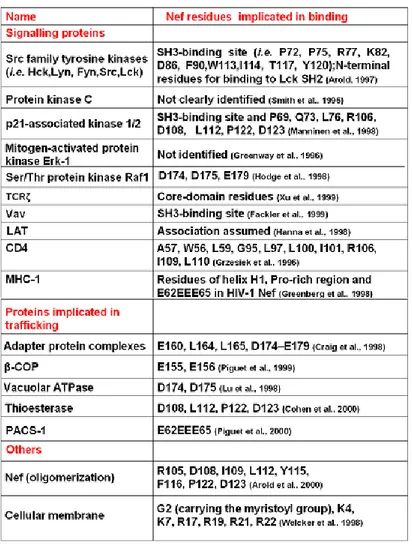
Table 1.1. The table shows a list, although non exhaustive, of proteins that have been found
associated with Nef.Abbreviations: β-COP, β subunit of COPI coatomers; LAT, linker for activation of T cells; MHC-1, major histocompatiblity complex 1; PACS-1, phosphofurin acidic cluster sorting protein 1; SH2/3, Src-homology domain 2/3; TCRζ, ζ chain of the T-cell receptor; vATPase, vacuolar ATPase. (Modified from Arold and Baur, 2001).
1.2.3. HIV-1 Nef functions.
How mentioned above, Nef is an important virulence factor able to interfere with the normal functions of the cells showing a wide range of phenotypes. In this paragraph we will consider what is known about the main Nef functions, even if their molecular mechanisms are still only partially solved.
Negative modulation of membrane receptors.
Nef interferes with the endocytic machinery decreasing, in the infected cells, the expression of certain membrane receptors such as CD4 and histocompatibility complex class I (MHC I) antigens.
Downregulation of CD4 is a common phenomenon, utilized also by other retroviruses, to avoid superinfection (Michel et al., 2005) and to enhance the release of viral progeny (Ross et al., 1999). Further, this could represent a strategy to control signalling events in the infected cells.
Nef promotes CD4 endocytosis recruiting clathrin molecules to the membrane, in a process that involves the Nef binding to CD4 and to the clathrin adaptor AP-2. Furthermore, it has been reported that the interaction between Nef and β-COP protein is responsible to direct CD4 molecules to the endosomal pathways leading to its degradation (Benichou et al., 1994; Landi et al., 2011).
MHC-I downregulation is a defense mechanisms involved in HIV immuno-evasion that, rendering infected cells less visible to circulating CD8+ cytotoxic T lymphocytes (CTL), confers resistance of infected cells to CTL killing (Adnan et al., 2006).
Despite to the Nef-CD4 interaction that happens when the receptor is already present on the cell surface, the Nef binding to MHC-I takes place early in the secretory compartment. Particularly, Nef with its acidic and polyproline domains interacts with the cytoplasmic tail of MHC-I to form a complex able to recruit and bind AP-1 (Roeth et al., 2004). The formation of this complex creates an alternative route for MHC-I that, instead to be directed to the cell surface, is transported to lysosomes to be degraded, in a process that also in this case involves the interaction with the protein β-COP (Schaefer et al., 2008).
Nef-mediated cell signalling deviation and activation of T-cell receptor (TCR).
In T-cells Nef activates TCR signalling pathways miming what activated by its exogenous stimulators, and thus is able to manipulate T-cell activation by interacting with several proteins downstream the TCR, which include PI3K, Vav or DOCK2-ELMO, small GTPases, Pak2 and PKC (Roeth et al., 2006).
It has been reported that Nef exerts modulatory opposite effects on TCR signalling in infected T-cells depending on its cellular localization. It has been observed, in fact, that Nef promotes T-cell activation when is myristoyled and thus anchored to the membrane while exerts an inhibitory effect on T-cell activation in its non-myristoyled cytosolic form (Baur et al., 1994).
On the basis of the current models, Nef mediates the control of T-cell activation both in a TCR-dependent and independent way. Nef can bind directly the ζ chain of TCR promoting, in concert with other signalling proteins (such as Lck, LAT and Vav), its phosphorilation and activation or can influence the process through the indirect activation of Inositol 1,4,5-trisphosphate receptor type 1 (Manninen and Saksela, 2002).
However it has been hypothesized that the strategy adopted by Nef to lead T-cell activation is in decreasing the threshold of T-cell activation instead to promote the activation of the resting cells (Schrager and Marsh, 1999). To conclude, activation of T-cells is a necessary event for integrated proviral genome transcription (Zack et al., 1990) and is evident the role of Nef in the regulation of this process.
Regulation of apoptosis.
Nef plays a central role in the regulation of apoptosis during the course of HIV-1 infection, protecting infected cells and inducing apoptosis mainly in bystander non-infected T-cells (Finkel et al., 1995). The relevance of this function is documented by the strong correlation existing between apoptosis and the massive CD4+ T-cell depletion typical of AIDS.
Nef, as already said, activates T-cell and it has been reported by several authors that the activation of T-cell is a central requisite that renders T-cells highly susceptible to apoptosis in a process known as activation-induced
cell death (Alimonti et al., 2003).
The induction of apoptosis in bystander T-cells can occur both in a direct way in which is involved the Nef interaction with the CXCR4 receptor
(James et al., 2004; Homann et al., 2009), and in indirect way in which Nef increases the expression of FasL on infected cells (Xu et al., 1999).On the other hand, Nef protects infected cells by apoptosis expounding an inhibitory effect on pro-apoptotic proteins such as ASK1, Bad or p53 (Greenway et al., 2002).
1.2.4. Nef and macrophages.
Several studies have highlighted on the important role that Nef exerts in macrophages that are used as an extremely valid in vitro model to better clarify the role of this protein in the HIV-1 infection context.
In macrophages HIV-1 induces the production and release of chemotactic factors able to activate and recruit T-cells to the infection site, rendering them more susceptible to virus attack. Studies carried out by infecting macrophages with HIV-1 M-tropic strains both containing nef gene or nef deleted have demonstrated that Nef is responsible for the chemokines production observed (Swingler et al., 1999). Indeed, the endogenous expression of HIV-1 Nef in human monocyte-macrophages has been reported to induce production and the release in the macrophages supernatants of inflammatory proteins such as MIP-1α and MIP-1β and other soluble factors (Swingler et al., 1999 Alessandrini et al., 2000). Further, Nef-mediated activation of the Signal Transducer and Activator of
Transcription STAT-1 has been demonstrated in monocyte-derived
macrophages (MDMs) infected with a Δenv HIV-1 strain. Interestingly, the STAT-1 activation doesn’t occur in MDMs upon infection with a Δenv/Δnef HIV-1 strain (Federico et al., 2001). Further studies, conducted by using an adenovirus-based vector to express Nef in macrophages, suggest that Nef is able to hijack the cellular signal transduction pathways intersecting the CD40/CD40L signalling pathway (Swingler et al., 2003).
Recently, on the basis of the fact that extracellular Nef has been found in the serum of infected patients at a significant range of concentration (from 0.5 to 10 ng/ml) (Fujii et al., 1996), the interest has been also focused in exploring the capability of macrophages to internalize Nef, also evaluating its effect on cell signalling (Olivetta et al., 2003).
Particularly, in vitro studies carried out in the laboratory where I have performed my Ph.D., have demonstrated that Nef is efficiently internalized by primary human non-infected MDMs and leads to effects very similar to those observed in cells endogenously expressing Nef.
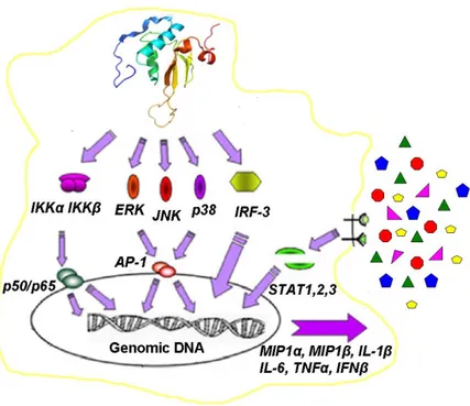
Here Nef mediates the cycloheximide-independent activation of the transcriptional activator NF-kB, the activation of specific MAPKs (i.e. ERK1/2, p38, and JNK) and of IRF-3 that is the main regulator of interferon β gene. These events lead to the synthesis and release of several cytokines and chemokines (i.e. IL-1β, IL-6, TNFα, MIP-1α, MIP-1β and IFN-β). These factors are in turn able to activate STAT-1, STAT-2 and STAT-3 in autocrine and paracrine manner (Fig. 1.6).
Figure 1.6. HIV-1 Nef mediated signalling in primary human non-infected monocytes derived macrophages (MDMs). Nef is efficiently internalized by MDMs, mediating the activation of NF-kB, specific MAPKs and IRF-3. These events lead to the synthesis and release of several cytokines and chemokines (i.e. IL-1β, IL-6, TNFα, MIP-1α, MIP-1β and IFN-β) able to activate the Signal Transducers and Activators of Transcription STAT1, STAT2 and STAT3 in an autocrine and paracrine manner. (Mangino et al, 2007).
These studies has been realized by using a myristoyled recombinant Nef (rNef) and a set of Nef mutants (Olivetta et al., 2003; Mangino et al., 2007).
1.2.5. The Nef anchor domain and its acidic cluster motif as potential targets to block Nef functions.
After the long series of Nef functions earlier described, it is not surprising ask about how to justify the intricate biology of Nef referring to its small size. Clearly, to answer this question we have to dwell in the Nef structure again. Particularly, in this paragraph we will focus on the N-Terminal anchor domain of Nef (also called Nef N-terminal arm), a protein region whose functions range over both structural and signalling tasks. This flexible unstructured region provides a large interaction surface that, in addiction to interact directly with the molecular effectors, seems involved in the conveying of binding molecules to the core domain interaction motifs or to other Nef binding site. Further, the anchor domain could behave as a regulative allosteric region involved in the exposure of specific Nef motifs through the induction of conformational changes (Baugh et al., 2008). It has been also assumed that the anchor domain could act as a spacer to consent, for example, a good juxtaposition between Nef and the membrane target receptors.
More over, the presence of a specific cleavage site for the viral protease between the two principal Nef domains suggests for this site a plausible protease-mediated regulative function (Freund et al., 1994) eliciting also the possibility that these two domains could work independently too, hence the importance of an in parallel modular study of the protein, that is object of this work.
As a matter of fact, the importance of the N-terminal anchor domain is documented also by the presence in this region of the most important sites subjected to post-translational modifications, such as myristoylation and phosphorylation sites, presumably responsible for the allosteric regulations mentioned previously.
Probing in detail the anchor domain it is possible to detect its two principal interaction motifs: the region including the first 22 amino acid residues (required for the membrane binding, mostly positively charged and containing the N term G fundamental for the myristic acid attachment) (Welcker et al., 1998), and the acidic cluster motif (AC).
The Nef acidic cluster is located at the border between the anchor domain and the core domain but is considered as a part of the anchor domain. It
encompasses, in Nef SF2 allele, the four glutamate residues from position 64 to 69 (A64QEEEE69) (Fig. 1.7).
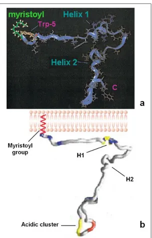
Figure 1.7. Structure of HIV-1 Nef anchor domain. Panel a shows the two principal secondary folded elements: helix 1 and helix 2. Panel b shows the localization of the acidic cluster domain (a: modified from Geyer et al., 1999; b: modified from Arold and Baur, 2001).
One of the first and highly important functions ascribed to the Nef acidic cluster is its involvement in the down-regulation of MHC-I mediated by the
AC binding with PACS-1 and PACS-2. This has been confirmed in studies carried out with Nef acidic cluster mutants, in which the substitution of the four glutamates in four alanines results in the loss of the Nef ability to down-regulates MHC-I (Greenberg et al., 1998). A large number of similar studies on SF2 Nef acidic cluster mutants has been carried out to define the AC role also for other functions of Nef (Baugh et al., 2008).
Recent studies, carried out by my co-workers, validate the involvement of the acidic cluster on the Nef signalling effects observed in macrophages that have been described in the paragraph 1.2.4.
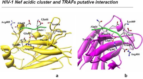
Particularly, it has been demonstrated that the synthesis and the release of inflammatory factors and IFNβ after treatment of MDMs with myristoyled rNef depends on the integrity of the acidic cluster region (Mangino et al., 2011). Particularly, on the basis of previous results indicating that Nef exerts its signalling functions intersecting the CD40 path (Swingler et al., 2003), it has been proposed a direct interaction between the acidic cluster and members of the Tumor Necrosis Factor Receptor-Associated Factor (TRAFs) adaptors family. This interaction is supported by modelling analyses performed using crystallographic data obtained on both TRAF2/4-1BB (Ye et al., 1999) and TRAF6/RANK (Ye et al., 2002) complexes indicating plausible the acidic cluster as a putative binding motif for TRAF2 and TRAF6 consensus binding sequences (Fig. 1.8).
Figure 1.8. Schematic representation of the modeled complexes formed by Nef acidic cluster with TRAFs. Panel a) putative interaction between Nef acidic cluster and TRAF2;
However, to really evaluate the effective involvement of TRAF in Nef mediated signalling and to confirm putative TRAFs-Nef acidic cluster interactions silencing studies and pull-down experiments have been carried out. Silencing experiments have been performed on human monocytic cell line THP-1 that presents a response to Nef very similar to that observed in MDMs. Three days post transfection of THP-1 with TRAF-2 or TRAF-6 specific siRNA pools, cells have been treated with rNef and the activation of STAT-1 and STAT-2 has been evaluated. As result, no STAT activation has been observed in transfected cells, suggesting that both TRAF-2 and TRAF-6 are involved in Nef mediated signalling.
On the other hand, pull-down experiments confirm the physical interaction between Nef and TRAF-2 even if the same result has not been reported for TRAF-6 (Mangino et al., 2011).
To resume, it has been demonstrated that the acidic cluster is responsible for the Nef signalling effects on macrophages and that in this process the acidic cluster plays a central role. Further, in this function is involved the direct interaction between the acidic cluster and TRAF-2.
Overall, considered the relevance of N-terminal anchor domain in Nef functions and consequently in HIV-1 pathogenesis, finding molecules able to block the main interaction sites of Nef could be of great therapeutic importance.
Starting from the results previously shown, the focus of my Ph.D. work has been directed on the search of Nef acidic cluster binding peptides through phage display methodology. This study has been carried out with two principal purposes: first, better elucidate the molecular mechanisms underlying the Nef interactions with its molecular partners and specifically the Nef/TRAF interactions dynamics and secondly, to find a potential Nef binding inhibitor. A peptide able to compete with TRAF in the acidic cluster binding, in fact, could represent a potential tool to control the Nef effects on the signalling and thus could represent a potential drug to contrast HIV-1 infection.
Phage display of random peptides, that consents to construct libraries with enormous molecular diversity and to select for molecules with predetermined properties, has been used in order to identify acidic cluster peptide ligands.
Further, this project has been also centred on the more generic study of Nef anchor domain and on its effects on cell signalling after internalization in macrophages, as it will be described in the “aim of the work”.
1.3. Phage display technology.
Phage display is a powerful laboratory technique largely used in a wide array of application such as the study of protein-protein, protein-peptide, protein-nucleic acids interactions, the individuation of newly enzymatic catalytic sites or activities and providing also the starting point for in vitro evolution studies. This methodology, introduced for the first time in 1985 (Smith, 1985), has had a great impact on several biologic fields such as immunology, cell biology, protein engineering, physiology and pharmacology.
Phage display uses bacteriophage as vehicle of peptides, and providing the coupling between phenotype and genotype, consents selection, recover and identification of the interest molecules from highly broad peptide libraries. Phage display, in fact, is centred on the ability of filamentous phage to display capsid protein-fused foreign peptide on their surface.
1.3.1. Biology of filamentous bacteriophage.
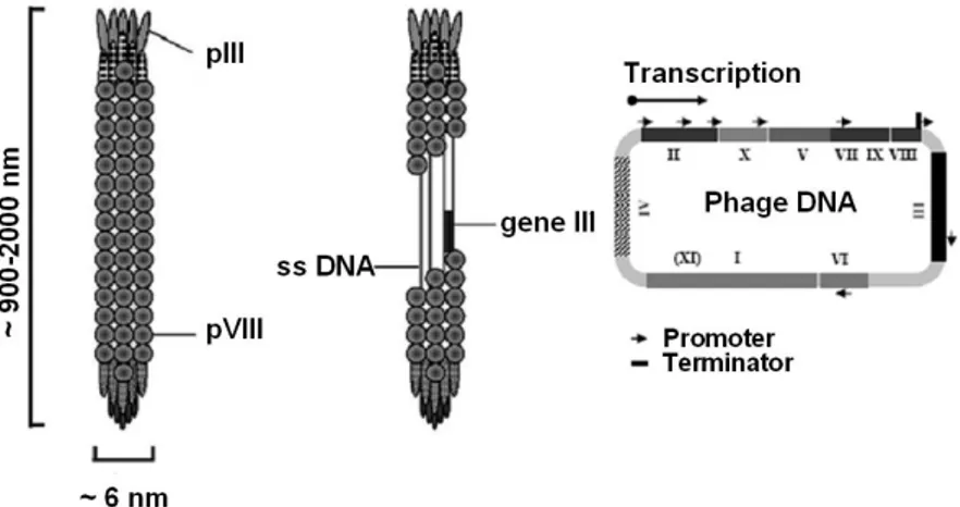
Filamentous phage have a single-stranded DNA genome which is encased in a long cylinder approximately 6 nm wide by 900 to 2000 nm in length. The entire genome of these phage consists of 11 genes (Fig. 1.9). Two of these genes, X and XI, overlap and are in-frame with the larger genes II and I (Rapoza and Webster, 1995; Model and Russel, 1988). The arrangement of the genes on DNA is based on their functions in the life cycle of the bacteriophage. Two genes (gII and gX) encode proteins required for DNA replication while a third one (gV) encodes for a protein necessary both at the assembly and DNA level; a group of three genes (gI, gIV and gXI) is involved in the phage assembly process at membrane level, while a last group encodes the capsid proteins. In addition to the regions which encode proteins, is the “Intergenic Region” which contains the sites of origin for the synthesis of the (+) strand (phage DNA) or (-) strand as well as a hairpin region which is the site of initiation for the assembly of the phage particles (packaging signal). A phage expresses about 2700 copies of the major coat protein (pVIII, 50 aa long), and 3 to 5 copies of the minor coat protein (pIII, a 406 aa long) (Russel, 1991).
Figure 1.9. Schematic representation of filamentous bacteriophage particle and phage genome organization.The single stranded DNA is encased inside the cylindrical coat. pVIII is the major coat protein presents approximately in 2700 copies per particle, while the minor capsid protein pIII is present at the tip of the phage. The phage genome of 6407 nucleotides contains 11 genes.
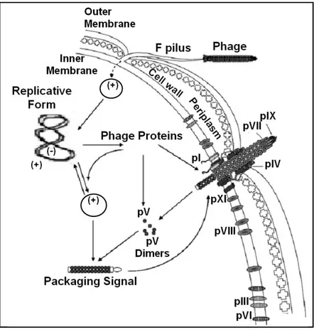
The best characterized filamentous phage are M13, fl and fd. These phage infect a variety of gram negative bacteria such as Escherichia coli that contain the F conjugative plasmid. Because of their dependence on the F plasmid for the infection these phage are known as Ff phage. The infection starts when one end of the phage interacts with the tip of the F pilus whose gene is located in the F conjugative plasmid. Afterwards, the capsid proteins integrate into membrane and the phage circular single stranded DNA is delivered in the cytoplasm. Here, the complementary strand synthesis, operated by bacterial enzymes, and its conversion in a double stranded super coiled replicative form molecule (RF) occurs. RF provides the template for transcription and translation of the phage proteins. Now, phage proteins together with bacterial enzymes lead the synthesis both of further RF and of new phage DNA molecules. New phage particles are produced when the phage specific single-stranded DNA binding protein pV, reaching a limit concentration, forms complexes with the newly synthesized phage single-stranded DNA. pV-DNA complexes are not converted to RF but assembled to form new phage particles (Fig. 1.10). The assembly of phage occurs at the bacterial envelope and continues until the end of DNA, this
fact renders Ff phage good cloning vectors because able to contain foreign DNA of big size (range from few to thousand nucleotides). Phage infection doesn’t alter significantly the host duplication and results in the production of about 1000 phage particles during the first generation and 100-200 phage particles in the next generations (Model and Russel, 1988).
Figure 1.10. Bacteriophage life cycle. After interaction between protein pIII and a tip of the
F pilus the phage DNA is translocated inside the cytoplasm. The phage DNA is converted in its replicative form (RF). pV protein phage bound phage ss DNA and direct it to the membrane. Here assembling of new phage particles occurs followed by particles extrusion from the cell.
1.3.2. Phagemid vector construction and phage display principles.
Phagemid is a particular kind of cloning vector specifically designed for the application of phage display. It consists of a double stranded circular DNA molecule containing the replication origin and the packaging signal (PS) of the filamentous phage together with the origin of replication and gene expression system of the chosen plasmid. Inside the phagemid is possible the cloning of the insert of interest. The phagemid is usually engineered to contain near to the cloning site a sequence codifying for a phage capsid protein in order to obtain after plasmid expression a chimeric protein consisting in the capsid protein fused with the protein of interest. E. coli cells are thus transformed with this construct. The infection of the transformed cells with a filamentous helper phage actives the viral origin of replication of the phagemid, further helper phage supplies the proteins for the assembly. As result, the newly synthesized phagemid single stranded DNA carrying the insert of interest is assembled to form new phage-like particles. These recombinant phage carry on their surface the chosen capsid protein (it is usually used the minor capsid protein PIII) fused with the peptide encoded by the foreign DNA, so that phenotype and genotype are linked and, after selection cycles, it is possible to identify the peptide that presents desired properties.
1.3.3. Phage display of random peptides.
How mentioned above, one of the features that render phage display a powerful methodology is the possibility to obtain a peptide with desired properties starting from enormously variegate peptide libraries. The starting point to carry out phage display of random peptides is to design and produce synthetic oligonucleotides fixed in length but with unspecified codons so as to have a highly diverse mixture of random nucleotide sequences (about 109 to 1012 different sequences). These sequences can be cloned in phagemid vectors as fusion to M13 genes codifying for capsid protein such as PIII to obtain a phagemid library. After infection of the bacterial cells transformed with the phagemid library the viral particles produced represent a phage library of random peptides in which each phage theoretically displays on its surface a different random peptide.
One of the most important application of phage display of random peptide is the selection of random peptides whose bind with a molecule target, and this can be used, as in the case of the project to which we refer here, to
select for a binding peptide that can exerts inhibitory activities on the target molecule.
The selection cycles are performed by using a form of affinity selection known as “bio-panning” (Parmely and Smith, 1988).
The selection through bio-panning consists in the immobilization of the molecule that is object of study on a solid surface and in the incubation of the target molecule with the phage library to start the selection cycles. During the selection cycles phage that display specific binding affinity for the target can be selected, recovered and submitted to further more stringent selection cycles. The process goes on until the finding of the peptide that best resemble that with the ideal features (Fig. 1.11).
Figure 1.11. Schematic representation of bio-panning cycle. 1: the target molecule is immobilized in the plate; 2: incubation with phage library; 3: selection of the phage that displays peptides with binding affinity for the target; 4: to remove unbound phage several washes are performed; 5: bound phage are eluted, and amplified by infection of susceptible E.
2. AIM OF THE WORK
Since, in 1983, HIV was declared the causative agent of AIDS scientists around the world have not yet been able to find a resolutive therapy for this disease that still remains a very significant public health and socio-economic emergency.
This doctoral project falls within the scope of contributing to the elucidation of the action mechanisms of HIV-1 Nef that currently represents one of the most interesting targets for the development of new therapeutic strategies, and further to search for inhibitors of Nef functions to be tested as drugs for HIV disease.
In detail, the study has been focused on the N-terminal anchor domain of Nef that, as already reported in chapter 1, is a region of the protein exceptionally flexible in terms of structure and of molecular interactions and absolves a key role in the Nef activity. Thus, this project aims at studying the role of Nef anchor domain in Nef activities, and at the investigation of the molecular basis underlying the Nef/TRAF interaction evaluating the role of the Nef acidic cluster in this process in order to clarify the enigmatic functions of Nef. Moreover, the project is aimed at searching for inhibitors of Nef functions.
To reach these goals it has been decided to produce and purify a target region of Nef designed as N-Term76-Nef, encompassing the Nef N-terminal anchor domain and the acidic cluster region, to be utilized both to elucidate the role of Nef anchor domain in Nef signalling and as target to phage display experiments.
Using phage display approach peptides will be selected, from a phage random peptide library, that display binding affinity for Nef. Finally, once found a N-Term76-Nef binding peptide it will be used to verify its potential inhibitory effects. The attention will be also focused on the responses of cell culture systems, such as primary human non infected MDMs and human monocytic cell lines to N-Term76-Nef treatment alone or in combination with the wild type protein.
3. RESULTS AND DISCUSSION
3.1. Expression and purification of HIV-1 Nef protein N-terminal anchor domain.
Here we want to acquaint the reader with the experimental procedures that have been set up for obtaining the production and the isolation of the N-terminus anchor domain of Nef protein.
The molecular characterization of the Nef anchor domain, the elucidation of the molecular mechanisms underlying its involvement in the biology of HIV-1 and the search for inhibitors of its functions to be tested as drugs for HIV disease are the goals of this project, and the first step towards the realization of these intentions has been the production of large amounts of this Nef target region.
The protocol here proposed consists of three main successive phases: the amplification of the region of the nef gene codifying for the first 76 amino acid residues here called N-Term76-nef, the expression of this nef region, and the purification of the expressed protein.
3.1.1. N-Term76-nef amplification and plasmids construction.
The entire nef gene (SF2 allele) was initially cloned inside pCDNA3 plasmid (Mangino et al., 2011). Starting from this construct, the amplification of the N-Term76-nef has been carried out by polymerase chain reaction (PCR). The trunked region of the nef gene includes the anchor domain (aa 2-61) and a further region that extends over the viral protease cleavage site (between Trp 57 and Leu 58), encompassing the Nef acidic cluster (A64QEEEE69) that is considered as an extension of the anchor domain (see chapter 1).
In detail, both the amplifications of N-Term76-nefWT (wild type A64QEEEE69) and of its mutant in the acidic cluster region
N-Term76-nef4EA (A64QAAAA69) have been performed in order to use this mutant as negative control during the experiments.
The design of three synthetic oligonucleotides (primers) has been based on the sequence of the specific nef gene region to amplify and on the type of plasmid utilized as cloning vector (how explained later, two different vectors have been used). The so designed primers are the follows: Nef-PshAI-Forward: 5’ ATG GGT GGC AAG TGG TCA 3’; Nef-NotI-Forward: 5’ ATA AGA ATG CGG CCG CAA TGG GCA AGT GGT CA
3’; Nef-BamHI-Reverse: 5’ AAC GAA TGG ATC CTA TAA AGG TAC CTG AGG TGT 3’.
The PCR reactions led to the amplification of four PCR products, deriving from the use of the two different forward primers and the two different DNA templates (nef WT and nef 4EA genes) (Fig. 3.1).
Figure 3.1. Amplification by PCR of N-Term76-nefWT and 4EA gene region. Lane: 1 and 2 N-Term76-nefWT PCR products, lane 3 and 4 N-Term76-nef4EA PCR products. Lane M: 50 bp
DNA Ladder.
How mentioned above, two different plasmids have been used for the cloning of the amplified PCR products in order to havethe possibility to use different methodologies of purification on the basis of the experimental requirements. Both N-Term76-nefWT and N-Term76-nef4EA PCR products have been cloned inside pET-14b (previously digested withPshAI and NotI restriction enzymes) or inside pET-42-CM (previously digested with PshAI and BamHI) expression vectors (Fig. 3.2).
Figure 3.2. Schematic representation of the plasmids used as expression vectors. Panel a:
pET-42-CM carrying two different tags: GST and two hexa histidine-tag (His-Tag) upstream
N-Term76-nef. Panel b: pET-14b carrying only a His-Tag upstream the cloning site.
pET-14b plasmid carries an N-terminal His-Tag sequence followed by a thrombin site and three cloning sites. On the other side, pET-42-CM vector, that has been modified from the original one (pET-42b) by adding two His-tag sequences upstream the cloning site and the not translating DMY sequence, that is used to check the enzymatic double digestion in the plasmid, consents a high expression level of the inserted sequences fused to the 220 aa GST (glutathione S-transferase) Tag protein (Fig. 3.3). As results, two different constructs have been realized which are genetically equipped for the expression of Nef anchor domain flanked by two different tags (His sequences and GST protein).
Figure 3.3. Schematic representation of pET-42-CM vector. After digestion with PshAI and
BamHI theDMY sequence (a) is removed and N-Term76-Nef gene sequence is cloned inside
the plasmid (b). a b N-Term76 Nef DMY sequence a b N-Term76 Nef DMY sequence N-Term76 Nef DMY sequence