UNIVERSITÀ DEGLI STUDI DI CATANIA
DIPARTIMENTO DI AGRICOLTURA, ALIMENTAZIONE E AMBIENTE
International Ph.D. Program in
“Plant Health Technologies and Protection of Agro-ecosystems”
Cycle 28
thEXPOSURE OF HONEYBEES (APIS MELLIFERA L.) AND OTHER
HYMENOPTERA POLLINATORS TO DIFFERENT BACILLUS
THURINGIENSIS BASED BIOPESTICIDES IN LABORATORY
CONTROLLED TRIALS
Dr. VALERIO VACCALLUZZO
Coordinator: Prof. CARMELO RAPISARDA Tutor: Prof. VERA D’URSO Co-Tutor: Prof. GAETANA MAZZEO Co-Tutor: Prof. SALVATORE TRAVALI Co-Tutor: Prof. MARIA ANTONIETTA TOSCANO
I
SUMMARY
ABSTRACT ... 1
RESEARCH GOAL ... 5
Chapter 1 – BEES AND ENVIRONMENT ... 7
1.1 INTRODUCTION ... 7
1.2 IMPORTANCE OF BEES FOR AGRICOLTURE ... 9
1.3 IMPORTANCE OF BEES FOR ECOSYSTEM CONSERVATION ... 12
Chapter 2 – BACILLUS THURINGIENSIS AND BIOPESTICIDES ... 13
2.1 INTRODUCTION ... 13
2.2 BACILLUS THURINGIENSIS B ... 14
2.3 BACILLUS THURINGIENSIS BIOPESTICIDE FORMULATIONS: STRAINS, SEROTYPE AND SUBSPECIES ... 21
2.4 RISK ASSESSMENT OF BACILLUS THURINGIENSIS BIOPESTICIDE FORMULATIONS ON NON-TARGET ORGANISMS AND ENVIRONMENT ... 22
Chapter 3 – THE ALIMENTARY CANAL OF HONEYBEE ... 24
3.1 THE ANATOMY AND DIGESTION ... 24
3.2 THE MIDGUT AS BARRIER AGAINST PATHOGENS AND AS MECHANISM OF INTERNAL DEFENSE ... 28
Chapter 4 – MATHERIALS AND METHODS ... 31
4.1 TOXICITY TEST ... 31
4.1.1 Honeybees (Apis mellifera) ... 31
4.1.2 Solitary bees (Osmia cornuta and Osmia rufa) ... 31
4.1.3 Biopesticides ... 34
4.1.4 Bioassay with honeybees ... 34
4.1.5 Bioassay with solitary bees ... 39
4.1.6 Statistical analyses and symptoms observation ... 41
4.2 MORPHOLOGICAL, HISTOLOGICAL AND ULTRASTRUCTURAL ANALYSIS 42 4.2.1 Preparation of honeybee workers for SEM, OM, and TEM observations ... 42
4.2.2 Scanning electron microscopy (SEM) preparation ... 43
4.2.3 Transmission electron microscopy(TEM) preparation ... 44
4.2.4 Optical microscopy (OM) preparation... 44
4.3 GENE EXPRESSION AND BIOCHEMICAL ANALYSIS ... 46
4.3.1 Preparation of biological samples ... 46
4.3.2 Protein extraction, one-dimensional SDS gel electrophoresis and protein identification by mass spectrometry (LC-MS) ... 47
4.3.3 Total RNA extraction and evaluation of gene expression by RT-PCR (Reverse Transcriptase) ... 48
II
4.3.4 Western blot analysis and immunoblotting ... 51
4.4 PRELIMINARY INVESTIGATION ON MICROFLORA ... 51
4.4.1 Preparation of honeybee workers ... 51
4.4.2 Culture media ... 53
Chapter 5 – RESULTS ... 55
5.1 TOXICITY TEST ... 55
5.1.1 Mortality of honeybees (Apis mellifera) ... 55
5.1.2 Survival of honeybees (Apis mellifera) ... 66
5.1.3 Behavior and symptoms observations on honeybees (Apis mellifera) ... 69
5.1.4 Mortality of solitary bees (Osmia cornuta and Osmia rufa) ... 71
5.1.5 Survival of solitary bees (Osmia cornuta and Osmia rufa) ... 78
5.2 SEM, OM AND TEM OBSERVATIONS OF THE HONEYBEES MIDGUT ... 79
5.3 GENE EXPRESSION AND BIOCHEMICAL ANALYSIS ... 85
5.4 PRELIMINARY INVESTIGATION ON MICROFLORA ... 89
Chapter 6 – DISCUSSION ... 92
Chapter 7 – CONCLUSION ... 102
BIBLIOGRAPHY ... 104
AKNOWLEDGEMENTS ... 126
- 1
-ABSTRACT
Apoidea play a fundamental and strategic role in the regulation of reproductive processes of many plants, including those of agricultural interest, due to their pollination activities. In Italy, Apoidea important for agriculture include: Apis mellifera Linnaeus 1758, known eusocial species with organization into castes (Tepedino, 1981; Vicens and Bosch, 2000), Osmia cornuta, Latreille 1805 and O. rufa Linnaeus 1758, two species of solitary bees that nest in gregarious form, profitable and easy to use on different crops in semi-field and open field (Accorti, 1988). O. cornuta is distributed in central and southern Europe, Turkey and parts of North Africa and the Middle East (Peters, 1977) and O. rufa can be found also in northern Europe.
However, populations of many species of Apoidea are in decline, and this phenomenon seems to be also related to the increase of the use of chemicals in agricultural practices (Oldroyd, 2007; vanEngelsdorp et al., 2009; Hamdi et al., 2011).
This decline not only representing a clear loss of biodiversity, but it has also a sensitive impact on productivity of agro-ecosystems, bringing serious economic damage to agricultural production (Porrini et al., 2003; Biesmeijer et al., 2006; Goulson et al., 2008; Gallai et al., 2009).
Biopesticides are a class of natural products used in agriculture to limit the damage caused by harmful organisms and include commercial formulations with microorganisms, entomopathogenic
- 2
-nematodes, natural and semiochemichal pheromones of insects and plant extracts (Copping and Menn, 2000; Warrior, 2006).
Biopesticidesshould be less harmful for the environment compared to traditional chemical pesticides, because they are effective at very low concentrations and have a rapid inactivation; furthermore, these products have a lower risk of non-specificity, acting mainly on the larval stages of some species of pests (Gupta and Dikshit, 2010). In the context of the Integrated Pest Management (IPM) (Dent, 1985), biopesticides are valuable feedback associated with products of synthesis bringing benefits to both the agricultural production and in integrated control programs of phytoparasites.
Bacillus thuringiensis-based biopesticides are often used in IPM
and they are dispersed in the environment (George and Crickmore, 2012). Bacillus thuringiensis (Bt) B. is one of the most common microorganisms which toxicity is due to the production of parasporal crystals exhibiting a highly specific insecticidal activity during sporulation of the bacterium (Höfte and Whiteley, 1989). These toxic crystals (Cry), consist of different protein families (insecticidal crystal
proteins or ICPs) not closely related, that are released into the
environment (Lambert and Peferoen, 1992) and, once arrived in the gut of the organism by via trophic, act specifically on the epithelium of the midgut. Since insects epithelial cells of midgut are involved in multiple processes such as digestion, absorption, formation of peritrophic membrane (Snodgrass, 1956), the alteration caused by the toxins of Bt would involve a functional impairment that could end with the death of the insect, according to the mechanisms of action
- 3
-(Percy and Fast, 1983; Cavados et al., 2004).
There are different strains of the bacterium that produce several combinations of Cry toxin proteins, which are considered selective against insects of a specific order (Knowles, 1994; Gupta and Dikshit, 2010; Bravo et al., 1992).
However, the natural compounds are not necessarily safer than synthetic ones because their biological properties and activity are in function of the structure rather than the origin, the applied dose in certain situations, and in relation to the safety of the product utilization (Coats, 1994).
Bt-based products are considered harmless for species of Apoidea
so far investigated, but it is believed that they could lead to sublethal effects (Coats, 1994; Han et al., 2010), thus reducing pollinator populations in the field. Despite the importance of information on this issue, investigations, are still limited (Mommaerts et al., 2010).
Given the crucial role played by both honeybees and solitary bees, not only for an ecological standpoint, but also for an agro-economic aspect, I thought it was interesting to undertake investigations leading to evaluation the toxic effects associated with the administration of various concentrations of three Bt-based formulations. For this purpose, I have focused my research on the effects of products at different levels: assessing the mortality of O. cornuta, O. rufa and A.
mellifera, and, for the latter species, it was observed a possible
behavior modification related to histological and ultrastructural impairment of the midgut epithelium of workers. Moreover, any biochemical alteration and gene expression was assessed in midgut of
- 4
-workers after ingestion of one of biopesticides tested.
Overall, results confirm such low toxicity of the tested Bt-based biopesticides on workers of A. mellifera and O. cornuta and O.rufa, at the concentrations presumably found in field-environment conditions, confirming that the biopesticides represent a category of commercial products certainly more convenient for the environment and the human health than the other agrochemicals, although any possible long-term and chronic effects should be taking into account.
- 5
-RESEARCH GOAL
Biopesticides are the most important and safe alternative to agrochemical, in particular Bt-based commercial formulation cause death due to septicemia in many species of susceptible insects.
Since Apoidea are beneficial arthropods playing a key role in pollinating wild and crop plants and despite the fact that Bt-biopesticides do not cause the death of the species of Apoidea so far investigated, the ingestion of them could represents a stress factor, from which bees must defend themselves and might affect or influence the fitness and the behavior of the individual and alter the normal microflora.
In my study I have conducted investigations aimed to the evaluation of the toxic effects following the administration of different concentrations of a Bt-based commercial formulation to honeybee specimens (Apis mellifera L.) that are generally considered as more sensitive to pesticides when compared to other bee species. The results obtained would be useful to make predictions for other species by extrapolation from the data on honeybees.
Since the insect midgut represents a target of Bt biopesticide, it has been detected possible midgut morpho-structural changes, by optical (OM) and electronic (SEM, TEM) microscopes, caused by Bt toxins, and their correlation with mortality/behavior in workers of A.
mellifera. Furthermore, it has been evaluated the gene expression and
- 6
-in the response to the treatment of specimens with Bt-biopesticide. Moreover, I try to perform a preliminary investigation of midgut microflora of honeybee workers evaluating if a change more or less accentuated of the normal state of colonization could be occur after the ingestion of a Bt-based biopesticide.
The oral toxicity has been evaluated not only on Apis mellifera but also on two species of solitary bees, Osmia cornuta and Osmia rufa.
Finally, the goal of this investigation is to make a contribution, in the Integrated Pest Management (IPM) context, to understanding the impact of Bt-based biopesticides may have on the environment and, furthermore, it could be an introduction for further studies on A.
mellifera and other Apoidea species, in order to limit the damage to
- 7
-Chapter 1
BEES AND ENVIRONMENT
1.1 Introduction
Sexual reproduction in plants involves pollination, that is the transfer of pollen grains from the anthers of a flower to the stigma of the same flower or another flower of the same species. Then, a pollen tube is developed from the stigma and proceeds along the stylus until it reaches the ovule in the ovary where fertilization occurs. Pollen can be moved by the wind or by animal pollinators. The animals, in particular insects, largely pollinate plants with flower. It is presumed, that the success of the Angiosperms is linked precisely to the development of these relations with insects (Gullan and Cranston, 2006).
Apoidea are considered the most important group of pollinators. They collect nectar and pollen for both their offspring and own consumption. Globally, about 20.000 species of Apoidea are known and they are all antophilous. The considarable trophic specialization has as consequence an intimate and profound interaction between Apoidea and Angiosperms. Indeed, plants pollinated by Apoidea often have many morphological characteristics of the floral parts to vexillum function with very bright colors (yellow or blue), with sweet smell and petals with guidelines to nectar (often visible only in the ultraviolet) (Gullan and Cranston, 2006). Even, the eco-physiological
- 8
-characters such as the synthesis of pheromones are capable of selectively attract pollinators and allow them collecting pollen to transport to other flowers as well as the nectar (Felicioli et al., 1998). In this regard, the chemical composition of the nectar varies greatly depending on the type of pollinator.
In addition, the pollinators are attracted, not only by the nectar, but also by pollen that is the vehicle through which it takes place the process of fecundation of the flower. The pollination is mediated primarily by female specimens that collect pollen as a protein source and to feed their larvae; males as well as females of the species that do not supply the nest, instead, bring with them the pollen almost involuntarily in the act to collect the nectar from the flowers that they visit and then play a less important role in pollination. An extremely diverse Apoidea fauna should have led to a general and widespread co-evolution with the different species of plants visited by them; indeed, many species gather nectar from the same flowers that provide their pollen. Most of the groups of Apoidea are defined generalists towards the plant species from which collect nectar, although they often prefer some groups that facilitate the achievement of the nectar.
The effectiveness of an Apoidea species as pollinator does not rely exclusively on the structures and characteristics of the flower itself, but also by factors related to the collection and transport of pollen by insect. Unlike social species, solitary bees living adult stage only for short periods and therefore may represent specialists for a plant species only for a few weeks every year. Apoidea generalists collect pollen from different types of flowers and are defined polylectic;
- 9
-others specialize on a particular pollen taxa are defined oligolectic, although seems to be a continuation from the larger group polylectic to the narrower oligolectic group. Although many oligolectic species seem to be dependent on particular types of flowers and don’t move from that flowering range, plants generally do not depend on oligolectic pollinators but they reproduce even outside of the given range in which their oligolectic pollinators are present; plants are able also to meet their reproductive needs thanks to the pollination of other polylectic Apoidea or other insects (Michener, 2007).
The role of bees in the ecosystem is, therefore, of undeniable ecological importance, mainly in tropical areas where most of the Angiosperms rely processes of pollination just to Apoidea. Instead, in temperate areas a variable part of Angiosperms (an average of 20%) has adopted anemophilous mechanisms, without a particular interaction with pollinator species. In any case, the ecological role of bees as pollinators is indisputable as well as the fact that the economic productivity of the surfaces of the crops pollinated by these insects is considerably increased (O'Toole, 1993; Klein et al. 2007).
1.2 Importance of bees for agricolture
Most of the plants of agricultural interest requires insect pollinators for their reproductive process. Honeybees are beneficial arthropods playing a key role in pollinating wild and crop plants. In the US, thanks to their pollination services in agriculture provide a contribution estimated at several billion dollars annually and the
- 10
-estimated value of pollination due to wild bees can reach 5-6 billion of dollar per year (Gullan and Cranston, 2006).
The role of bees in agricultural ecosystems is even more relevant than in the recent past if you consider that the populations of the most common species, Apis mellifera L., have recently undergone a drastic reduction in many temperate zones. This reduction is caused by both human indirect actions, such as the introduction of the parasite mite
Varroa destructor Anderson and Truemann in Europe and in North
and South America and the massive use of pesticides in intensive agriculture. Unfortunately, during their foraging activity honeybee workers can be exposed to pesticides (Devillers J. 2002). Pesticides have induced resistances in target organism, contamination of ecosystems, and producing adverse effects on non-target species.
In addition, exist several crops for which Apis mellifera appears to be a weak pollinator than the other species of Apoidea. Some examples are represented by two Megachilidae: Osmia cornifrons Radoszkowski, 1887 pollinator of many types of fruit tree crops in Japan; Megachile rotundata F., which effectively pollinate alfalfa in huge areas where this plant species is cultivated; also the social Apoidea of genus Bombus, especially the species Bombus terrestris L., pollinating in greenhouse many varieties and cultivars of tomato. The choice of the most suitable species for production goals, it must take into account the characteristics of eco-ethology of the species, and in particular the social structure, which can be quite variable, from small colonies with a few individuals tosociety who have thousands and even millions of specimens (subsocial and eusocial Apoidea) (fig
- 11
-1.1).
However, the modern industrialized agriculture, with the establishment of monoculture and the new common agricultural practices (weeding, removal of hedges, etc.), especially the indiscriminate use of chemical pesticides, above all insecticides, it made crops an inhospitable environment virtually for all insect pollinators. Although phytoiatric treatments made during the blooming are forbidden in many Italian regions, several reckless farmers still perform them, decimating numerous species of wild bees. An example is the disappearance from the fields of flowering alfalfa of Megachile rotundata and Nomia melanderi that assured pollination which manages to produce up to 2.000 kg/ha of seeds against a current Fig. 1.1 – Colony of the eusocial species Apis mellifera L.
- 12
-average of 220 kg/ha (Contessi, 2012). Furthermore, the introduction or increase of Apis mellifera populations is an important competitive factor for the ecological trophic resource (Banaszak, 1995; Matheson et al., 1996; O’Toole, 1993).
1.3 Importance of bees for ecosystem conservation
Apoidea have a significant role in the formation and preservation of the environment. About 80% of the wild plants is pollinated by several species of Apoidea and a lack in their pollination service can have drastic consequences leading in the worst case to their complete extinction (Contessi, 2012).
The extintion of a wild plant, maybe considered a weed or a pest, may have long-term consequences on the whole planet plant cover so difficult to predict. Every slightest disruption can have drastic consequences from the geological to the food levels (decrease in production areas). Whereas a number of wild plant species constitute the starting point for the life of many species of wild animals, defending and safeguarding bees means defending and protecting the vegetation and in other words the ecosystem in which we live (Contessi, 2012).
- 13
-Chapter 2
BACILLUS THURINGIENSIS AND BIOPESTICIDES
2.1 Introduction
Chemical pesticides were developed during and after the Second World War and they showed immediately effective and economic damage. The 50s and 60s of last century were so real periods of success for pesticides, especially for organochlorine compounds such as DDT. However, many pesticides are now banned by Rotterdam
Convention (DDT, Pentachlorophenol, Hexachlorobenzene,
Toxaphene, Anthophyllite), others, such as Azadirachtin, Indoxacarb, Spinosad, though allowed and potentially active against many pests, are subject to restrictions on use for their toxicity (Gullan and Cranston, 2006).
The current market for pesticides is characterized by a significant increase (about 15% per year) of the biopesticides, as reported by Global Industry Analysts, Inc., MCP-1573 (February 2012). Microbial insecticides are considered a successful alternative to chemicals and the bacterium Bacillus thuringiensis (Bt) represents the most successful insect pathogen used for insect control (presently 2% of the total insecticidal market) (Bravo et al. 2011). The global legislative scenario, in fact, encourages the development and pre-commercial registration of substances with less environmental impact, but effective for the pests control (EC Regulation No. 1107/2009).
- 14
-Moreover, it becomes compulsory the use of integrated control or Integrated Pest Management (IPM) in 2014, on international scale (EC Directive 2009/128). However, today, on the whole market of insecticides, biopesticides represent only about 3%, of which more than 90% consists of commercial products based on Bt (Ruiu and Floris, 2012).
2.2 Bacillus thuringiensis B.
Modern agriculture must incorporate the needs of companies such as productivity and profitability with food safety, respect for nature and biodiversity. For this reason, several research groups are undertake in the identification of new strategies for pests control and, in particular, in the identification of natural products with insecticidal activity. Interest is aimed to identify proteins and peptides produced by viruses, bacteria, fungi, plants, insects and their natural enemies, such as predators and parasitoids, which may interfere with the complex physiological processes of this class of arthropods (Bale et al., 2008; Dayan et al., 2009; Whetstone and Hammock, 2007).
Bacillus thuringiensis Berliner, 1915 (Bacteria, Bacilli, Bacillales,
Bacillaceae) was reported, for the first time, in 1901 in Japan, from Shigetane Ishiwata on larvae of Bombyx mori L. (Lepidoptera, Bombicidae) and subsequently described by Ernst Berliner that isolated it in Thuringia (central Germany) on infected larvae of
Ephestia kuehniella Zeller, 1879 (Lepidoptera, Pyralidae), the
- 15
-use of this bacterium in the fight against those insects. Bacillus
thuringiensis B. is a Gram positive, aerobic, spore-forming bacterium
found in soil and in different environments around the planet. It is a unicellular microorganism that multiplies by binary fission (Höfte and Whiteley, 1989).
As it regards the biological cycle, when the environmental conditions become unfavorable, in terms of humidity, temperature and nutrients, the cell produces endospores, becoming a sporangium. Spore, released by lysis of the cell wall, can remain vital for long periods, also for years (Dehò and Galli, 2012).
The peculiarity of Bacillus thuringiensis B., which it distinguishes from other species belonging to the same genus, is the production of one or more parasporal crystals in the cytoplasm of the cell, and at the same time the formation of the endospore. These parasporal corpuscles of crystalline appearance and protein nature (fig. 2.1), conferring the characteristic insecticidal activity of this microorganism (Claus and Berkeley, 1986); they are named δ-endotoxins and are highly toxic proteins for a great variety of insects and invertebrates (Feitelson, 1993), many of which are of agricultural importance. The simultaneous production of more δ-endotoxins may give rise to a higher insecticidal activity against a specific species, carrying out a synergistic effect (Gelernter and Schwab, 1993). The proteins of the crystal have a size that varies from 27 to 140 kDa and have the functional significance of protoxins. In fact, the insecticidal activity is due to their enzymatic degradation (Bravo et al., 2011).
- 16
-The δ-endotoxins are generally divided into two groups: Cry proteins, the most common and numerous;
Cyt proteins, mainly active against Diptera.
Although these molecules having parts of amino acid sequence conserved and in both cases structurally related, they have phylogenetic differences. At the same time, however, those differences, in the light of current knowledge of their mechanism of action, explain the great variability observed in their insecticide power and in their activity spectrum (Lereclus et al., 1993). Since 1985, when the first gene encoding a δ-endotoxin was discovered and sequenced (Schnepf and Whiteley, 1985), the number of proteins described grew enormously: today it comes to over 500 toxins, classified based on the amino acid sequence in 60 groups and, based on the mechanism of action, in 4 families (Bravo et al., 2011).
The largest family is represented by the Cry toxins with three Fig. 2.1 - Electron micrograph of B. thuringiensis during sporulation (from Agaisse, 1995).
- 17
-domains (3d-Cry). Domain I is implicated in membrane insertion, toxin oligomerization and pore formation responsible for altering transmembrane potential; domains II and III are, however, involved in the recognition of specific proteins present in the apical membrane of absorptive cells that serve as receptors for the toxin. Therefore, it comes to domains responsible for the species specificity of the different toxins (Bravo et al., 2011).
The toxin performs its action through several steps, which can be described as follows (fig. 2.2) (Schnepf et al., 1998; Bravo et al., 2011):
Crystal inclusions ingested by the larvae are solubilized in the intestinal lumen due to strongly reducing conditions and with highly alkaline pH;
Protoxins, the inactive form of the toxins, are released into the lumen and they are activated by proteolytic cleavage made by intestinal proteases;
The toxin, in monomeric form, binds proteins to the apical membrane of the cells, called primary receptors; in this case, for the protein Cry1A, mainly active against larvae of Lepidoptera, the primary receptors are proteins belonging to the family of cadherins. This has recently been demonstrated to insects belonging to the order Coleoptera and Diptera;
The interaction ligand-primary receptor makes possible a further proteolytic cleavage of the toxin that leads to the elimination of a short peptide at the N-terminus of the protein, namely corresponding to α-helix of domain I;
- 18
- The proteolytic cleavage induces oligomerization of structures that show a high binding affinity for secondary receptors, i.e. proteins anchored to the apical membrane of absorptive cells by means of glycosylphosphatidyl-inositol residues (GPI-anchored proteins). Among the secondary receptors identified in the Lepidoptera and Diptera we can mention the aminopeptidase N or alkaline phosphatase, which has recently been shown to be an enzyme that acts as a secondary receptor for Coleoptera;
Upon binding to the secondary receptor, the oligomers are inserted in the apical membrane of absorptive cells, at the level of membrane microdomains (lipid rafts), on which are expressed the same secondary receptors, with the consequent formation of pores selective for potassium ions;
Pores formation is responsible for the occurrence of osmotic shock phenomena in the intestinal cells, which determines colloidal osmotic lysis.
The effects on the insect are:
The intestinal muscles, the mouthparts and, sometimes, the entire body suffer from paralysis, for which the insect stops feeding.
The collapse of the intestinal epithelium creates the optimal conditions for germination of spores previously ingested; Bt can migrate into haemocoel and cause the death of the insect by septicemia.
Death typically occurs in a variable period, from a few hours to a few days, in relation to the susceptibility of the considered species
- 19
-(van Frankenhuvzen, 1993; Dubois and Dean, 1995).
As already said, given the fundamental physiological role played by the midgut and more in detail by the intestinal epithelium, the latter is the target for biopesticides orally administered (Casartelli, 2012). For example, a study of Rouis et al., 2007 shows histopathological effects of -endotoxin of Bacillus thuringiensis subsp. kurstaki in olive moth larvae, Prays oleae, that are vacuolization of the cytoplasm, hypertrophy of the epithelial cells and their nucleus, brush border membrane impairment, vesicle formation in the apical region Fig. 2.2 - Mode of action of Cry1A toxins in the lepidopteran M. sexta (from Bravo et al.
- 20
-of cells toward the midgut lumen, and disintegration -of the cells. The disintegration of the cells allows spores to pass into the lymphatic system of the insect, causing septicemic infection. The latter induces immediate paralysis of the alimentary canal resulting in interruption of trophic activity; the larva stops feeding from 30 minutes to 2 hours. The death of the larva occurs in a variable time (1-3 days) depending on the susceptibility of the species (Gullan and Cranston, 2006).
It is worth remembering the aforementioned proteins Cyt: these do not bind receptor proteins expressed on the apical membrane of intestinal cells; rather they directly interact with the lipid membrane, entering in the phospholipid bilayer and forming a pore, or by destroying the organization of the bilayer. As for the synergistic effect between certain Cry and Cyt proteins, the proposed mechanism is as follows: proteins Cyt inserted in the apical membrane of absorptive cells, exposing areas that are recognized by the Cry proteins, facilitating the oligomerization and the pore formation.
In conclusion, it can be concluded that the susceptibility of an insect to the toxic action of Bt depends on various factors, among which intestinal pH, presence of specific digestive enzymes, as well as specific receptors with a high binding affinity with the δ-endotoxins expressed on the apical membrane. These factors, of course, depend on the genetics of the insect and its stage of development (Schnepf et al., 1998; Bravo et al., 2011).
- 21
-2.3 Bacillus thuringiensis biopesticide formulations: strains, serotype and subspecies
Among the various classification criteria proposed to classify the different Bt strains, the H antigen immunoassay, described for the first time by de Barjac and Bonnefoi in 1962, is still one of the most commonly used. Based on the antigenic properties of bacterial flagella, through serological tests it can be classified Bt strains in serotypes, which divided into serovarieties. Thus, Bacillus
thuringiensis subsp. kurstaki belongs to serovariety H3: 3a 3b 3c of
H3 serotype, while Bacillus thuringiensis aizawai belongs to the only serotype H7. The most used strains for biopesticides in Western countries belong to the subspecies Bt kurstaki and aizawai for the control of Lepidoptera, Bt israelensis for the control of hematophagous Diptera and Bt tenebrionis for the control of Coleoptera (Caroli et al., 1998).
In Italy, experiments with Bt, began in the '60s and the registration of the first product dates back to 1984 (Deseö and Rovesti, 1992). The new genetic techniques developed from Ecogen Inc. as the "conjugation" (transfer of genetic material by means of a plasmid vector between two bacterial cells) and the "plasmid curing" (selection of the most effective genes from various strains expressed in a single strain, e.g. Bt var. kurstaki and aizawai) have allowed a more efficacy of the new generation formulations (Caroli et al., 1998). In Italy are commercialized many formulated products, registered at Ministry of Health, in various formulations such as: wettable powder (WP), water
- 22
-dispersible granules (WG) and suspension concentrate (SC).
The preparations power of Bt-based biopesticides is expressed in "international units" (IU), determining the power of the preparations (IU/mg) by comparison with a standard preparation adopted internationally and according to the following formula:
𝐹𝑜𝑟𝑚𝑢𝑙𝑎𝑡𝑖𝑜𝑛 𝑃𝑜𝑤𝑒𝑟 [𝑈𝐼
𝑚𝑔] =
𝐿𝐶50𝑆𝑡𝑎𝑛𝑑𝑎𝑟𝑑 × 𝑆𝑡𝑎𝑛𝑑𝑎𝑟𝑑 𝑃𝑜𝑤𝑒𝑟
𝐿𝐶50𝐹𝑜𝑟𝑚𝑢𝑙𝑎𝑡𝑖𝑜𝑛
where LC50 is the concentration of product that kills 50% of the
insects sample in a laboratory bioassay on semi-artificial substrate. The adoption of a standard in the calibration of the preparations is very important because it allows minimizing variations in the susceptibility of test insects and in the method of assay, both in the same laboratory and among different laboratories. Because of the diversity of products on the market, the calculation of IU has shown its limits; since, the formulations that differ in the range of quality and quantity of toxins Cry have a different spectrum of activity, and consequently their insecticide power is not unique but relative to the insect which is measured (Caroli et al., 1998).
2.4 Risk assessment of Bacillus thuringiensis biopesticide formulations on non-target organisms and environment
Indispensable prerogative for a formulation to reach commercial development is that it discharges the safety criteria regarding the
- 23
-absence of toxicity and the inability to produce unwanted side effects. In addition, it must adapt to the environment and persist in field during a production season. Therefore, it is necessary to develop methods capable of monitoring the production of any toxic metabolites and to ensure effective assessment of risks resulting from large-scale use. The active components of these formulations should not be toxic or pathogenic to humans, plants or other animals, including insects that don’t belong to the biopesticide targets (Caroli et al., 1998).
Actually, the data reported by the European Food Safety Authority (EFSA, 2013a), concerning different Bt strains, including Bt subsp.
aizawai GC-91, suggest the need for further investigations in terms of
ecotoxicology, since, no information is available on the persistence and multiplication in the soil of the aforementioned strain. Vettori et al., 2003 mention a resistance of about 88 months for the spores of Bt
kurstaki and about 28 months for its toxin: this doesn’t exclude that
the same will occur for other strains. In addition, the lack of information on the persistence and multiplication of bacteria in the water, the degradation path, its mobility in soil and the groundwater contamination represents a data gap (EFSA, 2013a).
Finally, the formation of residues in the soil represents a risk factor for those species of Apoidea nesting in the soil or using the mud as nesting material in addition to being a source of biopesticide accumulation: the death of a nesting female results in the end of the reproductive activity (Arena and Sgolastra, 2014).
- 24
-Chapter 3
THE ALIMENTARY CANAL OF HONEYBEE
3.1 The anatomy and digestion
In honeybees, just like all insects, the alimentary canal is a tube which extends through the entire length of the bodyand it is divided into three main regions: foregut, midgut and hindgut. Foregut and hindgut, being of ectodermal origin, are covered by a thin layer of cuticular intima; the midgut, instead, being of endodermal origin, has not such cuticolar layer (Gullan and Cranston, 2006).
The part of the foregut immediately following the mouth forms an enlargement, the pharynx, provided with muscles which able to make it dilate to facilitate the intake of the nutrient liquids. Esophagus follows the pharynx and, after going through the entire thorax, it enters into the abdomen where it widens to form the crop (or honey stomach) (fig. 3.1). This is where the nectar is stored in the workers during the harvest to be transported in the hive. Behind this tract there is a short, narrow, necklike division, with rigid walls constituting the proventriculus: honey stomach and proventriculus are divided by a X-shaped opening (Serrão and Cruz-Landim, 1995; Contessi, 2012). This opening is the mouth of the proventriculus, and its four triangular lips, which are thick and strong, mark four longitudinal ridges of the proventricular tube. This structure is commonly known as the ''stomach-mouth" and is supposed to be an apparatus designed
- 25
-especially to enable the worker to pick out pollen grains from the honey stomach and swallow them on down into the midgut or ventriculus, while the nectar is left to be stored in the hive (Snodgrass, 1956).
The midgut or ventriculus is the largest part of the alimentary canal in the bee and is bent into a U-shaped loop of which the posterior arm is dorsal (fig. 3.1). It is cylindrical and does not vary so much in shape and diameter according to its contents as do the other parts of the canal, although the numerous transverse constrictions which give it a segmented appearance are not at all constant. The ventriculus, when Fig. 3.1 – The alimentary canal and stinger with poison sac of honeybee (Apis mellifera). The image was
obtained using stereoscopic microscope Leica MZ 205A equipped with the software auto-montage pro, Syncroscopy.
- 26
-examined in the natural condition in a freshly killed or asphyxiated bee, is of a dark-brown color with lighter rings corresponding to the constrictions. The latter represent internal folds where the walls are really thicker than elsewhere, the color being due to the contents which naturally show more plainly through the thin parts (Snodgrass, 1956). In this tubular structure it is possible to distinguish from the outside towards the inside two layers of musculature: the outer is longitudinal while the inner is transversal; on the transversal musculature layer laying the epithelium of the midgut consisting of columnar cells, separate from the underlying muscle fibers by a basal membrane.
The surface of the epithelium is separated from the ventricular lumen by a thin sheath called peritrophic membrane, consisting of a meshwork of chitin placed in a glycoprotein matrix. The peritrophic membrane, which originates from the underlying epithelial cells, has the function of retaining solid residues of food within the lumen. It forms concentric rings which partly remain adherent to the surface epithelium, and partly fall off completely, pouring into the lumen of the midgut. You can distinguish, therefore, an endoperitrophic space and a ectoperitrophic space (Snodgrass, 1956).
In the endoperitrophic space, secretory cells of intestinal epithelium, producing numerous enzymes, are involved in digestion of the molecules introduced with food (Terra and Ferreira, 1994). The fluid containing molecules of food partially digested and enzymes produced by cells of intestinal epithelium, flows along the alimentary canal towards the posterior end within the ectoperitrophic space,
- 27
-instead, it flows towards the anterior end within the endoperitrophic space (Gullan and Cranston, 2006). In this way an endo-ectoperitrophic countercurrent flow is made which spreads the enzymes and nutrients efficiently throughout the midgut: the products of digestion can, therefore, pass through the porous wall of the peritrophic membrane and pour in the ectoperitrophic space to reach the surface of epithelial cells and to be absorbed (Jimenez and Gilliam, 1989; Giordana et al., 1998; Gullan and Cranston, 2006). The final stage of digestion usually takes place in the midgut on the surface of the microvilli, where some enzymes are trapped in a coating of mucopolysaccharides or attached to the cell membrane. Therefore, the peritrophic membrane form a permeable barrier and it helps to compartmentalize the various stages of digestion, in addition to providing mechanical protection to the cells of the midgut (Gullan and Cranston, 2006).
Following the midgut is a short, narrow, coiled ileum bringing a about one hundred of long, greatly coiled, blind, threadlike tubes opening into its anterior end (fig. 3.1). These latter are called the Malpighian tubules. Functionally they do not belong to the digestive tract, since they are excretory organs, corresponding with the nephridia of other invertebrates and with the kidneys of vertebrates. Following the ileum there is the rectum, whose lumen is coated by a cuticle that has its origin already in the ileum (fig. 3.1). The rectum is often distended by its contents into a great sac (rectal ampoule) more voluminous and extensible, in which can be accumulated for a long time the excrement, occupying a large part of the abdominal cavity.
- 28
-Six whitish bands on its anterior dome-shaped end are called the rectal glands, which function is to reabsorb water and minerals from the feces (fig. 3.1). The rectum opens to the exterior through the anus, which is situated at the end of the rudimentary tenth or last segment of the abdomen (Snodgrass, 1956).
3.2 The midgut as barrier against pathogens and as mechanism of internal defense
The anatomical and physiological barriers, including the cuticle, the tracheal system and the midgut, play an important role in honeybees’ defense as well as in all insects, against the penetration of microbial intruders into the haemolymph (Glinski and Jarosz, 1995). The peritrophic membrane in the midgut, acts as a sieve to rapid flow, with pores which permit the passage of small molecules, while inhibit the larger molecules, bacteria and food particles from a direct access to the intestinal epithelium (Lehane, 1997). Honeybees, during their life, are subjected to a continuous contact by different microorganisms, saprophytic and pathogenic bacteria, viruses, parasites (protozoa and metazoans) and mites. This leads them to develop a great variety of immune processes to counteract the development of infections (Dunn, 1986).Bacteria associated with bees are widely distributed in soil, water, air, stored bee food, surface plants and other living creatures. In most infections, the bacteria invade the body cavity of the bees through the gut, due to ingestion of
- 29
-contaminated food (Glinski and Jarosz, 1992). Symbiotic normal microflora of the digestive tract of honeybees consists of Gram-positive bacteria, Gram-negative, Gram-variable, moulds and yeasts. Microflora develops due to normal consumption of pollen, other food, and direct contact with bacteria. Several species of the genus
Lactobacillus, including lactic bacteria, and genera Gluconobacter, Acetobacter, Gluconacetobacter and Saccharibacter, including acetic
bacteria, Bacillus spp., Bifidusbacterium spp., Escherichia,
Enterobacter, Proteus, Hafnia, Klebsiella, Erwinia (Kačániová et al.
2004; Alma and Gonella, 2012) and numerous bacteria belonging to the family Enterobacteriaceae were found in the midgut of A.
mellifera. The latter are the most representative species of the
digestive system of honeybees (Rada et al., 1997), play a crucial role not only in the etiology of bacterial infections but also in the microbiological characteristics of pollen and beekeeping products.
Any injury of the alimentary canal cuticle is a potential access route for bacteria. In the adult bees, it is often reported septicemia by
Pseudomonas aeruginosa, Enterococcus faecalis and Hafnia alvei.
Moreover, saprophytic bacteria and plant pathogens can cause septicemia after invading the haemocoel. Bee body coverings and the biochemical environment of the midgut juice inhibit, thanks to bactericidal or bacteriostatic activity, the development of the most infections caused by bacterial saprophytes (Glinski and Jarosz, 1995).
Haemocytes represents the mechanisms of internal defense and the major cellular components in the immune system involved in the cell-mediated immune reactions and humoral defense, attributed to the
- 30
-activity of soluble protective factors both innate and inducible (Gliñski and Jarosz, 1994). Antimicrobial peptides are important components of the honeybee immune system. The majority of antimicrobial peptides is produced with infections and injuries of the midgut cuticle, and usually distinguished in the hemolymph. Defensins, for example, have a cytotoxic activity against gram-positive bacteria and several species of gram-negative bacteria (Ilyasov et al. 2012). In addition, serine proteases (SPs) present in hemolymph, in addition to their function in digestion food, could partecipate in regulatory cascade pathways in immune responses (Kanost and Clarke, 2005).
Furthermore, the production of ROS (reactive oxygen species) in case of bacterial infection is a key feature of the protective response besides the main molecular pathways in insects (NF-kB, Toll and
immune deficiency pathways). ROS, which are efficient antimicrobial
molecules, generally derived from oxidation-reduction process and their production is a general gut immune response to microorganism infection. A concurrent elimination of residual ROS is observed to protect the host, since the homeostasis of redox (reduction-oxidation) balance mediated by antioxidant enzymes is essential to the host survival (Dussaubat et al. 2012).
- 31
-Chapter 4
MATHERIALS AND METHODS
4.1 Toxicity test
4.1.1 Honeybees (Apis mellifera)
The acute oral toxicity assay for honeybees has been performed according to the OEPP-EPPO guidelines (OEPP-EPPO, 2010) considering not only mortality but also behavioral and sublethal effects.
Workers of different ages of Apis mellifera were caught during summer (June 2014), in agreement with Ladurner et al. (2005b), from a healthy queen-right colony of a previously prepared experimental apiary of Catania (Italy), in the morning, on the flight board at the hive entrance (Ladurner et al., 2005b; OEPP-EPPO, 2010) and brought to the laboratory. The caught workers didn’t show evident symptoms of diseases (e.g., varroasis, nosemiasis and virosis).
4.1.2 Solitary bees (Osmia cornuta and Osmia rufa)
As far as solitary bees are concerned, there are no official guidelines to perform oral toxicity tests on non-Apis species of Apoidea. In this study it was decided to adopt the guidelines drawn up by European Food Safety Authority (EFSA, 2013b) that, in fact, proposes Omia cornuta Latreille, 1805 and Osmia rufa Linnaeus,
- 32
-1758 as test species for the risk assessment of solitary bees. The choice fell on these species since they have much in common with the Palearctic species in terms of evolutionary history and behavioral habits (EFSA, 2013b). Furthermore, according to EFSA, 2013b, these two species are considered as suitable test species because:
1. Species of the genus Osmia are already used in ecotoxicological studies and some protocols are available in literature [EFSA Panel on Plant Protection Products and their Residues (PPR), 2012].
2. These species are quite easy to rear and it is possible to obtain large populations (Krunic and Stanisavljevic L.Z., 2006; Bosch, 2008).
3. compared with other species of solitary bees, the biology of these species is well known (Bosch et al., 2008).
4. They are economically important species and management methods have been developed to use various Osmia species as commercial pollinators in crop pollination in Asia, North America and Europe (Bosch and Kemp, 2002).
Specimens who have been employed in this study comefrom male cocoons of both species, O. cornuta and O. rufa, only available by the CRA-API (Consiglio per la Ricerca e la Sperimentazione in Agricoltura – Unità di Ricerca in Apicoltura e Bachicoltura) Bologna.
The cocoons were obtained by nest-trapping which consists of a number of reed segments that once tied together in a bundle, can be hung on any support (Felicioli and Pinzauti, 1994) (fig. 4.1).
- 33
-In October 2013, after the opening of the nests in Bologna at CRA-API, cocoons were put at 4°C (wintering) and remained at this temperature for about 150-180 days, which corresponds to the average duration of diapause of these insects (Felicioli and Pinzauti, 1994).
At the end of wintering (April 2014), the cocoons were individually incubated at 22°C in plastic glass (diameter 6,5 cm, height 8 cm) and sealed with lids from glass polystyrene to which has been inserted a network of durable nylon to ensure adequate ventilation. Wintering individuals (after 24 to 48 hrs of incubation), before being subjected to the test, were starved until the next morning.
Fig. 4.1 – Nest trapping; in particular, O.cornuta (▲) and O.rufa (■) cocoon inside the
- 34
-4.1.3 Biopesticides
Biopesticides commonly used in Italy tested in this study were:
Agree (Certis) with a 50% w/w, power 25.000 UI/mg, formulation of Bacillus thuringiensis var. aizawai and kurstaki (GC-91 strain) (46.2% -endotoxin and 3.8% spores in 100 g of wettable powder). Commonly used in grapevine, pepper, apple and pear plantation against lepidopteran pest.
DiPel DF (Valent BioSciences) power 32.000 UI/mg formulation of Bacillus thuringiensis var. kurstaki (ABTS-351 strain) (6,4 g in 100 g of wettable granules). Commonly used in grapevine, apple and pear plantation against lepidopteran pest.
VectoBac DT (Valent BioSciences) power 3.400 UI/mg formulation of Bacillus thuringiensis var. israeliensis (serotype H-14) (3,4% in 100 g of formulated product). The product is in tablets commonly used for civil use against dipteran pest such as mosquitoes and it can be dissolved in the saucers.
4.1.4 Bioassay with honeybees
In the bioassay, worker bees of A. mellifera were subdivided into five groups, in one of which, used as control group, each specimen has been fed with a 10 µl single dose of a sugary solution [distilled water – honey, 1:1 v/v, in agreement with Bailey et al. (2005)], previously
- 35
-sterilized by autoclave and devoid of the tested biopesticide; each specimen of the other four groups were fed with 10 µl of the same single dose of sugary solution mixed with different amounts of the
biopesticide, so preparing different biopesticide solution
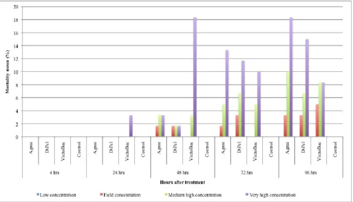
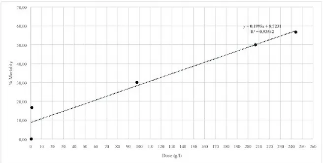
concentrations; more in detail: 1) "field concentration", it is not the concentration which normally found in the field after the use of product, but it is a concentration comparable to the field-dose in the label of the tested products i.e. 100.0 g/hl for Agree and DiPel specifically suggested to be used on the grapevine plantation to control the moths Lobesia botrana Denis and Schiffermüller, 1775 and Eupoecilia ambiguella Hübner, 1796 and 0,384 g/100 ml for VectoBac; 2) “low concentration” (40.00 g/hl for Agree and DiPel and 0,154 g/100 ml for VectoBac), assumed to be a possible concentration in field-environment conditions, of the aforementioned biopesticide concentration; 3) "very-high concentration" (24400,0 g/hl for Agree and DiPel and 37,5 g/100 ml for VectoBac), lethal to more than 50% of the tested foraging bees; 4) "medium-high concentration" (9765,00 g/hl for Agree and DiPel and 6,0 g/100 ml for VectoBac), between field and very-high concentrations. Low, medium-high and very-high concentrations, in particular, have been found, respectively, dividing (for the low one) and multiplying, by a factor of 2.5 fold, the field concentration, in a geometric progression. Furthermore, these concentrations were chosen in a suitable range in order to provide a regression line and LD50 oral i.e. the single dose of a substance that
can cause death in 50% of specimens when administred by the oral route.
- 36
-In bioassay, the trials were performed in triplicate for each group (including the control one) and product: 10 specimens were used in each group, for each of the three repetitions (total of 150 specimens for each product tested, 50 for each repetition), strictly in agreement with the recommendations of the same OEPP-EPPO guidelines. Before the exposition to the biopesticide, the 10 bees of each group were kept without food for 2 hrs so that all bees are equal in terms of their gut contents at the start of the test (OECD 213 guidelines, 1998), then anesthetized 30 minutes at 4 °C and placed individually in maintenance wooden cages (37x15x15 cm) divided into 10 cells (7x7x15 cm) created by removable dividing walls (fig. 4.2), at 25 ± 2 °C, 60% RH, in agreement with Ladurner et al. (2005b).
.
- 37
-The dose of the sugary solution (10 µl), without the biopesticide (for the control group) or with the biopesticide at the different concentrations (group-specific), was administered to each specimen only once in each of the three repetitions, according to the "natural flower" method (Ladurner et al., 2003) or the "artificial flower" method (Ladurner et al., 2005a) for A. mellifera until its total intake (fig. 4.3).
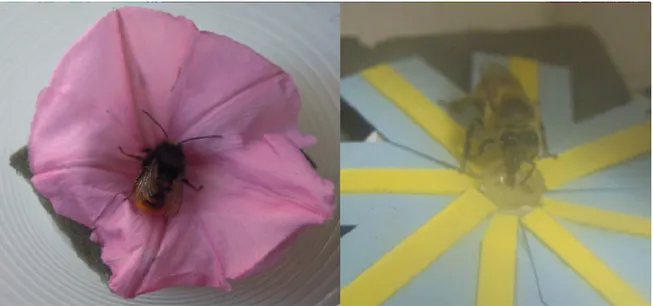
The "natural flower" method consist in the modification of a flower from which the reproductive structures were removed with the help of tweezers and entering into the calyx a small plastic ampoule (diameter 0.3 cm, height 0.5 cm) formed by the terminal part of a test tube, subsequently filled with the test solution. The flowers used, belonged to the species Convolvus althaeoides L. and Ipomoea acuminata Vahl as constituting the most abundant wild flowers during the study period.
In the “artificial flower” method, however, an artificial flower is made artificially, as described by Ladurner et al., 2005: eight petals of blue cardboard (2 x 1 cm), on which were pasted yellow cardbord guidelines nectar (2 x 0.5 cm) arranged in symmetrical order around a small ampoule of plastic (diameter 0.3 cm, height 0.5 cm), inside which the test solution was pipetted. Finally, both the natural and the artificial flower were mounted on a cube of green florists’ dry foam (5 x 3 x 1 cm). This no-choice dietary feeding protocol should guarantee, especially for the low and field concentrations administered in this assay, the highest exposure level to bees potentially occurring in natural conditions (worst case scenario) (Han et al., 2010).
- 38
-Each bee, exposed to an artificial light (cool white 15W), was kept isolated into the cell (about 1 hr) until the complete consumption of the 10 µl dose of solution on the flower (Ladurner et al., 2005b). The dividing walls were then removed, and a plastic container (group feeder), consists of two small Petridishes and overlapping of different dimensions, was added with the sugary solution (5 ml), devoid of the tested biopesticide and replaced every 24 hrs (OEPP-EPPO, 2010). Food consumption was measured weighing the group feeder before being inserted into the cage and after the time observations, the amount of sugary solution remaining was estimated weighing the group feeder and by difference and percentage ratio was obtained the value of food consumed. The workers were kept in the maintenance cages for the entire period of observation at 25 ± 2 °C, 60% RH and 0L:24D (Ladurner et al., 2005b). The bees considered dead, when
Fig. 4.3 – The two methos of feeding: the "natural flower" method (Ladurner et al., 2003) and the "artificial
- 39
-motionless for at least 10 seconds (Iwasa et al., 2004), were removed from the cages.
In accordance with the guidelines of OEPP-EPPO and OECD (OECD guidelines 213, 1998) for whichever acute toxicity oral assay, all the observations (about mortality, symptoms, behaviour and sublethal effects), made in the trials, on specimens from both the control group and the treated ones, were carried out at different time intervals after the start of the same trials (4, 24, 48, 72 and 96 hrs); in particular, although 48 hrs is suggested as a significant deadline as result of acute toxicity (OECD guidelines 213, 1998), the observations until 96 hrs after the start of the trials (possible recommended deadline by the same guidelines) have been aimed to assess any long-term effect.
4.1.5 Bioassay with solitary bees
Unlike honeybees, male specimens of O. cornuta e O. rufa were subdivided into four groups: field concentration, medium-high concentration, very high concentration and control group (see above).
The product tested was only DiPel and the low concentration was not tested because of the small number of cocoons available.
As reported above, wintering individuals of O. cornuta and O. rufa individuallyhave been incubated at 22°C in plastic glass, before being subjected to the test, and starved overnight after emergence of their cocoon (Ladurner et al. 2005b).
- 40
-specimen only once in each of the three repetitions using only the "natural flower" method until its total intake (fig. 4.3). This method was chosen because it was considered by Ladurner et al. 2003 highly effective in studies of oral toxicity in laboratory with solitary bees.
Similar to bioassay with A. mellifera, the trials with O. cornuta and
O. rufa were performed in triplicate for each group (the low
concentration was not tested): 10 specimens were used in each group, for each of the three repetitions (total sum of 120 specimens, 40 for each repetition). Also solitary bee was exposed to an artificial light (cool white 15W) at 22 ± 2 °C, 60% RH, and each of them was kept isolated into the plastic glass (about 1 hr) until the complete consumption of the 10 µl dose of solution on the natural flower (Ladurner et al., 2005b). Specimens of O. cornuta and O. rufa bees have been transferred into maintenance cages (600 ml clear plastic containers appropriately drilled and sealed with lids made from Petri dishes which have been inserted a network of durable nylon to ensure good ventilation), and within them was inserted the artificial feeder with the control solution (fig. 4.4).
- 41
-Specimens of O. cornuta and O. rufa were kept in the maintenance cages for the entire period of observation at 22 ± 2 °C, 60% RH and 12L:12D (Ladurner et al., 2005b).
As for honeybees, the observation of mortality made in the trials, on specimens from both the control group and the treated ones, were carried out at 4, 24, 48, 72 and 96 hrs after the start of the trials.
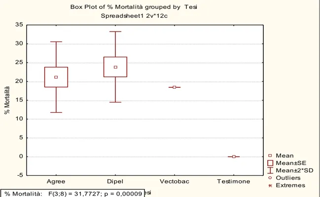
4.1.6 Statistical analyses and symptoms observation
Overall mortality analysis has been made by calculating the mortality rates (Malone et al., 1999; Ladurner et al., 2005b) observed Fig. 4.4 – Test with solitary bees: maintenance cages, plastic glass with natural
- 42
-in the different groups and species. Data on mortality rates were arcsinesquare root transformed and analyzed by means of an one-way ANOVA, followed by LSD test (Least Significant Difference) for homogenous groups to separate the mean obtained (Siscaro et al., 2006; Ruiu et al., 2007). Homogeneity of variance was assumed in all the assays. All the statistical analyses were performed with the STATISTICA software (Statsoft®). Mortality rates have not been corrected with the Abbott's formula (1925), since the death of the specimens of the control group was 0% (Ladurner et al., 2005b).
For each insecticide, regression lines, LD50 values, 95% fiducial
limits (FL) were determined using application tools for Microsoft Excel® software.
Direct observations on the behavior and symptoms of honeybees, treated specimens and control ones have been made at the considered time intervals (4, 24, 48, 72 and 96 hrs) as result of acute toxicity.
Furthermore, survival curves have been made to assess the survival in the different groups and species after 7 days from the treatment.
4.2 Morphological, histological and ultrastuctural analysis
4.2.1 Preparation of honeybees workers for SEM, OM and TEM observations.
Apis mellifera workers were collected and tested as described in
par. 4.1.1 and par. 4.1.4 using the product Agree at four different concentration (low, field, medium-high and very high concentration).
- 43
-The observations of morphology, histology and ultrastructure made in my study, on specimens from both the control group and the treated ones, were carried out at five different time intervals after the total intake of the test solution (4, 24, 48, 72 and 96 hrs).
The observations conducted to detect possible alterations in the midgut epithelium were carried out on five specimens, still alive, from each of the five groups, including the control one. The alimentary canal was extracted by decapitating the individual by bistoury with disposable sterile blade, removing the last abdominal segments using sterile scissors and with the help of tweezers, the alimentary canal was totally extract. Each alimentary canal was dissected in a Ringer’s solution and the various midgut samples were prepared according to the following methods for SEM, OM and TEM observations.
4.2.2 Scanning electron microscopy (SEM) preparation
Each midgut sample was fixed in 2.5% glutaraldehyde in a 0.1 M Sorensen’s phosphate buffer (SB), pH 7.4, for 4 hrs at room temperature (r.t.). The samples were then washed several times in the same buffer, dehydrated in ethyl alcohol, immersed in hexamethyldisilazane (HMDS) and air dried; finally, the sampleswere mounted on SEM stubs, metal coated and then observed by a ZEISS EVO LS10 microscope.
- 44
-4.2.3 Transmission electron microscopy (TEM) preparation
Each midgut sample was fixed in 2.5% glutaraldehyde and 3% sucrose, in a 0.1 M Sorensen’s phosphate buffer (SB), pH 7.4, for 4 hrs at room temperature (r.t.). The samples were then washed several times in the same buffer, post-fixed in 2% osmium tetroxide in the same buffer, at r.t. for 1 hr. their dehydration in ethyl alcohol, then immersed in propylene oxide and embedded in Embed 812. Ultra-thin sections (50-70 nm), placed on copper-rhodium grids (200/300 mesh), were contrasted in uranyl acetate and lead citrate (Reynolds, 1963) and then observed by a JEOL 1220 microscope.
4.2.4 Optical microscopy (OM) preparation
For OM observations of workers midgut sections (5-7 µm) were used three different fixatives and no obvious difference between them was shown. Furthermore, in order to prepare semi-thin sections (400-700 nm) for OM observations, midgut samples were fixed in 2.5% glutaraldehyde and 3% sucrose, in a 0.1 M Sorensen’s phosphate buffer (SB), pH 7.4 for 4 hrs at room temperature (see par. 4.2.3).
Three fixatives used are:
Formalin: after fixation in 4% formalin for 24 hrs at 4°C, the samples were washed with tap water and dehydrated in a series of increasing gradation ethanol (35°, 50°, 70°, 80°, 95°, absolute) and finally placed in xylene for 1 hr. Following the infiltration in paraffin
- 45
-for 4 hrs at 60°C.
Carnoy: fixation in Carnoy is faster than the formalin. Midgut samples, therefore, were placed in Carnoy for 4 hrs at room temperature (r.t.) and then directly dehydrated with 95° ethanol and absolute. Finally, samples are immersed in xylene and infiltrated in paraffin.
Bouin: it is one of the best fixatives. Being very penetrating, it serves to fix pieces of organs, even bulky, and allows the use of almost all staining methods. Fixation according to the thickness of the pieces from 12 to 48 hrs. After fixation with Bouin for 24 hrs at 4 ° C, the samples were washed with 50° ethanol up to eliminate the yellow color typical of the fixative. The samples were then dehydrated before in 70° ethanol then in 95° ethanol. Finally, samples are immersed in xylene and infiltrated in paraffin.
Samples included in paraffin blocks were cut with microtome REICHERT-JUNG; 1150/AutoCut, in 5-7 µm sections and the histological characterization was performed using the hematoxylin-eosin (HE) staining.
As far as semi-thin sections are concerned, these were cut with an Ultratome III LKB then mounted on microscope slides, deresined with a saturated solution of sodium hydroxide in absolute ethanol and stained with 0.5% Toluidine blue in SB.
- 46
-4.3 Gene expression and biochemical analysis
4.3.1 Preparation of biological samples
Apis mellifera workers were collected and tested as described in
par. 4.1.1 and par. 4.1.4 using the product Agree at two different concentration: 1) "field concentration", a concentration comparable to the field-dose in the label of the tested product i.e. 1 g/l and 2) “high concentration”, five times the field concentration corresponding to 5 g/l and represents an unlikely concentration in field established in order to make a comparison with specimens treated with field concentration and control.
The observations for the control group and for the two groups treated with the biopesticide were conducted at 4 hrs and 24 hrs after the single administration of the test solution. Specimens were taken from the maintenance wooden cages, where the toxicity test had been performed, and the midgut was completely extract as describe in par 4.2.1.
For each treatment (control, field concentration and high concentration) 15 midguts were extracted and grouped into 3 pools (total sum of 90 specimens, 45 for each observation), of which we proceeded to proteins and total RNA extraction.