1
An Immune Gene Expression Signature Associated With Development of Human Hepatocellular Carcinoma Identifies Mice That Respond to Chemopreventive Agents
Short-title: Immune-mediated field cancerization as target for HCC prevention
Authors: Agrin Moeini1, Sara Torrecilla1, Victoria Tovar1, Carla Montironi1,2, Carmen Andreu-Oller1, Judit Peix1, Mónica Higuera1,3, Dominik Pfister4, Pierluigi Ramadori4, Roser Pinyol1, Manel Solé1, Mathias Heikenwälder4 ,Scott L. Friedman2, Daniela Sia2,*, Josep M. Llovet1,2,5,*.
Affiliations:
1. Liver Cancer Translational Research Group, Institut d'Investigacions Biomèdiques August Pi i Sunyer (IDIBAPS)-Hospital Clínic, Liver Unit, Universitat de Barcelona, Barcelona, Catalonia, Spain.
2. Mount Sinai Liver Cancer Program, Department of Liver Diseases, Icahn School of Medicine at Mount Sinai, New York, USA.
3. Liver diseases, Vall d'Hebron Institut de Recerca (VHIR), Hospital Universitari Vall d’Hebron, Barcelona, Spain
4. Division of Chronic Inflammation and Cancer, German Cancer Research Center Heidelberg (DKFZ), Heidelberg, Germany
5. Institució Catalana de Recerca i Estudis Avançats (ICREA), Barcelona, Catalonia, Spain.
Grant Support:
JML is supported by the European Commission (EC)/Horizon 2020 Program (HEPCAR, Ref. 667273-2), U.S. Department of Defense (CA150272P3), an
Accelerator Award
(CRUCK, AECC, AIRC) (HUNTER, Ref. C9380/A26813)
, NCI Cancer Center Support Grant, National Cancer Institute, Tisch Cancer Institute (P30-CA196521), Samuel Waxman Cancer Research Foundation, Spanish National Health Institute (SAF2016-76390) and the Generalitat de Catalunya/AGAUR (SGR-1358). AM is supported by Spanish National Health Institute. ST and JP are funded by Centro de2 Investigación Biomedica en Red de Enfermedades Hepáticas y Digestivas (Ciberehd-ISCIII). CM is a recipient of Josep Font grant. CAO is supported by “la Caixa” INPhINIT Fellowship Grant (LCF/BQ/IN17/11620024). RP is supported by HEPCAR and AECC. DS is supported by the Gilead Sciences Research Scholar Program in Liver Disease. SLF is supported by the National Institutes of Health Research project grant (R01, DK5662) and U.S. Department of Defense (CA150272P3). M.H. was supported by an ERC Consolidator grant (HepatoMetaboPath), the SFBTR 209, 1335 and SFBTR179. This project has received funding from the European Union’s Horizon 2020 research and innovation program under grant agreement No 667273 and the Helmholtz future topic (Zukunftsthema) Immunology and Inflammation.
Abbreviations: ALT: alanine aminotransferase; α-SMA: α-smooth muscle actin; AKT:
protein kinase B; Asp: aspirin; AST: aspartate aminotransferase; CCl4: carbon tetrachloride; CD-HFD (choline-deficient high-fat diet); Clo: clopidogrel; DEN: diethylnitrosamine; EMT: epithelial-mesenchymal transition, ERK: extracellular signal-regulated kinase; FC: Fold change, FDR: false discovery rate; FFPE: formalin-fixed paraffin-embedded; FGFR: fibroblast growth factor receptor; GSEA: gene set enrichment analysis; HCC: hepatocellular carcinoma; HCV: hepatitis C virus; HSC: hepatic stellate cell; ICF: immune-mediated cancer field; IPA: ingenuity pathway analysis; mo: months; NTP: nearest template prediction; PDGFR: platelet-derived growth factor receptor; ssGSEA: single sample gene set enrichment analysis; TKIs: tyrosine kinase inhibitors; Treg: regulatory T cells; qRT-PCR: quantitative real-time polymerase chain reaction; VEGFR: vascular endothelial growth factor receptor.
*Correspondence: Josep M. Llovet, MD, Liver Cancer Translational Research
Laboratory, IDIBAPS-Hospital Clinic, Rosselló 153, 08039, Barcelona, Catalonia, Spain; Tel. 0034-932.279.155; Email address: [email protected]; Daniela Sia, PhD, Mount Sinai Liver Cancer Program, Division of Liver Diseases, Tisch Cancer Institute, Icahn School of Medicine at Mount Sinai, Madison Ave 1425. 11F-70. Box 1123, New York, NY10029. USA. E-mail: [email protected]
Disclosures: Part of the study was supported with an investigator-initiated research
grant by Boehringer Ingelheim. Prof. Josep M. Llovet has been a consultant, advisory board member and has received research funding from Boehringer Ingelheim; and is receiving research support from Bayer HealthCare Pharmaceuticals, Eisai Inc,
Bristol-3 Myers Squibb and Ipsen, and consulting fees from Bayer HealthCare Pharmaceuticals, Bristol-Myers Squibb, Eisai Inc, Celsion Corporation, Eli Lilly, Exelixis, Merck, Ipsen, Glycotest, Navigant, Leerink Swann LLC, Midatech Ltd, Fortress Biotech, Sprink Pharmaceuticals and Nucleix. Prof. Scott L Friedman has been a consultant for Abide Therapeutics, Allergan Pharmaceuticals, Angion Biomedica, Blade Therapeutics, Can-Fite Biopharma, Enanta Pharmaceuticals, Escient Therapeutics, Forbion, Galmed, Genfit, Glycotest, Glympse Bio, Metacrine Inc., Mistral Biosciences, Morphic Rock Therapeutics, North Sea Therapeutics, Novartis, Novo Nordisk, Pfizer Pharmaceuticals, Salix Pharmaceuticals, Scholar Rock, Seal Rock Therapeutics, Second Genome, Surrozen, Symic Bio, Viking Therapeutics and Kintai; has received research funding from Blade Therapeutics, Can-Fite Biopharma, Ferring Research Institute, Galmed; and has stock options from Intercept, Exalenz, Madrigal, Akarna Therapeutics, BirdRock Bio, Blade Therapeutics, Conatus, DeuteRx, Exalenz, Galectin, Galmed, Genfit, Glympse. The rest of the authors declare no conflict of interest relevant to the study reported.
Transcript profiling: Gene expression Omnibus accession number from previously
deposited data from our group (GSE63898, GSE10143, GSE15654) and others (GSE84044). Newly profiled mice samples are in GEO under accession number (GSE125975 and GSE133969).
Author contribution: Study concept and design: AM, VT, DS, JML; acquisition of data:
AM, ST, CM, JP, MH, MS; analysis and interpretation of data: AM, ST, VT, CM, CAO, MS; drafting of the manuscript: AM, DS, JML; critical revision of the manuscript for important intellectual content: RP, SF, DS, JML; obtained funding: JML; study supervision: JML, DS.
Acknowledgements: We thank Juan José Lozano for technical assistance in the
normalization of transcriptomic array of the animal model. This study has been developed at the building of Centre Esther Koplowitz from IDIBAPS/CERCA Programme/Generalitat de Catalunya. We also acknowledge Angelo Sangiovanni and Massimo Colombo for providing the seminal cohort of cirrhotic patients in our previous studies12,14.
4
ABSTRACT
Background & Aims: Cirrhosis and chronic inflammation precede development of
hepatocellular carcinoma (HCC) in approximately 80% of cases. We investigated immune-related gene expression patterns in liver tissues surrounding early-stage HCCs and chemopreventive agents that might alter these patterns to prevent liver tumorigenesis.
Methods: We analyzed gene expression profiles of non-tumor liver tissues from 392
patients with early-stage HCC (training set, n=167 and validation set, n=225) and liver tissue from patients with cirrhosis without HCC (n=216, controls) to identify changes in expression of genes that regulate the immune response that could contribute to hepatocarcinogenesis. We defined 172 genes as markers for this deregulated immune response, which we called the immune-mediated cancer field (ICF). We analyzed the expression data of liver tissues from 216 patients with cirrhosis without HCC and investigated the association between this gene expression signature and development of HCC and outcomes of patients (median follow-up 10 years). Human liver tissues were also analyzed by histology. C57BL/6J mice were given a single injection of Diethylnitrosamine (DEN) followed by weekly doses of carbon tetrachloride to induce liver fibrosis and tumorigenesis. Mice were then given orally the multiple tyrosine inhibitor nintedanib or vehicle (controls); liver tissues were collected and histology, transcriptome, and protein analyses were performed. We also analyzed transcriptomes of liver tissues collected from mice on a choline-deficient high-fat diet, which developed chronic liver inflammation and tumors, given orally aspirin and clopidogrel or the anti-inflammatory agent sulindac vs mice on a chow (control) diet.
Results: We found the ICF gene expression pattern in 50% of liver tissues from patients
with cirrhosis without HCC and in 60% of non-tumor liver tissues from patients with early-stage HCC. The liver tissues with the ICF gene expression pattern had 3 different features: increased numbers of effector T cells; increased expression of genes that suppress the immune response and activation of transforming growth factor beta signaling; or expression of genes that promote inflammation and activation of interferon gamma signaling. Patients with cirrhosis and liver tissues with the immunosuppressive profile (10% of cases) had a higher risk of HCC (hazard ratio, 2.41; 95% 1.21–4.80). Mice with chemically-induced fibrosis or diet-induced steatohepatitis given nintedanib or aspirin and clopidogrel downregulated the ICF gene expression pattern in liver and developed fewer and smaller tumors than mice given vehicle.
5
Conclusions: We identified an immune-related gene expression pattern in liver tissues
of patients with early-stage HCC, called the ICF, that associates with risk of HCC development in patients with cirrhosis. Administration of nintedanib or aspirin and clopidogrel to mice with chronic liver inflammation caused loss of this gene expression pattern and developed fewer and smaller liver tumors. Agents that alter immune regulatory gene expression patterns associated with carcinogenesis might be tested as chemopreventive agents in patients with cirrhosis.
6
INTRODUCTION
Liver cancer is the fourth leading cause of cancer-related mortality worldwide1. Hepatocellular carcinoma (HCC) accounts for more than 90% of liver cancers and is the main cause of death in patients with cirrhosis2,3. HCC arise from chronic liver inflammation, fibrosis and eventually cirrhosis in 70-80% of cases2. In developed countries, curative treatments are feasible in 30-40% of cases, but recurrence is high and no effective adjuvant therapies are available2,4. In addition, ~40-50% of patients are diagnosed at advanced stages when currently approved molecular therapies yield limited survival benefits (~1 year)3. Despite recent advances in the management and clearance of HCV infection, there is an unmet need for early detection and application of chemopreventive approaches in patients at high-risk of HCC development.
To date, there are no established preventive strategies for HCC in patients at risk beyond prevention with anti-viral therapies5. Once cirrhosis is established, anti-viral therapies reduce but do not eliminate the risk of HCC4,6,7. Individual risk assessment is a key first step in the successful development of any chemopreventive strategy. In this regard, increasing evidence suggests the existence of the so-called “cancer field-effect” or field cancerization which consists of predisposing oncogenic and inflammatory signals occurring during chronic liver injury and ultimately leading to malignant transformation8– 10. Gene signatures derived from the cirrhotic tissue adjacent to HCC tumors have been designed to predict poor outcome, particularly in HCV-infected cirrhotic patients at higher risk of HCC development9,11–14. Overall, these studies support the feasibility of using molecular scores of the carcinogenic field to identify patients at high risk of HCC development. However, the carcinogenic roles of inflammation and immune response in the context of the field cancerization have been poorly explored. Understanding the immune features governing the unresolved cancer field-effect is crucial for identifying potential therapeutic targets in patients at high risk of HCC development.
In this study, the analysis of the inflammatory milieu that characterizes the underlying liver disease in which HCC tumors arise has led to the identification of an immune-mediated cancer field (ICF) in 60% of early HCC patients and 50% of cirrhotic patients without HCC. This ICF comprises three distinct molecular subtypes including the High Infiltrate ICF subtype with increased infiltration of effector T cells, the Immunosuppressive ICF subtype with activation of stroma and TGF-b signaling, and the Pro-inflammatory ICF subtype with up-regulation of IFN-g signaling. These immune
profiles, particularly the Immunosuppressive cancer field, predict increased risk of HCC development in cirrhotic patients. Inhibition of this carcinogenic field significantly reduced HCC onset in two mouse models of chronic liver damage and hepatocarcinogenesis.
7 Overall, our study provides the rationale to explore chemopreventive strategies in cirrhotic patients at high-risk of HCC development.
8
MATERIALS AND METHODS Human cohort
Gene expression data from a cohort of 167 surgically resected fresh-frozen samples (Heptromic dataset, GSE63898) with matched tumor and adjacent non-tumor tissue were analyzed. Samples were previously collected (1998-2008) in the setting of the HCC Genomic Consortium upon institutional review board approval. Full description of the cohort and RNA profiling data are available in previous publications15,16. Supplementary
Table 1 provides a summary of the clinical-pathological variables of the samples used in
the current study (training cohort, n=167). Validation of the identified molecular profiles was then performed in an independent set of 225 adjacent non-tumor liver tissues previously characterized by our group (GSE10143)9. Finally, to identify those non-neoplastic patients at higher risk of HCC development and most likely to benefit from chemopreventive strategies, our findings were evaluated in a previously characterized cohort of patients with early cirrhosis (n=216, GSE15654)14 and a publicly available dataset of fibrotic liver tissues (n=124, GSE84044)17.
Modeling the immune-mediated cancer field
Enrichment scores of 4872 gene sets that represent cell states and perturbations of the immune system (Collection C7 of MSigDB, Broad Institute)18 were calculated by Single-sample Gene Set Enrichment Analysis (ssGSEA) in the non-tumor liver tissue of the training cohort. Next, unsupervised clustering analysis by non-negative matrix factorization (NMF consensus)19 method was performed to identify the presence of an immune-mediated cancer field. To characterize the samples presenting an ICF and to identify different immune-mediated field subtypes, a second unsupervised clustering was performed using ssGSEA scores obtained for a curated set of gene signatures representative of individual cell types20,21 ,cancer immune-related signaling pathways22, and inflammation- or immune-specific biological processes (Hallmark collection of MSigDB, Broad Institute).
Generation of an immune-mediated field gene signature
An ICF field gene signature was generated using top differentially expressed genes in each molecular group (FDR<0.05; Fold-change ≥2), which was then validated in an independent dataset using Nearest Template Prediction (NTP) analysis (p-value<0.05) (Gene Pattern modules)23.
9
Molecular characterization of the ICF subtypes and identification of candidate therapies
To characterize the ICF subtypes, gene expression signatures [available in MSigDB (Broad Institute) or previously reported (Supplementary Table 2)] were assessed by GSEA, ssGSEA, NTP and Ingenuity Pathway Analyses (IPA). CIBERSORT20 was used to estimate the relative fraction of 22 immune cell types within the leukocyte compartment of non-tumor liver tissues. The Immunophenoscore (IPS) algorithm24 was used to analyze the major immunogenic determinants. An in silico analysis based on ssGSEA scores of ~1230 gene sets (DSigDB) recapitulating targets of approved therapies was also performed for the screening of candidate targeted therapies.
Histological evaluation of infiltrating inflammation
Histopathological analysis was performed in 98 out of 167 cases. Specifically, hematoxylin and eosin (H&E) staining of formalin-fixed paraffin embedded (FFPE) tissue section of HCCs and their matched adjacent non-tumor livers were evaluated by two expert pathologists (CM and MS). The presence of inflammation (portal/septal, interface, pericentral and lobular) as well as the lymphoid aggregates were assessed in the non-tumor liver tissue sections. More details on the histological evaluation of the samples have been included in Supplementary material.
Animal models
We generated a chemically-induced model of HCC and fibrosis in male C57BL/6J mice (Harlan Laboratories, n=55) by a single injection of Diethylnitrosamine (DEN) followed by weekly dosing with carbon tetrachloride (CCl4), as previously described25. Once fibrosis was established, mice were randomized to receive vehicle or nintedanib (50 mg/kg, Boehringer Ingelheim). Mice were sacrificed at different time-points and liver and tumor tissue samples were collected and processed for histological, transcriptomic and protein expression analyses (see Supplementary material). All experimental procedures were carried out following the approval of the institutional ethical committee of the University of Barcelona and Hospital Clinic of Barcelona. Additionally, liver samples of a choline-deficient high-fat diet (CD-HFD) fed mouse model reported in a recent study26 were collected. A total of 25 samples were processed for transcriptomic profiling, including mice fed a chow diet (n=5) or CD-HFD for 12 months and given: vehicle (n=4); aspirin/clopidogrel (Asp/Clo) (n=6) or sulindac (n=10).
10
Statistical analysis
All analyses were performed using SPSS software version 23 (IBM) or GraphPad Prism version 5.00 (San Diego, CA). Correlations for categorical and continuous variables were analyzed by Fisher’s exact test and Wilcoxon rank-sum test, respectively. The prognostic value of the signatures was assessed using Kaplan-Meier estimates, log-rank test, and Cox regression models. In in vivo studies, the Mann-Whitney U test was used to compare differences in body weights, liver function, tumor number, tumor size and CD4/CD8 stained area in human samples. Fisher exact test was performed for analysis of HCC incidence and pERK staining. Student T-Test was used to compare the differences in Sirius Red quantification, CD31 staining, CD4/CD8 staining proportion of immune cell infiltrate in mice and relative gene expression.
11
RESULTS
Identification of a novel immune-mediated cancer field effect in non-tumor liver tissue of patients with early HCC.
In order to characterize the immune features governing the unresolved cancer-field in which new cancers arise, transcriptome-based analysis of a compendium of ~5,000 annotated immunology-specific gene-sets18 was performed in the non-tumor liver tissue of patients with early stage HCC. This analysis revealed the presence of an immune-mediated cancer field (ICF) in ~60% (98/167) of samples (Figure 1A and
Supplementary Figure 1). Specifically, these samples were characterized by
enrichment of several gene-sets recapitulating the presence of activated immune cells, up-regulation of core signaling pathways involved in immune response (both innate and adaptive) as well as those involved in the modulation of inflammatory response (i.e. IL2-STAT5, IL6-STAT3, IL17, IFN-γ, CSF, TNF-α, and TGF-β signaling) (Figure 1A-B and
Supplementary Figure 1). Moreover, histological evaluation confirmed that liver tissues
with ICF contained a higher frequency of moderate to marked inflammatory infiltrate (74% in ICF vs. 52% in non ICF, p=0.034) and lymphoid aggregates (80% in ICF and vs. 55% in non ICF, p=0.009) (Figure 1C-1D and Supplementary Table 3). Immunostaining for CD4+ and CD8+ further confirmed significantly higher levels of T cell infiltrates in the adjacent livers of patients with the ICF (Supplementary Figure 2A). In contrast, histological evaluation of the tumor showed no significant correlation between the presence of the ICF and the detection of intratumoral or peritumoral infiltration (Supplementary Table 3). This is in accordance with our recent publication15, where the tumor immune-based profile did not correlate with presence or absence of immune gene signatures in the surrounding non-tumor tissue.
While characterizing the ICF we detected that, in addition to immunogenic features, several well-known carcinogenic signals such as epithelial-to-mesenchymal transition, KRAS, EGFR, and VEGF signaling were also significantly enriched in liver tissues containing the ICF (Supplementary Table 4). In line with these oncogenic signals, a significant enrichment of previously reported prognostic signatures derived from the adjacent non-tumoral liver were also detected. These signatures included the 186-gene cancer-field signature9, activated hepatic stellate cells (HSCs)11, hepatic injury and regeneration (HIR)13, and multicentric occurrence of HCCs27 (Figure 1A). The presence of the ICF significantly correlated with HCV infection, features indicative of liver dysfunction such as high bilirubin, low platelet count and albumin levels (Supplementary
Table 5) and poor survival [median OS 43.4 mo in the ICF group vs 94.8 mo in non ICF;
12 data highlight the presence of an immune-mediated cancer field in 60% of early HCC patients. This ICF is characterized by activation of immunomodulatory signaling cascades (i.e. IFN-γ, TNF-α, TGF-β, IL6) along with cancer promoting signaling pathways (i.e. EMT, EGFR and VEGFR), and is associated with HCV infection and poor prognosis.
The immune-mediated cancer field contains 3 distinct molecular subtypes.
Further dissection of the key immune-modulating signaling pathways and immune-cell infiltrates in those samples harboring the immune-mediated cancer field revealed the existence of three distinct molecular subtypes. The first molecular subtype, henceforth called the “High Infiltrate ICF” subtype (23% of the ICF), showed a significant enrichment of several previously established gene signatures mirroring the presence and/or activation of immune cell infiltrates such as lymphocytes (T and B cells)22,28 or macrophages29 (Figure 2A-2B). Consistently, immunogenicity, herein captured either by the cytolytic activity score (Figure 2A)30 or using the immunophenoscore algorithm24 (Figure 2B), was also significantly higher in these samples (p<0.001). Specifically, non-tumor liver samples belonging to the High Infiltrate ICF subtype showed significant infiltration of effector T cells (Figure 2B, p≤0.001), including increased levels of cytotoxic CD8+ T cells assessed both by transcriptomic (p=0.03) and immunohistochemistry (p=0.0002) (Figure 2C and Supplementary Figure 2B). This subtype also was characterized by enrichment of the previously reported ectopic lymphoid structures (ELS) signature31 (Figure 2A). In addition, the High Infiltrate ICF was significantly associated with poor survival in comparison to the rest of the patients (Supplementary
Figure 1C), although there were no significant differences among the distinct ICF
subtypes (Supplementary Figure 1D). The second subtype, the so-called “Immunosuppressive ICF” (36% of the ICF), was characterized primarily by activation of stroma and HSCs, increased TGF-β signaling and T cell exhaustion (Figure 2A). Moreover, several immune-checkpoints (i.e. CTLA-4, TIGIT, LAG3) were significantly over-expressed (IPS, p<0.01) in this class, along with higher levels of M2 macrophages (p=0.04) and CD4+ memory resting cells (p=0.005), which are among main mediators of immune tolerance and inhibition (Figure 2B-2C). The third subtype (41% of the ICF) showed a clear predominance of IFN-γ signaling (p<0.001) and enrichment of the inflammatory M1 macrophages (p<0.0001), and was called the “Pro-inflammatory ICF” subtype (Figure 2A-2C). Interestingly, the High Infiltrate and Immunosuppressive subtypes shared several molecular features including the enrichment of key signaling pathways involved in modulating the immune response (i.e. IL2 and TNF signaling),
13 proliferation (i.e. KRAS signaling) and angiogenesis (Figure 2A, p<0.001).
In order to further confirm the presence and molecular traits of the identified ICF, we generated a transcriptome-based gene signature able to capture the three immune-mediated cancer field subtypes. Interestingly, this signature only showed minimal overlap (0-5%) with previously reported gene signatures of field cancerization in HCC (Supplementary Figure 3)9,12,14,32. The resulting 172-gene signature (Supplementary
Table 7) was then validated in the adjacent non-tumor tissue of 225 patients with early
HCC, previously characterized by our group9,33 (Supplementary Figure 4A). Similar to what was previously observed in the training cohort, 58% (130/225) of patients belonged to the ICF. Moreover, in this cohort, the presence of the ICF was an independent predictor of poor survival [HR=2.73; 95 CI: 1.1-6.8; p=0.03] (Supplementary Figure 4B,
Supplementary Table 8). Within the ICF group, ~31% (40/130) presented the High
Infiltrate ICF profile, ~27% (35/130) the Immunosuppressive ICF and ~42% (55/130) the Pro-inflammatory ICF subtype (Supplementary Figure 4A). Subsequent molecular characterization further confirmed the ability of the signature to capture the main molecular traits defining each subtype, such as increased infiltration of effector T cells in High Infiltrate subtype, activation of stroma and TGF-b signaling in Immunosuppressive subtype and up-regulation of IFN-g signaling in Pro-inflammatory subtype (Supplementary Figure 4A). Overall, our results highlight the presence of a poor prognosis-related immune-mediated cancer field comprised of 3 molecular subtypes with a high degree of lymphocyte infiltration (overall 16% of HCC patients) or predominance of either immunosuppressive (overall 20% of HCC patients) or pro-inflammatory (24% of HCC patients) signaling cascades.
The immune cancer-field, particularly the immunosuppressive subtype, predicts a high risk of HCC development in cirrhotic patients
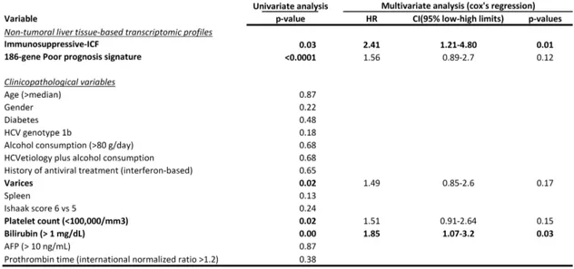
Following the identification of an immune-mediated cancer field in the livers of 60% of patients with early HCC, we next sought to assess its role in liver disease progression and HCC primary occurrence. To this end, the 172-gene signature was analyzed in a cohort of 216 non-malignant cirrhotic patients with a median follow-up of 10 years in the context of an HCC surveillance program14. Overall, 51% (110/216) of cirrhotic patients harbored the ICF, including the High Infiltrate ICF subtype in 28% (31/110), the Immunosuppressive ICF subtype in 19% (21/110), and the Pro-inflammatory ICF subtype in 53% (58/110) of cirrhotic patients harboring the ICF. Next, we tested the capacity of the ICF subtypes to predict the risk of HCC development in cirrhotic patients. Interestingly, the presence of the Immunosuppressive ICF subtype (10% of all cirrhotic
14 patients) was significantly associated with a higher risk of HCC development [median time to HCC development of 7.4 years (95% CI: 3.2-11.7) vs 17.1 years (95% CI: 10.6-23.7) in Rest, p<0.0001] and was found to be an independent predictor of HCC occurrence in cirrhotic patients in a multivariate analysis [HR 2.41 (95% CI: 1.2-4.8), p=0.012] (Figure 3A and Table 1). In addition, the Immunosuppressive ICF was also significantly associated with poor survival [median overall survival of 7.1 years (95% CI: 4.5-9.6) vs 16.3 years (95% CI: 9.1-23.5) in Rest, p<0.0001] and higher risk of hepatic decompensation [median time to hepatic decompensation of 6.5 years (95% CI: 4.3-8.6) vs >15 years in Rest, p<0.0001] (Figure 3B-3C). Cirrhotic patients harboring the other two ICF subtypes (High Infiltrate and Pro-inflammatory subtypes) also showed a non-significant trend towards a higher risk of HCC development compared to those patients lacking the ICF [mean time to HCC development of 12.8 years (95% CI:11.5-14.2) in Other ICF subtypes vs 16.3 years (95% CI: 14.2-18.5) in non ICF, p=0.06] (Supplementary Figure 5A).
Moreover, the analysis of an additional cohort of 124 non-neoplastic patients with liver fibrosis17 revealed that the immune-mediated cancer field may occur as a progressive event, as it significantly correlated with increasing levels of fibrosis stage and degree of inflammation (Supplementary Figure 5B). Particularly, the presence of the Immunosuppressive ICF significantly correlated with the presence of advanced liver fibrosis (Scheuer fibrosis S3-4 score17, p=0.034) (Supplementary Figure 5B).
In conclusion, the immune-mediated cancer field detected in patients with early HCC is also present in the livers of ~50% of cirrhotic patients and captures the presence of a damaging and continuous inflammatory response in the underlying liver disease. Furthermore, our results underscore the critical role of an Immunosuppressive ICF (overall, 10% of cirrhotic patients) in defining a 2.4 risk of HCC development, and to a smaller extent of the High Infiltrate and Pro-inflammatory subtypes.
The immune-mediated field as a target for chemoprevention in a mouse model recapitulating chronic liver inflammation and HCC development
Based on the compelling results described above, we hypothesized that the immune-mediated cancer field, and particularly the Immunosuppressive ICF subtype, may represent an ideal target for chemopreventive strategies in cirrhotic patients at high risk of HCC development. To this purpose, an in silico-based analysis was performed using our training cohort to identify those candidate therapies most likely to modulate the identified ICF. This analysis was based on the enrichment of a compendium of ~1230
15 gene sets (DSigDB collections D1 and D2)34 recapitulating the main targets of 1202 approved drugs. Among the top 10 most significantly enriched drugs (Supplementary
Figure 6), nintedanib was the only FDA-approved therapy indicated for a non-cancer
condition. Specifically, nintedanib is the first molecular targeted therapy with clinical efficacy in patients with idiopathic pulmonary fibrosis as both an fibrogenic and anti-inflammatory agent35. Given these considerations, the efficacy of nintedanib in reverting the pro-tumorigenic immune-mediated cancer field was tested in a mouse model of HCC development in the setting of chronic inflammation and liver fibrosis (Supplementary
Figure 7A). In this model, the macroscopic evaluation of explanted livers in DEN/CCl4 mice sacrificed at the age of 15, 17 and 18 weeks confirmed the development of numerous hepatic tumors (Figure 4A). Tumor penetrance and number of tumors progressively increased, ultimately reaching a 100% incidence at 18 weeks of age (Figure 4A and 4B). At all-time points, histological evaluation of the liver sections showed that a portion of the tumors were pre-neoplastic (dysplastic) nodules (Figure
4C). In mice sacrificed at 15 weeks of age, Supplementary Figure 7A), nintedanib
showed a clear trend towards reducing HCC incidence, number and size of tumors (Figure 4B, D and E). These differences reached significance at 17 weeks of age (Figure 4B), having a marked decrease in both overall tumor burden (30% in nintedanib vs 89% in vehicle group, p=0.019) and specifically in HCC incidence (7% vs 33%, p=0.04). Similarly, at 18 weeks of age, HCC incidence was significantly reduced in treated mice (Figure 4B, 22% vs 77%, p<0.001). In addition, nintedanib significantly reduced the overall tumor number and size both at 17 and 18 weeks of age (Figure
4D-E). Overall, nintedanib was well tolerated with no significant induction of body weight loss
or hepatotoxicity measured by serum ALT and AST levels (Supplementary Figure
7B-C). Taken together, our data suggest that nintedanib is safe and efficacious in preventing
HCC development in our experimental model.
Nintedanib treatment reverts the immune-mediated cancer field effect
Next, we sought to assess the impact of nintedanib treatment on the immune-mediated cancer field. For this purpose, we analyzed gene expression profiling of non-tumor liver samples from 17 weeks-old DEN/CCl4 mice given nintedanib (n= 10) or vehicle (n=9), and 3 healthy control mice. First, the comparison between the healthy control group and vehicle group revealed a profile of activated pathways compatible with HCC development within a fibrotic and inflammatory background. In this regard, functional analysis of differentially expressed genes (Supplementary Table 9) highlighted the activation of hepatic stellate cells and fibrogenesis, as well as immune system activation
16 (inflammatory response, chemotaxis, binding of myeloid and leukocytes) in vehicle treated DEN/CCl4 livers (Supplementary Table 10). Notably, our model presented a significant enrichment of the gene-set representing the ICF identified in humans (p=0.001) and faithfully recapitulated the human immune-mediated field subtypes described above (Figure 5A). The comparison of the gene expression profiles of adjacent non-tumor liver from mice treated with vehicle or nintedanib demonstrated that nintedanib significantly down-regulated the ICF subtypes and, more specifically the Pro-Inflammatory and the Immunosuppressive ICF phenotype, which predict risk of HCC development in cirrhotic patients (Figure 5A, p=0.02). A non-significant trend was also observed for the High Infiltration subtype (Figure 5A). Treatment with nintedanib led to a significant down-regulation of inflammatory cues (IL-6/STAT3, a, interferon-g) and immune-related signaling (IL-2/STAT5 activation, allograft rejection) (Figure 5A). Among the infiltrating immune cells, nintedanib significantly reduced the presence of B and T cells, activated macrophages, helper T cells and Tregs along with associated immune modulators (i.e. IL1, CCL5 and PDL1) (Figure 5A). Despite exhibiting similar global levels of inflammatory infiltrates, quantification of CD4 and CD8 positive infiltrating lymphocytes by IHC revealed a significant decrease of CD4+ T cells in nintedanib-treated mice compared to controls (Figure 5B, p<0.05).
Next, in order to further characterize the chemopreventive effects of nintedanib we assessed the activation status of the main nintedanib targets (i.e. VEGFR2 and PDGFR-β). Western blot of non-tumor liver tissue confirmed that nintedanib blocked the activation of VEGFR2 (Figure 5C) and its downstream effectors AKT and ERK (Supplementary
Figure 8A). Consistently, both liver parenchyma and liver tumors were pERK positive in
vehicle-treated mice and pERK negative in nintedanib treated mice (Supplementary
Figure 8B, p<0.05), indicating an anti-proliferative effect of nintedanib as well. Given the
strong inhibition of VEGFR signaling observed, we next assessed the anti-angiogenic effect of nintedanib in DEN/CCl4 mice. In this model, reduced CD31 staining was associated with diminished blood vessel area in both liver parenchyma and liver tumors of nintedanib-treated mice (Figure 5D). Altogether, these data suggest that nintedanib exhibits its chemopreventive effects in part by inducing vascular normalization and inhibiting hepatic proliferation. In contrast, no reduction of fibrosis degree, the pro-fibrogenic signaling pathway PDGFR-β, or collagen markers were detected in the livers of nintedanib-treated mice (Supplementary Figure 8C-E).
Overall, our data confirm that therapeutic targeting of the immune-mediated cancer field, accompanied by liver vascular normalization and suppression of hepatic proliferation, can prevent the development of HCC associated with advanced chronic liver disease.
17 Immunomodulatory effects of Asp/Clo treatment revert the immune cancer field effect and prevent hepatocarcinogenesis in vivo
To further support the concept of an ICF in promoting HCC development and its therapeutic immunomodulation as candidate strategy for chemoprevention, we performed gene expression profiling in non-tumor liver derived from the recently described mouse model of choline-deficient high-fat diet (CD-HFD) treated either with the immunomodulatory combination aspirin/clopidogrel (Asp/Clo) or the anti-inflammatory sulindac26. Of particular interest, in this model, which presents non-alcoholic fatty liver-related liver inflammation with various degrees of fibrosis and HCC development after 12 months of diet regimen26,36,37, HCC prevention was achieved only through the combination of the anti-inflammatory drug, aspirin, clopidrogel -an P2Y12 inhibitor-, (25% to 0% 12-mo HCC incidence control vs combo respectively, p=0.01)26 and not sulindac alone (25% to 20%12-mo HCC incidence control vs sulindac, respectively, p=ns, data not shown). In the context of our study, comparative analysis between the non-tumor liver of healthy control and CD-HFD mice showed a significant enrichment of the ICF signature in CD-HFD mice (p=0.002, Figure 5E). Notably, all 3 ICF subtypes were significantly up-regulated in CD-HFD mice compared to healthy controls (Figure 5E) along with the enrichment of signaling pathways regulating inflammation (i.e. IL6-STAT3, TNFα), immune infiltration and activation22,28, and epithelial-to-mesenchymal transition (i.e. TGFβ, p<0.05). These data were consistent with the high intrahepatic influx of metabolically activated CD8+ T cells and NK cells (CD3+NK1.1+) measured in CD-HFD fed mice by flow cytometry26. Overall, these data confirm the existence of an ICF in an independent model of chronic liver disease further suggesting a role in hepatocarcinogenesis.
Next, we compared the expression profiles of liver samples from CD-HFD vehicle-treated mice with CD-HFD mice treated with the combination Asp/Clo (n=6) or sulindac alone (n=10). Interestingly, only Asp/Clo, but not sulindac alone, was able to prevent HCC and revert the ICF within the liver microenvironment (p=0.05), being the Pro-inflammatory ICF subtype the most significantly down-regulated upon treatment (Figure 5B). Particularly, based on previous molecular characterization26, the inhibition of the ICF seemed to be accompanied by a significant reduction of the degree of liver damage, as well as a significantly reduced number of CD8+ and NKT cells in the liver.
Overall, these data support the role of the ICF in promoting carcinogenesis, and suggest that only those drugs able to simultaneously inhibit several components of the ICF by
18 targeting mitogenic, angiogenic and immunomodulatory kinases (i.e. nintedanib and Asp/Clo) present a more efficacious therapeutic index for HCC prevention.
DISCUSSION
This study represents an in-depth analysis of the inflammatory milieu associated with the “field cancerization” in the chronically injured liver, and investigates its clinical implications in the prediction and prevention of HCC occurrence in cirrhotic patients. The role of the “cancer field effect” in promoting neoplastic transformation has gained much interest in recent years and currently an altered microenvironment is considered a promoter of cancer8,10. Although, under physiological conditions, inflammation is an adaptive response to tissue injury, when the inflammatory stimuli persist, the non-resolved inflammation contributes to carcinogenesis38,39. In this line, activation of HSC as well as certain pathways, such as nuclear factor-KB and TGF-b signaling, have been previously associated with liver fibrogenesis, and eventually neoplastic transformation9,12. With this study, we move beyond the limits of current knowledge and provide a detailed description of the immune microenvironment underlying the field cancerization in the liver. To this end, we first characterized the immune profile of the non-tumor liver parenchyma of 392 early HCCs and then investigated its role in predicting HCC development in 216 cirrhotic patients with long-term surveillance for HCC (median of 10 years)14. The analysis revealed that up to 60% of HCCs and 50% of cirrhotic patients showed a deleterious immune-mediated response in the surrounding tissue, which was associated with impaired liver function, activation of specific oncogenic loops, angiogenesis and poor survival. Further characterization identified three distinct subtypes with different levels of lymphocyte infiltration and activation of either immunosuppressive or pro-inflammatory traits. In particular, the so-called Immunosuppressive ICF subtype (~10% of cirrhotic patients) was an independent predictor of HCC development, increasing 2.4 the risk of cancer development, whereas both the High-Infiltrate and the Pro-Inflammatory subtypes showed a trend towards higher risk of HCC occurrence in cirrhosis. The identification of distinct immune subtypes reflects the complex role of the immune system in hepatocarcinogenesis, with both an activated immune response and an exhausted immune-microenvironment contributing to create a pro-tumorigenic environment and increase the risk of HCC development40. Reducing the incidence and mortality of HCC requires advances in chemopreventive approaches at pre-neoplastic stages, in addition to curative treatment options for early
19 lesions. Universal immunization against HBV and antiviral therapies against HBV and HCV have been associated with very reduced HCC risk2,41,42. Once cirrhosis is established, the risk of HCC development remains despite achieving a sustained virologic response in HCV patients6,7. In addition, the incidence of other risk factors, such as non-alcoholic steatohepatitis (NASH), is dramatically increasing2. Thus, alternative HCC preventive strategies capable of interfering with molecular hepatocarcinogenesis are an unmet need. Furthermore, identifying those patients at high risk of HCC development should enable a cost-effective selection of patients most likely to benefit from chemopreventive approaches. In this scenario, our results are of clinical relevance since the ICF, and specifically the Immunosuppressive subtype, may provide a novel companion biomarker to enrich at-risk patients in chemoprevention clinical trials. Given these observations, we then sought to investigate if the molecular forces driving such cancer field could serve as target for chemopreventive strategies. Hence, we first verified that the molecular profiles observed in human cirrhosis were faithfully reproduced in two animal models of chronic liver injury. The DEN/CCl4 chemically-induced mouse model as well as the recently described NASH-HCC model26 reliably recapitulated the presence of a carcinogenic phenotype observed in liver tissues from patients belonging to the immune-mediated cancer field.
In order to identify the most promising candidate therapies for novel chemopreventive strategies, we conducted an in silico analysis using a large compendium of gene sets34 recapitulating the main targets of 1202 approved drugs. Among the top ten most significantly enriched drugs, we selected nintedanib, the only FDA therapy approved for non-neoplastic conditions. In the DEN/CCl4 animal model, oral administration of nintedanib reduced the immune-mediated cancer field, including the Immunosuppressive ICF subtype, ultimately reducing HCC incidence and growth. Reversion of the ICF induced by treatment with nintedanib was accompanied by reduction of CD4+ lymphocytes, which could be due to its mechanism of action inhibiting src family of kinases (i.e. LCK, FLT3 and SRC) . These findings are in line with previous reports suggesting that CD4+ cells propagate immune-mediated liver injury in models of chronic liver inflammation or autoimmune liver disease43,44. Pretreatment with T cell-specific Abs or immunosuppressive agents, such as anti-CD4 mAb, FK506 (Tacrolimus), or cyclosporine A, have shown to ameliorate hepatitis in these models, further supporting the role of CD4+ T cells in inducing liver damage43. Results in a second animal model treated with the combination of the anti-inflammatory drug, aspirin, and clopidrogel – a P2Y12 inhibitor-, confirmed the therapeutic potential of immunomodulating the ICF and supported the pro-tumorigenic role exerted by the immune response. Indeed, only the
20 treatment able to modulate the ICF, as indicated by the reduction of immune cells (i.e. CD8+ and NKT cells) and the reversion of the ICF signature, successfully reduced liver damage and prevented HCC development. Overall, our study identifies a novel promising chemopreventive strategy for HCC and confirms the validity of using the reversion of the ICF as reliable read-out of efficacy. This is of great clinical importance since there is currently no effective method to monitor the short-term effects of chemopreventive drugs5. Nintedanib belongs to a new generation of TKIs that, in addition to exerting immune modulation blocks the activation of main angiogenic receptors45. Many cytokines and growth factors are involved in modulating the formation of new vessels. Expression of VEGF and its receptors is elevated in HCC cell lines and tissues, as well as in the blood circulation of patients with HCC33,46–48. In our model, nintedanib exerted its chemopreventive mechanisms in part through the inhibition of VEGF signaling, a major driver of angiogenesis49.Thus far, independent studies had described that HCC prevention can be achieved in animal models by attenuating liver fibrosis through the inhibition of epidermal growth factor receptor (EGFR)50,51 or lysophosphatidic acid (LPA)32 signaling. With the current study, we demonstrate that modulation of the liver microenvironment by molecular targeted drugs, which simultaneously block liver inflammation and angiogenesis, might represent a powerful alternative strategy.
We recently defined the immune class of HCC15 and the Immune exclusion class (characterized by active Wnt/CTNNB1)3,15,52, which might predict response and primary resistance to checkpoint inhibitors, respectively3,52. We herein explore the immunomodulatory mechanisms underlying HCC occurrence by defining an immune-mediated field effect that conforms a cancer-permissive milieu, thus posing them at the highest risk of HCC development. In addition, our pre-clinical data with a drug approved in pulmonology and in non-small cell lung cancer treatment suggest that the permissive microenvironment can be reverted leading to a reduction in HCC occurrence. These data provide the rationale for testing this strategy in early chemoprevention trials targeting cirrhotic patients at high risk of HCC development. In addition, this strategy could also be further explored in the adjuvant setting considering that 60% of HCC undergoing resection also present this permissive milieu in the adjacent non-tumoral tissue.
21
FIGURE LEGEND
Figure 1. Identification of an ICF effect in non-tumoral liver tissue adjacent to early HCCs. A) Heatmap representation of the ICF present in 60% of HCC patients. High and
low ssGSEA scores are represented in red and blue, respectively. B) Top predicted upstream cytokine and transcription factors activated in liver tissues of ICF patients. C) Representative images of degree of Portal/Septal infiltrating inflammation. D) Representative images depicting presence or absence of lymphoid aggregates.
Figure 2. The ICF contains 3 distinct molecular subtypes. A) Heatmap representation
of the three ICF subtypes. Statistical significance is highlighted. B) Immunophenogram representing the enrichment of immunogenic determinants in the distinct ICF subtypes (MHC: Antigen presenting, EC: Effector cells, CP: Check-points, SC: Suppressor Cells). C) Comparison of estimated proportion of immune cells (CIBERSORT method) between the ICF subtypes, representing those immune populations with estimated average fraction >5% and significant differences between the ICF subtypes. Significant statistical differences observed among the different ICF subtypes are highlighted (High Infiltrate, Purple, Immunosuppressive, Orange; Pro-inflammatory, Green; and both High Infiltrate and Immunosuppressive, Black). *=p<0.05, **=p<0.01 and ***=p<0.001.
Figure 3. Association of the presence of the Immunnosuppressive ICF with HCC occurrence and prognostic variables in cirrhotic patients. (A) Kaplan-Meier
estimates of HCC development, (B) overall survival, (C) hepatic decompensation, according to the presence of the Immunosuppressive ICF subtype (orange).
Figure 4. Nintedanib reduces HCC onset in mice. A) Representative pictures of
macroscopic evaluation of hepatic tumors in mice given vehicle or nintedanib sacrificed at 15, 17 and 18 weeks of age. Arrows indicate macroscopically visible tumors. B) Evaluation of overall tumor burden and HCC incidence. (#) = statistical significance for overall tumor burden; (*) = statistical significance for HCC incidence. C) Microscopic evaluation of the number of tumors per mouse in each group. D) Number of macroscopic tumors per mouse given vehicle or nintedanib at the different time-points. E) Diameter size of the largest tumor per mouse given vehicle or nintedanib at the three different time-points. # or *=p<0.05, **=p<0.01 and ***=p<0.001.
Figure 5. Nintedanib and Asp/Clo reduce the ICF in animal models of chronic inflammation and HCC development. A) Heatmap representation of high and low
22 ssGSEA scores for the 172-gene signature and gene-sets recapitulating the ICF subtypes. B) Representative images and quantification of CD4+ and CD8+ infiltrating lymphocytes in the liver of 17 weeks old mice given vehicle (n=5) or nintedanib (n=5). C) Western-blot analysis of VEGFR2 activation in the non-tumor liver parenchyma of 17 weeks old mice given vehicle (n=6) or nintedanib (n=6). D) Morphometric quantification of blood vessel area by CD31 immunostaining in 5 randomly selected low magnification fields in mice given vehicle (n=5) or nintedanib (n=5). E) Single sample GSEA analysis of the ICF signature in the different treatment arms of the CD-HFD model. **=p<0.01 and ***=p<0.001.
23
TABLES
Table 1. Uni- and Multivariate Analysis of risk of HCC development in cirrhotic patients
24
REFERENCES
1. Bray F, Ferlay J, Soerjomataram I, et al. Global cancer statistics 2018: GLOBOCAN estimates of incidence and mortality worldwide for 36 cancers in 185 countries. CA Cancer J Clin 2018;68:394–424.
2. Llovet JM, Zucman-Rossi J, Pikarsky E, et al. Hepatocellular carcinoma. Nat Rev Dis Prim 2016;2:16018.
3. Llovet JM, Montal R, Sia D, et al. Molecular therapies and precision medicine for hepatocellular carcinoma. Nat Rev Clin Oncol 2018;15:599–616.
4. Galle PR, Forner A, Llovet JM, et al. EASL Clinical Practice Guidelines: Management of hepatocellular carcinoma. J Hepatol 2018.
5. Fujiwara N, Friedman SL, Goossens N, et al. Risk factors and prevention of hepatocellular carcinoma in the era of precision medicine. J Hepatol 2018;68:526– 549.
6. Calvaruso V, Cabibbo G, Cacciola I, et al. Incidence of Hepatocellular Carcinoma in Patients With HCV-Associated Cirrhosis Treated With Direct-Acting Antiviral Agents. Gastroenterology 2018;155:411–421.e4.
7. Kanwal F, Kramer J, Asch SM, et al. Risk of Hepatocellular Cancer in HCV Patients Treated With Direct-Acting Antiviral Agents. Gastroenterology 2017;153:996–1005.e1.
8. Lochhead P, Chan AT, Nishihara R, et al. Etiologic field effect: reappraisal of the field effect concept in cancer predisposition and progression. Mod Pathol 2015;28:14–29.
9. Hoshida Y, Villanueva A, Kobayashi M, et al. Gene expression in fixed tissues and outcome in hepatocellular carcinoma. N Engl J Med 2008;359:1995–2004. 10. Hernandez-Gea V, Toffanin S, Friedman SL, et al. Role of the microenvironment
in the pathogenesis and treatment of hepatocellular carcinoma. Gastroenterology 2013;144:512–27.
11. Ji J, Eggert T, Budhu A, et al. Hepatic stellate cell and monocyte interaction contributes to poor prognosis in hepatocellular carcinoma. Hepatology 2015;62:481–95.
12. Zhang DY, Goossens N, Guo J, et al. A hepatic stellate cell gene expression signature associated with outcomes in hepatitis C cirrhosis and hepatocellular carcinoma after curative resection. Gut 2016;65:1754–64.
25 13. Kim JH, Sohn BH, Lee H-S, et al. Genomic predictors for recurrence patterns of hepatocellular carcinoma: model derivation and validation. Beck AH, ed. PLoS Med 2014;11:e1001770.
14. Hoshida Y, Villanueva A, Sangiovanni A, et al. Prognostic gene expression signature for patients with hepatitis C-related early-stage cirrhosis. Gastroenterology 2013;144:1024–1030.
15. Sia D, Jiao Y, Martinez-Quetglas I, et al. Identification of an Immune-specific Class of Hepatocellular Carcinoma, Based on Molecular Features. Gastroenterology 2017;153:812–826.
16. Villanueva A, Portela A, Sayols S, et al. DNA Methylation-based prognosis and epidrivers in hepatocellular carcinoma. Hepatology 2015.
17. Wang M, Gong Q, Zhang J, et al. Characterization of gene expression profiles in HBV-related liver fibrosis patients and identification of ITGBL1 as a key regulator of fibrogenesis. Sci Rep 2017;7:43446.
18. Godec J, Tan Y, Liberzon A, et al. Compendium of Immune Signatures Identifies Conserved and Species-Specific Biology in Response to Inflammation. Immunity 2016;44:194–206.
19. Brunet J-P, Tamayo P, Golub TR, et al. Metagenes and molecular pattern discovery using matrix factorization. Proc Natl Acad Sci U S A 2004;101:4164–9. 20. Newman AM, Liu CL, Green MR, et al. Robust enumeration of cell subsets from
tissue expression profiles. Nat Methods 2015;12:453–7.
21. Bindea G, Mlecnik B, Tosolini M, et al. Spatiotemporal dynamics of intratumoral immune cells reveal the immune landscape in human cancer. Immunity 2013;39:782–95.
22. Thorsson V, Gibbs DL, Brown SD, et al. The Immune Landscape of Cancer. Immunity 2018;48:812–830.e14.
23. Reich M, Liefeld T, Gould J, et al. GenePattern 2.0. Nat Genet 2006;38:500–501. 24. Charoentong P, Finotello F, Angelova M, et al. Pan-cancer Immunogenomic Analyses Reveal Genotype-Immunophenotype Relationships and Predictors of Response to Checkpoint Blockade. Cell Rep 2017;18:248–262.
25. Dapito DH, Mencin A, Gwak G-Y, et al. Promotion of hepatocellular carcinoma by the intestinal microbiota and TLR4. Cancer Cell 2012;21:504–16.
26 potential interventional target for NASH and subsequent liver cancer. Nat Med 2019;25:641–655.
27. Okamoto M, Utsunomiya T, Wakiyama S, et al. Specific gene-expression profiles of noncancerous liver tissue predict the risk for multicentric occurrence of hepatocellular carcinoma in hepatitis C virus-positive patients. Ann Surg Oncol 2006;13:947–54.
28. Wolf DM, Lenburg ME, Yau C, et al. Gene Co-Expression Modules as Clinically Relevant Hallmarks of Breast Cancer Diversity Haibe-Kains B, ed. PLoS One 2014;9:e88309.
29. Beck AH, Espinosa I, Edris B, et al. The macrophage colony-stimulating factor 1 response signature in breast carcinoma. Clin Cancer Res 2009;15:778–87. 30. Rooney MS, Shukla SA, Wu CJ, et al. Molecular and genetic properties of tumors
associated with local immune cytolytic activity. Cell 2015;160:48–61.
31. Finkin S, Yuan D, Stein I, et al. Ectopic lymphoid structures function as microniches for tumor progenitor cells in hepatocellular carcinoma. Nat Immunol 2015;16:1235–1244.
32. Nakagawa S, Wei L, Song WM, et al. Molecular Liver Cancer Prevention in Cirrhosis by Organ Transcriptome Analysis and Lysophosphatidic Acid Pathway Inhibition. Cancer Cell 2016;30:879–890.
33. Chiang DY, Villanueva A, Hoshida Y, et al. Focal gains of VEGFA and molecular classification of hepatocellular carcinoma. Cancer Res 2008;68:6779–88.
34. Yoo M, Shin J, Kim J, et al. DSigDB: drug signatures database for gene set analysis: Fig. 1. Bioinformatics 2015;31:3069–3071.
35. Richeldi L, Bois RM du, Raghu G, et al. Efficacy and safety of nintedanib in idiopathic pulmonary fibrosis. N Engl J Med 2014;370:2071–82.
36. Wolf MJ, Adili A, Piotrowitz K, et al. Metabolic Activation of Intrahepatic CD8+ T Cells and NKT Cells Causes Nonalcoholic Steatohepatitis and Liver Cancer via Cross-Talk with Hepatocytes. Cancer Cell 2014;26:549–564.
37. Brown ZJ, Heinrich B, Greten TF. Mouse models of hepatocellular carcinoma: an overview and highlights for immunotherapy research. Nat Rev Gastroenterol Hepatol 2018;15:536–554.
38. Coussens LM, Werb Z. Inflammation and cancer. Nature 2002;420:860–7. 39. Hanahan D, Weinberg RA. Hallmarks of cancer: The next generation. Cell
27 2011;144:646–674.
40. Makarova-Rusher O V., Medina-Echeverz J, Duffy AG, et al. The yin and yang of evasion and immune activation in HCC. J Hepatol 2015;62:1420–1429.
41. Papatheodoridis G V, Dalekos GN, Yurdaydin C, et al. Incidence and predictors of hepatocellular carcinoma in Caucasian chronic hepatitis B patients receiving entecavir or tenofovir. J Hepatol 2015;62:363–70.
42. Morgan RL, Baack B, Smith BD, et al. Eradication of hepatitis C virus infection and the development of hepatocellular carcinoma: a meta-analysis of observational studies. Ann Intern Med 2013;158:329–37.
43. Tiegs G, Hentschel J, Wendel A. A T cell-dependent experimental liver injury in mice inducible by concanavalin A. J Clin Invest 1992;90:196–203.
44. Omenetti S, Brogi M, Goodman WA, et al. Dysregulated intrahepatic CD4+ T-cell activation drives liver inflammation in ileitis-prone SAMP1/YitFc mice. Cell Mol Gastroenterol Hepatol 2015;1:406–419.
45. Hilberg F, Roth GJ, Krssak M, et al. BIBF 1120: triple angiokinase inhibitor with sustained receptor blockade and good antitumor efficacy. Cancer Res 2008;68:4774–82.
46. Mas VR, Maluf DG, Archer KJ, et al. Angiogenesis soluble factors as hepatocellular carcinoma noninvasive markers for monitoring hepatitis C virus cirrhotic patients awaiting liver transplantation. Transplantation 2007;84:1262–71. 47. Poon RTP, Ho JWY, Tong CSW, et al. Prognostic significance of serum vascular endothelial growth factor and endostatin in patients with hepatocellular carcinoma. Br J Surg 2004;91:1354–60.
48. Horwitz E, Stein I, Andreozzi M, et al. Human and mouse VEGFA-amplified hepatocellular carcinomas are highly sensitive to sorafenib treatment. Cancer Discov 2014;4(6):730–743.
49. Carmeliet P. VEGF as a key mediator of angiogenesis in cancer. Oncology 2005;69 Suppl 3:4–10.
50. Schiffer E, Housset C, Cacheux W, et al. Gefitinib, an EGFR inhibitor, prevents hepatocellular carcinoma development in the rat liver with cirrhosis. Hepatology 2005;41:307–14.
51. Fuchs BC, Hoshida Y, Fujii T, et al. Epidermal growth factor receptor inhibition attenuates liver fibrosis and development of hepatocellular carcinoma. Hepatology
28 2014;59:1577–90.
52. Pinyol R, Sia D, Llovet JM. Immune exclusion-Wnt/CTNNB1 class predicts resistance to immunotherapies in HCC. Clin Cancer Res 2019.
Hallmark Inflammatory response Hallmark IL6-STAT3 signaling TNF-α signaling via STAT1 Hallmark TGF-β signaling Gene-Ontology regulation of IL2 biosynthesis
Reactome Immune system gene-set Hallmark allograft rejection Reactome Innate immune system gene-set
Kegg TOLL-like receptor signaling Reactome adaptive immune system gene-set Kegg Antigen processing and presentation Lymphocyte infiltration representative signature Activation of T and B cells Cytolytic activity Ectopic lymphoid structures Activation of monocytes/macrophages Immune enrichment score PD-1 response signature 186-gene Poor survival signature Poor survival activated HSC signature Late recurrence HIR signature Liver cancer multicentric occurrence signature
Immune-mediated cancer field (ICF) Non ICF
Figure 1
Inf la mm ati o n and imm un e -re la te d s ig n al in g p at h w ay s Imm un e i nf iltr ate and a cti va ti o n Pr o gno st ic si gna tu re sA
B
C
D
Presence Absence 40X 100X 40X 100X 40X 100XMild Moderate Marked
Por ta l/ Se pt al inf iltr ati ng inf la mm ati o n 40X 100X 40X 100X Ly mp ho id ag gr eg ate s
Figure 2
A
non-immune-mediated cancer fieldimmune-mediated cancer field (ICF) High
infiltrate Immunosuppressive Pro-inflammatory
B
C
High infiltrate ICF Immunosuppressive ICF Pro-inflammatory ICF
Immune enrichment score Lymphocyte infiltration representative signature Activation of T and B cells Ectopic lymphoid structures Cytolytic activity Hallmark IL2-STAT5 signaling Hallmark TNF-α signaling via NF-κβ Stromal wound repair and angiogenesis Hallmark angiogenesis Hallmark KRAS activation Activated stroma High enrichment of HSC signature Hallmark epithelial-to-mesenchymal transition TGF-β response representative signature Hallmark TGF-β signaling Wnt/TGF-β signature T cells exhaustion signature Upregulted genes in exhausted T cells IFN-γ response representative signature Hallmark IFN-γ response Hallmark IFN-α response 18-gene IFN-γ-related signature Cholesterol-induced inflammatory response 186-gene Poor survival signature Poor survival activated HSC signature
*** *** *** *** ** *** *** *** *** *** *** *** *** *** *** *** *** *** * ** ** *** ***
High Infiltrate ICF Immunosuppressive ICF Pro-Inflammatory ICF
Figure 3
A
B
P <.0001C
P <.0001 P <.0001 Immunosuppressive ICF 21 13 3 1 Rest 195 154 88 13 Immunosuppressive ICF 21 15 4 1 Rest 195 163 96 15 Immunosuppressive ICF 21 11 3 1 Rest 195 152 78 14Figure 4
A
Dysplastic Nodules HCC Hepatic tumors vehicle 15 we e ks 17 we e ks 18 we e ks nintedanib vascular invasion pushing margin Histological characteristic of HCCC
B
D
E
Veh. Nin. Veh. Nin. Veh. Nin. 0 20 40 60 80 100 % i n c id e n c e h e p a ti c t u m o rs HCC * #, ** 17 w 18 w 15 w Dysplastic
Figure 5
CD4 staining ve hi cl e ni nt e dani b CD8 stainingA
C
D
ve hi cl e ni nt e dani bNon-tumor liver Tumor CD31 staining p-VEGFR2 VEGFR2 Tubulin vehicle nintedanib
E
E
SUPPLEMENTARY MATERIAL
SUPPLEMENTARY METHODS
Histological analysis of human samples
Histological evaluation was performed in 98 tissue samples obtained from patients with early HCC included in the study cohort. The Portal/septal, pericentral and lobular Inflammation were assessed as follows: 0=absent; 1= mild; 2= moderate; and 3= marked. The presence or absence (1 or 0, respectively) of interface inflammation, as well as lymphoid aggregates was also evaluated. The latter structures were mainly found in the periportal/periseptal areas. An inflammatory infiltrate score was created by summing the values given by the portal/septal, lobular and interface inflammation. Pericentral inflammation was not considered for the scoring system since 57% of patients were cirrhotic. According to the final score, we defined two inflammatory categories: the absent-mild category if the score was < 3, and the moderate-marked category if the score was ³3. The presence or absence of cirrhosis was defined according to METAVIR algorithm1 (F0-1-2-3/F4). Ductular proliferation was also considered. Steatosis was assessed based on the size of the fat vesicles (macrovesicular or microvesicular) and the localization of the fat droplets in the liver parenchyma (periseptal/periportal, pericentral or lobular). The presence or absence of ballooning, apoptotic bodies and oncocytic change was also evaluated. Immunohistochemistry for CD4 and CD8 was performed in a subset of 70 patients of the training cohort. Staining was carried out on 3μm-thick FFPE tissue sections collected from an 40-44ºC flotation bath containing deionized water and mounted on 25 X 75 mm positively (+) charged slides. The slides were dried at 60ºC in convection oven for 30min. Deparaffinization was performed followed by standard cell conditioning 1 (ULTRA CC1 from Ventana Medical Systems, Inc.). Staining was performed using Ventana BenchMark ULTRA system with the primary antibody approximately 20min, 36ºC. Signal was captured using ultraview universal diaminobenzidine (DAB) detection kit and blocked with antibody diluent (REF 251-018) for 8 min. Samples were counterstained with hematoxylin for 4 min and post counterstained with Bluing Reagent for 4 min. The primary antibodies used were anti-CD4 (Roche, clone SP35) and anti-CD8 (Roche, clone SP57). The quantification was done automatically (Image J) by calculating the positive areas (μm2) -considering 5 random areas within lobular and portal/septal infiltrates of the non-tumor liver tissue- at 200X magnification and using the same threshold for all samples.