Università degli Studi di Napoli ‘Federico II’
Dipartimento di Agraria
Dottorato di Ricerca in Scienze Agrarie ed Agroalimentari
XXXI ciclo
Functional effects of dietary proteins and bioactive peptides
on satiety and metabolic response in humans
Supervisor:
Prof. Paola Vitaglione
Coordinator:
Prof. Guido D’urso
Ph.D. Student:
Nicolina Virgilio
Table of contents
Chapter 1 Introduction 1
Chapter 2 Functional dyspepsia:new evidence for an old problem 18
Chapter 3 Appetite and gastro-intestinal hormone response to a gluten free
meal in celiac and healthy subjects: a pilot study
32
Chapter 4 Milk protein enriched beverage reduces post-exercise energy
intakes in women with higher levels of cognitive dietary restraint
46
Chapter 5 Bioactive peptides and amino acids from casein and soy enhance
barrier integrity in a Caco-2 model of intestinal inflammation
67
Chapter 6 Potential functionality of protein hydrolysates for glycaemia control 86
1
Chapter 1
2
1.1 Oro-intestinal nutrient-sensing
The gastrointestinal tract (GIT) is the largest endocrine organ in the human body and it represents the proxy for communication between the human body and the external environment. The cells of the gut epithelium possess a subtle chemosensory system that collect the information about nutrient presence and composition in the lumen and activate other systems involved in the regulation of appetite, immune response, and gastrointestinal motility. Distress or adaptations in the communication of this sensory information may contribute to the development or maintenance of disease (Steensels & Depoortere 2018).
New findings support the existence of a functional continuum along the oro-intestinal tract that permanently senses ingested nutrients and non-nutrients to control ingestion, digestion, absorption, and the metabolic fate of energy nutrients. Taste buds and cells in the gut epithelium share common sensors, express similar hormones and receptors, and are connected to gustatory and vagal afferent nerve fibers involved in feeding behaviour (Roper & Chaudhari, 2017). Taste perception can be considered as a novel key player participating in the regulation of gut function, likewise specific diets or agonists that target these chemosensory signalling pathways may be considered as new therapeutic targets to tune adequate physiological processes in the gut, in health and/or disease condition. Particularly, taste buds represent the peripheral organs of taste, located mainly in the tongue epithelium. They sample the chemical makeup of foods and beverages for nutrient content, palatability and potential toxicity. The substantial diversity and redundancy of the molecular receptors for these compounds may reflect the importance of identifying nutrients and avoiding chemical threats from the environment. The molecular recognition of tastants, which occurs at the apical tips of taste bud cells, ultimately results in sensory perceptions (sweet, bitter, fat, umami, salty and sour) that guide appetite and trigger physiological processes for absorbing nutrients and adjusting metabolism.
The receptors on the chemo-sensitive apical side of taste bud cells confer specificity to gustatory stimuli. Taste receptors come in many types, including several classes of G protein-coupled receptors (GPCRs) and ion channels (Roper & Chaudhari, 2017).
After ingestion of the meal, the gut, will “taste” the macronutrient composition of the meal in order to elicit motor and secretory responses to assimilate nutrients. The chemosensory systems involved are similar to those present in oral cavity and are present on several cell types in the gut epithelium, such as enterocytes, enteroendocrine cells (EECs), tuft cells, Paneth cells, goblet cells, microfold cells, and cup cells. The epithelium plays a prominent role in the communication between the lumen, sub-epithelium, afferent nerve fibers, and the brain to trigger adaptive responses that affect
3
gastrointestinal function, food intake, glucose metabolism, and immune function (Stenseels & Depoortere, 2018).
Enterocytes are absorptive cells and are the major cell type lining the gut. On the apical side, they contain microvilli to enlarge the luminal contact surface and express several transporters that regulate the uptake of nutrient metabolites such as sugars, amino acids, and fatty acids (Figure 1a).
Figure 1 chemosensors in (a) enterocytes, (b) enteroendocrine cells of the gastrointestinal epithelium. Nutrients (carbohydrates,
proteins, and fatty acids) are sensed by different receptors and/or transporters. Abbreviations: AA, amino acid; CaSR, calcium-sensing receptor; CD36, cluster of differentiation 36; FATP4, fatty acid transport protein 4; FFAR1/2/3/4, free fatty acid receptor 1/2/3/4; GLUT2/5, glucose transporter 2/5; GPR119, G protein–coupled receptor 119; GPRC6A: G protein–coupled receptor family C group 6 member A; KATP, ATP-sensitive potassium; MCT, monocarboxylate transporter; OLFR78, olfactory receptor 78; PEPT1/2, peptide transporter 1/2; SCFA/HCO3 −, short-chain fatty acid bicarbonate exchanger; SGLT1/3, sodium-dependent glucose cotransporter 1/3; TAS1R1/2/3, taste 1 receptor family member 1/2/3. (Steensel & Depoortere 2018)
EECs represent the largest endocrine organ in the body. They are scattered throughout the GIT but only comprise <1% of the gut epithelium. At least 12 subtypes of EECs secrete a wide range of peptides (>20) to affect a number of physiological processes involved in the regulation of food intake and gastrointestinal motility (Roder et al., 2014) (Figure 1b).
EECs and absorptive epithelial cells are characterized by chemosensors that allow the detection of nutrient in the intestinal lumen (i.e. nutrient-sensing). ECCs act as chemosensory transducers that reply to dietary nutrients and other compounds by triggering the release of regulatory peptides to initiate humoral and vagal signalling cascades that convey information to the brain concerning the luminal milieu. As a consequence, a wide array of physiological responses, ranging from stimulation of gastric, intestinal, and pancreatic secretions to inhibition or stimulation of appetite and food intake, are triggered. Thus, intestinal chemosensing can regulate nutrient uptake and gut peptide secretion in order to control energy request and whole body metabolism (Stenseel & Depoortere, 2018).
4
Recent evidence suggests that nutrients can also directly interact with the nervous system via a neuroepithelial circuit. Moreover, peptide-secreting vesicles in EECs are contained within an axon-like basal process, called neuropod, that appears to guide the secretion of hormones to neurons innervating the small intestine and colon (Bohorquez et al., 2015), mainly serves to monitor the metabolic state and to relay hunger and satiety signals (Page et al., 2012). By synapsing with the vagus nerve, neuropod cells connect the gut lumen to the brainstem and they can transduce sensory stimuli in milliseconds by using glutamate as a neurotransmitter, providing a neuroepithelial circuit for fast sensory transduction (Kaelberer et al., 2018). It is hypothesized that the gut-brain neural circuit formed by neuropod cells and vagal nodose neurons could lead to: rapid computation of stimuli to distinguish their physical (e.g., volume) versus chemical (e.g., calorie) properties; precise sensory representation of specific gastrointestinal regions; localized plasticity encoded within the neural circuit; and timely vagal efferent feedback to modulate gastrointestinal sensory function (Kaelberer et al., 2018). The intestinal chemosensing can regulate the nutrient uptake and gut peptide secretion in order to control energy request and whole body metabolism (Janssen & Depoortere, 2013). GPCRs, expressed both in the oral cavity and in endocrine cells within the gut mucosa, coordinate the release of hormones-like peptides such as: ghrelin, cholecystokinin (CCK), glucagon-like peptide-1 (GLP-1), and peptide tyrosine-tyrosine (PYY (3–36)) that regulate food intake and glucose homeostasis as well as nutrient-sensing in the gut (Steinert et al., 2017).
Ghrelin secretion is stimulated mainly by neural control levels and is correlated with hunger sensations and meal size. Feedback from small-intestinal nutrient sensing inhibits ghrelin secretion during and after meals (Steinert et al., 2017). Ghrelin exerts physiological effects on brain, stomach and pancreatic β-cells stimulating eating, gastric emptying, inhibiting insulin secretion, respectively (Janseen & Depoortere 2013).
CCK is the best-established GI endocrine satiation signal in humans. CCK may contribute to the control of meal-related glycaemia both indirectly, via its effect on gastric emptying delay, and directly via control of hepatic glucose production (Steinert et al., 2017).
GLP-1 contributes to meal-related glycaemic control by stimulating insulin secretion, inhibiting glucagon secretion, slowing gastric emptying, and reducing hepatic glucose metabolism. GLP-1 may also contribute to glycaemic control in the fasting state. GLP-1, together with glucose-dependent insulinotropic polypeptide (GIP), mediates the incretin effect by exerting dose-related, glucose-dependent insulinotropic effects on β-cells (Steinert et al., 2017).
5
PYY(3–36) is secreted in response to carbohydrates, lipids, and proteins digestion during and after
meals. PYY may contribute to gastric emptying via the ileal brake mechanism, to the inhibition of eating, and to the control of meal-related glycaemia (Steinert et al., 2017).
1.2 Cross-talk between brain and gut: homeostatic/hedonic feeding and GI motility
Metabolic homeostasis is orchestrated in response to nutrient and vagal-dependent gut-initiated functions. Specifically, the sensory and motor fibres of the vagus nerve transmit intestinal signals to the CNS and exert biological and physiological responses (Waise et al., 2018).
Feeding control is a tightly regulated process at the brain level, requiring accurate information regarding the amount and nutrient content of food ingested into the GIT. Gut nutrient-sensing, hormone-derived satiety or hunger signals communicate with the CNS via the vagal afferent system, which expresses multiple receptors for orexigenic and anorexigenic peptides (Janseen & Depoortere 2013). Hormones also modulate mechanosensitive neurons that could potentially affect feeding (Kentish &Page 2014). For example, both CCK and leptin increase the firing of vagal afferent fibres (which affect feeding) that are also responsive to mechanical distension in rodents (Kentish et al., 2014). Moreover, 5-HT is secreted from gastric enterochromaffin cells in response to gastric distension to provide intake inhibitory signals by activating vagal mechanosensitive neurons, whereas ghrelin inhibits gastric tension receptors to lower the mechanosensitivity of the vagal afferent neurons (Page et al., 2007). Vagal mechanosensitive afferent activity is also modulated directly by volumetric gastrointestinal distention, which plays a pivotal role in controlling food intake behaviour triggering satiety or fullness sensations (Figure 2) (Waise et al., 2018).
6 Figure 2. Before a meal, gastrointestinal- derived orexigenic mediators (ghrelin) initiate a hunger drive through the vagal afferent
system. In response to a meal, nutrient- sensing and volumetric- stretch-sensing mechanisms in the stomach and the intestine trigger various signalling pathways within the vagal afferent nerves to regulate appetite and glucose homeostasis via the central nervous system. Branch points, where the parent vagal nerves intersect with the downstream neuronal branches, might facilitate the interaction of orexigenic and anorexigenic signals, thereby affecting feeding and glucose homeostatic control (Waise et al., 2018).
Extrinsic sensory pathways (vagal, thoracolumbar, lumbosacral, and viscerofugal) terminate in the gut and convey mechano- and chemosensory signals to target tissues within and outside the intestine to impact physiology and behavior (Brookes et al., 2013).
In addition, the gut is the only organ that contains its own intrinsic nervous system, the enteric nervous system (ENS). The ENS is often referred to as the second brain because it provides local control of the gastrointestinal tract and continues to function even when the primary neural connection with the vagus nerve is severed. Furthermore, the ENS contains sensory neurons, which are intrinsic primary afferent neurons that respond to mechanical and chemical stimuli and regulate the appropriate output to muscle and secretory motor neurons (Lasrado et al., 2017).
Many functions of the digestive system, and functions related to digestion, like satiety, involve both enteric innervation and the endocrine system. The ENS is one component of the neural control system of the digestive tract, working in concert with the CNS, interacting with both the gut endocrine and immune systems as well as having roles in modifying nutrient absorption and maintaining the mucosal barrier. In the small and large intestine, the ENS contains full reflex pathways that are essential to direct the movements of these parts of the digestive tract and to control fluid movement between the gut lumen and body compartments (Steensel & Depoortere, 2018).
7
Central regulation of food intake is a key mechanism contributing to energy homeostasis. Many neural circuits that orchestrate feeding behaviour overlap with the brain’s reward circuitry both anatomically and functionally. Numerous neural pathways can simultaneously influence food intake and reward, controlling homeostatic and hedonic feeding, whereas homeostatic feeding is necessary for basic metabolic processes and survival, while hedonic feeding is driven by sensory perception or pleasure (Rossi & Stuber, 2017). Despite much progress toward understanding how certain parts of the brain contribute to either feeding or reward, questions of motivated behaviour continue to be framed in terms of homeostatic feeding (food intake that is necessary to maintain typical body weight and metabolic function) or hedonic feeding (food intake driven by sensory perception or pleasure). Together, the systems involved in hedonic and homeostatic aspects of feeding provide a means by which the nervous system can dynamically coordinate intake of ‘rewarding’ stimuli in order to meet metabolic demands and ensure survival (Rossi & Stuber, 2017).
Nowadays, cognitive reasoning, impulsivity, and executive self-control related to enticing food are challenged on a daily basis (Spence et al., 2016). The shift from normal “liking” and “wanting” to
addictive behaviour has a pivotal role in neural mechanism for disordered eating. Stress-induced overeating can be seen as another disorder of reward mechanisms, and reward from comfort food is considered an attempt at self-medication to relieve the negative emotion and depressive state associated with chronic psychological stress (Berthoud et al., 2017).
The neural circuits controlling feeding and emotional behaviours are tightly and reciprocally connected (Sweeney & Yang, 2017). Since feeding is essential for survival, the brain has evolved multiple overlapping mechanisms to assure adequate levels of food intake during changing energy demands, involving several feeding centers distributed in the hypothalamus, hindbrain, and limbic brain regions conveying emotional information (Morton et al., 2014; Sternson et al., 2013; Schwartz & Zeltser, 2013; Williams & Elmquist, 2012). Consistently, feeding and emotions are known to be interrelated on a behavioural level (Sweeney & Yang, 2017). For example, psychiatric disorders are often associated with changes in feeding behaviour and metabolic disorders including obesity are associated with an increased risk for the development of mood and anxiety disorders (Foster et al., 2017).
The opioid signaling influences dietary behavior modulating individual ‘wanting’ of foods and ‘liking’. Opioid peptides bind μ-opioid receptors (MORs) involved in the food reward system by attributing a value of enjoyment to the taste of food and play also a key role in modulating food intake. MORs are well known to interfere with the mechanisms of regulation of pain. In particular, they are the targets of opioids, which act as agonists on MORs to promote analgesia (De Vadder et al., 2013).
8
It is noteworthy that, after the brain, the body site in which MORs are most widely expressed is the gastrointestinal area (neurons of the ENS), especially the small intestine, where they have been shown to control gut motility (i.e. delayed gastric emptying and slowing of intestinal transit) and may influence food intake (Ruscitto et al., 2015).
Gastrointestinal motility is known to be associated with neurotransmitters such as serotonin, dopamine, and γ-aminobutyric acid (GABA). Particularly, biochemical signalling from the GI tract to the CNS is mediated by GABA, a primary inhibitory neurotransmitter involved in GABAergic signalling process, occurring in the intestinal epithelium. Furthermore, gut microbiota can influence CNS activity through the production of molecules that function as local neurotransmitters, including serotonin, melatonin, GABA, acetylcholine and histamine as well as affect anxiety-like behaviour, responsiveness and activation of the hypothalamic-pituitary-adrenal axis (HPA) (Arneth, 2018). Calcium-sensing receptor (CaSR) is also present in the gastrointestinal tract, and is expressed on the apical and basolateral membranes of villous and crypt epithelial cells of the small intestine and colon, respectively, where it is involved in the regulation of intestinal homeostasis related intestinal absorption, secretion and motility. Furthermore, calcium-induced activation of CaSR trigger the regeneration of the intestinal barrier, suggesting that CaSR may be a promising target for treating intestinal inflammation (Zhang et al., 2015).
1.3 Food-derived bioactive peptides
Food is a source of bioactive compounds affecting human health. Altogether, public health professionals, consumers, food producers are becoming increasingly aware of the rapidly expanding body of epidemiological evidence linking the prevalence of diseases, such as obesity, cardiovascular disease, diabetes to dietary factors. This has led to an increased interest in the potential health effects of food derived bioactive compounds. Dietary proteins (including animals and plants sources) are characterized by peptides and amino acids encrypted within the primary structures of precursor protein molecule and both may be released either during food processing and/or during GI digestion (De Noni et al., 2009).
The process of GI digestion includes mechanical, chemical and enzymatic steps affecting the release of nutrients and promote their absorption. Bioactive peptides (BAPs) are short amino acid sequences that come from digestion of dietary protein and exert a measurable biological effect on body functions and health (Moller et al., 2008). Several food peptides are known to possess regulatory functions that can lead to health benefits as demonstrated mostly through in vitro, cell culture and animal studies.
9
Some dietary peptides show antihypertensive, antioxidant, anti-inflammatory, hypolipidemic, anticancer, antidiabetic and antimicrobial properties (Udenigwe & Fogliano, 2017).
BAPs need to be released from food matrix in the intestinal lumen to exert a biological effect and they can reach tissues through systemic circulation. Before this happens, BAPs undergo to hydrolysis during small intestinal passage and absorption. The GI tract is able to process a wide range of protein sources and the cascade of gastrointestinal proteolytic and peptidolytic enzymes very efficiently cleaves proteins into short- and medium-sized peptides as well as free amino acids. The amino acid absorption occurs in the form of di- and tripeptides at the apical side of enterocytes mediated by the proton-coupled peptide transporter 1 (PEPT1) whereas efflux of intact peptides via the basolateral membrane into systemic circulation seems to be negligible (Daniel & Zietek, 2015). Moreover, vascular endothelial tissue peptidases and soluble plasma peptidases further contribute to peptide hydrolysis, this is the reason why for most peptides the plasma half-life is limited to minutes (Foltz et al., 2010).
However, some peptides are fairly resistant to hydrolysis, and the extent and the velocity by which a dietary protein is broken down to its constituents is dependent on its composition (amino acid sequence) and on post-translational modifications such as glycosylation, which makes peptides more resistant to hydrolysis of proteases and peptidases (Daniel, 2004).
The action of dipeptidyl peptidase IV (DPP-IV) and dipeptidyl carboxypeptidase I, as rate-limiting enzymes in the GIT, determines the digestive breakdown of the peptides (Daniel, 2004).
Particularly, DPP-IV is a multifunctional type II transmembrane glycoprotein, expressed constitutively on epithelial cells of liver, intestine, kidney and in a soluble form as sCD26/DPPIV in the circulation. DPP-IV belongs to the prolyl oligopeptidase family, that preferentially remove N-terminal dipeptides from substrates and thereby either inactivates peptides and/or generates new bioactive compounds (Röhrborn et al., 2015). DPP-IV takes part in a number of biological processes as both a regulatory protease and a binding protein. The enzyme is involved in glucose homeostasis achieved by its catalytic activity against the incretin hormones GLP-1 and GIP, beyond that cleaves a number of molecules such as neuropeptides, chemokines and regulatory peptides (Lacroix & Li-chan, 2016). DPP-IV activity at intestinal mucosa level influences the amount and type of peptides passing into the bloodstream in intact form. Increased bioaccessibility and intestinal permeability, due to defective DPP-IV, can result in higher concentrations of bioactive peptides into the bloodstream.
Current research in nutrition field aims to gain insights in the physiological role of dietary constituents, among them proteins are well known for their nutritional and biological value. Different types of protein such as egg albumen, milk protein, soy protein, pea protein, and wheat gluten have
10
also been investigated for their potential effects on health and satiety. Proteins and peptides make up one of the main groups of food bioactive compounds, and the investigation of their nutritional value is enclosed in a new emerging field defined nutritional proteomics or nutriproteomics (Sauer & Luge, 2015). Recently, research in food science and nutrition changed the way food is considered. In fact, food is not just considerate as source of energy for the body, but it provides components with specific functions and nutritional properties, including potential benefits as well as possible adverse effects on health.
Dairy products and milk are potential sources of bioactive peptides with extra-nutritional physiological functions, influencing many regulatory systems as glucose and lipid metabolism, blood pressure, immune function, food intake and body weight (Sauer & Luge, 2015).
Furthermore, plant proteins represent valuable alternatives to animal proteins and are also cheaper and more sustainable. Still minor but nevertheless significant group of consumers, including vegetarian or vegan people, contributes to a trend of consuming plant protein sources (Capriotti et al., 2016).
Proteins influence appetite and food intake through the intestinal release of anorexigenic peptides such as PYY, GLP-1 and CCK. It is well known that taste may influence both appetite and food choice due to a strict interconnection between gustatory, metabolic and reward system (Steinert et al., 2016). Nutrient sensing in the mouth and in the gastro-intestinal tract is mediated by the same types of receptors (GPCRs) that trigger amino acid-sensing.
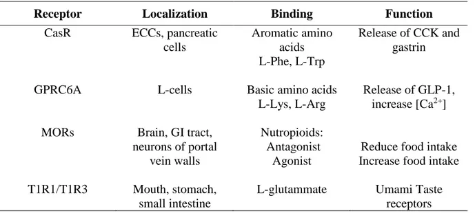
Some metabolites and amino acids contribute to the perception of postprandial satiety. This is at the basis of the aminostatic hypothesis (Veldorst et al., 2008). Accordingly, protein leverage hypothesis suggests that individual protein intake is regulated by individual protein target, emphasizing how ‘protein target’ can drive energy intake (Martens & Westerterp-Plantenga, 2014). The ingestion of proteins or amino acids increases serum amino acid concentration that elicits appetite reduction; inversely, a fall of the amino acid serum concentration enhances appetite. This happens because amino acids work as satiety signal (Veldorst et al., 2008). Taste buds in the oral cavity initiate gustatory signaling that influence food liking and choice. The amino acid-sensing in the mouth is mediated by GPCRs. The heterodimer T1R1/T1R3 is the umami taste receptor that binds the L-amino acids such as L-glutammate (eliciting the umami taste) (Zhang et al., 2008). Together with T1R1/T1R3, amino acids are also sensed by gastrointestinal EECs through other GPCRs such as CasR, MORs and GPRC6A (G protein-coupled receptor family C group 6 subtype A) (Table 1). The binding between a certain peptide or amino acid with the specific receptor activates a signaling pathway that culminates with a release of satiating peptides (GLP-1, CCK, PYY) as well as
11
neurotransmitters that directly reach the brain and fire the reward area (Spreckley & Murphy, 2015; Vancleef et al., 2015).
Table 1. Amino acid taste receptors
Receptor Localization Binding Function
CasR ECCs, pancreatic cells Aromatic amino acids L-Phe, L-Trp Release of CCK and gastrin
GPRC6A L-cells Basic amino acids
L-Lys, L-Arg
Release of GLP-1, increase [Ca2+]
MORs Brain, GI tract, neurons of portal
vein walls
Nutropioids: Antagonist
Agonist
Reduce food intake Increase food intake
T1R1/T1R3 Mouth, stomach, small intestine
L-glutammate Umami Taste receptors
In particular, MORs are expressed both in the brain where they are involved in food reward system and in the small intestine where they control gut motility. MORs are present in the neurons of the portal vein walls and can sense blood peptides coming from dietary protein digestion. These peptides can act as MOR agonists or antagonists. The latter activate gut-brain mechanisms inducing the intestinal gluconeogenesis controlling food intake (De Vadder et al., 2013). The opioid signaling influences dietary behavior modulating individual ‘wanting’ of foods and ‘liking’. Interestingly “nutropioids” can result from the digestion of dietary proteins. Casomorphin from milk casein and exorphin from gluten are opioid ligands acting as opioid agonists (Pfluger et al., 2012). Interestingly, it was recently demonstrated in animals that high-protein diets increased TAS2R/Gαtran/Gαgust expression in the pyloric mucosa possibly due to the bitter taste of compounds forming by protein digestion or to some amino acids (De Giorgio et al., 2016). This feature highlighted the chemosensory adaptation of gastro-intestinal tract to the dietary nutrients raising the hypothesis that diets may modulate both metabolism and dietary behavior through the fine network of taste receptors.
Consumption of dietary protein seems to decrease postprandial appetite and subsequent energy intake (EI) more than fat and carbohydrate. The type of protein ingested may also affect postprandial responses. A number of mechanisms have been proposed to explain this apparent satiety hierarchy of macronutrients, including higher thermogenic effect of dietary protein and post-absorptive small intestinal gluconeogenesis (which is associated with decreased EI in rats) (De Vadder et al., 2013).
12
Many oligopeptides coming from digestion of dietary proteins have opioid activity by acting as effectors of MORs (Pfluger et al., 2012). GIT, especially the small intestine, is the second body site, after brain, where MORs are most widely expressed and are involved in the control of gut motility and food intake (De Vadder et al., 2013).
Soymorphins-5, -6, and -7 are opioid agonist peptides derived from β-conglycinin β-subunit of soy. They are able to suppress food intake and delay bowel transit time via gut μ-opioid receptor after oral administration as well as through the activation of serotonin (5-HT1A), dopamine D2, and GABAB receptors (Kaneko et al., 2010).
Moreover, β-casomorphins (BCMs) are group of peptides with opioid properties arising by proteolytic digestion of β-casein (De Noni, 2008). Among the BCMs, BCM-7 is a typical peptide released during hydrolysis of the β-casein allelic variant A1, containing a histidine residue at position 67 (Nguyen et al., 2015). BCM-7 exerts various physiological effects, i.e., secretion of mucus, increased activity of superoxide dismutase and catalase, increased levels of prolactin, analgesic role, slows down the passage of food through the digestive system (as do other opioids) providing longer time for lactose fermentation (ul Haq, 2014).
In this scenario milk proteins are worthy of note because they are source of BAPs.
Milk proteins are often suspected to be the cause of nonspecific, undiagnosed GI symptoms in adults. The manifestation of stomach symptoms in healthy subjects is affected by meal composition, which influence gastric emptying and thereby lactose load in the gut. Also, differences in individual sensitivity and gut microflora may affect the tolerability of milk (Turpenein et al., 2016).
Opioid peptides are highly sensitive to hydrolysis by dipeptidyl peptidase IV (DPP-IV) thereby strongly limiting or preventing the transfer of these peptides in an intact form across the intestinal mucosa and the blood-brain barrier (De Noni et al., 2009). Increased intestinal permeability for digested food proteins and defective DPP-IV can result in biological active peptides circulating in the bloodstream, that can traverse the blood–brain barrier and reach the CNS (Cieślińska et al., 2015).
1.4 Intestinal permeability and GI disorders
Intestinal permeability can be defined as the facility with which intestinal epithelium allows molecules to pass through by non-mediated passive diffusion. This concept mainly refers to the passage of ions and inert molecules of low molecular weight. The intestinal transport of molecules from the intestinal lumen to the lamina propria can occur through two distinct mechanisms: paracellular diffusion through tight junctions (TJs) between adjacent intestinal epithelial cells (IECs) and transcellular transport involving endocytosis/exocytosis (transcytosis) mediated or not by membrane receptors (Ménard et al., 2010). There are a number of diseases that are known to increases
13
the mucosal permeation of macromolecules, such as celiac disease, Crohn’s disease, type 1 diabetes and patients with food allergies (Mishra & Makharia, 2012).
A cross talk between gut and brain is consolidated. In addition, mounting evidence shows that gut microbiota can influence host appetite and eating behaviour by directly affecting nutrient sensing and appetite, for this reasons the microbiota-gut-brain axis has been coined (van de Wouw et al., 2017). The microbiota functions in tandem with the host’s defences and the immune system to protect against pathogen colonisation and invasion. It also performs an essential metabolic function, acting as a source of essential nutrients and vitamins and aiding in the extraction of energy and nutrients. (Carding et al., 2015). Alterations in the bowel flora and its activities are now believed to be contributing factors to many chronic and degenerative diseases. The intestinal dysbiosis hypothesis suggests a number of factors associated with modern Western living that have a detrimental impact on the microbiota of the GIT. In comparison to diets high in overall protein, diets especially high in animal protein have specific effects on intestinal microbiota (Myers, 2004). Intestinal permeability reflects just one function of the barrier that is intimately related to and interacts with luminal contents, including the microbiota. The mucosal immune response also influences barrier integrity although changes in barrier function have been described in several gastrointestinal disorders. It is important to note that the gut microbiota has a key regulatory role in both host metabolism and central appetite, which together can modify host eating behaviour in metabolic disorders and eating disorders. (Carding et al., 2015).
Functional gastrointestinal disorders (FGIDs), are characterized by morphologic and physiological abnormalities that often occur in combination with motility disturbance, visceral hypersensitivity a well as altered mucosal and immune function, gut microbiota and CNS processes (Drossman, 2016). A complex interrelationship of predisposing genetic factors, influenced by life events as well as psychosocial factors lead to abnormalities in motility, visceral sensation, and brain – gut interactions, manifesting clinically as GI symptoms (Chey, 2013).
The relationships between celiac disease (CD), Non-Celiac Gluten Sensitivity (NCGS), and irritable bowel syndrome (IBS) remain unclear. However, it has been a matter of debate whether barrier function contributes to the development of FGIDs or if it is merely a consequence (Barbara et al., 2016).
14
References
Arneth, B. M. 2018. Gut–brain axis biochemical signalling from the gastrointestinal tract to the central nervous system: gut dysbiosis and altered brain function. Postgraduate medical journal,94(1114), 446-452.
Barbara, G., Feinle-Bisset, C., Ghoshal, U. C., Santos, J., Vanner, S. J., Vergnolle, N., ... & Quigley, E. M. 2016. The intestinal microenvironment and functional gastrointestinal disorders. Gastroenterology, 150(6), 1305-1318.
Berthoud, H. R., Münzberg, H., & Morrison, C. D. 2017. Blaming the brain for obesity: integration of hedonic and homeostatic mechanisms.Gastroenterology,152(7), 1728-1738.
Bohorquez DV, Shahid RA, Erdmann A, Kreger AM, Wang Y, et al. 2015. Neuroepithelial circuit formed by innervation of sensory enteroendocrine cells. Journal of Clinical. Investigation. 125:782–86
Brookes SJ, Spencer NJ, Costa M, Zagorodnyuk VP. 2013. Extrinsic primary afferent signalling in the gut. Nature Reviews Gastroenterology. Hepatology. 10:286–96
Capriotti, A.L., et al., 2016. Recent trends in the analysis of bioactive peptides in milk and dairy products. Analytical and bioanalytical chemistry. 408 (11), 2677–2685.
Carding, S., Verbeke, K., Vipond, D. T., Corfe, B. M., & Owen, L. J. 2015. Dysbiosis of the gut microbiota in disease.Microbial ecology in health and disease,26(1), 26191.
Chey, W. D. 2013. The role of food in the functional gastrointestinal disorders: introduction to a manuscript series.The American journal of gastroenterology,108(5), 694.
Cieślińska, A., Sienkiewicz-Szłapka, E., Wasilewska, J., Fiedorowicz, E., Chwała, B., Moszyńska-Dumara, M., ... & Kostyra, E. 2015. Influence of candidate polymorphisms on the dipeptidyl peptidase IV and μ-opioid receptor genes expression in aspect of the β-casomorphin-7 modulation functions in autism.Peptides,65, 6-11.
Daniel, H., 2004. Molecular and integrative physiology of intestinal peptide transport. Annu. Rev. Physiology 66 (1), 361–384.
Daniel, H., Zietek, T., 2015. Taste and move: glucose and peptide transporters in the gastrointestinal tract. Experimental. Physiology. 100 (12), 1441–1450.
De Giorgio R, Mazzoni M, Vallorani C, Latorre R, Bombardi C, Bacci ML, et al. 2016 Regulation of α-Transducin and α-Gustducin Expression by a High Protein Diet in the Pig Gastrointestinal Tract. PLoS ONE 11(2): e0148954
De Noni, I. 2008. Release of [beta]-casomorphins 5 and 7 during simulated gastro-intestinal digestion of bovine [beta]-casein variants and milk-based infant formulas. Food Chemistry 110(4):897– 903.
De Noni, I., et al., 2009. Review of the potential health impact of b-casomorphins and related peptides. EFSA J. 7 (2), 231r. Available at: http://doi.wiley.com/10.2903/j.efsa. 2009.231r.
15
De Vadder F, Gautier-Stein A, Mithieux G 2013 Satiety and the role of μ-opioid receptors in the portal vein.Current Opinion in Pharmacology13.6 959-963.
Drossman, D. A. 2016. Functional gastrointestinal disorders: history, pathophysiology, clinical features, and Rome IV.Gastroenterology,150(6), 1262-1279.
Foltz, M., van der Pijl, P.C., Duchateau, G.S., 2010. Current in vitro testing of bioactive peptides is not valuable. The Journal of. Nutrition. 140 (1), 117–118.
Foster, J. A., Rinaman, L., & Cryan, J. F. 2017. Stress & the gut-brain axis: regulation by the microbiome.Neurobiology of stress.
Kaelberer, M. M., Buchanan, K. L., Klein, M. E., Barth, B. B., Montoya, M. M., Shen, X., & Bohórquez, D. V. 2018. A gut-brain neural circuit for nutrient sensory transduction.Science,361(6408), eaat5236.
Kaneko, K., Iwasaki, M., Yoshikawa, M., & Ohinata, K. 2010. Orally administered soymorphins, soy-derived opioid peptides, suppress feeding and intestinal transit via gut μ1-receptor coupled to 5-HT1A, D2, and GABAB systems.American Journal of Physiology-Gastrointestinal and Liver Physiology,299(3), G799-G805.
Kentish, S. J. & Page, A. J. 2014. Plasticity of gastrointestinal vagal afferent endings. Physiology. Behavior. 136, 170–178
Kentish, S. J., O'donnell, T. A., Frisby, C. L., Li, H., Wittert, G. A., & Page, A. J. 2014. Altered gastric vagal mechanosensitivity in diet-induced obesity persists on return to normal chow and is accompanied by increased food intake. International journal of obesity, 38(5), 636..
Janssen , S. & Depoortere, I. 2013. Nutrient sensing in the gut: new roads to therapeutics? Trends in Endocrinology and. Metabolism. 24, 92–100
Lacroix, I.M.E., Li-chan, E.C.Y., 2016. Trends in Food Science & Technology Food-derived dipeptidyl-peptidase IV inhibitors as a potential approach for glycemic regulation e Current knowledge and future research considerations. Trends in Food Science and Technology. 54, 1– 16. Available at: https://doi.org/10.1016/j.tifs.2016.05.008
Lasrado, R., Boesmans, W., Kleinjung, J., Pin, C., Bell, D., Bhaw, L., ... & Berghe, P. V. 2017. Lineage-dependent spatial and functional organization of the mammalian enteric nervous system.Science,356(6339), 722-726.
Martens EA, & Westerterp-Plantenga, MS 2014 Protein diets, body weight loss and weight maintenance.Current Opinion in Clinical Nutrition 17(1), 75-79.
Möller, N. P., Scholz-Ahrens, K. E., Roos, N., & Schrezenmeir, J. 2008. Bioactive peptides and proteins from foods: indication for health effects. European journal of nutrition, 47(4), 171-182. Morton, G. J., Meek, T. H., & Schwartz, M. W. 2014. Neurobiology of food intake in health and
disease. Nature Reviews Neuroscience, 15(6), 367.
16
Nguyen, D. D., Johnson, S. K., Busetti, F., & Solah, V. A. 2015. Formation and degradation of Beta-casomorphins in dairy processing.Critical reviews in food science and nutrition,55(14), 1955-1967.
Page, A. J., Slattery, J. A., Milte, C., Laker, R., O'Donnell, T., Dorian, C., ... & Blackshaw, L. A. 2007. Ghrelin selectively reduces mechanosensitivity of upper gastrointestinal vagal afferents. American Journal of Physiology-Gastrointestinal and Liver Physiology, 292(5), G1376-G1384.
Page AJ, Symonds E, PeirisM, Blackshaw LA, Young RL. 2012. Peripheral neural targets in obesity. British. Journal of. Pharmacology. 166:1537–58
Pfluger PT, Schriever SC, Tschöp MH 2012. Nutropioids, hedonism in the gut?Cell metabolism 16(2), 137-139.
Roder PV, Geillinger KE, Zietek TS, Thorens B, Koepsell H, Daniel H. 2014. The role of SGLT1 and GLUT2 in intestinal glucose transport and sensing. PLOS ONE 9:e89977
Röhrborn, D., Wronkowitz, N., Eckel, J., 2015. DPP4 in diabetes. Frontiers in. Immunology. 6, 1– 20.
Roper, S. D., & Chaudhari, N. 2017. Taste buds: cells, signals and synapses.Nature Reviews Neuroscience,18(8), 485.
Rossi, M. A., & Stuber, G. D. 2017. Overlapping brain circuits for homeostatic and hedonic feeding.Cell metabolism.
Ruscitto, A., Smith, B. H., & Guthrie, B. 2015. Changes in opioid and other analgesic use 1995– 2010: Repeated cross‐sectional analysis of dispensed prescribing for a large geographical population in S cotland. European Journal of Pain, 19(1), 59-66..
Sauer, S., Luge, T., 2015. Nutriproteomics: facts, concepts, and perspectives. Proteomics 15 (5–6), 997–1013.
Schwartz, G.J. and Zeltser, L.M. 2013 Functional organization of neuronal and humoral signals regulating feeding behavior. Annual. Review of. Nutrition. 33, 1–21 4.
Sobczak, M.,Sałaga, M.,Storr, M. A.&Fichna, J 2014..Physiology, signaling, and pharmacology of opioid receptors and their ligands in the gastrointestinal tract: current concepts and future perspectives.Journal of Gastroenterol.49, 24–45
Spence, C., Okajima, K., Cheok, A. D., Petit, O., & Michel, C. (2016). Eating with our eyes: From visual hunger to digital satiation. Brain and cognition, 110, 53-63.
Spreckley &, Murphy KG 2015 The L-cell in nutritional sensing and the regulation of appetite. Frontiers in Nutrition, 2
Steensels, S., & Depoortere, I. 2018. Chemoreceptors in the Gut.Annual review of physiology,80, 117-141.
17
CCK, GLP-1, and PYY (3–36): Secretory controls and physiological roles in eating and glycemia in health, obesity, and after RYGB.Physiological reviews,97(1), 411-463.
Sternson, S. M., Betley, J. N., & Cao, Z. F. H. 2013. Neural circuits and motivational processes for hunger. Current opinion in neurobiology, 23(3), 353-360.
Sweeney, P., & Yang, Y. 2017. Neural circuit mechanisms underlying emotional regulation of homeostatic feeding.Trends in Endocrinology & Metabolism,28(6), 437-448.
Szigethy, E., Knisely, M., & Drossman, D. 2018. Opioid misuse in gastroenterology and non-opioid management of abdominal pain.Nature Reviews Gastroenterology & Hepatology,15(3), 168.
Turpeinen, A., Kautiainen, H., Tikkanen, M. L., Sibakov, T., Tossavainen, O., & Myllyluoma, E. 2016. Mild protein hydrolysation of lactose-free milk further reduces milk-related gastrointestinal symptoms.Journal of Dairy Research,83(2), 256-260.
Udenigwe, C.C., Fogliano, V., 2017. Food matrix interaction and bioavailability of bioactive peptides two faces of the same coin ? J. Funct. Foods 35, 9–12. Available at: https://doi. org/10.1016/j.jff.2017.05.029.
ul Haq, M. R., Kapila, R., Shandilya, U. K., & Kapila, S. 2014. Impact of milk derived β-casomorphins on physiological functions and trends in research: a review.International Journal of Food Properties,17(8), 1726-1741.
van de Wouw, M., Schellekens, H., Dinan, T. G., & Cryan, J. F. 2017. Microbiota-Gut-Brain Axis: Modulator of Host Metabolism and Appetite.The Journal of Nutrition,147(5), 727-745.
Vancleef, L., Van Den Broeck, T., Thijs, T., Steensels, S., Briand, L., Tack, J., & Depoortere, I. 2015. Chemosensory signalling pathways involved in sensing of amino acids by the ghrelin cell. Scientific reports,5, 15725.
Veldhorst M, Smeets AJPG, Soenen S, Hochstenbach-Waelen A, Hursel R, Diepvens K, Westerterp-Plantenga M (2008) Protein-induced satiety: effects and mechanisms of different proteins. Physiology Behavior, 94(2), 300-307.
Waise, T. Z., Dranse, H. J., & Lam, T. K. 2018. The metabolic role of vagal afferent innervation. Nature Reviews Gastroenterology & Hepatology, 1.
Williams, K.W. and Elmquist, J.K. 2012 From neuroanatomy to behavior: central integration of peripheral signals regulating feed-ing behavior. Nature. Neuroscience. 15, 1350–1355
Zhang F, Klebansky B, Fine RM, Xu H, Pronin A, Liu H, Li X 2008 Molecular mechanism for the umami taste synergism. Proceedings of the National Academy of Sciences USA, 105(52), 20930-20934.
Zhang, H., Kovacs-Nolan, J., Kodera, T., Eto, Y., & Mine, Y. 2015. Glutamyl cysteine and γ-glutamyl valine inhibit TNF-α signaling in intestinal epithelial cells and reduce inflammation in a mouse model of colitis via allosteric activation of the calcium-sensing receptor.Biochimica et Biophysica Acta (BBA)-Molecular Basis of Disease,1852(5), 792-804.
18
Chapter 2
Functional dyspepsia: new evidence for an old problem
Nicolina Virgilio, Paola Vitaglione
Department of Agricultural Sciences, University of Naples ‘‘Federico II’’ Portici,
Italy
19
1.1 Functional dyspepsia: definition, incidences and main symptoms
Functional gastrointestinal disorders (FGIDs), are characterized by morphologic and physiological abnormalities that often occur in combination with motility disturbance, visceral hypersensitivity and altered mucosal and immune function, gut microbiota and central nervous system (CNS) processes. FGDIs are one of the leading causes for referral to emergency care units and represent 40% of diagnoses in gastroenterological settings (Drossman 2016). Although only about 25% of symptomatic individuals seek medical support, the frequency of FGDIs drains substantial amounts of healthcare resources (Stanghellini 2017).
According to the Rome IV committee, FGIDs can be classified into six groups: esophageal disorders, gastroduodenal disorders, bowel disorders, centrally mediated disorders of gastrointestinal pain, gallbladder and sphincter of Oddi disorders, and anorectal disorders (Oshima & Miwa 2018).
Digestive function abnormalities and their symptoms, including functional dyspepsia (FD) and irritable bowel syndrome (IBS) are the most common FGIDs.
FD refers to upper abdominal chronic symptoms arising from the gastroduodenal region mainly triggered by ingestion of food (Talley 2017). The prevalence of FD in the community ranges between 5 and 11% (Vanheel et al.,2016).
According to the Rome IV criteria, the diagnosis of FD is based on the presence of any combination of 4 symptoms such as postprandial fullness, early satiety, epigastric pain, and epigastric burning. Moreover, to get a positive diagnosis symptoms have to be severe enough to interfere with the usual activities with a frequency of at least 3 days per week over a previous 3 months-period with an onset of at least 6 months in advance (Drossman 2016). That definition identifies patients suffering from 3 specific categories of disorders such as (1) postprandial distress syndrome (PDS), (2) epigastric pain syndrome (EPS), and (3) overlapping of PDS and EPS. PDS is characterized by meal-induced dyspeptic symptoms suggestive of a motility disturbance. EPS refers to epigastric pain or epigastric burning that do not necessarily occur after meal ingestion, can be even improved by meal, and is not associated with peptic ulcer or gastro-esophageal reflux disease. Overlapping of PDS and EPS is characterized by concomitance of meal induced dyspeptic symptoms and epigastric pain or burning (Stanghellini 2017).
According to the Rome IV definition, the prevalence of FD is higher in USA (12%) than Canada (8%) and UK (8%) with a distribution of PDS and EPS similar in the combined population and across the three countries and genders, even if a higher prevalence of FD was found in women compared to men across all age groups (Aziz et al.,2018). In particular, most of the participants with FD fulfilled criteria for PDS (61%), followed by EPS (18%) and 21% overlapping variant with both syndromes. In
20
addition, subjects with PDS had overlap with irritable bowel syndrome (IBS 15%, vs. 42% with EPS) (Aziz et al., 2018), supporting the idea that the PDS subtype of FD is distinct from other FGIDs. A meta-analysis of 100 population-based studies comprising over 312,000 subjects showed that the pooled prevalence of uninvestigated dyspepsia was 21% (95% confidence interval, 18% to 24%) and that the risk of dyspepsia was increased in females and those with Helicobacter pylori infection, smokers, and nonsteroidal anti-inflammatory drug users (Talley 2017).
The prevalence of FD is higher among women than men and this correlation could be due to sex-specific biological differences in gastrointestinal function (for example, sex hormone-driven alterations in intestinal motility) or the processing of (visceral) pain in the CNS, but also to sex-specific health care behaviour (Enck et al.,2017).
1.2 Pathophysiology and biopsychosocial perspective of FGIDs
The pathophysiology of FD is multifactorial and conclusive associations between symptoms and functional as well as psychological abnormalities are still a cause of debate among experts. It is well known that FD impacts on quality of life in a manner depending from symptom severity and comorbid depression. On the other hand, psychosocial disorders such as anxiety, depression, as well as physical and emotional abuse and difficulty in coping with life events are very frequent among FD patients (Stanghellini 2017).
While a variety of peripheral candidate biomarkers related to FGIDs continue to be investigated, none appear to account for a large proportion of the symptom variance in this diversified set of syndromes. At the same time, a model for FGIDs that includes a prominent role for brain-gut interactions has been emerged over time. Brain-gut axis may explain the complex interconnections between gastrointestinal sensation, motility, immune function, and gut microbes with sensory, cognitive, and affective circuitry in the brain (Tillish 2018).
In this framework, a biopsychosocial perspective is needed to shed light on how the complex interactions of environmental, psychological, and biological factors contribute to the development and maintenance of FGIDs as well as for an appropriate treatment of these comorbidities (Van Oudenhove 2016).
The biopsychosocial model suggests that a complex interrelationship of predisposing genetic factors, influenced by life events as well as psychosocial factors lead to abnormalities in motility, visceral sensation, and brain – gut interactions, manifesting clinically as GI symptoms (Chey 2013).
Psychological distress is a considerable risk factor for FGIDs development and, when present, can perpetuate or exacerbate symptoms. Comorbid anxiety and depression are independent predictors of
21
post-infectious IBS and FD but, at the same time, also occur as a consequence of bodily symptoms and related quality of life impairment. The absence of formal psychiatric comorbidity does not exclude a role of dysfunctional cognitive and affective processes (van Oudenhove 2016), likewise represent a big bias in the context of diagnosis of FGIDs. In this frame it is essential clarify how each of these factors—the environment, the individual’s own psychological states and traits, and the individual’s (neuro)physiological make-up-interact to ultimately result in the generation of FGID symptoms (van Oudenhove 2016).
1.3 Involvement and metabolic interactions of microbiome-gut-brain axis in
FGIDs development
A bidirectional communication takes place between gastrointestinal (GI) tract and central nervous system (CNS) through multiple pathways involving neural, endocrine, and immune cells. The gut– brain axis (GBA) allows the CNS to regulate GI functions, including motility and secretion, and the GI tract to signal sensations such as hunger, pain or discomfort to the CNS. New emotional, cognitive and behavioural functions concerning the GBA have been discovered, including affective mood, memory formation and food intake respectively (Mazzoli & Pessione, 2016). FGIDs symptoms are not as easy to localize and are influenced more by overarching effects resulting from CNS–enteric nervous system (ENS) dysregulation of symptom control pathways (Drossman 2016). Brain imaging studies using functional magnetic resonance imaging (fMRI), positron-emission tomography (PET) or other emerging imaging technologies have identified alterations in several interconnected brain networks, including sensorimotor, emotional arousal and salience networks, in patients with FGIDs (Enck et al.,2017).
Genetics and environmental factors as well as sociocultural influences may affect one’s psychosocial development in terms of personality traits, susceptibility to life stresses, psychological state, and cognitive and coping skills (Holtman et al., 2017).These factors influence the susceptibility to gut dysfunction: abnormal motility or sensitivity, altered mucosal immune dysfunction or inflammation, and the microbial environment, as well as the effect of food. This complex interactions and emotional distress may feed back to perpetuate and amplify symptoms (Sahan 2018). After exposure to stress, an increased basal activation of hypothalamic-pituitary-adrenal axis (HPA) and autonomic nervous system (ANS) associated with secretion of the corticotrophin releasing hormone (CRH) lead to physical and psychological symptoms, linked to GI system as well (Sahan 2018). The mechanisms underlying gut-brain communications involve neuro-immuno-endocrine mediators. This bidirectional communication network includes the CNS, both brain and spinal cord, the ANS, the
22
ENS and HPA axis. The HPA axis is considered the core stress efferent axis that coordinates the adaptive responses of the organism to stressors of any kind (Holtman et al.,2017).Environmental stress, as well as elevated systemic pro-inflammatory cytokines, activate this system that, through secretion of the corticotropin-releasing factor (CRF) from the hypothalamus, stimulates adrenocorticotropic hormone (ACTH) secretion from pituitary gland that, in turn, leads to cortisol release from the adrenal glands. Cortisol is a major stress hormone that affects many human organs, including the brain. Thus, both neural and hormonal mediators allow brain to influence the activities of intestinal functional effector cells, such as immune cells, epithelial cells, enteric neurons, smooth muscle cells and enterochromaffin cells (Carabotti et al., 2015). Both clinical and experimental evidence suggest that gut microbiota has an important impact on GBA, interacting not only locally with intestinal cells and ENS, but also directly with CNS through neuroendocrine and metabolic pathways involving serotonergic and GABAergic signalling systems (Barbara et al., 2016).
FGIDs symptoms seem to be affected by microbiota in terms of microbial dysbiosis within the gut and its role in influencing anxiety and depressive-like behaviours (Foster & Neufeld 2013). Dysbiosis, that occurs in FGIDs, is highly associated with mood disorders as well. The break down communication of the GBA induce changes in intestinal motility and secretion, causes visceral hypersensitivity and leads to cellular alterations of the entero-endocrine and immune system. Gut microbiota may interplay with multiple of these different pathophysiological FGIDs targets and its role is supported by varying lines of evidence (Dupont 2014).
The absence of microbial colonization is associated to an altered expression and turnover of neurotransmitters in both nervous systems (Clarke et al., 2013, Stilling et al., 2014) and also to alterations of gut sensory-motor functions, consisting in delayed gastric emptying and intestinal transit altered mucosal immune function, altered gut signalling (visceral hypersensitivity) and CNS dysregulation of the modulation of gut signalling and motor function (Carabotti et al.,2015).
It was speculated that the high prevalence of psychiatric comorbidities in FGID patients reflects the fact that FGID may be a primary manifestation of brain dysfunction, or even primary somatization, with the brain driving the gut manifestations (Tanaka et al., 2011). Epidemiological data from prospective studies (Jones et al., 2012; Koloski et al., 2012; Koloski et al., 2016) suggest that in at least half of the cases, GI symptoms arise first and incident mood disorders occur later. Other studies emphasize the role of (intestinal) inflammation and cytokine response, and the gut microbiome in driving gut to brain alterations (Holtman 2017). If these findings hold true, reversing GI dysfunction, could allow targeting and potentially curing not only the FGID but also concomitant mood disorders. Moreover, mounting evidence show that duodenal low-grade inflammation may be involved in the etiopathogenesis of FD inducing mucosal immune activation, duodenal barrier dysfunction, and
23
sensory-motor dysfunction. An altered duodenal gut microbiota, food antigens or infection may trigger duodenal micro-inflammation in a subset of FD patients (Jung & Talley 2018).
The duodenum regulates acid secretion from the stomach and the nutrient absorption in the small intestine via local signalling pathways, and connects with the CNS, via neuronal and endocrine mediators. Duodenal eosinophils and in some cases mast cells may play a key role in immune activation in FD. Low-grade intestinal inflammation in patients with FD may provoke impairment in motor-sensory abnormalities along the gastrointestinal neural axis. Among FD patients, the risk of developing dyspeptic symptoms after a bout of gastroenteritis is 2.54 odds ratio (95% confidence, 1.76-3.65) at more than 6 months after acute gastroenteritis (Futagami et al.,2015). Albeit causation is not established, the hypothesis that FD is a disorder of small intestinal inflammation in a major subset of patients is gaining acceptance, opening the possibility of novel and targeted treatment approaches.
1.4 FGIDs and foods
Food is associated with symptom onset or exacerbation in a significant proportion of FGID patients. Currently 80% of patients report that the symptoms are aggravated by ingestion of a meal (Page & Li 2018) and particularly approximately two-thirds of patients report symptoms within 15–45 min of food ingestion (Pilichiewicz et al., 2009). Despite this, the role of food in the pathogenesis of the FGIDs has remained poorly understood. For this reason, diet has largely played an adjunctive rather than a primary role in the management of FGID patients, underestimating the role of food both in GI function and sensation as how food relates to GI symptoms in FGID patients as well (Chey 2013). Increasing evidence show that diet contributes to functional digestive symptoms (Feinle-Bisset, 2004; Gibson et al., 2015; Pilichiewicz et al., 2009), and dietary restrictions are frequent among patients affected by FGIDs, especially those suffering from IBS (Bohn et al.,2013; Gibson et al., 2015). The most common nutrients supposed of being attributable to FGIDs symptoms are dietary fibers, dietary fats, and carbohydrates (Feinle-Bisset 2013(b); Saito et al., 2005; Yang 2012, Moayyedi et al., 2014). Nowadays, most of people adopt unhealthy dietary patterns, characterized by high consumption of fat and sugary products, sodas, snacks, breakfast cereals and other ultra-processed foods (UPFs, high density of saturated fatty acids, sugar, sodium and low content of fibers). Several studies evaluate the possibility that UPFs could be related with increased risk of IBS or contribute to the induction and/or exacerbation of digestive symptoms (Buscail et al., 2017; Khayyatzadeh et al., 2016). At this regard, Schnabel and colleagues (2018) investigated the possible association between UPFs consumption and FGIDs (Schnabel et al., 2018).
24
A prospective observational cohort study including 3516 adult participants was carried out. In the total population UPFs consumption accounted for 33.0% of total energy intake and the incidence of FGIDs was 10.5% for IBS, followed by functional constipation (5.4%), FD (3.9%), functional diarrhea (1.1%) respectively. Moreover, an increased proportion of UPFs in the diet was linked with a higher prevalence of IBS risk (aOR Q4 vs. Q1 [95% CI]: 1.25 [1.12–1.39], p < 0.0001), likewise an association between increased share of UPF in the diet and higher risk of FD when concomitant with IBS was observed (Schnabel et al., 2018).
In dietary intervention studies, specific amounts of solid or liquid meals were served to determine the meal-related dyspeptic symptoms, gastric accommodation, or hormonal changes in FD patients (Lee et al., 2018). Although it is already known that FD patients tolerate only small amounts of food, evidence on the extent of nutritional intake of daily meals remains inconclusive (Feinle-Bisset 2013). Dietary recommendations in FD include eating smaller meals and avoiding high-fat meals which have been reported to aggravate clinical symptoms such as nausea and abdominal pain, more than isocaloric high-carbohydrate meals (Yamawaki et al.,2018).
Recent studies investigating the role of dietary habit and nutritional intake in FD patients suggest that fat ingestion influences symptom development (Goktas et al., 2016, Khodarahmi et al., 2016). In particular, Goktas et al (2016) showed that FD subjects had symptoms mainly triggered by fried and fatty foods (27.1%), hot spices (26.4%), and carbonated drinks (21.8%) (Göktaş et al., 2016). In general, lipids in the duodenum can trigger gastric filling symptoms through a direct neuronal stimulation, higher lipid sensitivity of enteroendocrine cells or nerves, increased levels of systemic or local cholecystokinin (CCK, secreted by lipid-activated enteroendocrine cells, stimulates the release of digestive enzymes and bile and induces satiety) and/or increased sensitivity to CCK involving type A CCK-receptors (Enck et al.,2017).
Evidence shows that in FD patients the intra-duodenal infused lipids (but not glucose) stimulate the stomach distension more than in healthy controls, thus suggesting a cross-sensitization between mechano-sensors and chemo-sensors (Barbera et al.,1995).
Thus early satiety and intolerance of fatty foods in FD subjects could be related to gastrointestinal hypersensitivities to distension and/or small-intestinal fat (Feinle-Bisset 2013).
Hypersensitivity to mechanical stimulation of the stomach is frequent in patients with FD, however, the underlying mechanisms for this hypersensitivity are unclear. There is some evidence that transient receptor potential (TRP) channels may be involved in the visceral hypersensitivity associated with FGIDs (Balemans et al., 2017). In particular, the TRP vanilloid receptor 1 (TRPV1) expressed on vagal and spinal sensory nerve endings in the gut wall and activated by low pH, high temperature, painful stimuli, exogenous irritants such as capsaicin (active component of hot chilli peppers;
25
Caterina et al., 1997) and endocannabinoids like anandamide seems the most involved (Zygmunt et al., 1999). Indeed, the consumption of spicy capsaicin containing food was positively associated with scores of stomach fullness in FD patients (Lee et al., 2018) and a hypersensitivity to capsaicin in patients with FD compared to healthy controls has been recently reported (Hammer et al., 2018). The chemical hypersensitivity to capsaicin characteristic of FD patients was used to develop a simple and minimally invasive oral test to identify subjects with FD (Hammer et al., 2018).
Carbohydrates have been also associated to FGIDs as abdominal symptoms can be specifically induced by challenges with sugar (lactose or fructose), sorbitol, and oligosaccharides (fructans) alone or in combination and because exclusion diet-re-challenge tests in patients with FGIDs identified many cereal-based foods as those responsible to induce symptoms (Sheperd et al., 2013).
Short-chain carbohydrates containing up to 10 monosaccharide units vary in their digestibility and subsequent absorption. Those that are poorly absorbed exert osmotic effects in the intestinal lumen, attract water, and are rapidly fermented by bacteria with consequent gas production (Sheperd et al., 2013). As all dietary poorly absorbed short-chain carbohydrates have similar and additive effects in the intestine, a concept has been developed to regard them collectively as FODMAPs (fermentable oligosaccharides, disaccharides, monosaccharides and polyols) and to evaluate a dietary approach that restricts them all. Observational and comparative studies, and randomized-controlled trials support that FODMAPs trigger gastrointestinal symptoms in patients with functional bowel disorders, and that a diet low in FODMAPs offers considerable symptom relief in the majority of patients who use it (Yamawaki et al.,2018). Dairy or lactose intolerances and hypersensitivity to sour, acid-secreting or irritant foods (for example, citrus, spices, coffee or alcohol) could also play a role in stimulating GI symptoms. In addition, protein-rich foods could have an effect as thy can contain hidden fat (not readily identified by patients) or cause specific immune responses (Enck et al.,2017). Studies using magnetic resonance imaging (MRI) confirm that when FODMAPs are administered to healthy volunteers, small bowel distension occurs due to increased small bowel water content. However, water retention in the intestine cause discomforts only in patients with FGIDs and altered sensory functions(Holtman et al.,2017).
If FODMAPs are poorly absorbed and have been shown to induce symptoms in FGIDs, likewise gluten intolerance, even in the absence of coeliac disease, needs to be considered.
In fact, IBS and FD patients, without evidence for celiac disease based on serological markers and histology, experience substantial improvement in symptoms upon withdrawal of gluten from their diet (Holtman 2017). The relationships between celiac disease (CD), “gluten-sensitivity,” and IBS remain unclear, with various studies reporting increased or expected rates of CD among IBS subjects
26
(Barbara et al.,2016). An overlap between IBS and Non-Celiac Gluten Sensitivity (NCGS) has been detected. NCGS is a syndrome characterized by intestinal and extra-intestinal symptoms related to the ingestion of gluten-containing food, in subjects that are not affected by either celiac disease or wheat allergy (Catassi et al.,2015).
Epidemiology studies on IBS provide an indirect estimation of intestinal NCGS frequency. According to recent population-based surveys performed in Northern Europe, the prevalence of IBS in the general adult population was 16%–25% (Breckan et al., 2012; Krosgaard et al., 2013). In a selected series of adults with IBS, the frequency of NCGS, documented by a double -blind, placebo-controlled challenge, was 28% (Biesiekierski et al., 2011). Another study showed that 276 out of 920 (30%) subjects with IBS-like symptoms, according to the Rome II criteria, suffered from wheat sensitivity or multiple food hypersensitivity, including wheat sensitivity (Carroccio et al., 2012). Should a consistent proportion of IBS patients be affected with NCGS, the prevalence of NCGS in the general population could well be higher than CD (1%) (Catassi et al., 2015). A recent systematic review of Duncanson et al., (2018) consolidated the already known relationship between dietary fats and FD. The apparently disparate set of foods reported as inducing symptoms are each high in either fermentable carbohydrate (some soft drinks, fruit, fruit juice, watermelon, milk), wheat/gluten (grain/pasta/wheat products, takeout/processed foods) or natural food chemicals (fruit, fruit juice, red pepper, soft drink, tea). The findings in relation to lactose and fructose intolerance suggest that FODMAPs may induce symptoms via the small or large bowel. From this review conducted from January 1982 to February 2016 only 16 studies out of 6451 studies met the inclusion criteria and they dealt with investigation about the effect of nutrients, foods and food components in adults FD patients. Of note, of this 16 studies only 5 were case-control studies and 1 randomized controlled trial (RCT) characterized by a crossover gluten or placebo challenge.
In two years, despite the relevance of food triggering FGIDs-symptoms, there is still the lack of data in literature related to RCT. Previous findings highlighted that FD is, at least in part, a disorder related to food ingestion per se, in which symptoms can be induced by specific foods or food components and not simply a postprandial gastrointestinal motility disorder. This highlights the need for well-designed clinical studies that involve randomising patients to a wheat/gluten-free diet as well as controlling for FODMAP/fat content, aiming to investigate specific dyspeptic symptom associations. Furthermore, several lines of evidence suggest the involvement of the intestinal microbiota in the pathogenesis of FGIDs in general and IBS in particular. Gastrointestinal infections are strong risk factors for the development of FD and IBS, enhancing the intestinal permeability (leaky gut), within lifestyle and diet that are crucial determinants of microbiota composition and function in humans (Barbara et al., 2016). The impact of diet on the microbiota can be direct, through changes in its