Journal Name
COMMUNICATION
This journal is © The Royal Society of Chemistry 20xx J. Name., 2013, 00, 1-3 | 1Please do not adjust margins
Please do not adjust margins
Received 00th January 20xx, Accepted 00th January 20xx DOI: 10.1039/x0xx00000x www.rsc.org/A multifunctional toolkit for target-directed cancer therapy
Montserrat Terrazas,*ab Dani Sánchez,ab Federica Battistini,ab Núria Villegas,ab Isabelle Brun-Heathab and Modesto Orozco*abc
Here we present 2shRNA, a shRNA-based nanobinder that can simultaneously attack two therapeutic targets involved in drug resistance pathways, and that can additionally bind accessory molecules such as cell targeting peptides or fluorophores. We create 2shRNAs designed to specifically kill HER2+ breast cancer cells in the absence of transfecting agent.
Acquired resistance to molecularly targeted therapies is a major limitation for the successful treatment of cancer,1 as in many cases,
rescue pathways are activated in response to perturbation of the major pathway,2 leading to patient relapse. Approaches to fight
resistance consist in changes of treatment or in the administration of combination of drugs used to attack primary and rescue pathways.3 Unfortunately, most drug combinations approved to
date face major hurdles,3b such as toxicity effects, which hampers
their clinical use forcing the development of new efficient and non-toxic combinatorial approaches. An alternative strategy consists in the simultaneous inactivation of both primary and rescue pathways by using RNA interference (RNAi) machinery (siRNAs and shRNAs).4
Following this approach undruggable proteins can be tackled with large specificity.4cd Unfortunately, shortcomings exist, the most
important ones related to poor bio-disponibility4cd and to the
difficulty of controlling the concentration of the two RNAs inside the cell.4cd
In the search of innovative and efficient RNAi chemistries, we recently developed a N-hexyl-N dimeric nucleoside unit (BC6; Fig. 1A) with very interesting structural properties, such as high flexibility, which makes possible its use in many RNA constructs,5
and the presence of anchoring points, which allows the conjugation of accessory molecules such as peptide carriers or fluorophores. We explore here the BC6 dimer in the context of combined RNAi-based
therapies of breast cancer (BC), a complex disease6 that represents the second leading cause of death in women in developed countries.7 In recent years targeted therapies against BC have gained importance,2 especially those targeting HER2,8 a transmembrane tyrosine kinase belonging to a surface receptor family that includes epidermal growth factor receptor.9 The use of monoclonal antibody trastuzumab (Herceptin®),10 and the tyrosine kinase inhibitor Lapatinib (Tykerb®),11 which interfere HER2 activity, results in stopping proliferation, leading to major improvements in patient survival.10,11 Unfortunately, long term treatment12 leads to the overexpression of oncogenes involved in BC metastatic spread, such as GRB7,13 STARD313c,14 and Hsp27,15 leading to resistance. It is tempting to believe that combination of siRNAs in a branched structure16 could result in a multi-targeted drug able to block two different pathways.
The flexibility of BC65 prompted us to investigate its ability to act as a joint between two double-stranded RNA fragments of different sequence in a branched structure, which might have great potential to inhibit two different genes simultaneously. To this end, we devised a bifunctional shRNA-based branched nanobinder (2shRNA; Fig. 1B) composed of three building blocks: two BC6-loop RNA hairpins (arms; An and Bn) with 3'-overhangs (sticky ends) that form base-pairs with the 3'-sticky ends of a central two-way junction (Cn) formed by a natural RNA strand (CBn) and its complementary
counterpart (CTn),which possesses an internal BC6 bulge. The
building blocks are designed to spontaneously self-assemble into a branched structure after hybridization.
Molecular Dynamics (MD) simulations revealed that the presence of the BC6-loops and the BC6 internal bulge introduce little changes in the structure of the two double-stranded arms (Fig. S10, ESI†), but produces a local bending at the bulge generating a two-way branched architecture with very well-defined arms (Fig. 1C), which should make them accessible to RNAi machinery. Encouraged by these simulations we synthesized two branched RNAs specifically designed to attack two combinations of targets involved in drug resistance pathways in HER2+ BC: 2shSG and
2shHG (Fig. 1D), targeting STARD3/GRB7 and Hsp27/GRB7
COMMUNICATION Journal Name
2 | J. Name., 2012, 00, 1-3 This journal is © The Royal Society of Chemistry 20xx
Please do not adjust margins
Please do not adjust margins
(CT1:CB1) and in the 3'-sticky ends (Fig. S5, ESI†), differing only in
the base-paired sequence of their hairpin components [A1 (targeting STARD3) and B1 (targeting GRB7) in 2shSG, and A2 (targeting Hsp27) and B1 in 2shHG]. This design allows us to test an unlimited number of target combinations in a straightforward manner by changing only the hairpin components each time we need to target a different combination of genes. To further validate this design, we prepared 2shRG, targeting the Renilla luciferase (A3)/GRB7(B1) combination (Fig. 1D). Finally, as a non-targeting negative control, we constructed 2shRR, with both arms (A4 and
B2) targeting Renilla mRNA and with central joint (CT2:CB2) and
sticky ends different to those of the other three systems (Fig. 1D). Formation of the branched nanostructures proceeded in a very straightforward and specific manner (Fig. S1A and S2, ESI†) by combination of the four building blocks of each of the branched RNAs (which possess specifically designed complementary sticky ends) under annealing conditions. As an example, analysis of the formation of 2shSG by native PAGE revealed a single product (~80 bp; Fig. S1A) that migrated more slowly than each of the four separated RNA building blocks and similarly to its “covalent” branched RNA analogue (with each of the four components linked between them by phosphodiester bonds; Fig. S3, ESI†;).
2shSG turned out to be a good substrate of Dicer, which slowly converted it into sequences ~19 bp (Fig. S1B). The central part
CT1:CB1 remained unaffected (Fig. S1C), whereas hairpin B1 was
completely converted into ~19 bp fragments (Fig. S1D), suggesting that Dicer recognizes only the hairpin components of the 2shRNA.
Encouraged by these observations, we next studied the effect of 2shSG and 2shHG (40 nM) on target protein levels (by western blot) after treating the HER2+ BC cell line SK-BR-3 with these constructs in the presence of Lipofectamine 2000. 2shSG caused strong inhibition of expression of STARD3 and GRB7 (85% and 95% knockdown; Fig. 2A; Fig. S6, ESI†), with activities similar to those of the corresponding linear siRNAs. Similarly, 2shHG induced 100% and 97% of Hsp27 and GRB7 knockdown, respectively (Fig. 2B; Fig. S6, ESI†). Satisfactory results were also obtained for Fig. 1 (A) BC6 dimer and its potential applications. (B) Construction of the 2shRNA nanobinder. (C) Representative snapshot from the MD trajectory of the 2shRNA. (D) 2shRNAs used in this study.
2shRG, with 97% of GRB7 knockdown in the same cell line (Fig. 2C;
Fig. S6, ESI†). The same construct caused a (86 ± 0.5)% inhibition of
Renilla expression in HeLa cells previously transfected with plasmids encoding Renilla and Firefly. Remarkably, Hsp27 protein levels remained unaffected (Fig. 2B) after treatment of SK-BR-3 cells with 2shRG, confirming the specificity of the observed effects. Similarly, levels of GRB7 remained intact after treatment of SK-BR-3 cells with non-targeting 2shRR (Fig. 2B) which in contrast, induced significant
Renilla knockdown [(95 ± 0.5)% Fig. 2C].
RNAi-mediated silencing of GRB7, STARD3 and Hsp27 (independently) is known to decrease the viability of HER2+ BC cells such as SK-BR-3 and BT-474 [Lapatinib (Lap)-sensitive).13ac,15 Moreover, it has been found that suppression
of GRB7 by siRNAs increases the activity of Lap in these cell lines13a and that Hsp-27 removal increases the susceptibility of
HER2+ drug-resistant BC cells to HER2 inhibitors.15 To investigate the potential beneficial effect of the branched-mediated knockdown of GRB7/STARD3 and GRB7/ Hsp27 gene combinations on BC therapy, we carried out a cell viability study, quantified 72 h (Fig. 2D-G) after transfection (in the presence of Lipofectamine) using the crystal violet assay in the presence and in the absence of Lap.
The viability of SK-BR-3 cells transfected with branched systems
2shSG and 2shHG was significantly reduced (Fig. 2D), compared with cells transfected with mixtures of natural targeting RNAs [II (GRB7) + III (STARD3) and II + IV (Hsp27)], with (52 ± 2)% and (46 ± 2)% decrease in cell proliferation for 2shSG and 2shHG versus (32 ± 2)% and (25 ± 5)% for mixtures of siRNAs II + III and II + IV. As expected, cells were unaffected when treated with non-targeting branched RNA (2shRR) (Fig. 2D). The combination of Lap with the targeting 2shRNAs leads to a dramatic decrease on cell proliferation [(71 ± 1)% and (60 ± 1)% decrease in proliferation for 2shSG and
2shHG versus (64 ± 1)% and (55 ± 1)% for mixtures of siRNAs II+ III
and II + IV], outperforming treatments based on the combination of Lap and siRNAs.
Similar results were obtained with the BT-474 cell line (Fig. 2E), thus confirming the higher anti-proliferative effect of the branched systems, both alone, or in combination with Lap. Very interestingly, treatment of the Lap-resistant HER2+ BC cell line UACC-73217 with 2shSG and 2shHG caused a significant decrease in cell proliferation, compared with untreated cells and cells treated with non-targeting 2shRR (Fig. 2F) or with the central part CB1:CB1 (Fig. S8, ESI†). Very encouragingly, the decrease in proliferation caused by 2shSG and 2shHG is significantly larger than that obtained with the corresponding mixtures of individual siRNAs (II + III or II + IV). Again encouragingly, Lap which alone does not have effect in this cell line recovers antiproliferative activity when combined with 2shHG and 2shSG. Remarkably, the anti-proliferative activity of mixtures of the targeting BC6-loop shRNAs (A1 + B1 and A2 + B1) was significantly higher than that of the corresponding mixtures of siRNAs (II + III and II + IV) and significantly lower than the corresponding 2shRNAs containing them (2shSG and 2shHG; Fig. S8, ESI†), confirming the efficiency of the BC6-loop shRNA design and the synergistic effect achieved with our branched co-delivery strategy. Finally, non-malignant HEK-293 cells, that express very low levels of GRB7, were unaffected when treated with branched
Journal Name COMMUNICATION
This journal is © The Royal Society of Chemistry 20xx J. Name., 2013, 00, 1-3 | 3
Please do not adjust margins
Please do not adjust margins
Fig. 2 (A-C) Western blot analysis and plot of inhibition of Renilla expression of SK-BR-3 and HeLa cells (respectively) treated with branched or control siRNAs in the presence of Lipofectamine. (D-G) Cell viability after treatment with RNAs, in the absence/precence of Lap. Vehicle: cells treated with Lipofectamine and DMSO alone. ** (P < 0.01), *** (P < 0.001) and **** (P < 0.0001) versus indicated samples; # (P < 0.05), ## (P < 0.01) and #### (P < 0.0001) versus vehicle in the absence of Lap.
RNAs
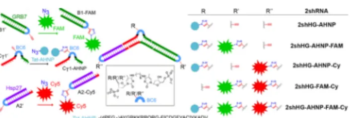
(Fig. 2G), confirming the low toxicity of our RNA design.To further increase the therapeutic potential of the branched construct, we proceeded to functionalize it with accessory molecules. With this aim, we converted one hydroxyl group of BC6 into an ethynyl group (ESI†) and used the resulting derivative to synthesize ethynyl-bearing CT1', A2'
and B1' (Fig. 3). CT1' was clicked18 with the azido-derivative of
a peptide carrier based on an anti-HER2 peptide (AHNP) linked to a Tat cell-penetrating peptide (Tat-AHNP; to give C1-AHNP; Fig. 3, Fig. S4, ESI†), which has been reported to enter HER2+ cancer cells,19 and A2' and B1' were reacted with the azido-derivative of two different fluorophores: Cy5 and carboxy-fluorescein (FAM) (red and green fluorescent signals, respectively), to give A2-Cy and B1-FAM (Fig. 3, Fig. S4, ESI†). We used these conjugates to construct a variety of mono-, di- and tri-functionalized 2shHG derivatives, by hybridization of the appropriate building blocks (Fig. 3 and Fig S5, ESI†): (i)
2shHG-AHNP, (ii) 2shHG-AHNP-FAM, (iii) 2shHG-AHNP-Cy5,
(iV) 2shHG-AHNP-FAM-Cy, and (v) 2shHG-FAM-Cy.
To confirm the co-delivery efficiency of our technology, SK-BR-3 cells were incubated with dual-labelled 2shHG-FAM-Cy (Fig. 4A-D; 40 nM) in the presence of Lipofectamine. As a control, we used a mixture of equimolar amounts of fluorescently labelled siRNA analogues siRNA IV-Cy (red) and siRNA II-FAM (green) (Fig. 4E-H; Fig. S5, ESI†; 40 nM each). In the case of cells treated with 2shHG-FAM-Cy, both dyes co-localized in a single point (Fig. 4D), thus confirming that the
branched RNA enters the cell and delivers the two shRNAs simultaneously. In contrast, in the case of cells treated with the mixture of siRNAs (Fig. 4E-H) an uneven and random distribution of both dyes is observed, due to different transfection efficiencies. Moreover, fluorescence intensities are lower than in the case of cells treated with 2shHG-FAM-Cy [Fig. 4E versus Fig. 4A (recorded using the same instrument settings) and Fig. 4F versus Fig. 4B (recorded using the same instrument settings)]. These results confirm the robustness, efficiency and potential advantages of our technology, compared with classical siRNA approaches.
We next explored the ability of Tat-AHNP-bearing 2shRNAs to enter HER2+ BC cells in the absence of transfecting agent. As indicated by the green fluorescence signal detected in Fig. S9C, 2shHG-AHNP-FAM (100 nM) efficiently internalized into HER2+ SK-BR-3 cells. Similar results were obtained with its Cy5 analogue 2shHG-AHNP-Cy (Fig. S9D). In contrast, no appreciable entry was observed when the same cell line was treated with 2shHG-FAM-Cy, lacking the AHNP carrier (Fig. S9F,G). Tat-AHNP-mediated specificity was further confirmed when BC cells expressing negligible levels of HER2 (MCF7) were treated with 2shHG-AHNP-FAM, as indicated by lower fluorescent intensities (Fig. S9E) than it did in SK-BR-3 cells (Fig. S9C). Finally, treatment of SK-BR-3 cells with
2shHG-AHNP-FAM-Cy (100 nM) further confirmed the efficient entry and the
robustness of our Tat-AHNP-bearing constructs, as both Cy5 and FAM dyes are detected in all the target cells (Fig. 4I-L). Western blot analysis of SK-BR-3 cells treated with
2shHG-AHNP (100 nM) in the absence of transfecting agent
demonstrated the ability of the construct to impart the desired RNAi effect after cell entry (65% and 59% of GRB7 and Hsp27 knockdown; Fig. 4M), which also induced significant levels of cancer cell death in the absence of transfecting agent [(43 ± 2)% decrease in cell proliferation; Fig. 4N]. In contrast, cells remained unaffected when treated with a mixture of siRNAs II and IV and with nacked branched 2shHG (Fig. 4N). Taken together, these results demonstrate the ability of AHNP-bearing 2shRNA conjugates to enter HER2+ cells in the absence of transfecting agent, which is translated in a significant anti-proliferative activity, due to specific GRB7/Hsp27 silencing. The specificity of this approach offers clear advantages over cationic-liposome based drug delivery, due to its ability to selectively deliver the two RNA drugs to the target tumor cell.
In summary, we have created a new RNA nanobinder of great potential in the treatment of complex diseases that rely on the use of multiple drugs. This bifunctional RNA design acts as a co-delivery system able to administer two different shRNAs simultaneously, improving therapeutic efficacy. The functionalization of the construct with potentially reactive groups (alkynyl) opens the door to the attachment of accessory molecules which can, for example, induce delivery to desired cells. As a proof of concept, we have created 2shRNA derivatives designed to target wild type and Lap-resistant HER2+ BC cells and functionalized with a peptide carrier targeting the HER2 receptor and two differently colored fluorescent dyes. Good anti-proliferative activity and null toxicity are observed, even in the absence of transfecting
COMMUNICATION Journal Name 4 | J. Name., 2012, 00, 1-3 This journal is © The Royal Society of Chemistry 20xx
Please do not adjust margins
Please do not adjust margins
Fig. 3 Functionalization of the three building blocks of 2shHG, and decorated 2shHG derivatives used in this study.Fig. 4 (A-L) Confocal microscopy images of SK-BR-3 cells treated with 2shHG-FAM-Cy (A-D), siRNA II-FAM + siRNA IV-Cy (E-H) in the presence of Lipofectamine, and with 2shHG-AHNP-FAM-Cy in the absence of Lipofectamine (I-L). (A,E,I) Green channel (FAM), (B,F,J) red channel (Cy5), (C,G,K) blue channel (Hoechst (nuclei)), (D,H,L) merged views. (M,N) Western blot analysis and anti-proliferative studies of SK-BR-3 cells treated with 2shRNAs in the absence of Lipofectamine.
agent. As the BC6 modification is located at the ends and central part of the branched structure, it could be compatible
with conventional biostable chemistries at internal positions (like phosphorothioate chemistries) to give rise to extremely robust constructs, thus further improving the properties of our 2shRNA technology.
This work was supported by the Instituto de Salud Carlos III [Miguel Servet I Program; CP13/00211; 205024141 to M.T.], the Spanish MINECO (BIO2015-64802-R; BFU2015-61670-EXP to M.O.) and the ERC Council (SimDNA, grant 291433, to M.O.). IRB is the recipient of a Severo Ochoa Award of Excellence from MINECO. We thank Dr. R. Eritja, Dr. M. Royo, Dr. A. Grandas, Dr. E. Pedroso and the Advanced Digital Microscopy Facility (IRB Barcelona), in particular A. Lladó, for their help and valuable comments.
Conflicts of interest
There are no conflicts to declare.
Notes and references
1 (a) J. Li, F. Chen, M. M. Cona, Y. Feng, U. Himmelreich, R. Oyen, A. Verbruggen and Y. Ni, Targ. Oncol., 2012, 7, 69; (b) K. Masui, B. Gini, J. Wykosky, C. Zanca, P. S. Mischel, F. B. Furnari and W. K. Cavenee, Carcinogenesis, 2013, 34, 725. 2 C. Holohan, S. Van Schaeybroeck, D. B. Longley and P. G.
Johnston, Nat. Rev. Cancer, 2013, 13, 714.
3 (a) K. C. Bulusu, R. Guha, D. J. Mason, R. P. I. Lewis, E. Muratov, Y. K. Motamedi, M. Cokol and A. Bender, Drug
Discov. Today, 2016, 21, 225; (b) B. Al-Lazikani, U. Banerji and P. Workman, Nat. Biotechnol., 2012, 30, 679; (c) M. J. Higgins and J. Baselga, J. Clin. Invest., 2011, 121, 3797. 4 (a) A. Fire, S. Xu, M. K. Montgomery, S. A. Kostas, S. E. Driver
and C. C. Mello, Nature, 1998, 391, 806; (b) S. M. Elbashir, W. Lendeckel and T. Tuschl, Genes Dev., 2001, 15, 188; (c) J. K. Watts, G. F. Deleavey and M. J. Damha, Drug Discov. Today, 2008, 13, 842; (d) G. F. Deleavey and M. J. Damha, Chem.
Biol., 2012, 19, 937; (e) D. Siolas, C. Lerner, J. Burchard, W.
Ge, P. S. Linsley, P. J. Paddison, G. J. Hannon and M. A. Cleary, Nat. Biotechnol., 2005, 23, 227.
5 M. Terrazas, I. Ivani, N. Villegas, C. Paris, C. Salvans, I. Brun-Heath and M. Orozco, Nucleic Acids Res., 2016, 44, 4354. 6 P. Eroles, A. Bosch, J. A. Pérez-Fidalgo and A. Lluch, Cancer
Treat. Rev., 2012, 38, 698.
7 R. L. Siegel, K. D. Miller and A. Jemal, CA Cancer J. Clin., 2017, 67, 7.
8 (a) F. Schettini, G. Buono, C. Cardalesi, I. Desideri, S. De Placido and L. Del Mastro, Cancer Treat. Rev., 2016, 46, 20; (b) D. J. Slamon, G. M. Clark, S. G. Wong, W. J. Levin, A. Ullrich and W. L. McGuire, Science, 1987, 235, 177.
9 M. Osaki, M. Oshimura and H. Ito, Apoptosis, 2004, 9, 667. 10 M. J. Piccart-Gebhart, M. Procter, B. Leyland-Jones, A.
Goldhirsch, M. Untch, I. Smith, L. Gianni, J. Baselga, R. Bell, C. Jackisch et al., N. Engl. J. Med., 2005, 353, 1659.
11 G. E. Konecny, M. D. Pegram, N. Venkatesan, R. Finn, G. Yang, M. Rahmeh, M. Untch, D. W. Rusnak, G. Spehar, R. J. Mullin et al., Cancer Res., 2006, 66, 1630. 12 (a) R. Y. Tsang and R. S. Finn, Br. J. Cancer, 2012, 106, 6; (b) C. M. Perou, T. SØrlie, M. B. Eisen, M. van de Rijn, S. S. Jeffrey, C. A. Rees, J. R. Pollack, D. T. Ross, H. Johnsen, L. A. Akslen et al., Nature, 2000, 406, 747; (c) S. Jaeger, A. Igea, R. Arroyo, V.
Alcalde, B. Canovas, M. Orozco, A. R. Nebreda and P. Aloy,
Cancer Res., 2017, 77, 459.
13 (a) A. Nencioni, M. Cea, A. Garuti, M. Passalacqua, L. Raffaghello, D. Soncini, E. Moran, G. Zoppoli, V. Pistoia, F. Patrone et al., PLos One, 2010, 5, e9024; (b) B. Ramsey, T. Bai, A. H. Newel, M. Troxell, B. Park, S. Olson, E. Keenan and S. -W. Luoh, Breast Cancer Res. Treat., 2011, 127, 659; (c) J. Kao and J. R. Pollack, Gene Chromosome Canc, 2006, 45, 761. 14 T. Z. Parris, A. Kovács, S. Hajizadeh, S. Nemes, M. Semaan, M. Levin, P. Karlsson and K. Helou, Oncogenesis, 2014, 3, e95. 15 S. H. Kang, K. W. Kang, K. -H. Kim, B. Kwon, S. -K. Kim, H. -Y.
Lee, S. -Y. Kong, E. S. Lee, S. -G. Jang and B. C. Yoo, BMC
Cancer, 2008, 8, 286.
16 (a) Y. Nakashima, H. Abe, N. Abe, K. Aikawa and Y. Ito, Chem.
Commun., 2011, 47, 8367; (b) D. Shu, Y. Shu, F. Haque, S.
Abdelmawla and P. Guo, Nat. Nanotechnol., 2011, 6, 658; (c) P. Tarapore, Y. Shu, P. Guo and H. Shuk-Mei, Mol. Ther., 2011, 19, 386. (d) C. Chang, H. S. Kang, C. Ban, S. Kim, D. –k. Lee, Mol. Cells, 2009, 27, 689. (e) W. Peng, J. Chen, Y. Qin, Z. Yang, Y. Y. Zhu, Nucleic Acids Ther., 2013, 23, 281. (f) H. J. Chung, C. A. Hong, S. H. Lee, S. D. Jo, T. G. Park, Bioconjugate Chem., 2011, 22, 299. 17 S. S. Kim, H. -J. Cho, J. -M. Cho, J. Y. Kang, H. -W. Yang and T. -K. Yoo, Scientif. World J., 2013, 174392. 18 J. E. Hein and V. V. Fokin, Chem. Soc. Rev., 2010, 39, 1302. 19 M. Tan, K. -H. Lan, J. Yao, C. -H. Lu, M. Sun, C. L. Neal, J. Lu, D. Yu, Cancer Res., 2006, 66, 3764.