A comparative study of silver amalgam and compomer
as retrograde filling materials in periapical surgery
Jordi Gargallo Albiol 1, José Manuel Aguirre Urízar 2, Cosme Gay Escoda 3
(1) Doctor of Odontology. Professor of the Master of Oral Surgery and Implantology. School of Dentistry of the University of Barcelona
(2) Professor and Chairman of Oral Medicine. Department of Stomatology. University of de Basque Country. EHU. Bilbao (3) Professor and Chairman of Oral and Maxillofacial Surgery. Director of the Master of Oral Surgery and Implantology. School of Dentistry of the University of Barcelona. Oral and Maxillofacial Surgeon of the Teknon Medical Center, Barcelona. (Spain)
Correspondence: Prof. Cosme Gay-Escoda Centro Médico Teknon Vilana 12
08022 – Barcelona. Spain E-mail: [email protected]
Received: 4/01/2007 Accepted: 6/12/2007
Gargallo-Albiol J, Aguirre-Urízar JM, Gay-Escoda C. A compa-rative study of silver amalgam and compomer as retrograde filling materials in periapical surgery. Med Oral Patol Oral Cir Bucal. 2008 Feb1;13(2):E133-7.
© Medicina Oral S. L. C.I.F. B 96689336 - ISSN 1698-6946
URL: http://www.medicinaoral.com/medoralfree01/v13i2/medoralv13i2p133.pdf
Summary
Objective: A comparative study is made of the histological effects of silver amalgam versus compomer (Dyract®) 90 days after placement as retrograde filling materials in experimental animals.
Method: Six Beagle dogs were used, with total pulpectomy and orthograde material filling followed by periapical surgery of the 6 upper and 6 lower incisors (for a total of 72 teeth). Thirty-six teeth corresponded to the right side and were filled with the control material (silver amalgam), while the 36 teeth on the left side were filled with the compomer study material (Dyract®). After three months the animals were sacrificed and the histological study was carried out, with evaluation of bone formation, inflammation, and the tissue in contact with the filler material. The results obtained were subjected to a descriptive and comparative statistical analysis (chi-square test).
Results: The samples retrogradely filled with compomer showed significantly greater percentage inflammation (76.19% versus 26.66% in the control group). On the other hand, a large proportion of samples with root cement growth were found in the compomer group. Filler material expulsion was also significantly more common when compomer was used.
Conclusions: the comparative study of the histological findings showed greater inflammation but also greater root cement growth in the compomer group versus the controls.
Key words: Amalgam, apicoectomy, compomer, retrograde filling, periapical surgery. Indexed in:
-Index Medicus / MEDLINE / PubMed -EMBASE, Excerpta Medica -SCOPUS
-Indice Médico Español -IBECS
Introduction
Retrograde filling in periapical surgery fundamentally aims to secure good apical sealing, thereby improving the prognosis of the operated tooth (1-3).
The search for adequate materials for retrograde filling continues to characterize much of research in periapical surgery – thus reflecting the fact that no ideal material has yet been developed. Silver amalgam has been the most widely used retrograde filler material over the years (2-6), though it poses a series of inconveniences such as
corro-sion, which can give rise to chronic inflammation; patient exposure to mercury, with its still uncertain but possibly systemic repercussions; allergies reactions; and a tendency to disperse around the periapical zone. Lastly, silver amal-gam is able to tattoo the adjacent tissues (7-10).
Glass ionomer shows good marginal sealing capacity in vitro, and has been found to offer excellent biocompati-bility in most studies in vivo (11-18). Unlike ionomers, compomers tend to be more resistant to humidity, and have yielded good results in different studies (19-22).
The present study was designed to histologically compare the effects of silver amalgam and compomer (Dentsply®, NY, USA) upon the periapical tissues 90 days after their placement as retrograde filler material in an experimental animal model.
Material and Methods
Six female Beagle dogs aged 2-3 years (body weight 12-15 kg) with fully formed permanent dentition were used. Six upper and 6 lower incisors from each dog were included (for a total of 72 teeth).
Anesthesia was induced by a cephalic vein injection of sodium pentothal. General anesthesia was maintained with inhalatory 2% fluothane and intravenous sodium pentothal. Oral intubation and mechanical ventilation were used.
Each surgical session comprised root canal treatment and periapical surgery of 6 incisors in one jaw. Total pulpec-tomy included biomechanical preparation followed by root canal filling using the lateral condensation technique with guttapercha and Endomethasone® sealer cement (Septodont®, Saint-Maur-des-Fossés, Cedex, France). The first phase of treatment was completed by coronal filling with silver amalgam (Inibsalloy®, Laboratorios Inibsa, Barcelona, Spain).
The second phase of treatment comprised periapical sur-gery of the endodontically treated teeth. A mucoperiosteal flap was raised, followed by trepanation of the external cortical layer of the jaw to locate the apical extremities of the 6 incisors. Use was made of the micromotor, handpie-ce and a rounded number 8 tungsten carbide drill under abundant irrigation with sterile bidistilled water. After horizontal resection of the apical end, measuring no more than 2 mm and controlled by the periodontal probe, the apical retrograde cavities measuring 2 mm in depth and 1 mm in width were prepared, using the microsurgery piece and truncoconal drill. Of the 6 incisors operated upon in each session, three were filled with the control material (i.e., silver amalgam without zinc non-gamma two and a high copper content (Inibsalloy®, Laborato-rios Inibsa, Barcelona, Spain)). The other three incisors were filled with compomer (Dentsply®, NY, USA). The compomer was placed with the applicator syringe and photopolymerized for 45 seconds. After carefully cleaning the surgical field, discontinuous sutures were placed using Vicryl® 4/0 and a TC-16 atraumatic needle (Laboratorios Aragó®, Barcelona, Spain). Postoperative antibiotic and analgesic treatment was provided: amoxicillin (Clamoxyl® GlaxoSmithKline, S.A.) 1 ml/10 kg b.w. via the intramus-cular route every 48 hours during 10 days, and magnesium metamizol (Nolotil®, Boehringer Ingelheim, Barcelona, Spain) 4 ml via the intramuscular route every 12 hours during 4 days.
Three months after the last operation, the animals were sacrificed by an intravenous sodium pentothal overdose
in the cephalic vein. The jaws were carefully dissected, and once thoroughly cleaned of soft tissues, they were submerged in 10% formalin solution.
Sagittal sections were cut along the longitudinal axis of the teeth, to facilitate examination of the retrograde filling and periapical area (Figure 1).
The bone-tooth specimens were processed according to the usual technique for light microscopy, with standard methodology for embedding in paraffin. Transverse sections measuring 5-10 µm in thickness were obtained using a Nahita® rotary microtome (Nahita Limited, Kingston-Upon-Hull, UK), followed by sample staining with hematoxylin-eosin. The histological study was then carried out, with evaluation of the following:
(a) Bone formation: based on the dichotomic classification used by a series of authors (12,23,24), where grade 0 = no bone formation, and grade 1 = bone formation.
(b) Inflammation: a global assessment of inflammation was made in relation to the intensity of the latter throug-hout the periapical zone (no inflammation, mild <30% periapical region, moderate 50-70% periapical region, and severe >70% periapical region). Use was also made of the Pertot grading system (20), which contemplates a series of concrete histological criteria (grade 1 = present, grade 0 = absent). The scores are added to yield a final nume-rical value corresponding to the inflammatory response, classified into 4 grades (Table 1).
(c) Tissue in contact with the filler material: bone, fibrous tissue, granulation tissue or epithelial tissue - classified as absent or present, and excluding the presence of tissues not in direct contact with the retrograde filler material. (d) Other histological parameters: these comprised any his-tological observations of relevance not addressed above. A descriptive study was made of the different variables, with application of the chi-square test with Yates correction for evaluation of the qualitative variables – statistical signifi-cance being considered for p < 0.05. The Epi Info® version 5.01 statistical package was used throughout (Centers for Disease Control and Prevention, Atlanta, GA, USA).
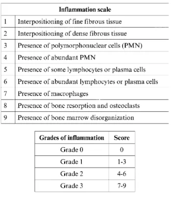
Fig. 1. Macroscopic section of the periapical area with silver
Results
Of the total 72 treated teeth, 22 (30.55%) were rejected prior to the histological analysis because of processing effects that made it impossible to correctly visualize the apical zone, and particularly the retrograde filling material. Of the 50 samples examined, 23 belonged to the control group (silver amalgam)(46%) and 27 to the experimental group (compomer)(54%).
Table 2 shows the results in relation to bone formation - no statistically significant differences being observed (p>0.05).
In relation to general inflammation as classified in our stu-dy, important differences were seen in severe inflammation (Table 3)(p=0.006) – the samples subjected to retrograde filling with compomer exhibiting a significantly greater percentage of severe inflammation than the controls (76.19% versus 26.66%)(Figure 2). In contrast, no statis-tically significant differences were observed on evaluating moderate inflammation and grouped moderate and severe inflammation (p>0.05).
Likewise, no statistically significant differences between the two filler materials were observed in terms of the Pertot grading system, or on evaluating the tissue in direct contact with the filler material surface (p>0.05).
Regarding the rest of histological data, only retrograde filling material expulsion proved statistically significant – with greater expulsion following retrograde filling with compomer versus silver amalgam (29.6% versus 0%) (p=0.013)(Figure 3).
Discussion
The exclusion of 22 teeth from our series was due to the small size of the teeth and of the periapical region, together with the position and inclination of the roots, and the fact that the only visible dental region was the crown - the root being covered by bone. All this compli-cated the obtainment of valid longitudinal sections for histological analysis, since the anatomical sections were required to pass through the periapical area, including the retrograde filling.
Table 1. Pertot et al. (20) classification of inflammation.
Table 2. Bone formation in relation to filler material employed.
As to bone formation, the results obtained indicate that little bone (10%) was found in the periapical area three months after obturation. Other authors have reported greater formation of bone tissue. However, such studies were carried out under different circumstances, with place-ment of the study material in rabbit tibial or femoral bone (10,11,20), or involving evaluation after 6 months (13). In relation to the formation of other tissues, over half of the specimens presented granulation tissue in direct contact with the surface of the retrograde filling material (64%) – in coincidence with the observations of DeGrood et al. (10), Zetterqvist et al. (13), and Maher et al. (23). On evaluating the growth of root cement, no statistically significant differences were observed between the two groups (silver amalgam and compomer), though retro-grade filling with compomer did yield a comparatively larger proportion of samples with cement growth (48.14% versus 34.78%). In concordance with these results, different authors have shown glass ionomer to favor the formation of bone tissue and root cement in the vicinity (14,25). Root cement growth is an important histological finding,
On the other hand, in relation to inflammatory response, and in the same way as for bone formation, the results reported by other investigators tend to be better than in our own– with superior performance in the case of glass ionomer and compomer versus silver amalgam (10-13). Our compomer contained 30% resin composite - a fact that could account for the differences observed with respect to those studies using glass ionomer (17,26).
While filler material expulsion is not an actual histological finding, it is an important factor when evaluating a filler material for application in periapical surgery. This coin-cides with the doubts expressed by Pitt Ford and Roberts (26) regarding the use of glass ionomers for retrograde filling. Possibly, the greater number of compomer expul-sions in our study reflects the greater difficulty of using this material under routine conditions of periapical surgery, where despite periapical isolation measures (particularly before retrograde filling), adequate dryness of the apical cavity proves difficult. If the cavity is not dry, then material adhesion does not occur and, secondly, contamination takes place before filler polymerization - thus leading to alterations in the properties of the material, with defective adhesion and adaptation.
In relation to biocompatibility, most studies involving animal models have shown tissue tolerance to be greater with glass ionomer versus silver amalgam (11-13,18). Our findings are more in line with those of clinical stu-dies conducted in humans (15,16), where in most cases no differences are observed between teeth subjected to retrograde filling with silver amalgam, glass ionomer, or compomer. It would be interesting to determine whether the histological differences observed in animal models are also found in clinical studies involving humans.
Conclusions
(a) The histological study of the samples involving retro-grade filling with silver amalgam in our animal model revealed average biocompatibility, with limited bone formation and moderate inflammation. In comparison, compomer (Dyract, Dentsply International, NY, USA) was associated to low biocompatibility, limited bone for-mation and severe inflamfor-mation.
(b) The comparative study of the two groups showed greater inflammation but also greater root cement growth in the compomer group versus the controls.
(c) Changes in this protocol would be required to reduce the frequency of compomer expulsion.
References
1. Torabinejad M, Hong CU, Pitt Ford TR, Kettering JD. Cytotoxicity of four root end filling materials. J Endod. 1995 Oct;21(10):489-92. 2. Tronstad L, Trope M, Doering A, Hasselgren G. Sealing ability of dental amalgams as retrograde fillings in endodontic therapy. J Endod. 1983 Dec;9(12):551-3.
3. Bondra DL, Hartwell GR, MacPherson MG, Portell FR. Leakage in vitro with IRM, high copper amalgam, and EBA cement as retrofilling
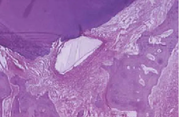
Fig. 2. Periradicular area showing retrograde filling
(compo-mer) and an intense inflammatory infiltrate (Hematoxylin-eosin, x10).
Fig. 3. Fragment of expulsed compomer surrounded by
4. Friedman S, Rotstein I, Koren L, Trope M. Dye leakage in retrofilled dog teeth and its correlation with radiographic healing. J Endod. 1991 Aug;17(8):392-5.
5. Penarrocha Diago M, Sanchis Bielsa JM, Gay Escoda C. Periapical sur-gery of 31 lower molars based on the ultrasound technique and retrograde filling with silver amalgam. Med Oral. 2001 Nov-Dec;6(5):376-82. 6. Peñarrocha Diago M, Boronat López A, Lamas Pelayo J. Update in dental implant periapical surgery. Med Oral Patol Oral Cir Bucal. 2006 Aug 1;11(5):E429-32.
7. Mjör IA, Hensten-Pettersen A, Skogedal O. Biologic evaluation of fi-lling materials. A comparison of results using cell culture techniques, im-plantation tests and pulp studies. Int Dent J. 1977 Jun 2;27(2):124-9. 8. Waikakul A, Punwutikorn J. Gold leaf as an alternative retrograde fi-lling material. Oral Surg Oral Med Oral Pathol. 1989 Jun;67(6):746-9. 9. Ferracane JL, Nakajima H, Okabe T. Enhanced evaporation of mercury from amalgams in non-oxidizing environments. Dent Mater. 1993 Sep;9(5):300-5.
10. DeGrood ME, Oguntebi BR, Cunningham CJ, Pink R. A com-parison of tissue reactions to Ketac-Fil and amalgam. J Endod. 1995 Feb;21(2):65-9.
11. Zmener O, Dominguez FV. Tissue response to a glass ionomer used as an endodontic cement. A preliminary study in dogs. Oral Surg Oral Med Oral Pathol. 1983 Aug;56(2):198-205.
12. Callis PD, Santini A. Tissue response to retrograde root fillings in the ferret canine: a comparison of a glass ionomer cement and gutta-percha with sealer. Oral Surg Oral Med Oral Pathol. 1987 Oct;64(4):475-9. 13. Zetterqvist L, Anneroth G, Nordenram A. Glass-ionomer cement as retrograde filling material. An experimental investigation in monkeys. Int J Oral Maxillofac Surg. 1987 Aug;16(4):459-64.
14. Blackman R, Gross M, Seltzer S. An evaluation of the biocompati-bility of a glass ionomer-silver cement in rat connective tissue. J Endod. 1989 Feb;15(2):76-9.
15. Jesslén P, Zetterqvist L, Heimdahl A. Long-term results of amalgam versus glass ionomer cement as apical sealant after apicectomy. Oral Surg Oral Med Oral Pathol Oral Radiol Endod. 1995 Jan;79(1):101-3. 16. Zetterqvist L, Heimdahl A. Long-term results of amalgam versus glass ionomer cement as apical sealant after apicectomy. Oral Surg Oral Med Oral Pathol Oral Radiol Endod 1995; 79: 101-3.
16. Jesslén P, Zetterqvist L, Heimdahl A. Long-term results of amalgam versus glass ionomer cement as apical sealant after apicectomy. Oral Surg Oral Med Oral Pathol Oral Radiol Endod. 1995 Jan;79(1):101-3. 17. Trope M, Lost C, Schmitz HJ, Friedman S. Healing of apical perio-dontitis in dogs after apicoectomy and retrofilling with various filling materials. Oral Surg Oral Med Oral Pathol Oral Radiol Endod. 1996 Feb;81(2):221-8.
18. Peltola M, Salo T, Oikarinen K. Toxic effects of various retrograde root filling materials on gingival fibroblasts and rat sarcoma cells. Endod Dent Traumatol. 1992 Jun;8(3):120-4.
19. Tarim B, Hafez AA, Suzuki SH, Suzuki S, Cox CF. Biocompatability of compomer restorative systems on nonexposed dental pulps of primate teeth. Oper Dent. 1997 Jul-Aug;22(4):149-58.
20. Pertot WJ, Stephan G, Tardieu C, Proust JP. Comparison of the intraosseous biocompatibility of Dyract and Super EBA. J Endod. 1997 May;23(5):315-9.
21. Bohsali K, Pertot WJ, Hosseini B, Camps J. Sealing ability of super EBA and Dyract as root-end fillings: a study in vitro. Int Endod J. 1998 Sep;31(5):338-42.
22. Greer BD, West LA, Liewehr FR, Pashley DH. Sealing ability of Dyract, Geristore, IRM, and super-EBA as root-end filling materials. J Endod. 2001 Jul;27(7):441-3.
23. Maher WP, Johnson RL, Hess J, Steiman HR. Biocompatibility of retrograde filling materials in the ferret canine. Amalgam and IRM. Oral Surg Oral Med Oral Pathol. 1992 Jun;73(6):738-45.
24. Torabinejad M, Ford TR, Abedi HR, Kariyawasam SP, Tang HM. Tissue reaction to implanted root-end filling materials in the tibia and mandible of guinea pigs. J Endod. 1998 Jul;24(7):468-71.
25. Sasanaluckit P, Albustany KR, Doherty PJ, Williams DF. Biocom-patibility of glass ionomer cements. Biomaterials. 1993 Oct;14(12):906-16.
26. Pitt Ford TR, Roberts GJ. Tissue response to glass ionomer retrograde root fillings. Int Endod J. 1990 Sep;23(5):233-8.