1
“FEDERICO II”
UNIVERSITY OF NAPLES
P
hD PROGRAM IN NEUROSCIENCE
XXXI CYCLE
PhD Thesis:
“
Identification of Epigenetic Mechanisms Regulating
NCX3 in in Vivo Model of Brain Ischemic
Preconditioning
”
Coordinator
Prof. Maurizio Taglialatela
Tutor
Candidate
Dr. LUIGI FORMISANO
Dr. LUIGI MASCOLO
2
INDEX
Summary Introduction
I.A. ISCHEMIC PRECONDITIONING IN BRAIN ... 6
I.A.1. Mechanisms of Brain Ischemic Preconditioning ... 7
I.A.2. Clinical Applications and Future Perspectives ... 9
I.B. EPIGENETIC ... 9
I.B.1. Nucleosome ... 11
I.B.2. Epigenetic Modification ... 13
I.B.3. Histone Lysine Methylation and Demethylation ... 16
I.B.4. Histone Lysine Methyl Transferase (KMT) Families ... 19
I.B.5. Histone Lysine Demethylases (KDM) Families ... 22
I.B.6. Drugs Regulating Lysine Methylation ... 24
I.B.7. The Role Of Kmts in Models of Neurological Disease... 24
I.B.7.1.Huntington’s disease and Friedreich’s Ataxia ... 25
I.B.7.2.Stroke inVivo and Vitro ... 26
I.B.7.3 Weidemann Steiner Syndrome ... 26
I.B.7.4.Kabuki Syndrome ... 27
I.B.7.5.Schizophrenia ... 27
I.C. SODIUM CALCIUM EXCHANGER ... 28
I.C.1. State of Art of Na+\Ca2+ Exchanger (NCX) ... 29
I.C.2. Molecular Biology of NCX Isoforms ... 32
I.C.3. Distribution of NCX Isoforms ... 33
I.C.4. Regulation of NCX Isoforms ... 35
I.D. NCX3 Na+/Ca2+ EXCHANGER 3 ... 37
I.D.1. Role and Mechanism of NCX3 in Brain Ischemic Preconditioning ... 39
I.D.2. Transcriptional Regulation of ncx3 Gene in the Brain ... 41
I.D.2.1. ncx3 brain promoter region and its putative transcription factor in binding sites ... 44
I.D.2.2. ncx3 Is Transcriptionally Up-regulated by CREB ... 45
I.D.2.3. ncx3 Is Transcriptionally Down-regulated by DREAM ... 47
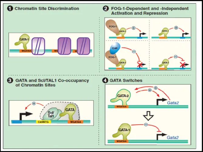
I.E. GATA TRANSCRIPTION FACTORS ... 47
I.E.1. The GATA Transcription Factor Family ... 48
3
I.E.3. Physiological Function and Tissue Specificity of GATA Protein ... 55
I.E.4. Specificity in the Regulation of Transcription by GATA Factor ... 55
I.E.4.1. Post-translational Modification ... 57
Aim of the Thesis II. AIM OF THE STUDY ... 59
Materials and Methods III.1. Materials ... 61
III.2. Primary Cortical Neurons ... 61
III.3. Transfection with Expression Plasmids or Small Interfering RNA (siRNA) and Luciferase Reporter Assay in Cortical Neurons ... 62
III.4. Quantitative Real-Time PCR (qRT-PCR) Analysis ... 62
III.5. Western Blotting ... 63
III.6. Chromatin Immunoprecipitation (ChIP) ... 64
III.7. In Vivo Studies ... 65
III.7.1. Experimental groups ... 65
III.7.2. Transient Focal Ischemia and Ischemic Preconditioning ... 65
III.8. Statistical Analysis ... 65
Results IV.1. GATA3 binds and activates ncx3 by a specific GATA sequence ... 71
IV.2.GATA3,KMT2A,NCX3 increase in the temporoparietal cortex after preconditioning+ischemia ... 73
IV.3.GATA3,KMT2A and NCX3 increase in the temporoparietal cortex after preconditioning and preconditioning+ischemia but not after ischemia ... 76
Discussion V. Discussion ... 79 Conclusion VI. Conclusion ... 80 References VII. References ... 86
4
SUMMARY
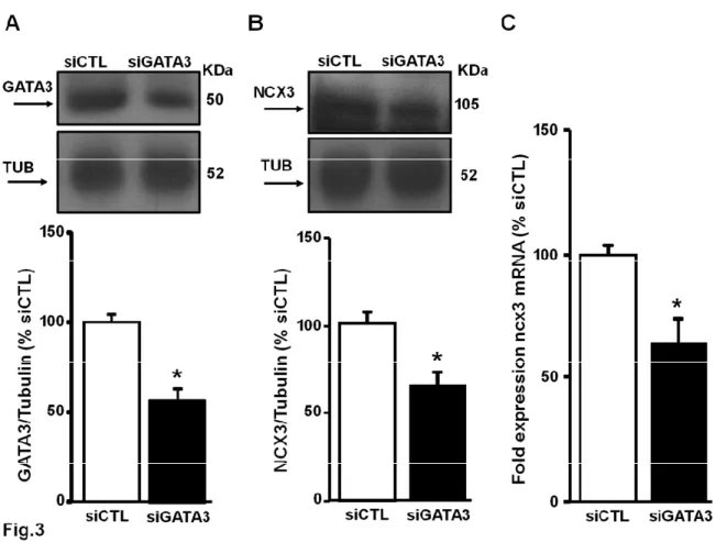
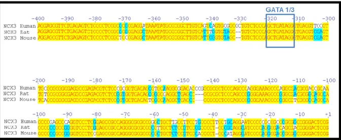
Variations of the isoform 3 expression of the sodium / calcium exchanger play an important role in the response to neuronal damage after an ischemic insult. I found that the transcription factor (GATA binding protein) 3 GATA3 activates the transcription of ncx3 in rat cortical cultures. In fact, the overexpression of GATA3, obtained as a result of transient transfection of the plasmid containing the GATA3 cDNA in neurons, leads to a significant increase in the luciferase activity of the ncx3 promoter, in parallel with an increase in mRNA and protein expression of ncx3. In contrast, the transfection of a siRNA capable of reducing the protein expression of GATA3 by about 60% causes a reduction of the luciferase activity of the ncx3 promoter and a decrease in the ncx3 mRNA. The site-specific mutagenesis of the binding sequence of GATA3 on the ncx3 promoter demonstrates that the mechanism by which GATA3 activates NCX3 is site-specific. More important, in vivo, GATA3 recruitment to the ncx3 gene was increased in the temporoparietal cortex of rats subjected to Preconditioning (PC) followed by transient middle cerebral artery occlusion (tMCAO), with an increase of histone 3 lysine 3 trimethylation of the ncx3 promoter region. Interestingly, in the same experimental conditions histone acetylation on ncx3 promoter was unmodified. Furthermore, Re-ChIP experiments demonstrated that GATA3 forms a functional complex with the histone lysine methyl transferase KMT2A on the ncx3 gene during PC+tMCAO. Therefore, increasing KMT2A expression or activity might represent a new possible strategy in stroke intervention.
5
I. INTRODUCTION
I.A. ISCHEMIC PRECONDITIONING IN BRAIN
Brain ischemia is one of the most common causes of death and the leading cause of adult disability in the world. Brain ischemic preconditioning (BIP) refers to a transient, sublethal ischemia which results in tolerance to later, otherwise lethal, cerebral ischemia. neuroprotective mechanisms may involve a series of molecular regulatory pathways. Generally speaking, any stimulus capable of causing injury to a tissue or organ can, when applied close to (but below) the threshold of damage, activate endogenous protective mechanisms and thus potentially lessen the impact of a subsequent, more severe attack. a phenomenon known as ischemic preconditioning (IP) or ischemic tolerance (IT)(Liu et al. 2009). The terms 'tolerance' and 'preconditioning' were introduced for the first time by Janoff in 1964 (JANOFF 1964) ischemic preconditioning is an adaptive reaction to a potentially noxious stimulus, such as ischemia, hypoxia, hypoglycemia, or inflammation(Liu, Sheng et al. 2009) .The phenomenon of ischemic preconditioning has been observed in numerous organs such as brain (Kitagawa et al. 1991) and heart (Meldrum et al. 1997), as well as in a wide range of species like the gerbil (Kirino et al. 1991), the rat and the mouse. Various brain regions, hippocampus, cerebral cortex, basal ganglia and thalamus were often reported to acquire ischemic tolerance (Kitagawa, Matsumoto et al. 1991) The brain is one of the most sensitive organs to injury. A constant flow of blood to the brain is essential for delivering oxygen and glucose to neurons. If this flow is disrupted for even a short period of time, the result is cell damage or death. Neurons are rarely replaced once they have died, so the damage to affected regions may be permanent (Caplan 2000). there are two threshold values for cerebral blood flow. Reduction in blood flow below the first threshold results in electrical failure within neurons, and further reduction in blood flow below the second threshold leads to the failure of metabolism and ion pumps. Cells with lower perfusion than the second threshold are designated to die. Between the two thresholds, cells are electrically silent but maintain a low level of metabolic activity and can be stable for hours. If normal blood flow is restored within a reasonable amount of time, they may recover with no apparent damage. Longer periods of ischemia, however, will result in their death. So these cells have a variable fate, and constitute what is known as the ischemic penumbra.(Dirnagl et al. 1999) It is the cells within the penumbra that
6
receive the most benefit from ischemic preconditioning endogenous neuroprotective mechanisms and potentially, a window of opportunity to utilize these mechanisms in the clinic to treat patients with stroke and other CNS disorders.(Liu, Sheng et al. 2009)
I.A.1. Mechanisms of Brain Ischemic Preconditioning
Two temporally distinct types of ischemic tolerance are afforded by sublethal pretreatment: early and delayed tolerance (Bhuiyan and Kim 2010),The first phase, named rapid or acute preconditioning ,starts 3–5min after the preconditioning Stimulus and ends 1h later and is due to rapid post-translational Protein modifications .The second phase, named delayed preconditioning ,starts2–3days after preconditioning And ends 1week later and mainly involves de novo protein synthesis (Cuomo et al. 2015).To induce tolerance by means of ischemic episodes, three factors should be considered: 1) the duration, 2) the time interval, and 3) the number of episodes. First, the preconditioning stimuli must be severe enough to initiate a response, but not so severe as to cause permanent damage. Second, some interval of time must exist between sublethal and lethal stress. Third, the number of short ischemic episodes should be considered for sufficient stimulation of the protective response against a lethal ischemic insult. In general, the process of tolerance induction can be divided into the following elements: sensors of the stress signal, transducers of the stimulus, and effectors of the tolerance. First, the preconditioning stimulus must be recognized by cellular sensors so that the cells can be prepared for upcoming stress. Neurotransmitter and cytokine receptors, ion channels and redox-sensitive enzymes generally work as molecular sensor of stress stimuli. These sensors activate enzymes, such as kinase protein Ras, Raf, mitogen-activated protein kinase (MAPK) kinase (MEK), extracellular regulated kinase (ERK), Akt, and protein kinase C, and signaling molecules, such as nitric oxide (NO), diacylglycerol, inositol triphosphate, Ca2+, and ceramide, which transduce the signal and initiate an adaptive response. Finally, effectors of the preconditioning response confer tolerance to cells or tissues through excitotoxicity, apoptosis, anti-inflammation, protection of mitochondria and increased anti-oxidant mechanisms (Bhuiyan and Kim 2010). Ischemic tolerance thus reflects a fundamental change in the cellular response to injury that shifts the outcome from cell death to cell survival
7
(Dirnagl et al. 2003). Although transcription factors, such as HIF-1, CREB and NF-κB are already known to be driving neuroprotective gene expression upon an ischemic preconditioning stimulus, we are today more aware that apart from the transcription factors and DNA sequence, regulation of such transcriptional activity requires the cooperation of a third party, namely epigenetic alterations of the DNA and histones. Indeed, these modifications crucially regulate the accessibility of specific regulatory DNA elements for transcription machinery yet involvement of these mechanisms in brain ischemic preconditioning and neuroprotection is mostly unknown.
I.A.2. Clinical Applications and Future Prospective
The preconditioning phenomenon has been successful as an experimental procedure for identifying the mechanisms responsible for brain protection and regeneration. Important examples of strategies to modulate these mechanisms include erythropoietin, activators of mitochondrial KATP channels, and volatile anesthetics. The phenomenon of ischemic tolerance has not only been found in cells, organs and animal experimental models; some clinical observational data indicate that this phenomenon may occur naturally in the human brain in the form of short episodes of ischemia without infarction, known as TIA. In a retrospective clinical study, evaluated 148 stroke patients with and without antecedent TIA and found that TIA before stroke is associated with significantly less severe stroke on admission and improved outcomes on follow-up. Another retrospective study, which included more than 2000 patients, confirmed these results 1 year later . In support of these findings, studies using magnetic resonance imaging and neuroradiological analysis showed that ischemic stroke patients with prodromal TIA have significantly smaller ischemic lesions after stroke than those patients without TIA . These observations suggest that endogenous preconditioning triggered by TIA is present in the human brain. Induction of ischemic tolerance in the brain has been suggested to be a promising clinical strategy for preparing the brain for situations of possible ischemia, such as cardiac or brain surgery and in patients with a high risk of stroke. However, because of ethical and safety concerns associated with ischemic preconditioning, researchers are trying to identify a safer preconditioning stimulus that would be both practical and effective or a biological agent that can mimic preconditioning pharmacologically. Many candidate pharmacological regulators of the stress response and inducers of
8
ischemic tolerance have been proposed, one of which is erythropoietin. Erythropoietin is approved for the treatment of anemia and seems safe and effective for critically ill patients who are anemic and have experienced trauma. The iron chelator desferrioxamine is clinically approved for various indications, including thalassemia and other iron-overload syndromes. Various inhalational anesthetics used in human beings (e.g., sevoflurane) induce tolerance against brain ischemia and act as brain protectants after ischemia in preclinical experiments. These compounds are safe and effective at eliciting early preconditioning in patients undergoing coronary artery bypass graft surgery in randomized controlled trials. These drugs also elicit delayed preconditioning in human beings. Another promising approach is remote preconditioning in which preconditioning of one organ or system leads to protection of a different (remote) organ. The prototypical approach for remote preconditioning is the initiation of short ischemic insult(s) to a limb to protect organs such as the heart and the brain. Remote preconditioning might indicate a crosstalk between the brain and the rest of the body in response to stress through the peripheral nervous system or paracrine signal. Randomized clinical trials have already shown the efficacy of this strategy for the heart. Remote preconditioning is a particularly attractive strategy for protecting organs that are highly susceptible to damage but that are difficult to target, such as the brain. Although many researchers are actively characterizing the signaling mechanisms of ischemic preconditioning in the nervous system, our knowledge of cerebral ischemic tolerance is still in its infancy and insufficient to be able to translate the laboratory results into application. Many issues need to be resolved to avoid disappointing results from the clinical application of ischemic preconditioning, e.g., whether the tolerant state can be maintained long term and provide chronic neuroprotection. Thus far, it appears that ischemic tolerance persists for approximately 1 week in the brain. The threshold for tolerance induction and for cell injury needs to be determined. Pharmacological substances used for tolerance induction need to be safe. In conclusion, the past two decades have provided interesting insights into the mechanisms and potential applications of ischemic tolerance in the brain. Current knowledge suggests that the preconditioning strategy and related interventions, such as remote preconditioning and pharmacological preconditioning, can protect neurons and improve neuronal survival after critical ischemia, and, thus, have promise for practical application in cases of vascular neurosurgery and endo-vascular therapy and possibly in the management
9
of brain trauma. As knowledge in this field advances, the unresolved issues concerning the preconditioning cascade will likely be resolved and will lead to pharmacological strategies for protecting the brain from ischemic injury, traumatic brain injury, and other neurodegenerative disorders.(Bhuiyan and Kim 2010).
I.B. EPIGENETICS
Conrad Waddington introduced the term epigenetics in the early 1940s.He defined epigenetics as the branch of biology which studies the causal interactions between genes and their products which bring the phenotype into being. In the original sense of this definition, epigenetics referred to all molecular pathways modulating the expression of a genotype into a particular phenotype. Epigenetics is generally accepted as ‘‘the study of changes in gene function that are mitotically and/or meiotically heritable and that do not entail a change in DNA sequence. "In particular the epigenetic modifications described in current literature generally comprise histone variants, posttranslational modifications of amino acids on the amino-terminal tail of histones, and covalent modifications of DNA bases.(Dupont et al. 2009)
I.B.1. Nucleosome
Chromatin is the complex of DNA wrapped around histonic proteins found in the eukaryotic nuclei. The functional unit of chromatin is the nucleosome. Nucleosomes elicit an initial ~7-fold linear compaction of genomic DNA. They provide a critical mechanism for stable repression of genes and other DNA-dependent activities by restricting binding of trans-acting factors to cognate DNA sequences. They are engineered to be nearly meta-stable and disassembled (and reassembled) in a facile manner to allow rapid access to the underlying DNA during processes such as transcription, replication and DNA repair. Nucleosomes protect the genome from DNA damaging agents and provide a lattice onto which a myriad of epigenetic signals are deposited. Moreover, vast strings of nucleosomes provide a framework for assembly of the chromatin fiber and higher-order chromatin structures. (Figure 1-A). Nucleosomes constitute the basic repeating subunit of chromatin. Each nucleosome can be considered as composed of a nucleosome ‘core’, linker DNA, and in most instances, a linker histone. The structure of the nucleosome core
10
includes a 147 bp segment of DNA and two copies each of four core histone proteins (H2A, H2B, H3 and H4).The core histones assemble into a spool-like structure onto which the core DNA is wrapped, in about 1¾ left-handed superhelical turns, forming a squat disc-like structure about 5.5 nm in height and 11 nm in diameter .The core DNA is in tight association with the core histones and is protected from nuclease digestion whereas the linker DNA is rapidly digested.(Cutter and Hayes 2015). Histones, enriched in basic amino acids, are proteins which structure is made of a globular domain and an N- terminal tail protruding from the nucleosome. Although the histones are classified among the most evolutionarily conserved proteins, they represent the most variable in terms of posttranslational modifications.The N- terminal tails of histones are usually targets for various covalent posttranslational modifications, including acetylation, phosphorylation,methylation, sumoylation and ubiquitination (Figure 1). The specific combinations in posttranslational modifications generate a sort of “histone code”. The role of these modifications is found in their particular combinatorial pattern since they decode for a selective chromatin affinity to the associated proteins, which determine whether the chromatin is active (relaxed state) or silent (condensed state) (Thiagalingam et al. 2003).Thus, histone code influences the structure and pattern of chromatin condensation and consequently it has been found involved in the gene regulation (Jenuwein and Allis 2001).
11
Figure 1. Chromatine organization into nucleosomes (A) and epigenetic modifications (B).
I.B.2. Epigenetic Modifications
Two types of chromatin modification that regulate transcription of the protein-encoding genome are listed in (Figure2). These modifications have been broadly classified into repressing and activating — in other words, they correlate with, and perhaps directly regulate, gene repression and induction. DNA methylation occurs at cytosine residues usually within CG dinucleotides or CNG trinucleotides, and generally opposes transcription. DNA hypermethylation is a feature of certain human cancers, causing aberrant repression of tumour suppressor genes through methylation of the CpG islands in promoters. There are various histone PTMs,
12
including acetylation, phosphorylation, methylation, ubiquitylation and SUMOylation. These modifications decorate the canonical histones (H2A, H2B, H3 and H4), as well as variant histones (such as H3.1, H3.3 and HTZ.1). Most modifications localize to the amino- and carboxy-terminal histone tails, and a few localize to the histone globular domains. Lysine is a key substrate residue in histone biochemistry, because it undergoes many exclusive modifications, including acetylation, methylation, ubiquitylation and SUMOylation. Acetylation and methylation involve small chemical groups, whereas ubiquitylation and SUMOylation add large moieties, owing to their bulk, may lead to more profound changes in chromatin structure. Another degree of complexity is that methylation can occur several times (mono-, di- or trimethylation) on one lysine side chain, and each level of modification can have different biological outcomes. For example, there is abundant evidence that acetylation is activating, whereas SUMOylation seems to be repressing, and these two types of modification may mutually interfere. By contrast, methylation and ubiquitylation have variable effects, depending on the precise residues and contexts. For example, trimethylation of lysine 4 in histone H3 (H3K4me3) occurs at the 5ʹ ends of ORFs as genes become induced, whereas H3K9me3 occurs in compact pericentromeric heterochromatin, which is transcriptionally inert. In H3 and H4, arginine residues can also be mono- or dimethylated, and in the latter case the methyl groups can be placed symmetrically or asymmetrically on the side chain. Arginine methylation seems to be strictly activating to transcription Serine/threonine phosphorylation is also involved in transcription. All histone PTMs are removable. Histone deacetylases (HDACs) remove acetyl groups and Ser/Thr phosphatases remove phosphate groups. Ubiquitin proteases remove mono-ubiquitin from H2B. Arginine methylation is altered by deiminases, which convert the side chain to citrulline. Two classes of lysine demethylase have recently been identified: the LSD1/BHC110 class (which removes H3K4me1 and me2) and the jumonji class (which removes H3K4me2 and me3, H3K9me2 and me3, and H3K36me2 and me3). This finding put an end to heated debate about the reversibility of histone lysine methylation and whether it is the only ‘true’ epigenetic histone PTM. As mentioned above, the functional consequences of histone PTMs can be direct, causing structural changes to chromatin, or indirect, acting through the recruitment of effector proteins. In addition to the idea that histone PTMs directly alter nucleosome fibre structure is an expanding body of evidence that histone PTMs serve as binding surfaces for the
13
association of effector proteins. The initial finding was for acetyl lysine, which has been shown to associate with bromodomains. In this case, acetylated H3 stabilizes binding of the histone acetyltransferase GCN5 through its bromodomain14. Lysine methylation provides an important switch for binding of representatives of the ‘royal family’ of domains, including chromodomains and tudordomains. (Berger 2007)
Figure2 Chromatin modifications art the complex language of chromatin
I.B.3. Histone Lisine Methilation and Histone Lisine Demethylation
The enzymes involved in lysine methylation were first found to target histone and thus were initially named histone methyltransferases and histone demethylases, following the naming model for histone acetyltransferase and histone deacetylase. With accumulating evidence that these modifications are not histone specific, a new nomenclature has been advocated for more generic names for these enzymes . The enzymes that add or remove the methylation mark on lysine residues are now named
14
lysine methyltransferases (KMTs) and lysine demethylases (KDMs).(Zhang et al. 2012). The SET domain is a 130 amino acid catalytic domain initially found to be conserved in Su(var)3-9, E(z) (enhancer of zeste) and trithorax. SET domain-containing enzymes are currently the larger of the two classes of KMTs. The second class of KMTs is represented solely by KMT4 (also known as Dot1p in yeast and Dot1L in human), which does not have a SET domain. Even though the catalytic domains of these two enzyme classes are distinct, both use S-adenosyl-Lmethionine (SAM) as the methyl group donor (Figure 3B). KMTs observe a high degree of enzymatic specificity for the lysine within the substrate and for the degree of methylation (Figure 3C). Therefore, KMTs can be highly specific, but their interacting partners can alter their target lysine or degree of activity. the first histone KDM, LSD1/KDM1A (Figure 3A and Table 1), as part of the C-terminal binding protein 1 (CtBP1) corepressor complex. KDM1A was found to be associated with other similar corepressor complexes, suggesting that this protein was a candidate repressor. KDM1A contains a flavin adenine dinucleotide (FAD)-dependent amine oxidase domain that demethylates H3K4me2 and H3K4me1 and modulates gene expression (Figures 3B and 3C). Subsequent to the discovery of KDM1A, an additional class of KDMs was discovered. This enzyme class utilizes the JmjC domain (Figures 3A and 3C) to catalyze demethylation through the oxidation of methyl groups. JmjC proteins rely on α- ketoglutarate, molecular oxygen, and Fe(II) as cofactors for demethylation (Figure 3B). In some cases, the JmjN domain is observed with the JmjC domain and is essential for enzymatic activity. The KDM2 and KDM3 families were unable to demethylate trimethylated lysines; however, this lack of activity was remedied by the discovery of the first tri-demethylase family, KDM4AKDM4D (also known as JMJD2A-JMJD2D) (Figure3A). KDM4A-KDM4D remove H3K9me3/H3K9me2, H3K36me3/H3K36me2, and H1.4K26me3/H1.4K26me2, but are unable to remove H3K9me1 or H3K36me1, emphasizing the specificity for both the site and degree of methylation. Subsequent to these discoveries, a multitude of groups proceeded to identify additional amine oxidase- and JmjC-containing KDMs (Figure 3C). However, no enzyme that is capable of demethylating H4K20me3 or H3K79me1/ H3K79me2/H3K79me3 has been discovered. Recently, LOXL2 has been demonstrated to remove methyl groups by deaminating lysine. Therefore, we hypothesize that LOXL2 or other LOX family members may catalyze demethylation of H4K20me3 or H3K79, which is an exciting area for future research (Black et al.
15
2012).Unlike acetylation, where positive charges on histones are removed, relaxing chromatin and activating genes, methylation or its removal does not affect charges on histones. Histone lysine residues can be monomethylated, dimethylated, or trimethylated (Bannister and Kouzarides 2004), while arginine residues can be monomethylated or dimethylated symmetrically or asymmetrically(Bedford and Richard 2005).The methylation of lysines 4, 36, or 79 of histone H3 is typically associated with active transcription, while the methylation of lysines 9 or 27 on histone H3 and lysine 20 on histone H4 contributes to repressed transcription.
16
17
I.B.4. Histone Lisine Methilation Family (KMT)
KMTs catalyze mono-, di-, or tri-methylation by transferring one, two, or three methyl groups, respectively, from S-adenosyl-L-methionine to the 1-amino group of a lysine residue. Except for KMT4/DOT1L, all known KMTs contain a conserved SET (Su(var)3-9, Enhancer of Zeste, Trithorax) domain harboring the enzymatic activity. Besides the SET domain, most KMTs also contain some other defined protein domain or homologous sequence that is used to classify KMTs into distinct subfamilies. The enzymatic activities and substrate specificities of the eight KMT subfamilies are summarized here (Figure 4). Most KMT1 group proteins are members of the SUV39 family that specifically methylate H3K9. This family includes KMT1A/SUV39H1, KMT1B/SUV39H2, KMT1C/G9a, KMT1D/EHMT1/GLP, KMT1E/SETDB1, and KMT1F/SETDB2. They are the dominant enzymes generating H3K9 trimethylation (H3K9me3) at pericentric heterochromatin, a highly compacted, transcriptionally silent chromatin domainThe KMT2 family includes Drosophila Trithorax homologs MLL family proteins (MLL1–MLL5, named KMT2A–KMT2E, respectively). All KMT2 family proteins are H3K4 methyltransferases, and they share a distinct SET domain with an essential post-SET region at the C-terminus. KMT2A/MLL1 is a major regulator of hematopoiesis and embryonic development through regulation of HOX gene expression, and the MLL gene is frequently rearranged in human acute leukemias. The KMT3 family includes yeast Set2 homolog SETD2/KMT3A, nuclear receptor binding SET domain protein 1, NSD1/KMT3B, and SET and MYND domain-containing proteins SMYD2/KMT3C, SMYD1/KMT3D, and SMYD3/KMT3E. This group of proteins methylates mainly histone H3K36, but their KMT activities are not restricted to H3K36. KMT3A is a Huntingtin interacting protein, and it is responsible for global transcription-dependent H3K36 trimethylation, a specific epigenetic mark for transcriptional activation enriched at the gene body. KMT3B is involved also shown to methylate H4K20. A recurrent translocation of KMT3B fused to nucleoporin-98 has been reported in childhood acute myeloid leukemia. KMT4/DOT1L is a H3K79-specific methyltransferase and represents the only class of KMT without a SET domain. KMT4-mediated H3K79 di- and tri-methylation are evolutionarily conserved from yeast to human and ubiquitously correlated with active transcription. It is the sole
18
enzyme responsible for H3K79 methylation.In mammals, KMT4 is an essential gene for embryogenesis, hematopoiesis, and cardiac function. Its H3K79 methylation activity is essential for MLL-AF4 and MLL-AF9 fusion-induced leukemias, suggesting that KMT4 could serve as a potential therapeutic target for MLL-rearranged leukemias. The KMT5 family consists of enzymes that methylate H4K20. KMT5A/SET8 specifically mono-methylates H4K20, which has been shown to associate with both gene repression and activation, depending on its chromatin context. KMT5A and H4K20me1 are also essential for multiple other chromatin-associated processes, such as cell cycle progression, DNA replication, and DNA damage response. KMT5B/SUV420H1 and its homolog KMT5C/ SUV420H2 catalyze di- and tri-methylation of H4K20, which, along with H3K9me2/3, is essential for maintenance of repressive heterochromatin at pericentric and telomeric regions. KMT6A/EZH2, and KMT6B/ EZH1 belong to the KMT6 family. The methylation activity of KMT6A and KMT6B requires other core components of their associated protein complex, polycomb repressive complex 2 (PRC2). As the catalytic subunit of PRC2, KMT6A participates in maintaining the transcriptional repressive state of chromatin and is upregulated in a broad range of human cancers. This family contains only one protein, SET7/9, which monomethylates histone at H3K4. KMT7 was found to methylate, besides histone, a number of non-histone proteins, including p53, DNA methyltransferase 1 (DNMT1), estrogen receptor alpha (ERa), nuclear factor kappaB (NFkB), and components of the TATA binding protein (TBP) complex, TBP-associated factors TAF10 and TAF7. The consensus recognition sequence in substrates for SET7-mediated lysine methylation, K/R–S/T–K ,is also recognized by the H3K4 demethylase KDM1A/LSD1, which is capable of removing the methyl mark on most of these substrates. Therefore KMT7/KDM1A has emerged as a classic model for dynamic lysine methylation of both histone and non-histone proteins. Currently this family comprises only one member, PRDM2/RIZ1. KMT8 was identified as retinoblastoma (RB) protein-interacting zinc-finger protein (RIZ1), and it belongs to the PRDM family of proteins which are characterized by the presence of a N-terminal positive regulatory (PR) domain (PRDI-BF1 and RIZ). The PR domain is a homolog of the SET domain and shares 20%–30% identity with the SET module. Some PR domains show intrinsic methyltransferase activity, whereas the methylation activity of most other PRDM proteins has not been identified. KMT8 possesses H3K9 methylation activity and functions as corepressor for gene regulation. Many other
19
PRDM proteins, although lacking methylation activity, also play a role in regulating chromatin dynamics during stem cell self-renewal, differentiation, and development.(Zhang, Wen et al. 2012).
20
I.B.5. Histone Lisine Demethylation Family (KDMs)
Histone lysine methylation was regarded as enzymatically irreversible for decades until the recent discovery of the first histone KDM, LSD1/KDM1A. Soon after, Jumonji (JmjC) domain was identified as another module that possesses enzymatic activity in removing methyl groups from lysine residues. As a large number of proteins in the human genome contain the JmjC domain, numerous JmjC domain-containing KDMs were discovered in the past few years, which in turn provide novel insights into the mechanisms of histone modification and epigenetic regulation. Like the KMTs, KDMs are classified according to the new nomenclature into several distinct groups based on their substrate specificities and protein domain organization (Figure 5). KDM1A/LSD1 is a flavin adenine dinucleotide-dependent monoamine oxidase that can remove mono- and di-methyl, but not tri-methyl, groups from methylated lysines such as H3K4. Other core components of its protein complex, such as CoREST and BHC80, are essential for its demethylating activity on nucleosomes. KDM1 has been shown to alter its substrates’ specificity toward H3K9 methylation when associated with nuclear receptors, there by acting as a transcriptional coactivator. Furthermore, KDM1 has a broad spectrum of non-histone substrates, such as p53 and DNMT1. As a homolog of KDM1A, KDM1B/LSD2 was recently identified and characterized as another H3K4 demethylase. In contrast to KDM1A, which functions at promoters, KDM1B removes intragenic H3K4 methylation for gene activation. Like KDM1A, KDM1B was reported to be able to remove mono- and di-methylation at histone H3K9. KDM2A and KDM2B demethylate mono- and di-methylation from H3K36, while KDM2B is also implicated in demethylation of H3K4. This family of proteins has been shown to function as transcriptional corepressors for regulation of several tumor-associated genes, including c-Jun and p15Ink4b. KDM3 family of proteins was identified as the second family of JmjC histone demethylases (JHDM2). KDM3A/JHDM2A and KDM3B/JHDM2B have specific action toward mono- and di-methylation of H3K9, and they therefore function as transcriptional coactivators for gene expression. KDM3A and 3B have been shown to be involved in multiple biological processes such as androgen receptor (AR) signaling and spermatogenesis. Another JHDM2 family member, JHDM2C/ TRIP8, has not yet been shown to have enzymatic activity. The KDM4 family encompasses four
21
homologous demethylases, KDM4A–4D (JMJD2A–2D, respectively). KDM4 proteins are the first demethylases that show demethylation activity on trimethylation. All KDM4 family members are able to remove di- and tri-methylation from H3K9 and/or H3K36. Besides the JmjC domain, they share a highly conserved JmjN domain, and three of them (all except KDM4D) contain tandem PHD fingers and Tudor domains that read distinct histone methylation. KDM4 family proteins function in hormone response. KDM5 family members KDM5A–5D/JARID1A–1D specifically remove di- and tri-methylation from H3K4. They are multi domain-containing proteins characterized by a combination of JmjC and JmjN catalytic domains with an ARID DNA-binding domain, a C5HC2 zinc finger, and two to three PHD fingers. Distinct PHD fingers of KDM5A and KDM5C were shown to bind methylated H3K4 or H3K9, respectively.KDM6 This family comprises two H3K27-specific histone demethylases, KDM6A/UTX and KDM6B/JMJD3, which are capable of removing di- and tri-methylation from H3K27. As H3K27 tri-methylation is a repressive epigenetic mark elevated in multiple cancers, both KDM6A and KDM6B function as tumor suppressors, implicated in gene transcriptional activation, epigenetic reprogramming, and RB-dependent cell fate control. The KDM7/ PHF2 family consists of three members: KDM7A/JHDM1D, KDM7B/PHF8, and KDM7C/PHF2, which are involved in regulation of the expression of ribosomal RNA and genes involved in X-linked mental retardation. This family of proteins possesses strong demethylation activity toward H3K9 and H3K27 mono and di-methylation. Furthermore, PHF8 is able to remove mono-methylation from H4K20. All three KDM7 family proteins contain a PHD finger that binds to histone H3K4me3, and this recognition is essential for their substrate specificity, genomic occupancy, and regulation of target gene expression.(Zhang, Wen et al. 2012)
22
Figure5 Human KDMs and their substrates( Zhang et al., 2012)
23
I.B.6. Drugs Regulating Lysine Methylation
The emerging fundamental roles have implicated that development of inhibitors for KMTs are a new frontier for drug discovery. However, so far, only a few compounds targeting KMTs are available for preclinical and clinical development due to their toxicity. Some of the first-generation inhibitors for KMTs are derived from natural products KMT enzymes catalyze the transfer of one to three methyl groups from Sadenosylmethionine (SAM) to specific lysine residues on histones. Targeting the cofactor (SAM) binding site of protein methyltransferases appears to be the first approach for KMT inhibition. Sinefungin A, is the first SAMcompetitive and nonselective inhibitor of KMTs identified. Another natural KMT inhibitor is the Chaetocin a fungal metabolite, it is inhibitor of the Drosophila melanogaster Suv39 family including Suv39h1. A recent attempt at total synthesis of (+)-chaetocin enantiomers has showed that they also have inhibitory activity towards G9a. Chaetocin has also been reported to exhibit anti-myeloma activity, and can inhibit Suv39h1 in acute myeloid leukemia cells with hypermethylated tumor suppressor genes. These results support the potential of developing chaetocin H3K9 methyltransferase inhibitors as therapeutics to target reactivation of silenced genes. Gliotoxin analogs with a disulfide bond, show potent inhibitory activity of G9a and Suv39h1without affecting SET7/9. In another high throughput screen against a preselected chemical library, a highly selective small inhibitor of G9a, BIX-01294 (diazepinquinazolin-amine derivative) has been identified to lower bulk H3K9 me2 levels in mouse ES cells and fibroblasts, with levels restored upon removal of the inhibitor. BIX-01294 binds at the protein substrate channel of G9A and GLP1. Recently, second-generation inhibitors, such as E72, UNC321, UNC0638, and UNC0646 that are based on a 7-alkoxyamine tethered to the quinazoline core, have been developed with a marked improvement of potency and specificity against G9a/GLP. Among them, UNC0646 has also demonstrated improved potency of this quinazoline series in cell based assays. In addition, reported that BRD4770, a compound from a focused library of 2-substituted benzimidazoles as a potential SAM mimetic, reduced cellular levels of di- and trimethylated H3K9, induced senescence and inhibition of cell growth in the pancreatic cancer cell line PANC-1The DOT1L inhibitor EPZ004777 is also a SAM analogue and binds to the SAM binding site.EPZ004777 has been shown to kill mixed lineage leukemia cells with little effect
24
on non-MLL-translocated cells. In addition, EPZ004777 increases the survival of mice bearing tumors with MLL translocation. EZH2 is essential for cancer stem cell self-renewal. A potent SAM hydrolase inhibitor, 3- Deazaneplanocin A (DZNep), has been shown to selectively inhibit EZH2, leading to H3K27 demethylation and induction of apoptosis in breast cancer cells but not in normal breast epithelial cells. SMYD2 exhibits oncogenic properties by repressing the functional activities of p53 and retinoblastoma protein. Therefore, SMYD2 is an attractive drug target for the development of small-molecule inhibitors.(Tian et al. 2013)
I.B.7. The Role of KMTs in Models of Neurological Disease
I.B.7.1. Huntington’s Disease (HD )and Friedreich's Ataxia
From a genome-wide perspective, other types of histone modifications show a distribution that is highly complementary to most (histone) acetylation marks. For example, the di- and tri-methylated forms of histone H3-lysine 9 are typically enriched in heterochromatin and, when present at the sites of promoters, these marks are typically involved in transcriptional repression and silencing. These mechanisms likely contribute to the neurobiology of disease. For example, dysregulation of H3-methyl-lysine 9 was reported in postmortem brain studies of subjects diagnosed with Huntington’s disease or Friedreich’s Ataxia ,both of which are triplet repeat disorders. If the upregulation of histone acetylation generally seems to be beneficial for neuronal functions and behaviors, then what phenotype would be expected in genetically engineered animals with neuron-specific elevations of repressive chromatin marks such as H3-tri(di-)-methyl-lysine 9? This was examined using transgenic mice overexpressing the H3K9-specific histone methyltransferase, SET domain bifurcated 1 (Setdb1), also known as Erg-associated protein with SET domain. When expression in adult brain is upregulated (via transgenes expressed under control of neuron-specific promoters), levels of trimethylated H3 lysine 9 in heterochromatin surrounding pericentromeric repeat DNA became significantly elevated. Preliminary results from ongoing studies indicated that gross neurological function in these mice, as evaluated by rotarod and locomotor assays, body weight, and breeding behaviors were either normal or showed only subtle changes. However, preliminary findings suggest that Setdb1-overexpressing mice outperform their wild-type littermates in the Morris Water Maze and the Object Recognition tests,
25
which are thought to relate to hippocampus- and cortex-related memory functions . These findings were unexpected and suggest that therapeutic benefits in preclinical models of memory disorders are not limited to HDAC-mediated histone acetylation, but the mechanisms by which Setdb1- mediated H3-lysine 9 methylation alters neuronal function and behavior remain to be explored.(Jiang et al. 2008).
I.B.7.2. Stroke In Vivo and Vitro
Stroke, caused due to the interruption of cerebral blood supply, is a major cause of death after heart disease, and cancer. It has worldwide prevalence and is a highly debilitating disorder where most of the survivors suffer from permanent neurological disorders. Unfortunately, the treatment options for alleviating the stroke-associated conditions are limited both in terms of available drugs and the narrow time window for the therapeutic intervention. In rodents, various reliable models for inducing cerebral ischemia have been developed to study pathophysiology, cellular, and molecular processes involved in the cerebral infarction and also in evaluating the efficacy of potentially therapeutic molecules. During last few decades, most of the cellular and molecular investigations employed the middle cerebral artery occlusion (MCAO) model in rodent that represents stroke conditions in human. However, many clinical studies have shown that 13– 25% stroke cases are due to the internal carotid artery occlusion -a cerebral ischemic condition that may be asymptomatic to symptomatic. So, they established an Internal Carotid Artery Occlusion (ICAO) model in CD1 mouse that resulted in mild to moderate level of neural damage mostly localized to the striatum. Recent molecular studies using various models of stroke in rodents have implicated epigenetic modifications that control transcription events such as DNA methylation and histone lysine acetylation and deacetylation, in ischemia-induced damage and recovery. Administration of compounds that attenuate the methylation of DNA and increase the lysine (K) acetylation of histones H3 and H4 by blocking histone deacetylases (HDACs), have been shown to slow down ischemia-induced neural damage. However, the role of other epigenetic mechanisms such as histone H3 and H4methylation and demethylation, in particular H3K9, H3K27 and H4K20 methylations, the transcriptionally repressive epigenetic modifications recently implicated in the etiopathology of most of the neurodegenerative and neuropsychiatric disorders, have little been investigated in ischemia or stroke models. Formisano et al. have recently shown that global ischemia induces
26
deacetylation of histones H3 and H4 and enhances H3K9me2 on MOR-1 (μ-opioid receptor) promoter in CA1 region of hippocampus causing a significant decrease in MOR-1 mRNA expression, leading to neurodegeneration. Another study by Schweizer et al., has also implicated H3K9 methylation and the role of histone lysine methyl transferases (KMTs) acting on this epigenetic modification, Suv39h1, and G9a, in hypoxia-induced oxidative stress and neuronal survival and death, using an in vitro OGD (oxygen glucose deprivation) model. The inhibition of transcriptionally repressive KMTs Suv39h1, and G9a using RNAi approach and specific inhibitor chaetocin, promoted neuronal survival partly mediated by enhancement of transcriptionally activating epigenetic mark H3K9ac (acetylation) on the promoter of brain-derived neurotrophic factor (BDNF) and its transcription in neurons.(Chakravarty et al. 2017)
I.B.7.3. Wiedemann-Steiner Syndrome
Mutations in KMT2A were reported to be associated withWiedemann-Steiner syndrome (WDSTS; OMIM 605130), an extremely rare neurodevelopmental condition accompanied by microcephaly, short stature, autism-like phenotype, and aggression. Interestingly, these abnormal brain functions were recapitulated in KMT2A heterozygous mutant mice, which displayed profound deficits in long-term contextual fear memory. In particular, neuronal ablation of KMT2A in the postnatal forebrain and adult prefrontal cortex exhibited increased anxiety and robust cognitive deficits in mice. In the same study, the analyzing H3K4me3 level and the gene expression profiles in KMT2A-deficient cortical neurons revealed that the homeodomain transcription factor, MEIS2, was repressed in these mice. Moreover, MEIS2 knockdown in prefrontal cortex phenocopied memory defects elicited by the deletion of KMT2A, thus proposing a critical role of MEIS2 in the pathogenesis of WDSTS.(Kim et al. 2017)
I.B.7.4. Kabuki Syndrome 1
The most well-studied neurodevelopmental disorder associated with dysregulated H3K4me is Kabuki syndrome 1 (KABUK1; OMIM 147920), which is a rare congenital syndrome characterized by a distinctive face (a reminiscent of the make-up of actors Kabuki, traditional Japanese music-drama) and mental retardation with additional features including autism, seizure, and microcephaly. Heterozygous mutations in
27
KMT2D were found in more than 50% of patients with KABUK1, with the majority of mutations resulting in the premature termination of the protein product. In addition, mutations in KDM6A, an H3K27me demethylase gene, were also reported to contribute to less than 10% of this syndrome, and this type is referred as Kabuki syndrome 2 (KABUK2; OMIM 300867). Recently, Bögershausen et al. identified two mutations in RAP1A/B, which encode the Ras family small GTPases, in patients with KABUK1 by whole exome sequencing. The authors also demonstrated that mutant RAP1 morphant phenocopied KDM6A and KMT2D mutants in zebrafish, and that the MEK/ERK pathway signaling was perturbed in RAP1- and KMT2D-defective cells. Interestingly, these phenotypes were rescued by treatment with an MEK inhibitor. On the other hands, the reduction in neurogenesis and hippocampal memory defects exhibited in a KABUK1 mouse model were ameliorated by the treatment with a histone deacetylase (HDAC) inhibitor, AR-42. Furthermore, a ketogenic diet rescued hippocampal memory defects through the elevation of beta-hydroxybutyrate, an endogenous HDAC inhibitor, in the same mice model. Taken together, these results potentially provide diverse therapeutic directions to treat, or at least mitigate, the symptoms of KABUK1.(Kim, Lee et al. 2017)
I.B.7.5. Schizophrenia
Extensive exome sequencing from over 200 patients with schizophrenia (SCZD; OMIM 181500) revealed two de novo mutations in SETD1A, which likely cause malfunction of SETD1A activity. Furthermore, a strong association between the loss-of-function mutation of SETD1A and SCZD was confirmed by analyzing the whole exome sequencing of over 4000 patients with SCZD. Interestingly, a recent bioinformatic analysis demonstrated that in addition to mutations in the protein coding region, mutations in the regulatory elements of SETD1A also contributed to the etiology of SCZD. De novo synonymous mutations within frontal cortex-derived DNase I-hypersensitive sites were enriched in SCZD, and SETD1A was identified as the highest statistical significant gene.(Kim, Lee et al. 2017)
28
I.C. SODIUM CALCIUM EXCHANGER
I.C.1. State of Art of Na+/Ca2+ Exchanger (NCX)
The Na+/Ca2+ exchanger (NCX) consists of 9 transmembrane segments that can mediate Ca2+ and Na+ fluxes across the plasma membrane(Blaustein and Lederer 1999) (Figure 6). In particular it is distributed throughout the brain and the heart. These laboratories individually discovered the presence of a countertransport mechanism that exchanged Na+ and Ca2+ ions across the plasma membrane of different excitable but also non excitable cells. In 1988 and in 1990, Philipson and colleagues successfully performed the purification and cloning of the first isoform of NCX, the so-called NCX1, and some years later, the same investigation group cloned NCX2 (Li et al. 1994) and NCX3 (Nicoll et al. 1996). Among NCX isoforms, NCX2 and NCX3 are selectively expressed in the brain (Lee et al. 1994) and in the skeletal muscle (Nicoll, Quednau et al. 1996) .The regulation of intracellular ionic concentrations of the previously mentioned cations, plays important roles in several cellular homeostasis mechanisms in excitable cells. In fact, sodium regulates cellular osmolarity, plays a crucial role in the induction of action potential (Lipton 1999), and also acts in transducing signaling pathways (Yu and Colvin 1997). Importantly, also calcium is involved in several cytosolic intracellular signaling mechanisms as second messenger. The sodium calcium exchanger works in association with other selective ionic channels and ATP-dependent pumps involved in the physiological mechanism of regulation of cytosolic ions concentrations (Blaustein and Lederer 1999). Depending on the intracellular concentrations of Ca2+ [Ca2+]i, and Na+, [Na+]i, NCX
can operate either in the forward mode, coupling the uphill extrusion of Ca2+ to the influx of Na+ ions, or in the reverse mode, mediating the extrusion of Na+ and the influx of the Ca2+ ions (Blaustein and Lederer 1999; Philipson and Nicoll 2000). The stechiometry of NCX is generally accepted to be three Na+ ions/one Ca2+ ion; however, at a later time, in addition to the major 3:1 transport mode, it has been demonstrate that ion flux ratio can vary from 1:1 to a maximum of 4:1, depending on [Na+]i and [Ca2+]i (Fujioka et al. 2000; Kang and Hilgemann 2004).
29
Figure 6. Molecular topology of NCX (Annunziato et al., 2004).
I.C.2. Molecular Biology of NCX
The Na+/Ca2+ exchanger belongs to the superfamily of membrane proteins comprising the following members:
1. the NCX family, which exchanges three Na+ ions for one Ca2+ ion or four Na+ ions for one Ca2+ ion depending on [Na+]i and [Ca2+]i (Fujioka, Hiroe et al.
2000; Kang and Hilgemann 2004)
2. the Na+/Ca2+ exchanger K+-dependent family, which exchanges four Na+ ions for one Ca2+ plus one K+ ion (Lytton et al. 2002)
3. the bacterial family which probably promotes Ca2+/H+ exchange(Cunningham and Fink 1996)
4. the nonbacterial Ca2+/H+ exchange family, which is also the Ca2+ exchanger of yeast vacuoles(Pozos et al. 1996)
5. the Mg2+/H+ exchanger, an electrogenic exchanger of protons with Mg2+ and Zn2+ ions (Shaul et al. 1999)
These membrane proteins are all peculiarly characterized by the presence of α-repeats, the regions involved in ion translocation. About the NCX family, three dominant genes coding for the three different NCX1, NCX2 and NCX3 proteins have been identified in mammals. These three genes appear to be dispersed, since NCX1, NCX2 and NCX3 have been mapped in mouse chromosomes 17, 7, and 12,
2
3 4 5 6 7
8 9
1
N D V D Q D G 101 130 825 826 829 820 COOH COOH NH 2 1 repeats CHO 2 repeats Intra Extra 562 508 371 Ca2+-regulatory site 2602
3 4 5 6 7
8 9
1
N D V D Q D G 101 130 825 826 829 820 COOH COOH NH 2 1 repeats CHO 2 repeats Intra Extra 562 508 371 Ca2+-regulatory site 26030
respectively (Nicoll, Quednau et al. 1996). At the post-transcriptional level, at least 17 NCX1 and 4 NCX3 proteins are generated through an alternative splicing of the primary nuclear transcripts (Quednau et al. 1997). These variants arise from a region of the large intracellular f loop, are encoded by six small exons defined A to F, and are used in different combinations in a tissue-specific manner (Lee, Yu et al. 1994). To maintain an open reading frame, all splice variants must include either exon A or B, which are mutually exclusive. Excitable tissues, such as those of the brain and heart, are usually characterized by the presence of exon A, whereas kidney, stomach, and skeletal muscle tissues comprise NCX with exon B (Quednau, Nicoll et al. 1997).
NCX1 is composed of 938 aminoacids, in the canine heart, having a theoretical molecular mass of 120 KDa and containing nine transmembrane segments (TMS). NCX1 amino-terminus is located in the extracellular space, whereas the carboxyl terminus is located intracellularly (Figure 6). The nine transmembrane segments can be divided into an N-terminal hydrophobic domain, composed of the first five TMS (1-5), and into a C-terminal hydrophobic domain, composed of the last four TMS (6-9). These two hydrophobic domains are important for the binding and the transport of ions. The first (1-5) TMS are separated from the last four (6-9) TMS through a large hydrophilic intracellular loop of 550 amino acids, named the f loop (Nicoll et al. 1999). Although the f loop is not implicated in Na+ and Ca2+ translocation, it is responsible for the regulation of NCX activity elicited by several cytoplasmic messengers and transductional mechanisms, such as Ca2+ and Na+ ions, NO, phosphatidylinositol 4,5 bisphosphate (PIP2), protein kinase C (PKC), protein kinase A (PKA), and ATP. In the center of the f loop, a region of approximately 130 amino acids in length has been reported to exert a Ca2+ regulatory function. This region is characterized by two Ca2+ binding domain (CBD1 and CBD2) that undergo conformational changes upon binding of Ca2+ and look very similar in the Ca2+ bound form, whereas in the absence of Ca2+, the domains show dramatic structural differences. CBD1 binds Ca2+ with Kd
values of 120 and 240 nM, whereas the respective values of CBD2 are 820 nM and 8,6 µM (Figure 7) (Hilge et al. 2006). At the N-terminal end of the f loop near the membrane lipid interface, an autoinhibitory domain, rich in both basic and hydrophobic residues, named exchange inhibitory peptide (XIP)(Matsuoka et al. 1997), has been identified. The f loops also characterized by alternative splicing sites named β1-repeat and β2-repeat. These β-repeats are characterized by similar
31
regions comprising 60 to 70 amino acids for which no functional role has been proposed (Hilgemann 1990).
Figure 7. Ca2+ binding in CBD1 and CBD2 domains. (Hilge et al., 2006).
The NCX protein aminoacid sequence found between TMS2 and TMS3 is called α-1 repeat, whereas the one found between TMS7 and TMS8 is named α-2 repeat. Both these regions are located on the opposite site of the membrane and include two segments composed of 12 and 9 highly conserved residues separated by a non conserved segment of 18 to 20 amino acids (Nicoll, Quednau et al. 1996; Nicoll et al. 2002). Since the putative α-helices of the α-repeats are amphipathic, the hydrophilic faces of these helices may form a portion of the ion translocation pathway (Nicoll, Quednau et al. 1996).
Interestingly, NCX2 and NCX3 have been found only in the brain and in the skeletal muscle. These two gene products consist of 921 and 927 amino acids and are characterized by molecular masses of 102 and 105 KDa, respectively. In addition, NCX2 displays a 65% sequence identity with NCX1, whereas NCX3 possesses a 73% sequence identity with NCX1 and 75% sequence identity with NCX2 (Nicoll, Quednau et al. 1996). All three NCX gene products share the same membrane topology.
32
In the central nervous system (CNS), the Na+/Ca2+ exchanger plays a fundamental role in controlling the changes in the intracellular concentrations of Na+ and Ca2+ ions. These cations are known to regulate neurotransmitter release, cell migration and differentiation, gene expression, and neurodegenerative processes. The NCX protein aminoacid sequence found between TMS2 and TMS3 is called α-1 repeat, whereas the one found between TMS7 and TMS8 is named α-2 repeat. Both these regions are located on the opposite site of the membrane and include two segments composed of 12 and 9 highly conserved residues separated by a non conserved segment of 18 to 20 amino acids (Nicoll, Ottolia et al. 2002). Since the putative α-helices of the α-repeats are amphipathic, the hydrophilic faces of these α-helices may form a portion of the ion translocation pathway (Nicoll, Quednau et al. 1996). Interestingly, NCX2 and NCX3 have been found only in the brain and in the skeletal muscle. These two gene products consist of 921 and 927 amino acids and are characterized by molecular masses of 102 and 105 KDa, respectively. In addition, NCX2 displays a 65% sequence identity with NCX1, whereas NCX3 possesses a 73% sequence identity with NCX1 and 75% sequence identity with NCX2 (Nicoll, Quednau et al. 1996). All three NCX gene products share the same membrane topology. In the central nervous system (CNS), the Na+/Ca2+ exchanger plays a fundamental role in controlling the changes in the intracellular concentrations of Na+ and Ca2+ ions. These cations are known to regulate neurotransmitter release, cell migration and differentiation, gene expression, and neurodegenerative processes. I.C.3.Distribution of NCX in Different Tissues
NCX1 is the most expressed protein of the SLC8 gene (solute carrier gene) family encoding for the Na+- Ca2+ exchangers. It was chiefly characterized and cloned as cardiac protein, then it has been disclosed in brain and kidney and minimally in other tissues (Quednau et al. 2004). NCX1 gene is alternatively spliced into two well-known sites giving tissue specificity (Lipton 1999). The first site is located at the 5' untranslated region (5′ UTR) and does not alter the structure of the encoded protein. Otherwise, the presence of three different promoters independently drives the tissue specific expression for NCX, supposedly in response to different physiological requirements. (Nicholas et al. 1998).The second site of splicing takes place into the coding region of NCX transcript, whereas two mutually exclusive and four cassette
33
exons encode for a huge number of isoforms that differs just for the cytosolic inner portion of the exchanger (Lee, Yu et al. 1994).
The two mutually exclusive exons includes the exon A in the transcript of excitable cells, primarily muscular and nervous cells, and the exon B in non-excitable cells. Nowadays, a combinatory pattern for cassette exons in tissues is not still available. Unlike NCX1, the other members of the SLC8 family show a more stringent tissue-specific expression pattern. In fact, NCX2 is present in neurons, but minimally in other sites and NCX3 was found mainly expressed in the brain and skeletal muscle(Papa et al. 2003). Recently, NCX3 has been detected also in the immune system and bones. Remarkably, NCX3, but not NCX2, is alternatively spliced and these spicing variants differ for the cytoplasmic region as it has already been demonstrated for NCX1. Since the three Na+ - Ca2+ exchangers, NCX1, NCX2 and NCX3 isoforms display a high homology of sequence, it is widely accepted that structure and the functional role of NCX1 can be generally extended also to NCX2 and NCX3 isoforms (Quednau, Nicoll et al. 2004).
I.C.4. Regulation of Na+/Ca2+ Exchanger Isoforms
Several factors are involved in the regulation of Na+/Ca2+ exchanger activity: the intracellular pH; metabolic related compounds, ATP, PA, PIP2, PKA, and PKC; redox agents, hydroxyl radicals, H2O2, dithiothreitol (DTT), O2- ,Fe3+, Fe2+, Cu2+, OH,
glutathione reduced (GSH), and glutathione oxidized (GSSG); and the gaseous mediator, NO. The site level at which [Ca2+]i regulates NCX activity is different from
the one required for Ca2+ transport (Levitsky et al. 1994). The removal of intracellular Ca2+ ions completely blocks NCX activity (Philipson and Nicoll 2000). The location of such regulatory site has been identified in the 134-amino acid region, situated in the center of the f loop. In addition to intracellular Ca2+ regulatory site, an increase in [Na+]i can also regulate the Na+/Ca2+ exchanger. In particular, when intracellular Na+
increases, it binds to the transport site of the exchanger molecule, and after an initial fast outward the Na+/Ca2+ current, an inactivation process occurs (Hilgemann et al. 1992). This inactivation process, very similar to the phenomenon occurring in voltage-dependent ionic channels, is named Na+-dependent inactivation. The region of the intracellular f loop, in which this regulatory site is located, has been identified in a 20-aminoacid portion of the N-terminal part of the loop named XIP (Matsuoka, Nicoll et al. 1997). Regarding on the mechanism by which XIP inhibits NCX activity, it
34
has been proposed that when the XIP-binding site is ligand-occupied, a conformational change is induced in C-terminal portion of the f loop, thus resulting in the inhibition of the ion transport (Li et al. 1991). XIP is provided with relevant pharmacological implications. In fact, those exogenous peptides, having the same amino acid sequence as XIP, act as potent inhibitors of NCX activity (Annunziato et al. 2004b); (Pignataro et al. 2004b). Interestingly, Ca2+ ions, at low micromolar concentrations, binding its regulatory site, decrease the extent of this Na+-dependent inactivation. In fact, mutations in the Ca2+ regulatory binding site alter the activation and inactivation kinetics of exchange currents by modulating Na+-dependent inactivation (Matsuoka et al. 1995). H+ strongly inhibits NCX activity under steady-state conditions (Doering and Lederer 1993). The action exerted by H+ ions is pathophysiologically relevant with regards to brain and heart ischemia. In fact, when intracellular H+ and Na+ ion homeostasis is deregulated, the anoxic conditions resulting in these cells may selectively interfere with the activity of the different NCX gene products. ATP may increase the activity of the exchanger in a number of ways. Firstly, ATP directly participates in the NCX molecule phosphorylation process by PKA and PKC (Caroni and Carafoli 1983). Secondly, it increases PIP2 production. This mechanism of activation is related to the relevant PIP2 influence on Na+ -dependent inactivation of NCX. In fact, PIP2 directly interacts with the XIP region of the exchanger, thus eliminating its inactivation and stimulating NCX function (Hilgemann and Ball 1996). Finally, by activating G-protein-coupled receptors, via endogenous and exogenous ligands, ATP can stimulate NCX activity through the pathway involving PKC or PKA activation (DiPolo and Beaugé 1998) .The mechanism underlying the phosphorylating effect on the exchanger seems to be related to an increase in its affinity for both internal Ca2+ and external Na+ and to a decrease in its inhibition by internal Na+. In addition, ATP cellular depletion inhibits NCX1 and NCX2, but does not affect NCX3 activity (Secondo et al. 2007). Moreover, phosphoarginine (PA) can stimulate NCX activity. In particular, PA present in millimolar concentrations in the cytosol, activates Na+/Ca2+ exchanger function in the forward mode of operation by intracellular Mg2+- and Ca2+-dependent way (DiPolo et al. 2004). Several groups of investigators also have found that NCX is sensitive to different combinations of redox agents (Amoroso et al. 2000); (Santacruz-Toloza et al. 2000). In particular, the stimulation of the exchanger activity requires the combination of a reducing agent with an oxidizing agent. The effects of both agents
35
are mediated by metal ions. The antiporter’s sensitivity to changes in the redox status can assume particular relevance during oxidative stress. In fact, in this condition, the modulation of reactive oxygen species (ROS) could affect the transport of Na+ and Ca2+ ions through the plasma membrane (Santacruz-Toloza, Ottolia et al. 2000). The ubiquitous gaseous mediator NO seems to be involved in the modulation of NCX activity. In fact, there are several evidences that NO, released by NO donors, is able to stimulate NCX in the reverse mode of operation in neuronal preparations and astrocytes. By contrast, in C6 glioma cells, the stimulatory action on NCX reverse
mode of operation, elicited by the sodium nitroprusside (SNP), is not elicited by NO
release but by the presence of iron in SNP molecule (Amoroso, Tortiglione et al. 2000). In addition, the relationship between the constitutive form of nitric oxide synthase (NOS) and NCX has recently been demonstrated. Indeed, heat stress by inducing NOS phosphorylation causes NOS complexation with NCX, thus decreasing its activity (Kiang et al. 2003).
I.D. NCX3 Na
+/Ca
2+EXCHANGER 3
The cloning of the Na+- Ca2+ exchanger isoform 3 (NCX3) was achieved from rat at the Philipson’s laboratory in 1996 (Nicoll, Quednau et al. 1996).NCX3 displays approximately the 80% sequence homology with the other isoforms of the Na+-Ca2+ exchanger family NCX, including NCX1 and NCX2. The NCX structure expresses a different percentage of homology within the isoforms sequence. For istance, the 9 transmembrane domains share more than the 75% of homology within NCX1 and NCX2, but not the cytoplasmic f-loop with just the 60%, thus suggesting that the transport mechanism requires the conservation of the transmembrane regions, whereas the NCX3 capacity is specifically restricted to the cytoplasmic loop. The exact structure of calcium-binding domain (CBD1) of NCX3 remains unknown. Nonetheless, the conservation of some key acidic and basic residues leads to the same Ca2+ binding sites and a structure most likely similar to NCX1. Therefore, the regulation of NCX3 capacity of exchange by CBD1 is probably comparable to NCX1 (Hilge et al. 2009).The comparison of CBD2 has proven to be more difficult because of the alternative splicing of the NCX family causing various possibilities of sequence for CBD2. The NCX gene is spliced in different variants. It has been described more than 15 splicing variants for the isoform 1 and a exiguous number of variants for