For Peer Review ONLY/ Not for Distribution
Relevance of BRAFV600E mutation testing versus RAS point mutations and RET/PTC rearrangements evaluation in the
diagnosis of thyroid cancer
Journal: Thyroid
Manuscript ID: THY-2014-0338.R2
Manuscript Type: Clinical or Basic Original Study Date Submitted by the Author: n/a
Complete List of Authors: Rossi, Martina; Section of Endocrinology and Internal Medicine, Dept. of Medical Sciences
Buratto, Mattia; Section of Endocrinology and Internal Medicine, Dept. of Medical Sciences
Tagliati, Federico; Section of Endocrinology and Internal Medicine, Dept. of Medical Sciences
Rossi, Roberta; Section of Endocrinology and Internal Medicine, Dept. of Medical Sciences
Lupo, Sabrina; Section of Endocrinology and Internal Medicine, Dept. of Medical Sciences
Trasforini, Giorgio; Section of Endocrinology and Internal Medicine, Dept. of Medical Sciences
Lanza, Giovanni; University of Ferrara, Dept of Morphology, Surgery and Experimental Medicine
Franceschetti, Paola; Section of Endocrinology and Internal Medicine, Dept. of Medical Sciences
Bruni, Stefania; Section of Endocrinology and Internal Medicine, Dept. of Medical Sciences
degli Uberti, Ettore; Section of Endocrinology and Internal Medicine, Dept. of Medical Sciences
Zatelli, Maria Chiara; University of Ferrara, Section of Endocrinology, Department of Biochemical Sciences and Advanced Therapies Keyword: Thyroid Cancer- clinical, Thyroid Nodules, Molecular Biology
Abstract:
Background: A molecular profile including BRAF and RAS mutations as well as RET/PTC rearrangement evaluation has been proposed to provide an accurate pre-surgical assessment of thyroid nodules and to reduce the number of unnecessary diagnostic surgeries, sparing patient’s health and saving healthcare resources. However, the application of such molecular analyses may provide different results among different centers and populations in real life settings. Our aim was to evaluate the diagnostic utility of assessing the presence of BRAF and RAS mutations and RET/PTC1 and RET/PTC3 rearrangements in all cytological categories in an Italian group of thyroid nodule patients assessed prospectively and to understand whether and which mutation testing might be helpful in cytologically indeterminate nodules.
For Peer Review ONLY/ Not for Distribution
Methods: 911 patients were submitted to ultrasound and fine needleaspiration biopsy examination. Cytological evaluation was performed in parallel with molecular testing and compared to pathological results in 940 thyroid nodules, including 140 indeterminate lesions.
Results: BRAF mutation testing provided the best contribution to cancer diagnosis, allowing to detect the disease at an early stage and to identify indeterminate nodules in which diagnostic lobectomy could be spared. On the contrary, RAS and RET/PTC analysis did not further increase diagnostic sensitivity for thyroid cancer. In addition, we found RET/PTC
rearrangements in benign lesions, indicating that this molecular marker might not be useful to detect thyroid cancer.
Conclusion: BRAFV600E mutation analysis is superior to RAS point mutations and RET/PTC rearrangements evaluation in the diagnosis of thyroid cancer even in indeterminate lesions.
For Peer Review ONLY/ Not for Distribution
Relevance of BRAFV600E mutation testing versus RAS point mutations and RET/PTC1
rearrangements evaluation in the diagnosis of thyroid cancer 2
3
Martina Rossi1, Mattia Buratto1, Federico Tagliati1, Roberta Rossi2, Sabrina Lupo1, Giorgio 4
Trasforini2, Giovanni Lanza3, Paola Franceschetti2, Stefania Bruni2, Ettore degli Uberti1, Maria 5
Chiara Zatelli1 6
7
1
Section of Endocrinology and Internal Medicine, Dept of Medical Sciences, University of Ferrara 8
Via Savonarola 9, 44121 Ferrara, Italy 9
2
Endocrinology Unit, Azienda Ospedaliero-Universitaria di Ferrara, Via Aldo Moro 8, 44124 10
Ferrara, Italy 11
3
Section of Pathology and Biomolecular Diagnostics, Dept of Morphology, Surgery and 12
Experimental Medicine, University of Ferrara, Via Savonarola 9, 44121 Ferrara, Italy 13
14
Martina Rossi, MD; e-mail address: [email protected] 15
Mattia Buratto, BS; e-mail address: [email protected] 16
Federico Tagliati, MS; e-mail address: [email protected] 17
Roberta Rossi, MD; e-mail address: [email protected] 18
Sabrina Lupo MD; e-mail address: [email protected] 19
Giorgio Trasforini, MD; e-mail address: [email protected] 20
Giovanni Lanza, MD; e-mail address: [email protected] 21
Paola Franceschetti, MD; e-mail address: [email protected] 22
Stefania Bruni, BS; e-mail address: [email protected] 23
Ettore degli Uberti, MD; e-mail address: [email protected] 24
Maria Chiara Zatelli, MD, PhD; e-mail address: [email protected] 25
26
Page 2 of 24 Thyroid
For Peer Review ONLY/ Not for Distribution
Running title: BRAF is superior to RAS and RET/PTC in PTC diagnosis27
Key words: BRAF V600E mutation, RAS mutations, RET/PTC rearrangements, papillary thyroid 28 cancer, diagnosis 29 30 ABSTRACT 31
Background: A molecular profile including BRAF and RAS mutations as well as RET/PTC 32
rearrangement evaluation has been proposed to provide an accurate pre-surgical assessment of 33
thyroid nodules and to reduce the number of unnecessary diagnostic surgeries, sparing patient’s 34
health and saving healthcare resources. However, the application of such molecular analyses may 35
provide different results among different centers and populations in real life settings. Our aim was 36
to evaluate the diagnostic utility of assessing the presence of BRAF and RAS mutations and 37
RET/PTC1 and RET/PTC3 rearrangements in all cytological categories in an Italian group of
38
thyroid nodule patients assessed prospectively and to understand whether and which mutation 39
testing might be helpful in cytologically indeterminate nodules. 40
Methods: 911 patients were submitted to ultrasound and fine needle aspiration biopsy 41
examination. Cytological evaluation was performed in parallel with molecular testing and 42
compared to pathological results in 940 thyroid nodules, including 140 indeterminate lesions. 43
Results: BRAF mutation testing provided the best contribution to cancer diagnosis, allowing to 44
detect the disease at an early stage and to identify indeterminate nodules in which diagnostic 45
lobectomy could be spared. On the contrary, RAS and RET/PTC analysis did not further increase 46
diagnostic sensitivity for thyroid cancer. In addition, we found RET/PTC rearrangements in benign 47
lesions, indicating that this molecular marker might not be useful to detect thyroid cancer. 48
Conclusion: BRAFV600E mutation analysis is superior to RAS point mutations and RET/PTC 49
rearrangements evaluation in the diagnosis of thyroid cancer even in indeterminate lesions. 50
51
Abtract word count: 243 52
For Peer Review ONLY/ Not for Distribution
INTRODUCTION53
The diagnostic and therapeutic approach to thyroid cancer has been highly debated in the last 54
years. Ultrasound (US), cytology and molecular profiling (by mRNA gene expression platforms, 55
protein immunocytochemistry, miRNA panels, and by screening for somatic mutations including 56
BRAFV600E and RAS mutations as well as RET/PTC1, RET/PTC3, PAX8/PPARγ, TK and ALK
57
rearrangements) have been employed in order to provide the most accurate pre-surgical 58
assessment of thyroid nodules with the aim of increasing the sensitivity for cancer detection and of 59
avoiding surgery for lesions erroneously identified as malignant (1, 2, 3). The availability of pre-60
surgical information improved preoperative risk stratification and often influenced the extent of 61
surgery (4, 5, 6, 7). The revised American Thyroid Association (ATA) guidelines indicate that 62
thyroid cancer should be treated according to risk stratification, assessed on the basis of disease 63
stage (8). The provided evidence indicates that treatment needs to be tailored according to the risk 64
of recurrence, suggesting that a more conservative attitude, avoiding radioiodine ablation, may be 65
indicated for patients with very low risk of recurrence (9, 10). As a consequence, early diagnosis 66
is crucial in order to detect the disease at an early stage and to guide the patient to a less 67
aggressive treatment thereby avoiding unnecessary risks for the patient’s health and saving 68
healthcare resources (11, 12). The main diagnostic tool consists in fine needle aspiration biopsy 69
(FNAB), which, however, cannot provide a definitive diagnosis in cases with non diagnostic (ND) 70
or indeterminate cytology. The latter may represent a malignant lesion in ~20% of the cases, that 71
are not accurately predictable by ultrasound (US) risk factors and thus lead to the need for 72
diagnostic surgery (13). The preoperative use of molecular markers is still highly debated, among 73
other reasons because the incidence of mutations in the different categories outlined in the 74
Bethesda System for Reporting Thyroid Cytopathology (BSRTC) (14) is still unknown. To date, 75
the ATA guidelines suggest considering molecular testing only to refine a cytological 76
indeterminate result (8). Moreover, genetic, environmental and clinical background may 77
profoundly impact the incidence of mutations and hence there is a need to explore the applicability 78
Page 4 of 24 Thyroid
For Peer Review ONLY/ Not for Distribution
of molecular testing of thyroid nodules in different populations in the clinical setting. The aim of79
our study was to evaluate the diagnostic utility of assessing the presence of three previously 80
employed thyroid cancer molecular markers, including BRAF and RAS mutations, as well as 81
RET/PTC1 and RET/PTC3 rearrangements, in FNAB material from all cytological categories in a
82
“real life” context involving an Italian group of thyroid nodule patients, in order to improve 83
patient management and surgical treatment. In addition, we aim to assess mutation incidence in 84
each Bethesda category and to understand whether and which mutation testing might be helpful in 85
indeterminate nodules. 86
We therefore assessed the feasibility to obtain reliable results from FNAB material for the search 87
for these molecular markers (BRAF V600E, RAS mutations, RET/PTC rearrangements) in daily 88
clinical practice employing previously reported methods with slight modifications. 89
90
MATERIALS AND METHODS 91
Subjects 92
From January 2007 to July 2013, a total of 6500 thyroid nodules from 5800 patients were 93
submitted to FNAB procedure at the Section of Endocrinology of the University of Ferrara. 94
Among these, 940 FNAB specimens from 911 consecutive patients, displaying at least 2 clinical 95
and/or US characteristics of suspected malignancy, prospectively underwent the evaluation for 96
somatic mutations, including BRAF V600E and RAS point mutations and RET/PTC1 and 97
RET/PTC3 rearrangements, a panel partially overlapping the approach described previously by
98
Nikiforov et al. (15). Patients gave written informed consent for molecular analysis and data 99
collection. 100
101
Medical and US examination 102
All 911 patients recruited in this study were submitted to a careful US examination by a single 103
operator (S.L.) during routine medical care. The collected US features included nodule size (<or 104
For Peer Review ONLY/ Not for Distribution
>1 cm), structure (solid, mixed, or cystic), echogenicity (iso-, hypo-, or hyperechoic), presence or105
absence of micro calcifications, and margins. In addition, the patients’ clinical information 106
regarding age, sex, family history of thyroid cancer or history of previous external beam radiation 107
exposure was collected. 108
109
FNAB procedures 110
All 940 US-guided FNAB procedures were performed by two experienced endocrinologists (G.T 111
and P.F.) using a standardized protocol, as previously described (16). Cytological evaluation was 112
performed in parallel with molecular testing. All FNAB results were categorized according to the 113
BSRTC (14), including class III (atypia of undetermined significance/follicular lesion of 114
undetermined significance: AUS/FLUS), IV (follicular neoplasm or suspicious for a follicular 115
neoplasm: FN) and V (suspicious of malignancy: SM) categories in the group of indeterminate 116
lesions. 117
118
DNA and RNA isolation 119
FNAB material from a needle pass through the nodule was used for cytology (performed at the 120
Section of Pathology of the University of Ferrara) and a second pass was collected in 5 ml of RNA 121
Later solution (Resnova) for molecular analysis, performed at the Laboratory of the Section of 122
Endocrinology of the University of Ferrara. Genomic DNA for BRAF and RAS somatic mutation 123
analysis was obtained as previously described (16, 17). Total RNA isolation for RET/PTC1 and 124
RET/PTC3 rearrangements evaluation was performed by centrifuging 2 ml of FNAB sample for 5
125
minutes at 5000 x g and the pellet was suspended in 350 µl of RLT Lysis Buffer (Qiagen, Hilden, 126
Germany). Later, the samples were processed in the QIAcube instrument (Qiagen) using the 127
RNeasy micro kit (Qiagen) according to manufacturers protocol, obtaining 30 µl of purified total 128
RNA. Samples were then processed as described in the following paragraphs. All samples 129
displaying a genetic variation were tested in a second assay by a different technician. 130
Page 6 of 24 Thyroid
For Peer Review ONLY/ Not for Distribution
BRAF and RAS mutation analysis131
BRAFV600E mutation analysis was performed as previously described (16, 17), employing a well
132
established methodology. 133
A first evaluation of RAS mutations was performed by applying Real Time Polymerase Chain 134
Reaction amplification followed by High Resolution Melting (HRM) analysis. Amplification of 135
RAS gene targets (codon 12, 13 and 61 of N-RAS, H-RAS and K-RAS gene isoforms) was
136
performed by using the MeltDoctor HRM Mastermix (Life Technologies, Carlsbad, CA, USA) 137
and specific primers (N-RAS exon 2 FOR 5’ – TTGCTGGTGTGAAATGACTGAGT – 3’ and 138
REV 5’ – TAGCTGGATTGTCAGTGCGC – 3’; N-RAS exon 3 FOR: 5’ – 139
CAGAAAACAAGTGGTTATAGATGGTGA – 3’ and REV 5’ –
140
CAAATACACAGAGGAAGCCTTCG – 3’; H-RAS exon 2 FOR: 5’ – 141
GGAGCGATGACGGAATATAAGC – 3’ and REV 5’ – GTATTCGTCCACAAAATGGTTCTG 142
– 3’; H-RAS exon 3 FOR 5’ – GGAAGCAGGTGGTCATTGATG – 3’ and REV 5’ – 143
GCATGTACTGGTCCCGCAT – 3’; K-RAS exon 2: FOR 5’ – 144
TCACATTTTCATTATTTTTATTATAAGGC – 3’ and REV 5’ – GA 145
TTCTGAATTAGCTGTATCGTCAAG – 3’; K-RAS exon 3: FOR 5’ – 146
TCCAGACTGTGTTTCTCCCTTC – 3’ and REV 5’ – TACACAAAGAAAGCCCTCCC – 3’). 147
Mutated samples were then genotyped by direct sequencing using the same primers on the 3130 148
Genetic Analizer (Life Technologies) employing the Ready Reaction Cycle Sequencing 1.1 mix 149
(Life Technologies). This approach, which is very similar to that previously employed (18), 150
allowed to obtain reliable results from FNAB material with a turn-around time of 72 hours. 151
152
RET/PTC rearrangement analysis 153
For the evaluation of RET/PTC1 and RET/PTC3 rearrangements, total RNA from FNAB samples 154
was analyzed by One Step Real Time RT-PCR, performed on a 7900 HT Real Time System (Life 155
Technologies, Carlsbad, CA USA), employing a modified method as compared to Nikiforov et al. 156
For Peer Review ONLY/ Not for Distribution
(15). The presence of RET/PTC1 and RET/PTC3 rearrangements has been assessed using two157
different custom Taqman Gene Expression assays (Life Technologies), each represented by a 158
rearrangement specific primer-probe set; probes have been designed centred on the rearrangement 159
site, in order to avoid false positive results. Sequences of primers and probes for RET/PTC1 were: 160
FOR: 5’- CGCGACCTGCGCAAA – 3’, REV 5 – CAAGTTCTTCCGAGGGAATTCC – 3’, and 161
PROBE: 5’ - FAM-CCAGCGTGACCATCGAGGATCCAAAGT-NFQ – 3’. Sequences of 162
primers and probes for RET/PTC3 were: FOR: 5’ – CCCCAGGACTGGCTTACCC – 3’, REV 5’ 163
– CAAGTTCTTCCGAGGGAATTCC – 3’ and PROBE: 5’ – FAM-164
AAAGCAGACCTTGGAGAACAGTCAGGAGG-NFQ – 3’. All runs were multiplexed with 165
Eukaryotic 18S rRNA Endogenous Control (Life Technologies). The reaction mix included iScript 166
One-Step RT-PCR Kit for probes (Bio-Rad, Hercules, CA USA) and the appropriate Taqman 167
assays, described above. To test the method sensitivity, each target sequence assay was diluted 168
1:10, 1:100, 1:1000 and 1:10000 in not-rearranged cDNA. Both rearrangements were correctly 169
identified up to a 1:1000 dilution by the employed method. To exclude the possibility of 170
crossreactions, RET/PTC1 and RET/PTC3 assays were employed to amplify RET/PTC3 and 171
RET/PTC1 targets respectively, and no signal was obtained. RNA from one or more tumors or cell
172
lines known to carry a particular rearrangement was used as a positive control. This approach 173
allowed obtaining reliable results from FNAB material with a turn-around time of 24 hours. 174
175
Statistical analysis 176
Sensitivity, specificity, positive predictive value (PPV) and negative predictive value (NPV) were 177
calculated for each detection method and for combined methods, considering histology as the gold 178
standard. Statistical analysis was carried out using the R Software package 3.0.2 (R Foundation 179
for Statistical Computing, Vienna, Austria). The chi square test (with Yates continuity correction) 180
was employed to compare the diagnostic sensitivity of cytology with that observed performing 181
Page 8 of 24 Thyroid
For Peer Review ONLY/ Not for Distribution
both cytology and genetic analysis and to assess the presence of a significant association between182
the presence of each mutation and US features. A p<0.05 was considered significant in all tests. 183 184 RESULTS 185 Patient findings 186
Among the 911 patients who participated in the study, 51 had a family history of thyroid cancer, 187
712 were female and 199 males, with a mean age of 59 ± 0.46 years (age range 25 – 81 years). 188
Patients with BSRTC class V and VI lesions, or with a nodule displaying BRAF V600E mutation 189
(independently of cytology results), or with large goiters underwent total thyroidectomy (TT). 190
Patients with repeatedly class I cytology and patients with BSRTC class IV lesions, or with a 191
nodule displaying either RAS mutations or RET/PTC rearrangements underwent lobectomy (LT), 192
independently of US nodule features, in line with the previously demonstrated increased cancer 193
risk associated with these mutations (18). Patients with class III lesions without a genetic variation 194
in the studied genes underwent a second FNAB and then underwent lobectomy if the cytological 195
diagnosis was confirmed; otherwise the patients were managed according to the new BSRTC 196
system. Finally, patients with class II lesions underwent clinical follow-up. 197
198
Cytology, molecular testing, US and pathology findings 199
Cytological results and genetic alteration frequencies are displayed according to BSRTC classes in 200
Table 1. Among 940 FNAB, 134 displayed at least one mutation (14.2%), specifically a 201
BRAFV600E mutation in 4.2% of all nodules, RAS mutations in 3.4% (25 at N-RAS codon 61, 1 at
202
H-RAS codon 13, 1 at H-RAS codon 61, 2 at K-RAS codon 12, 1 at K-RAS codon 13, 2 at K-RAS
203
codon 61), and RET/PTC rearrangements in 7.3% (3.9 % RET/PTC1 and 3.4 % RET/PTC3). The 204
highest incidence of RAS mutations was found within BSRTC class III and class VI samples, 205
while the highest incidence of RET/PTC rearrangements was found among BSRTC class I 206
For Peer Review ONLY/ Not for Distribution
samples (of which about 30% was operated on and had a benign histology) and among BSRTC207
class III and VI samples (Table 1). 208
The presence of a BRAFV600E mutation was significantly associated (p<0.01) with 209
hypoechogenicity, microcalcifications and a diameter <1 cm. RAS mutations were significantly 210
(p<0.01) associated with isoechogenicity and a diameter >1 cm. RET/PTC3 rearrangements were 211
significantly (p<0.01) associated with isoechogenicity on US. 212
Overall, 72 patients underwent TT and 45 patients underwent LT, which was completed in 5 213
patients (11.1% of LT), for a total of 117 operated patients. Among these, 62 patients (52.1%) had 214
an indeterminate lesion on cytology: 23 AUS/FLUS (class III), 17 FN (class IV) and 22 SM (class 215
V). The presence of a cancer was histologically confirmed in 72 patients (61.5% of operated 216
patients), including 70 papillary thyroid cancers (PTC; 96.05%), 1 follicular thyroid cancers 217
(FTC) and 1 anaplastic thyroid cancer (ATC). Among the patients with a final malignant 218
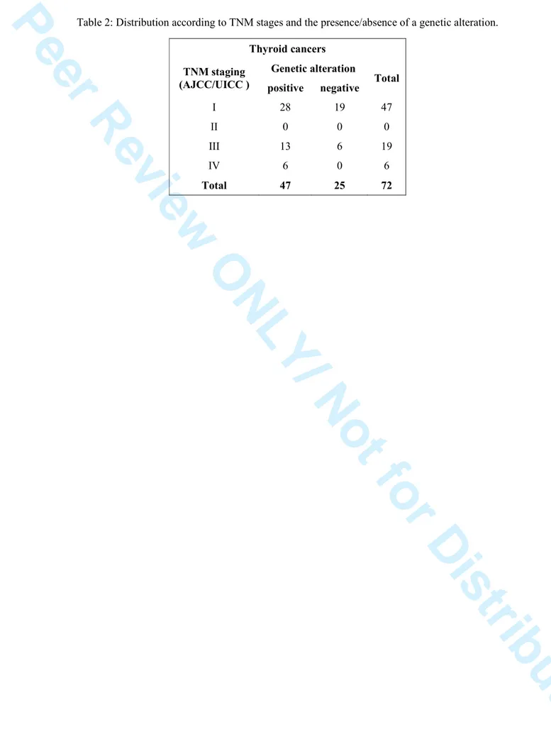
histology, more than half carried one or more somatic genetic alteration and displayed stage I 219
disease (Table 2). 220
In particular, 40 patients who displayed a somatic BRAF V600E mutation (including 6 who also 221
displayed a RET/PTC rearrangement) underwent TT and had a PTC on final histology. 222
Among the 31 patients who displayed an isolated somatic RAS mutation, 10 were submitted to LT 223
and 1 to TT. Histology revealed the presence of a cancer in 2 cases, including 1 ATC and 1 FTC 224
(the latter initially submitted to LT and then to completion thyroidectomy). The remaining 9 225
patients that were operated on showed a follicular adenoma (FA) in 6 cases and hyperplastic 226
nodules (HN) in 3 cases. Moreover, one patient with a malignant cytology, displaying a somatic 227
RAS mutation, was not operated on due to several co-morbidities. The remaining 19 patients
228
refused surgery, mostly because of the finding of a benign cytology. 229
The presence of a RET/PTC rearrangement was found in 69 FNAB, 6 of which also harbored a 230
BRAFV600E mutation and were therefore submitted to TT; one patient carried also a RAS
231
mutation and was submitted to LT with final histology of a FA; one was to have both RET/PTC 232
Page 10 of 24 Thyroid
For Peer Review ONLY/ Not for Distribution
rearrangements and was submitted to TT with a final histology of PTC. Among the 62 patients233
displaying an isolated RET/PTC rearrangement, 5 underwent TT (in the presence of a BSTRC 234
class V in 2 patients and class VI in 3 patients) and 19 underwent LT. Histology revealed the 235
presence of a cancer in 5 cases (all PTC), while 11 lesions were FA and 8 HN. The remaining 38 236
patients refused surgery, mostly because of the finding of a benign cytology. No correlation was 237
found between the presence of a malignant lesion and the amount of RET/PTC rearranged mRNA, 238
preventing the identification of a threshold value that discriminates benign from malignant lesions. 239
240
Indeterminate lesions 241
We then evaluated cytology, molecular testing and pathology findings in the group of 242
indeterminate nodules, which were included in the whole group described above. 243
We found that 37 (26.4%) of the 140 cytologically indeterminate lesions (corresponding to 14.8 % 244
of all FNAB), including 19 class III, 7 class IV and 11 class V lesions, displayed at least one 245
genetic alteration. Among these patients, 2 refused LT (class III cytology) and 35 underwent TT. 246
Final histology showed 24 thyroid cancers (23 PTC and 1 FTC), 8 FA and 3 HN. Among the 23 247
identified PTCs, 21 carried a somatic BRAF V600E mutation. 248
Among the 103 patients with a cytologically indeterminate lesion not displaying a genetic 249
alteration, all the 11 patients with a class V lesion underwent TT, with a final histology of 10 PTC 250
and 1 HN. Ten out of 30 patients with class IV lesions accepted to undergo LT, with a final 251
histology of 3 PTC (then submitted to completion thyroidectomy) and 7 FA. All 62 patients with a 252
class III lesion underwent a second FNAB that confirmed an indeterminate lesion in 33 cases; 6 of 253
these patients accepted to undergo LT, and the final histology showed 1 FTC, 4 FA and 1 HN. 254
Cytology showed a benign lesion in the other 29 patients who were then re-classified as BSRTC 255
class II and subsequently followed with US. The management of these patients was chosen 256
according to the ATA guidelines (8), in order to avoid unnecessary surgery in keeping with the 257
For Peer Review ONLY/ Not for Distribution
low cancer risk of BSRTC class III nodules (in contrast with the higher cancer risk of BSRTC258
class IV and V nodules). 259
Taken together, in our series malignancy rates in each BSRTC class overlap those described by 260
Cibas et al. (14). The cancer risk in thyroid nodules with indeterminate cytology according to 261
BSRTC classification and genetic alterations is shown in Table 3. 262
263
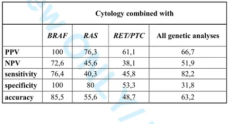
Diagnostic value of cytology and molecular analyses 264
The diagnostic value of cytology and of the studied mutational analyses is reported in Table 4a, 265
which also reports the results obtained by performing the three available genetic analyses in 266
combination. Our data show that cytology displays optimal PPV and specificity, while sensitivity 267
for thyroid cancer is low. When performed alone, BRAFV600E analysis shows, as compared to 268
cytology, a significantly higher diagnostic sensitivity (p<0.05), which increases by 20.8% 269
(p<0.01) when the two evaluations are performed together (Table 4b). On the other hand, the 270
presence of RAS mutations and RET/PTC rearrangements shows a very low sensitivity for thyroid 271
cancer when evaluated alone (Table 4a) and does not significantly increase the diagnostic 272
sensitivity of cytology (Table 4b). In addition, the increased sensitivity recorded when all three 273
genetic analyses are performed in combination is not significantly higher as compared to the 274
sensitivity obtained by performing BRAFV600E analysis alone, even when combined with 275
cytology. These data indicate that, in our setting, BRAFV600E analysis suffices to increase the 276
diagnostic sensitivity of cytology for thyroid cancer. 277
We then evaluated the diagnostic sensitivity of the genetic analysis panel in the subset of the 278
indeterminate lesions, in order to understand whether and which mutation testing might be helpful 279
in this group. We found that the diagnostic sensitivity for thyroid cancer of the three genetic 280
analyses in the indeterminate group, performed alone or in combination, overlaps that identified in 281
the whole group. We then analyzed each BSRTC class included in the indeterminate group (Table 282
4c) and found that the diagnostic sensitivity for thyroid cancer reaches 90% in class III when 283
Page 12 of 24 Thyroid
For Peer Review ONLY/ Not for Distribution
BRAFV600E analysis is performed. This value does not change when RAS mutations and284
RET/PTC rearrangements are simultaneously included. In class IV and V samples, when all three
285
genetic abnormalities are analysed in combination, the diagnostic sensitivity for cancer is greater 286
as compared to BRAFV600E alone, but the difference is not statistically significant. In addition, 287
the analysis of RAS mutations and RET/PTC rearrangements does not seem to be important to 288
further increase the high NPV of BRAFV600E analysis in class III and IV samples. 289
290
DISCUSSION 291
This prospective study confirms the diagnostic utility of assessing the presence of a BRAFV600E 292
mutation (16). On the other hand, the investigation of two additional genetic abnormalities (RAS 293
mutations and RET/PTC rearrangements) did not significantly increase the diagnostic sensitivity 294
of cytology towards thyroid cancer in this cohort, even in the category with indeterminate lesions. 295
Despite the fact that the techniques employed in our study are very similar to those employed by 296
others (5, 15, 18), the results do not overlap. It should be noted that the method employed here to 297
assess RET/PTC rearrangements displayed a 10-fold higher sensitivity as compared to that 298
employed by Nikiforov et al. (15, 18), but provided low sensitivity and specificity in detecting 299
malignant lesions. Therefore, the identification of RET/PTC rearrangements by a very sensitive 300
method may not be useful to increase FNAB diagnostic sensitivity for thyroid cancer. These data 301
suggest that the contribution of this genetic marker to pre-surgical diagnosis of thyroid nodules 302
may not be so relevant, since we found a very high incidence of RET/PTC rearrangements also in 303
benign lesions. 304
US characteristics provide the basis to perform FNAB (8) and often accurately predict the 305
presence of a BRAFV600E mutation (20). In our hands, the presence of a BRAFV600E mutation 306
was significantly associated with hypoechogenicity, microcalcifications and a diameter <1 cm, 307
strengthening the evidence that nodules displaying these US characteristics very likely reflect the 308
presence of a cancer. Our study highlights, for the first time, that RAS mutations and RET/PTC 309
For Peer Review ONLY/ Not for Distribution
rearrangements correlate with specific US findings (i.e. isoechogenicity and diameter >1 cm).310
However, these genetic abnormalities do not indicate the presence of a cancer with high accuracy 311
in our population, and therefore the related US characteristics cannot be taken into account as 312
predictive of cancer. 313
The distribution of our samples among BSRTC classes is in line with literature data, indicating 314
that the investigated nodules had been selected according to the indications of the ATA guidelines 315
(8). In particular, more than 80% of FNAB cytologies turned out to be a benign lesion and ~12% 316
of the samples displayed an indeterminate cytology. The latter result is very similar to the 317
percentage of indeterminate lesions that were retrieved in our previous study (17) which included 318
an unselected nodule population, indicating that the application of strict selection criteria for 319
FNAB does not influence the number of indeterminate lesions. While the percentage of malignant 320
lesions identified by cytology in our series (2.9%) is comparable to the literature data, the 321
incidence of ND reports is quite high (3.5%). This may be due to the fact that the retrieved FNAB 322
material was used for several diagnostic procedures, which may have reduced the sample quantity 323
dedicated to cytology. 324
The present series shows that 14.2% of the investigated nodules harbored at least one mutation, a 325
higher incidence than the previously reported ~9% (18), probably due to the different inclusion 326
criteria. In addition, 6% of mutated FNAB samples displayed more than one genetic alteration, 327
confirming that BRAF and RAS mutations, as well as RET/PTC rearrangements, are not mutually 328
exclusive, as previously indicated (21). Our data also show that the applied FNAB criteria allowed 329
diagnosing thyroid cancers at an early stage of disease, since 65.3% of the diagnosed cancers were 330
Stage I. In addition, nearly 50% of Stage I cancers had a negative cytology but displayed at least 331
one genetic alteration, most commonly a BRAFV600E mutation, which allowed to establish a 332
correct diagnosis. These data indicate that BRAFV600E mutation analysis helps in identifying 333
PTC at an earlier stage, possibly resulting in a more conservative treatment with potential 334
consequences on patient health and healthcare resources. Moreover, 76% of Stage III and IV 335
Page 14 of 24 Thyroid
For Peer Review ONLY/ Not for Distribution
cancers displayed a genetic alteration, in line with the hypothesis that the latter may characterize a336
more aggressive behavior (22, 23), as previously indicated (24). Last, but not least, the applied 337
protocol allowed to correctly diagnose 31 out of 46 false negative lesions on cytology as cancers, 338
corresponding to 43% of the diagnosed malignant lesions. Among these 31 patients, 21 harbored a 339
BRAFV600E mutation and an indeterminate cytology, and were therefore submitted to TT rather
340
than to a diagnostic LT. Moreover, 7 patients were submitted to TT only on the basis of positivity 341
for a BRAFV600E mutation and turned out to have a PTC (6 Stage I and 1 Stage III). The latter 342
finding strengthens the evidence that BRAFV600E mutation analysis facilitates early diagnosis. On 343
the other hand, in our settings RAS mutations have a poor diagnostic value, in keeping with their 344
rarity, and are predominantly associated with follicular lesions, mainly represented by FA that 345
may, in part, be considered as precursors of malignant lesions (25). In keeping with the latter 346
hypothesis, RAS mutated cancers were characterized by an aggressive histology and a high disease 347
stage. In our patients, each RET/PTC rearrangement was nearly as frequent as BRAFV600E 348
mutations, but had a poor diagnostic value since the rearranged lesions were mostly found in 349
benign nodules (64.5% of the cases), contrary to what observed by Cantara et al. (5) and Nikiforov 350
et al. (18), but in line with Marotta et al. (26), even if a prognostic significance cannot be ruled out 351
(27). These differences may be due to different genetic backgrounds and to geographic factors, but 352
may also be due to the applied selection criteria. Among the samples harboring RET/PTC 353
rearrangements, the 11 PTC cases had a BRAFV600E mutation and/or a suspicious or malignant 354
cytology, and were therefore submitted to TT independently of the presence of a RET/PTC 355
rearrangement. 356
A previous report (18) showed an increased diagnostic sensitivity for thyroid cancer in a large 357
group of indeterminate nodules submitted to multiple genetic analyses (including BRAFV600E 358
and RAS mutations as well as RET/PTC1, RET/PTC3 and PAX8/PPARγ rearrangements). The 359
study showed a high NPV for this panel of molecular markers, indicating that the absence of a 360
genetic mutation very likely excludes the presence of a malignant lesion. On the contrary, we did 361
For Peer Review ONLY/ Not for Distribution
not obtain high NPV values in the indeterminate group when performing the three analyses362
together (BRAFV600E and RAS mutations, as well as RET/PTC1 and RET/PTC3 rearrangements), 363
but we found a high NPV for BRAFV600E mutation analysis alone, which is even higher in class 364
III nodules. The latter finding, together with the low cancer risk, suggests that in the absence of a 365
BRAFV600E mutation, diagnostic LT may not be necessary in class III nodules. In class IV
366
nodules without mutations, we found a slightly higher cancer risk, which importantly increased 367
when a RAS mutation was present. These data, together with a suboptimal NPV of BRAFV600E 368
analysis in class IV lesions, do not support a conservative management in these settings (i.e. 369
avoiding a LT). On the other hand, cancer risk is high in class V nodules, indicating that an 370
aggressive surgical management (i.e. TT) is justified in these patients, independently of the 371
presence of a mutation, like in class VI lesions. Taken together, these data demonstrate that, 372
among the investigated molecular markers, only BRAFV600E mutation may modify patient 373
management and has an impact on the surgical approach. Therefore, our data concerning 374
indeterminate lesions are only partially in keeping with previous findings (18), probably due to the 375
different inclusion criteria, that may play an important role in molecular studies. 376
In conclusion, our results confirm that BRAFV600E analysis performed in all BSRTC classes 377
increases the diagnostic sensitivity of cytology for thyroid cancer, which is not further enhanced 378
by investigating the presence of RAS mutations or RET/PTC rearrangements, even among 379
indeterminate nodules. In addition, our data demonstrate that BRAFV600E analysis, when 380
negative, may be useful to identify class III nodules at very low risk of being cancerous, 381
suggesting that these cases may be treated more conservatively and do not need to be submitted to 382
a LT. Moreover, we conclude that BRAFV600E analysis is useful to diagnose thyroid cancer at an 383
early stage, possibly reducing the clinical impact of a delayed diagnosis, which also implicates 384
higher costs for the patients and for the healthcare system. 385
386
387
Page 16 of 24 Thyroid
For Peer Review ONLY/ Not for Distribution
ACKNOWLEDGEMENTS388
This work was supported by grants from the Italian Ministry of Education, Research and University 389
(FIRB RBAP11884M, FIRB RBAP1153LS, 2010TYCL9B_002), Fondazione Cassa di Risparmio 390
di Ferrara, in collaboration with Laboratorio in rete del Tecnopolo “Tecnologie delle terapie 391
avanzate” (LTTA) of the University of Ferrara 392
393
DISCLOSURES 394
EdU received consulting fees from Novartis and Pfizer. MCZ received consulting fees from 395
Novartis and Genzyme. The other Authors have nothing to disclose and have no conflict of 396 interest. 397 398 CORRESPONDING AUTHOR 399
Maria Chiara Zatelli, MD PhD 400
Section of Endocrinology and Internal Medicine, Dept of Medical Sciences, University of Ferrara 401
Via Savonarola 9, 44121 Ferrara, Italy. Phone: +39 0253 239618; Fax: +39 0532 236514; e-mail: 402 [email protected] 403 404 REFERENCES 405
1. Ferraz C, Eszlinger M, Paschke R 2011 Current state and future perspective of molecular 406
diagnosis of fine-needle aspiration biopsy of thyroid nodules. J Clin Endocrinol Metab 96: 407
2016-2026. 408
2. Keutgen XM, Filicori F, Fahey TJ 3rd 2013 Molecular diagnosis for indeterminate thyroid 409
nodules on fine needle aspiration: advances and limitations. Expert Rev Mol Diag 13:613-623. 410
3. Freitas BC, Cerutti JMc 2010 Genetic markers differentiating follicular thyroid carcinoma from 411
benign lesions. Mol Cell Endocrinol 321:77-85 412
For Peer Review ONLY/ Not for Distribution
4. Xing M, Haugen BR, Schlumberger M 2013 Progress in molecular-based management of413
differentiated thyroid cancer. Lancet 381:1058-1069. 414
5. Cantara S, Capezzone M, Marchisotta S, Capuano S, Busonero G, Toti P, Di Santo A, Caruso 415
G, Carli AF, Brilli L, Montanaro A, Pacini F 2010 Impact of proto-oncogene mutation 416
detection in cytological specimens from thyroid nodules improves the diagnostic accuracy of 417
cytology. J Clin Endocrinol Metab 95:1365-1369. 418
6. Eszlinger M, Paschke R 2010 Molecular fine-needle aspiration biopsy diagnosis of thyroid 419
nodules by tumor specific mutations and gene expression patterns. Mol Cell Endocrinol 420
322:29-37. 421
7. Mehta V, Nikiforov YE, Ferris RL 2013 Use of molecular biomarkers in FNA specimens to 422
personalize treatment for thyroid surgery. Head Neck. 35:1499-1506. 423
8. Cooper DS, Doherty GM, Haugen BR, Kloos RT, Lee SL, Mandel SJ, Mazzaferri EL, McIver 424
B, Pacini F, Schlumberger M, Sherman SI, Steward DL, Tuttle RM 2009 The American 425
Thyroid Association (ATA) Guidelines Taskforce on Thyroid Nodules and Differentiated 426
Thyroid Cancer. Revised American Thyroid Association management guidelines for patients 427
with thyroid nodules and differentiated thyroid cancer. Thyroid 19:1167-1214. 428
9. Durante C, Costante G, Filetti S 2013 Differentiated thyroid carcinoma: defining new 429
paradigms for postoperative management. Endocr Relat Cancer 20:R141-R154 430
10. Tuttle RM, Sabra MM 2013 Selective use of RAI for ablation and adjuvant therapy after total 431
thyroidectomy for differentiated thyroid cancer: a practical approach to clinical decision 432
making. Oral Oncol 49:676-683. 433
11. Durante C, Attard M, Torlontano M, Ronga G, Monzani F, Costante G, Ferdeghini M, Tumino 434
S, Meringolo D, Bruno R, De Toma G, Crocetti U, Montesano T, Dardano A, Lamartina L, 435
Maniglia A, Giacomelli L, Filetti S; Papillary Thyroid Cancer Study Group 2010 Identification 436
and optimal postsurgical follow-up of patients with very low-risk papillary thyroid 437
microcarcinomas. J Clin Endocrinol Metab 95:4882-4888. 438
Page 18 of 24 Thyroid
For Peer Review ONLY/ Not for Distribution
12. Roti E, degli Uberti EC, Bondanelli M, Braverman LE 2008 Thyroid papillary439
microcarcinoma: a descriptive and meta-analysis study. Eur J Endocrinol 159:659-673. 440
13. Batawil N, Alkordy T 2014 Ultrasonographic features associated with malignancy in 441
cytologically indeterminate thyroid nodules. Eur J Surg Oncol 40:182-186. 442
14. Cibas ES, Ali SZ 2009 The Bethesda System for Reporting Thyroid Cytopathology. Thyroid 443
19:1159-1165. 444
15. Nikiforov YE, Steward DL, Robinson-Smith TM, Haugen BR,Klopper JP, Zhu Z, Fagin JA, 445
Falciglia M, Weber K, Nikiforova MN 2009 Molecular testing for mutations in improving the 446
fine-needle aspiration diagnosis of thyroid nodules. J Clin Endocrinol Metab 94:2092–2098. 447
16. Zatelli MC, Trasforini G, Leoni S, Frigato G, Buratto M, Tagliati F, Rossi R, Cavazzini L, Roti 448
E, degli Uberti EC 2009 BRAF V600E mutation analysis increases diagnostic accuracy for 449
papillary thyroid carcinoma in fine-needle aspiration biopsies. Eur J Endocrinol 161:467-473. 450
17. Rossi M, Buratto M, Bruni S, Filieri C, Tagliati F, Trasforini G, Rossi R, Beccati MD, degli 451
Uberti EC, Zatelli MC 2012 Role of ultrasonographic/clinicalprofile, cytology, and BRAF 452
V600E mutation evaluation in thyroid nodule screening for malignancy: a prospective study. J 453
Clin Endocrinol Metab 97:2354-2361. 454
18. Nikiforov YE, Ohori NP, Hodak SP, Carty SE, LeBeau SO, Ferris RL, Yip L, Seethala RR, 455
Tublin ME, Stang MT, Coyne C, Johnson JT, Stewart AF, Nikiforova MN 2011 Impact of 456
mutational testing on the diagnosis and management of patients with cytologically 457
indeterminate thyroid nodules: a prospective analysis of 1056 FNA samples. J Clin Endocrinol 458
Metab 96:3390-3397. 459
1 9 . Guerra A, Carrano M, Angrisani E, Puzziello A, Izzo G, Di Crescenzo V, Vatrella A, Vitale M. 460
Detection of RAS mutation by pyrosequencing in thyroid cytology samples. Int J Surg. 2014 461
May 24. pii: S1743-9191(14)00144-7 462
For Peer Review ONLY/ Not for Distribution
20. Kabaker AS, Tublin ME, Nikiforov YE, Armstrong MJ, Hodak SP, Stang MT, McCoy KL,463
Carty SE, Yip L 2012 Suspicious ultrasound characteristics predict BRAFV600E-positive 464
papillary thyroid carcinoma. Thyroid 22:585-589. 465
2 1 . Guerra A, Zeppa P, Bifulco M, Vitale M 2014 Concomitant BRAF(V600E) mutation and 466
RET/PTC rearrangement is a frequent occurrence in papillary thyroid carcinoma. Thyroid 467
24:254-259. 468
22. Fugazzola L, Mannavola D, Cirello V, Vannucchi G, Muzza M, Vicentini L, Beck-Peccoz P 469
2004 BRAF mutations in an Italian cohort of thyroid cancers. Clin Endocrinol (Oxf) 61:239-470
243. 471
23. Xing M, Alzahrani AS, Carson KA, Viola D, Elisei R, Bendlova B, Yip L, Mian C, Vianello F, 472
Tuttle M, Robenshtok E, Fagin JA, Puxeddu E, Fugazzola L, Czarniecka A, Jarzab B, O'Neill 473
CJ, Sywak MS, Lam K, Riesco-Eizaguirre G, Santisteban P, Nakayama H, Tufano RP, Pai SI, 474
Zeiger MA, Westra WH, Clark DP, Clifton-Bligh R, Sidransky D, Ladenson PW, Sykorova V 475
2013 Association between BRAF V600E mutation and mortality in patients with papillary 476
thyroid cancer. JAMA 309:1493-1501. 477
2 4 . Xing M, Clark D, Guan H, Ji M, Dackiw A, Carson KA, Kim M, Tufaro A, Ladenson P, Zeiger 478
M, Tufano R. BRAF mutation testing of thyroid fine-needle aspiration biopsy specimens for 479
preoperative risk stratification in papillary thyroid cancer. J Clin Oncol. 2009 Jun 480
20;27(18):2977-82 481
25. Schulten HJ, Salama S, Al-Ahmadi A, Al-Mansouri Z, Mirza Z, Al-Ghamdi K,Al-Hamour OA, 482
Huwait E, Gari M, Al-Qahtani MH, Al-Maghrabi J 2013 Comprehensive survey of HRAS, 483
KRAS, and NRAS mutations in proliferative thyroid lesions from an ethnically diverse 484
population. Anticancer Res 33:4779-4784. 485
26. Marotta V, Guerra A, Sapio MR, Vitale M 2011 RET/PTC rearrangement in benign and 486
malignant thyroid diseases: a clinical standpoint. Eur J Endocrinol 165:499-507. 487
Page 20 of 24 Thyroid
For Peer Review ONLY/ Not for Distribution
27. Sapio MR, Guerra A, Marotta V, Campanile E, Formisano R, Deandrea M, Motta M, Limone488
PP, Fenzi G, Rossi G, Vitale M. High growth rate of benign thyroid nodules bearing RET/PTC 489
rearrangements. J Clin Endocrinol Metab. 2011 Jun;96(6):E916-9 490
For Peer Review ONLY/ Not for Distribution
Table 1: Genetic alterations and their frequencies in each BSTRC class nodulesGenetic alteration (n) BSTRC classes
I II III IV V VI Total
BRAF V600E 4 3 7 4 6 10 34
BRAF and RET/PTC 1 0 0 2 0 1 1 4
BRAF and RET/PTC 3 0 0 0 0 1 1 2
RAS 1 21 4 2 1 2 31
RAS and RET/PTC 3 0 1 0 0 0 0 1
RET/PTC-1 2 25 3 0 1 1 32
RET/PTC-3 4 19 3 1 1 1 29
RET/PTC-1 and -3 0 0 0 0 0 1 1
Total samples with genetic alteration(s) 11 69 19 7 11 17 134
None 22 699 33 30 11 11 806
All samples 33 768 52 37 22 28 940
Genetic alteration frequence (%)
BRAF V600E 12.1 0. 4 17.3 10.8 36.3 42.8 4.2
RAS 3 2.8 7.7 5.4 4.5 7.1 3.4
RET/PTC-1 and RET/PTC-3 18.2 5.8 15.4 2.7 18.2 17.8 7.3
Total (s) 33.3 9 36.5 18.9 50.0 60.7 14.2
Page 22 of 24 Thyroid
For Peer Review ONLY/ Not for Distribution
Table 2: Distribution according to TNM stages and the presence/absence of a genetic alteration.
Thyroid cancers TNM staging (AJCC/UICC ) Genetic alteration Total positive negative I 28 19 47 II 0 0 0 III 13 6 19 IV 6 0 6 Total 47 25 72 Page 23 of 24 Thyroid
For Peer Review ONLY/ Not for Distribution
Table 3: Cancer risk in thyroid nodules with indeterminate cytology according to BSTRCclassification and genetic alteration
*The patients with a PTC displaying RET/PTC rearrangements also had a BRAFV600E mutation or a class V or a class VI BSTRC cytology.
% class III class IV class V Indeterminate cytology
Cytology alone 19.2 21,6 90,9 27,1 Any mutation 47,3 71,4 90,9 63,1 BRAF 100 100 100 100 RAS 0 50 0 14,2 RET/PTC-1 RET/PTC-3 40 0 - 0 100* 100* 57,1 33,3 No mutations 3 10 90,9 13,5 Page 24 of 24 Thyroid
For Peer Review ONLY/ Not for Distribution
Table 4a: Diagnostic value of cytology and of genetic analyses in all 940 samples
Cytology BRAF RAS RET/PTC All genetic analyses
PPV 100 100 25 34,4 63,3
NPV 50 58,4 34,3 28,2 34,2
sensitivity 37,5 55,6 4,2 15,3 66,7
specificity 100 100, 0 80 53,3 31
accuracy 61,5 72,6 33,3 29,9 53,8
Table 4b: Diagnostic value of cytology combined with genetic analyses in all 940 samples
Cytology combined with
BRAF RAS RET/PTC All genetic analyses
PPV 100 76,3 61,1 66,7
NPV 72,6 45,6 38,1 51,9
sensitivity 76,4 40,3 45,8 82,2
specificity 100 80 53,3 31,8
accuracy 85,5 55,6 48,7 63,2
Table 4c: Diagnostic value of genetic analyses in the 140 indeterminate lesions according to BSRTC classification
Class III Class IV Class V
BRAF all genetic analyses BRAF all genetic analyses BRAF all genetic analyses
PPV 100 52,9 100 71,4 100 90,9 NPV 92,9 83,3 69,2 70 14,3 9,1 Sensitività 90 90 50 62,5 40 50 Specificità 100 38,5 100 77,8 100 50 Accuracy 95,7 60,9 76,5 70,6 45,5 50 Page 25 of 24 Thyroid