UNIVERSITÀ DEGLI STUDI DI SASSARI
CORSO DI DOTTORATO DI RICERCA IN SCIENZE BIOMEDICHE
Coordinatore del Corso: Prof. Andrea Fausto PianaCURRICULUM IN ONCOLOGIA MOLECOLARE
Responsabile di Curriculum: Prof.ssa Rosa Maria Pascale
XXIX CICLO
Identification and characterization of cytogenetic
profile in olfactory neuroblastoma by array
comparative genomic hybridization
Coordinatore:
Prof. Andrea Fausto Piana
Tutor:
Prof. Francesco Meloni Prof.ssa Rosa Maria Pascale
Tesi di dottorato di: Dott. Luca Volpi
Luca Volpi - Identification and characterization of cytogenetic profile in olfactory neuroblastoma by array comparative genomic hybridization - Tesi di dottorato in Scienze biomediche – Curriculum di Oncologia molecolare XXIX ciclo - Università degli studi di Sassari
2 CONTENTS 1. INTRODUCTION ... 6 2. OLFACTORY NEUROBLASTOMA……… 7 2.1 Clinical features……… 7 2.2 Imaging Studies ... 9 2.3 Staging system... 10 2.4 Histology ... 11
2.5 Treatment and results ... 14
2.6 Surgery……….………15
2.7 Radiotherapy ……….17
2.8 Chemotherapy ... 18
3. ARRAY COMPARATIVE GENOMIC HYBRIDIZATION ... 19
3.1 a-CGH definition and technique... 19
3.2 Principles and research applications of Array CGH……….21
3.3 a-CGH and olfactory neuroblastoma... 23
4. MATHERIALS AND METHODS... 28
4.1 Patients... 28 4.2 Hystological method ... 29 4.3 Technique of a-CGH ... 31 5. RESULTS ... 47 5.1 Clinical results ... 47 5.2 Hystopatological results ... 48 5.3 a-CGH results ... 49
Luca Volpi - Identification and characterization of cytogenetic profile in olfactory neuroblastoma by array comparative genomic hybridization - Tesi di dottorato in Scienze biomediche – Curriculum di Oncologia molecolare XXIX ciclo - Università degli studi di Sassari
3 6. DISCUSSION ... 65 7. REFERENCES ... 69
Luca Volpi - Identification and characterization of cytogenetic profile in olfactory neuroblastoma by array comparative genomic hybridization - Tesi di dottorato in Scienze biomediche – Curriculum di Oncologia molecolare XXIX ciclo - Università degli studi di Sassari
4 FIGURES INDEX
Figure 1: Endoscopic view of olfactory neuroblastoma ... 8
Figure 2: MR- aspects of ONB ... 9
Figure 3:Schematic representation of CGH microarray technology ... 20
Figure 4: copy number aberrations in ONBs in Guled's paper ... 27
Figure 5: histological features of ONB ... 30
Figure 6: a-CGH workflow diagram for sample preparation and microarray processing…………..32
Figure 7: Slide in slide holder for SureScan microarray scanner………...…43
Figure 8: Agilent scanner ... 46
Figure 9: histologic features of ONB ... 33
Figure 10: a-CGH workflow ... 35
Figure 11: Slide in slide holder for SureScan microarray scanner... 43
Figure 12: agilent scanner ... 46
Figure 13: percentage of patients with DNA gain or loss for each chromosome ... 52
Figure 14: chromosomal gains ... 53
Figure 15: chromosomal loss ... 54
Figure 16: percentage of patients with DNA gain or loss for each chromosome (FRESH) ... 55
Figure 17: percentage of patients with DNA gain or loss for each chromosome (FFPE). ... 58
Figure 18: chromosome 1 FFPE vs FRESH ... 63
Figure 19: chromosome 1 FFPE vs FRESH: scattered plot profile………63
Figure 20: chromosome 20 FFPE vs FRESH………...………..64
Luca Volpi - Identification and characterization of cytogenetic profile in olfactory neuroblastoma by array comparative genomic hybridization - Tesi di dottorato in Scienze biomediche – Curriculum di Oncologia molecolare XXIX ciclo - Università degli studi di Sassari
5 TABLES INDEX
Table 1: Kadish staging system... 10
Table 2: Dulguerov and Calcaterra staging system... 10
Table 3: Hyams’ grading system... 12
Table 4: Immunohistochemical features of olfactory neuroblastoma ... 13
Table 5: Features for differential diagnosis…...……….13
Table 6:Olfactory neuroblastoma: comparison of survival results (1992 – 2008) according to Devaiah’s meta-analysis………...………..16
Table 7: Median Follow-up Times (months) for olfactory neuroblastoma………17
Table 8: Most common alterations in ONB found in Bockmuhl’s paper………...………24
Table 9: cytogenetic features showed in Holland’s paper……….……….25
Table 10: Clinical data on olfactory neuroblastomas (personal case-series)……….28
Table 11: amplificated regions for patient and for chromosomes showed by the a-CGH ... 50
Table 12: loss regions for patients and for chromosomes showed by the a-CGH ... 51
Table 13: amplificated regions in fresh tissue samples showed by the a-CGH ... 56
Table 14: loss regions in fresh tissue samples showed by the a-CGH ... 57
Table 15: amplificated regions in FFPE tissue samples showed by the a-CGH ... 59
Luca Volpi - Identification and characterization of cytogenetic profile in olfactory neuroblastoma by array comparative genomic hybridization - Tesi di dottorato in Scienze biomediche – Curriculum di Oncologia molecolare XXIX ciclo - Università degli studi di Sassari
6 1. INTRODUCTION
Olfactory neuroblastoma (ONB) is a rare malignant neoplasm arising from the olfactory neuroepithelium1,2. Its incidence is 0.4 patients per million per year, and it represents about 3% of
malignant tumours of the nasal cavity and paranasal sinuses3,4. Basal cells of the olfactory
neuroepithelium are presumably the progenitors of the ONB. The neoplasm usually arises in the olfactory vault of the nasal fossa and it is intimately related to the cribriform plate. Often, diagnosis is made late because early stage ONBs are usually asymptomatic. The incidence of cervical metastasis varies from 5 to 12% at the time of diagnosis, whereas distant metastases occur in 12 to 25% of patients. To date, no universally accepted classification systems have been adopted; the Hyams’ grading system is usually used to histologically grade the tumors and the Kadish system is used to stage them5,6.
Cytogenetic data on ONB are scarce; in one study by Guled et al. (2008) the authors analysed tumor samples from 13 patients by array comparative genomic hybridization (a-CGH). The authors observed a number of recurrent chromosome region gains and losses, which might also be
related to the tumor stage. In particular, gains of 20q and 13q were suggested to be important in cancer progression7. Here we present data on 13 DNA samples from12 patients with ONB analyzed
by a-CGH. The aim of our study was to better define the cytogenetic profile of ONB by a-CGH, and identify possible correlations with clinical and pathological features, evaluating the advantages and limits of the technique.
Luca Volpi - Identification and characterization of cytogenetic profile in olfactory neuroblastoma by array comparative genomic hybridization - Tesi di dottorato in Scienze biomediche – Curriculum di Oncologia molecolare XXIX ciclo - Università degli studi di Sassari
7 with a careful follow-up. At the Department of Pathology tumor samples of the patients were reviewed with the use of immunohistochemical techniques to confirm the diagnosis of ONB. At the Section of Biology and Medical Genetics, Department of Clinical and Experimental Medicine of Varese, the DNA analysis of tumors were carried out using a-CGH technique.
2. OLFACTORY NEUROBLASTOMA
Olfactory neuroblastoma (ONB) is an uncomm malignant neuroectodermal nasal tumour. ONBs are thought to arise from the specialized sensory neuroepithelial (neuroectodermal) olfactory cells that are normally found in the upper part of the nasal cavity, including the superior nasal concha, the upper part of septum, the roof of nose, and the cribriform plate of ethmoid8.
2.1 Clinical features
Those tumors usually present with unilateral nasal obstruction and epistaxis. Impairment or loss of the sense of smell is not common as a clinical symptom as might be expected, in part because olfaction is preserved on the contra lateral side in some tumors and in part because some patients fail to notice its gradual loss. Extension into the intracranial cavity rarely causes neurological symptoms as invasion of the frontal lobe only produces symptoms after massive involvement. Large tumors can invade the orbit and produce ocular symptoms.
Luca Volpi - Identification and characterization of cytogenetic profile in olfactory neuroblastoma by array comparative genomic hybridization - Tesi di dottorato in Scienze biomediche – Curriculum di Oncologia molecolare XXIX ciclo - Università degli studi di Sassari
8 Figure 1: Endoscopic view of olfactory neuroblastoma
Luca Volpi - Identification and characterization of cytogenetic profile in olfactory neuroblastoma by array comparative genomic hybridization - Tesi di dottorato in Scienze biomediche – Curriculum di Oncologia molecolare XXIX ciclo - Università degli studi di Sassari
9
2.2 Imaging Studies
The imaging features of olfactory neuroblastoma are non-specific. Nevertheless, this neoplasm should be suspected when a mass is detected in the superior nasal cavity, causing either remodeling or destruction of adjacent bony structures, and erosion of the cribriform plate or of the fovea ethmoidalis9.
The stage at initial presentation is highly predictive of survival and accurate staging is essential. Appropriate evaluation includes both computed tomography (CT) and magnetic resonance
imaging (MRI), to help define the likely extent of the disease both at the primary site and in the neck. However, thosee imaging modalities are complimentary and should be performed in all cases. Fine-cut spiral overlapping CT scan in all three dimensions is the radiological study of choice. Olfactory neuroblastoma does not have any specific radiological appearance but its site in the olfactory cleft makes it a likely diagnosis in its early stages. It is seen is a homogenous soft-tissue mass in the nasal vault, with uniform and moderate contrast enhancement and occasionally the adjacent bone can be hyperostotic. CT images are essential for correct staging and should be carefully examined for erosion of the lamina papryacea, cribriform plate, and fovea ethmoidalis. MRI enables a better estimate of tumor spread into surrounding soft tissue and areas in particular it can indicate whether the tumor has involved or traversed the dura and/or the orbit and can differentiate mucus from tumor10.
Luca Volpi - Identification and characterization of cytogenetic profile in olfactory neuroblastoma by array comparative genomic hybridization - Tesi di dottorato in Scienze biomediche – Curriculum di Oncologia molecolare XXIX ciclo - Università degli studi di Sassari
10
a b
c
Figure 2: MR - aspects of ONB
a. Coronal MR T1 with contrast shows a mass with a low- to intermediate signal, epicenter within the right ethmoid and intracranial extension. b On sagittal plane post contrast MR demonstrates that the tumor extends into the anterior cranial fossa c. A post contrast coronal MR of a case without intracranial spread into the anterior cranial fossa
Luca Volpi - Identification and characterization of cytogenetic profile in olfactory neuroblastoma by array comparative genomic hybridization - Tesi di dottorato in Scienze biomediche – Curriculum di Oncologia molecolare XXIX ciclo - Università degli studi di Sassari
11
2.3 Staging system
Different staging systems based on the extension of the lesion11 have been specifically proposed for
olfactory neuroblastoma (Table 1 and 2).
Table 1: Kadish staging system Kadish staging system (1976) Stage Features
A Tumor confined to the nasal cavity
B Tumor confined to the nasal cavity, involving one or more paranasal sinuses
C Tumor extending beyond the nasal cavity and paranasal sinuses. Includes involving of the orbit, skull base, intracranial cavity, cervical lymph nodes and distant metastatic sites
Table 2: Dulguerov and Calcaterra staging system Dulguerov and Calcaterra staging system (1992) Stage Features
T1 Tumor involving the nasal cavity and/or paranasal sinuses, sparing the most superior ethmoidal cells
T2 Tumor involving the nasal cavity and/or paranasal sinuses, including the sphenoid, with extension to and erosion of the cribriform plate
T3 Tumor extending into the orbit or protruding into the anterior cranial fossa T4 Tumor involving the brain
Kadish and co-workers11 were the first to propose a staging classification, using three categories,
Group A, B and C (Table1). A further system was proposed with the advent of advances in imaging (241), based on the TNM system (Table 2). Although a system of classification has been proposed, various attempts have been made to modify the Kadish system11,12. Other authors suggest that by
Luca Volpi - Identification and characterization of cytogenetic profile in olfactory neuroblastoma by array comparative genomic hybridization - Tesi di dottorato in Scienze biomediche – Curriculum di Oncologia molecolare XXIX ciclo - Università degli studi di Sassari
12 using the Kadish staging system and the Hyams grading system independently they can predict patients outcome with more accuracy13. Hyams grading system is based on histology and will be
referred in the next section.
2.4 Histology
Olfactory neuroblastoma is an uncommon sinonasal tumour, and can often been confused with other neoplasms, e.g. sinonasal undifferentiated carcinoma, neuroendocrine carcinoma, melanoma, lymphoma, plasmacytoma, embryonal rhabdomyosarcoma, Ewing’s sarcoma, peripheral primitive neuroectodermal tumours, vascular tumours etc.. It was this frustrating feature that prompted Ogura and Schenek14 to describe olfactory neuroblastoma as the “great impostor”. The diagnosis of
olfactory neuroblastoma can be problematic and consequently, histochemical, immunochemical and ultrastructural investigations are often needed to support the diagnosis15. Westra and colleagues
from Johns Hopkins reviewed 37 cases of diagnosed olfactory neuroblastoma and excluded 8 (21.6%) because they did not meet diagnostic inclusion criteria16. Hirose et al.15 from the Mayo
Clinic Tissue Registry reviewed 30 cases of olfactory neuroblastoma and found that 4 (13.3%) were excluded because they lacked neural or neuroendocrine markers. Exhibiting only epithelial markers, they were considered examples of sinonasal undifferentiated carcinoma.
It is reported that a diagnosis of olfactory neuroblastoma by light microscopy is not difficult when the tumor is well differentiated and consists of homogenous small cells with uniform round to oval nuclei, with rosette or pseudorosette formation, and eosinophilic fibrillary intercellular back ground material. Hyams et al.17 proposed a grading system for olfactory neuroblastoma, Grades I being
well differentiated to IV undifferentiated, based on several tumour histological parameters: preservation of lobular architecture, mitotic index, nuclear polymorphism, fibrillary matrix, the presence of HW (Horner Wright) or FW (Flexner-Wintersteiner) rosettes, and the presence or absence of tumour necrosis.
Luca Volpi - Identification and characterization of cytogenetic profile in olfactory neuroblastoma by array comparative genomic hybridization - Tesi di dottorato in Scienze biomediche – Curriculum di Oncologia molecolare XXIX ciclo - Università degli studi di Sassari
13 Table 3: Hyams’ grading system
Olfactory neuroblastoma grading (based on Hyams’ grading system)
Microscopic features Grade I Grade II Grade III Grade IV Architecture Lobular Lobular ±Lobular ±Lobular
NF matrix Prominent Present May be present Present
Rosettes HR HR FW FW
Mitoses Absent Present Prominent Marked
Necrosis Absent Absent Present Prominent
Glands May be present May be present May be present May be present Calcification Variable Variable Absent Absent
NF neurofibrillary, HR Homer Wright pseudorosettes, FW Flexner-Wintersteiner rosettes
When the tumor is undifferentiated with anaplastic hyperchromatic small cells that show many mitotic figures and scant cytoplasm, differentiating the tumor from other small-cell nasal neoplasms by light microscopy becomes difficult. To summarise, the pathologic distinction of poorly differentiated small cell neoplasms of the nasal cavity is difficult and can only be based on the results of antigen expression using a panel of antibodies by immunohistochemistry and if necessary confirmed by electron microscopy. There is still a lack of consensus relating to these prognostic histological features. Support for the prognostic value of Hyams and co-workers grading17 has been
published18. Histopathological staging according to Hyams has also been advocated more recently
Luca Volpi - Identification and characterization of cytogenetic profile in olfactory neuroblastoma by array comparative genomic hybridization - Tesi di dottorato in Scienze biomediche – Curriculum di Oncologia molecolare XXIX ciclo - Università degli studi di Sassari
14 immunopositivity and a low (<10%) Ki-67 labelled index (a marker for proliferation) was associated with better survival. There are conflicting data on the prognostic role of p53 tumor suppressor gene mutations13 .
Table 4: Immunohistochemical features of olfactory neuroblastoma
positive negative
neuron specific enolase (NSE) synaptophysin
cromogranine A S-100
neurofilament protein (NFP)
Cytokeratin
epithelial membrane antigen (EMA) carcinoembryonic antigen (CEA) CD45
CD99 HMB45 Melan A Desmin
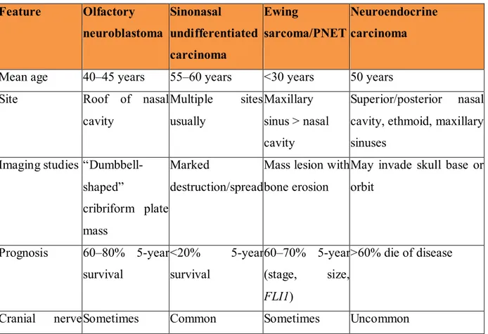
Table 5: Features for differential diagnosis Feature Olfactory neuroblastoma Sinonasal undifferentiated carcinoma Ewing sarcoma/PNET Neuroendocrine carcinoma
Mean age 40–45 years 55–60 years <30 years 50 years Site Roof of nasal
cavity Multiple sites usually Maxillary sinus > nasal cavity Superior/posterior nasal cavity, ethmoid, maxillary sinuses
Imaging studies “Dumbbell-shaped”
cribriform plate mass
Marked
destruction/spread
Mass lesion with bone erosion
May invade skull base or orbit Prognosis 60–80% 5-year survival <20% 5-year survival 60–70% 5-year (stage, size, FLI1) >60% die of disease
Luca Volpi - Identification and characterization of cytogenetic profile in olfactory neuroblastoma by array comparative genomic hybridization - Tesi di dottorato in Scienze biomediche – Curriculum di Oncologia molecolare XXIX ciclo - Università degli studi di Sassari
15 Feature Olfactory neuroblastoma Sinonasal undifferentiated carcinoma Ewing sarcoma/PNET Neuroendocrine carcinoma involvement
Pattern Lobular Sheets and nests Sheets, nests Ribbons, islands Cytology Salt and pepper
chromatin, small nucleoli (grade dependent) Medium cells, inconspicuous nucleoli Medium, round cells, vacuolated cytoplasm, fine chromatin
Salt and pepper, granular chromatin
Anaplasia Occasionally and focally
Common Minimal Moderate
Mitotic figures Variable High Common High Necrosis Occasionally Prominent Frequent Prominent Vascular
invasion
Occasionally Prominent Rare Present
Neurofibrillary stroma
Common Absent Absent Absent
Pseudorosettes Common Absent Present Present Keratin Focal, weak >90% Rare Positive
CK 5/6 Negative Negative n/a n/a
EMA Negative 50% n/a n/a
NSE >90% 50% Positive Positive
S-100 protein + (sustentacular) <15% Rare Positive Synaptophysin >90% (can be
weak)
<15% Positive Positive
In situ EBER Absent Absent Absent Absent Neurosecretory
granules (EM)
Numerous Rare Absent Present
EBER Epstein barr virus encoded RNA (EBV-encoded RNA) Modified From: Thompson 2009
Luca Volpi - Identification and characterization of cytogenetic profile in olfactory neuroblastoma by array comparative genomic hybridization - Tesi di dottorato in Scienze biomediche – Curriculum di Oncologia molecolare XXIX ciclo - Università degli studi di Sassari
16
2.5 Treatment and results
The Primary Site
A combination of surgery and radiotherapy is the most frequently used approach, and the one that achieved the highest cure rates20. Despite the lack of support for single-modality treatment
regimes21, a substantial number of patients are treated by surgery or radiotherapy alone. The
difference in survival between the combined treatments and radiotherapy alone is significant. The 5-year disease-specific survival in the literature is reported between 52–90%1. Surgery alone was
associated with lower survival combined with a combination of radiotherapy and chemotherapy, or triple modality treatment (surgery, radiotherapy and chemotherapy). Although the results were 15-20% better, the differences from the best combination were not statistically significant, probably because of the limited number of patients2. These results were complied from the MEDLINE
database from 1990–2000, without language tags. There were 26 treatment studies that formed the basis for the above tabulations and data extracted from these studies comprised the total number of patients, the staging system used, the patients’ distributions by stage and the histological grade and the treatment used. Outcome data consisted of recurrence free survival at 3 years and 5 years, overall survival at 5 and 10 years, and the results by stage, grade and treatment modality. In five studies, olfactory neuroblastoma were histo-pathologically graded according to Hyams et al., the mean 5 year survival was 56% in patients with grades I or II tumours and 25% in those with tumours of grade III or IV. This difference was significant (odds ration 6.18 {95% CI 1.30 – 29.3}). In 25 studies that used the Kadish classification, the mean 5 year survival for these three groups was Group A 72%, Group B 59% and Group C 47%, respectively. On average, 5% (SD 7) of patients presented with cervical lymph node metastases. In the studies of survival data according to N stage, only 29% of N+ patients were treated successfully, compared with 64% of N0, a significant difference (Odds ratio 5.1 {95% CI 1.6 – 17.0}).
Luca Volpi - Identification and characterization of cytogenetic profile in olfactory neuroblastoma by array comparative genomic hybridization - Tesi di dottorato in Scienze biomediche – Curriculum di Oncologia molecolare XXIX ciclo - Università degli studi di Sassari
17
2.6 Surgery
Most institutions favour surgery as the first treatment modality, followed by radiotherapy22,23,24.
Endocranial extension and a close relation to the ethmoid roof and cribriform plate have conventionally led to a combined transfacial and neurosurgical approach. Craniofacial resection allows for an en bloc resection of the tumour with better assessment of any intracranial extension and protection of the brain and optic nerve. The resection should include the entire cribriform plate and crista galli. It is said that the olfactory bulb and overlying dura should be removed with the specimen although there is no clear evidence to support the assertion that the whole of the bulb should be removed25. Open surgery has long been regarded as the gold standard, with results
available for decades. A craniotomy is probably not justified for T1 tumours where there is clear radiological evidence of a normal cribriform plate and no involvement of the upper ethmoidal cells although this clinical picture is seldom seen. The evolution of surgical techniques has created another surgical option in the form of endoanasal endoscopic surgery. The use of endoscopic surgery for olfactory neuroblastoma followed by the use of the stereotactic radiosurgical gamma knife therapy has recently been used26. One report of 10 cases with a mean follow-up of 38 months
used endoscopic resection alone without any recurrence although only 2 had Kadish stage C 27. In
the last decade, numerous articles with small numbers have been published on the endoscopic resection of olfactory neuroblastoma. Devaiah et al. reviewed the literature with a meta-analysis and showed that an endoscopic approach gave a better survival rate28. The aim of this study was to
compare results of open, endoscopic, endoscopic-assisted, and nonsurgical treatments since the first publication that mentioned an endoscopic removal in the literature. This analysis extracted sufficient data in 361 subjects and the statistically significant results for the full cohort are summarized in Table 6.
Luca Volpi - Identification and characterization of cytogenetic profile in olfactory neuroblastoma by array comparative genomic hybridization - Tesi di dottorato in Scienze biomediche – Curriculum di Oncologia molecolare XXIX ciclo - Università degli studi di Sassari
18
Table 6: Olfactory neuroblastoma: comparison of survival results (1992 – 2008) according to Devaiah’s meta-analysis
Endoscopic surgery produced overall better survival rates than open surgery with no significant difference between follow-up times in the endoscopic and open surgery groups (Table 7).
Table 7: Median Follow-up Times (months) for olfactory neuroblastoma
As the gold standard open procedure considerably predated endoscopic treatment, they also grouped the data according to the publication year. The endoscopic surgery group maintained better survival rates. This data shows evidence for the efficacy of endoscopic surgery in olfactory neuroblastoma. There are more cases of long-term follow-up in the open surgery group than the endoscopic treatment group and most of the open surgery tumors belonged to the Kadish C stage, whereas the endoscopic techniques were used more commonly for Kadish A and B tumors. This reflects how endoscopic surgery has mainly been used for less extensive lesions. This might not only be a
Luca Volpi - Identification and characterization of cytogenetic profile in olfactory neuroblastoma by array comparative genomic hybridization - Tesi di dottorato in Scienze biomediche – Curriculum di Oncologia molecolare XXIX ciclo - Università degli studi di Sassari
19 reflection of the size of the tumor but their pathology as more extensive lesions might be expected to be more invasive and less differentiated although this cannot be ascertained from the data available 28. The most recent publication on endoscopic endonasal resection for all Kadish groups
has just been published by Folbe et al. 29. This is a retrospective, multicenter study with 23 patients
operated endoscopically with postoperative radiotherapy in 16 patients. The mean follow-up was 45.2 months with one recurrence. The authors conclude that endoscopic surgery is replacing craniofacial resection and that oncologic control is not sacrificed when good endoscopic resection techniques are used.
2.6 Radiotherapy
Standard radiotherapeutic techniques include external megavoltage beam and a three-field technique; an anterior port is combined with wedge later fields to provide a homogeneous dose distribution. The doses range from 55 Gy to 65 Gy with the majority receiving above 60 Gy. Currently it is considered that radiotherapy should play a role in the management of olfactory neuroblastoma, particularly in patients who have had incomplete surgical resection or who present with residual disease . In a small retrospective series, a comparison was made between conventional radiotherapy and stereotactically guided conformal radiotherapy (SCRT). It was concluded that SCRT improved target coverage and sparing of organs at risk30.
2.7 Chemotherapy
Olfactory neuroblastoma is regarded as a chemosensitive tumour based on multiple reported responses to treatment31. Neoadjuvant therapy is seldom curative on its own and it may be of no
benefit in some patients. Individuals who respond to preoperative chemotherapy have a greater chance of long term disease-free surviva. It has been proposed that Hyams’ grading is an important
Luca Volpi - Identification and characterization of cytogenetic profile in olfactory neuroblastoma by array comparative genomic hybridization - Tesi di dottorato in Scienze biomediche – Curriculum di Oncologia molecolare XXIX ciclo - Università degli studi di Sassari
20 predictor of response to chemotherapy31, and it has been suggested that cisplatinbased
chemotherapy is helpful in advanced, high-grade olfactory neuroblastoma and should be considered the choice in the systemic treatment of these patients.
Neoadjuvant chemotherapy has been advocated for patients with advanced disease at the University of Virginia over a 20 year period32. In thirty four consecutive patients, two thirds showed a
significant reduction of tumour burden with adjuvant therapy and patients that showed a response to neoadjuvant therapy demonstrated a significantly larger disease- free mortality rate. Preoperative chemotherapy consisted of cyclophosphamide (650 mg/m2) and vincristine (1.5 mg/m2; maximal dose, 2 mg) there were administered every 3 weeks for 6 cycles. Adriamycin was used in combination with cyclosphosphamide in two patients. Most patients also received a total dose of 50 Gy of preoperative fractionatedradiation therapy.
3. ARRAY COMPARATIVE GENOMIC HYBRIDIZATION
3.1 a-CGH definition and technique
Chromosomal CGH was first introduced by Kallioniemi et al.33 and has rerevolutionized
cytogenetic studies over past decades. The precept of chromosomal CGH is competitive hybridization of equal amounts of test and normal genomic DNA onto metaphase chromosome spreads. Subsequent quantitative analysis delineates chromosomal aberration (gain or loss). A major limitation of this method, however, is its restricted resolution of 10-20 Mb34.
This problem was overcome by the introduction of array CGH, in which a collection of mapped and annotated genomic clones replace metaphase chromosomes35, allowing for higher resolution and
Luca Volpi - Identification and characterization of cytogenetic profile in olfactory neuroblastoma by array comparative genomic hybridization - Tesi di dottorato in Scienze biomediche – Curriculum di Oncologia molecolare XXIX ciclo - Università degli studi di Sassari
21 The application of microarray-based comparative genomic hybridization (array CGH) to diagnostics is transforming the field of clinical cytogenetics. Array CGH compares DNA content from two differentially labeled genomes. The two genomes, a test (or patient) and a reference (or control), are cohybridized onto a solid support (usually a glass microscope slide) on which cloned or synthesized DNA fragments have been immobilized (Figure 3).
Luca Volpi - Identification and characterization of cytogenetic profile in olfactory neuroblastoma by array comparative genomic hybridization - Tesi di dottorato in Scienze biomediche – Curriculum di Oncologia molecolare XXIX ciclo - Università degli studi di Sassari
22 Figure 3.Schematic representation of CGH microarray technology. Whole genomic DNA from a control or reference (left) and genomic DNA from a test or patient (right) are differentially labeled with two different fluorophores. The two genomic DNA samples are competitively cohybridized with large-insert clone DNA targets that have been robotically printed onto the microarray (middle). Computer imaging programs assess the relative fluorescence levels of each DNA for each target on the array (lower left). The ratio between control and test DNA for each clone can be linearly plotted using data analysis software to visualize dosage variations (lower right), indicated by a deviation from the normal log2 ratio of zero.
Arrays have been built with a variety of DNA substrates that may include oligonucleotides, cDNAs, or bacterial artificial chromosomes (BACs). The resolution of the array is limited only by the size of the cloned DNA targets and the natural distance between these sequences located on the chromosome. The primary advantage of array CGH over fluorescence in situ hybridization (FISH) is the array’s ability to detect DNA copy changes simultaneously at multiple loci in a genome. These changes may include deletions, duplications, or amplifications at any locus as long as that region is represented on the array. Thus, array CGH is a coordinated and concurrent FISH experiment over hundreds or thousands of loci. In contrast, FISH on metaphase or interphase cells is limited by the number of probes that can be used simultaneously. In addition, FISH requires clinical suspicion that a specific locus in the genome has undergone copy-number change. This knowledge dictates the choice of probe for the FISH analysis and the examination of either interphase nuclei or metaphase chromosomes. Finally, FISH analysis on metaphase chromosomes detects only microdeletions, since FISH even on interphase nuclei may fail to identify duplications.
3.2 Principles and research applications of Array CGH
Array CGH is based on the same principle as traditional metaphase CGH. In both techniques, whole genomic DNA from a control (or reference) and genomic DNA from a test (or patient) are differentially labeled with two different fluorophores and used as probes that are cohybridized competitively onto nucleic acid targets. In traditional metaphase CGH, the target is a reference metaphase spread. In array CGH, these targets can be oligonucleotides, cDNAs, or genomic
Luca Volpi - Identification and characterization of cytogenetic profile in olfactory neuroblastoma by array comparative genomic hybridization - Tesi di dottorato in Scienze biomediche – Curriculum di Oncologia molecolare XXIX ciclo - Università degli studi di Sassari
23 fragments that are cloned in a variety of vectors such as plasmids, cosmids, BACs, or P1 artificial chromosomes.
The resolution of array CGH is defined by two main factors: 1) the size of the nucleic acid targets and 2) the density of coverage over the genome; the smaller the size of the nucleic acid targets and the more contiguous the targets on the native chromosome, the higher the resolution of the array. The use of array CGH in research has accelerated the pace of gene discovery in human genetics, deepened the understanding of genomic changes in cancer, and furthered the study of fundamental concepts related to chromosome conformation, DNA methylation, histone acetylation, gene silencing, replication timing, and many other basic mechanisms pertaining to DNA structure and function36,37. The high resolution afforded by array CGH has been used to define candidate regions
for putative genes responsible or human genetic diseases. For example, Vissers et al38 hybridized
cell lines from two individuals with CHARGE syndrome onto a genome-wide array with a 1-Mb resolution. The authors used a 918-BAC tiling resolution array to narrow a candidate region for CHARGE syndrome on 8q12 based on data from two individuals, one with a 5-Mb deletion and another with a more complex rearrangement comprising two deletions that overlapped that of the first deletion subject. These results allowed the authors to focus on only nine genes in the region and detect heterozygous mutations in the gene CHD7, which was eventually shown to be the gene for CHARGE syndrome. The high resolution of that array was crucial in refining the critical region for this diseaseand in reducing the number of candidate genes to be investigated further. Array CGH has proven useful in providing DNA copy number “signatures” or profiles for various cancers. Many cancers are associated with multiple gains and losses of chromosomes and chromosomal segments. Given the difficulties associated with culturing and obtaining quality metaphases from most solid tumors, approaches that directly examine the DNA content and link any dosage changes to chromosome abnormalities are highly desired.
Luca Volpi - Identification and characterization of cytogenetic profile in olfactory neuroblastoma by array comparative genomic hybridization - Tesi di dottorato in Scienze biomediche – Curriculum di Oncologia molecolare XXIX ciclo - Università degli studi di Sassari
24 clinical treatments. Array CGH has been applied to a large number of cancer studies with reproducible results35.
Luca Volpi - Identification and characterization of cytogenetic profile in olfactory neuroblastoma by array comparative genomic hybridization - Tesi di dottorato in Scienze biomediche – Curriculum di Oncologia molecolare XXIX ciclo - Università degli studi di Sassari
25
3.3 a-CGH and olfactory neuroblastoma
The cytogenic data on ONB from the literature are rather sparse: therefore we decided to compare our results with those Guled et al. (2008), who reported a-CGH results obtained on 13 patients, and reviewed all previous reports in the literature. Early suggestions that ONB is a form of peripheral neuroectodermal tumour have been subsequently disproven in multiple studies. In these studies, reverse transcriptase-PCR or FISH failed to find the EWS-FLI1 fusion transcript or EWS rearrangement in any candidate ONB. These findings explain the demonstrated absence of CD99 immunohistochemical staining in ONB39.
In 1997 Szymas et al describe genomic imbalances of olfactory neuroblastoma in a 46-year-old woman by using the molecular cytogenetic technique - comparative genomic hybridization (CGH) for the first time in order to define the spectrum of genetic abnormalities in the tumor.
The CGH analysis showed multiple changes including DNA overrepresentations of chromosomes 4, 8, 11 and 14, partial DNA gains of the long arms of chromosomes 1 and 17, deletions of the entire chromosomes 16, 18, 19 and X, and partial losses of chromosomes 5q and 17p. This study represents an early utilisation of the CGH technique in olfactory neuroblastoma and demonstrates that the tumour carries complex chromosomal aberrations40.
In the study of Riazimand the genomic imbalances of three ONB were analyzed by CGH to evaluate a recurrent pattern of imbalances and its relation to the pPNET family. The CGH analysis revealed multiple recurrent aberrations including DNA overrepresentations of chromosomal material of the entire chromosome 19, partial gains of the long arms of chromosomes 8, 15, and 22, and deletions of the entire long arm of chromosome 4. Beside these common aberrations, several single gains and losses occurred, that is, gains on 6p, 10q, 1p, 9q, and 13q. Their findings confirmed the former observation of amplified genetic material on chromosome 8 and found several new, currently not described recurrent genetic aberrations distinct from those described for pPNET and
Luca Volpi - Identification and characterization of cytogenetic profile in olfactory neuroblastoma by array comparative genomic hybridization - Tesi di dottorato in Scienze biomediche – Curriculum di Oncologia molecolare XXIX ciclo - Università degli studi di Sassari
26 give evidence that ONB is not part of the pPNET family. They suggest that the combined gain of genetic material on 15q, 22q, and chromosome 8 might be indicative for ONB 41.
Bockmuhl et al. reported on findings in ONB by conventional comparative genomic hybridization including frequent deletions of 1p, 3p/q, 9p, and 10p/q, and amplifications of 17q, 17p13, 20p, and 22q. They also noted a deletion on chromosome 11 and gain on chromosome 1p, which were apparently associated with metastasis and a worse prognosis. The study included 12 patients42.
Table 8: Most common alterations in ONB found in Bockmuhl’s paper
Another study by Holland et al. has similarly shown complex cytogenetic changes in ONB. They performed comprehensive cytogenetic analyses of an ONB, Hyam's grade III-IV, using trypsin-Giemsa staining (GTG banding), multicolor fluorescence in situ hybridization (M-FISH), and locus-specific FISH complemented by molecular karyotyping using high-density single nucleotide polymorphism arrays. Therefore, their study supported the usefulness of applying complementary methods for cytogenetic analysis43 (Table 9).
Luca Volpi - Identification and characterization of cytogenetic profile in olfactory neuroblastoma by array comparative genomic hybridization - Tesi di dottorato in Scienze biomediche – Curriculum di Oncologia molecolare XXIX ciclo - Università degli studi di Sassari
27 Table 9: cytogenetic features showed in Holland’s paper
Guled et al. performed an oligonucleotide-based aCGH analysis on 13 olfactory neuroblastoma samples, which was the first time that array-based CGH was applied to study the copy number changes in this neoplasm. Several copy number changes reported in previous studies were observed in their study, with identical, overlapping, or slightly different minimal common regions of alteration. Gains at the distal parts of 1p, 4, 9p, 13q, 15q, 22q, and 21q, and deletions at 4p and X, were reported in at least one study. Gains at 7q11 and 20q and deletions at 2q, 5q, 6p, 6q, and 18q were detected in two studies.
Overall, olfactory neuroblastomas have highly complex copy number changes that occur over the entire genome. All samples analyzed showed genomic imbalances with slightly more gains than losses. a-CGH revealed more copy number changes than previous studies that used conventional CGH. Furthermore, their results showed novel aberrations, which were not described in previous reports. In accordance with at least two previous studies, they found gains at 7q11.2 and 20q13, and
Luca Volpi - Identification and characterization of cytogenetic profile in olfactory neuroblastoma by array comparative genomic hybridization - Tesi di dottorato in Scienze biomediche – Curriculum di Oncologia molecolare XXIX ciclo - Università degli studi di Sassari
28 losses at 2q31–q37, 5q, 6p, 6q, and 18q. In addition to these previously reported alterations, they identified novel gains in their samples at 5q34–q35, 6p12.3, 10p12.31, 12q23.1–q24.31, and all of chromosome X. Losses at 15q11.2–q24.1, 15q13.1, 19q12–q13, 22q11.1–q11.21, 22q11.23, and 22q12.1 have not been described previously.
They identified a 770 kb region of chromosomal gain at 7q11.2. This region has been implicated in other cancers, and is overexpressed in prostate carcinomas, adenoid cystic carcinomas, head and neck squamous cell carcinomas, and pancreatic endocrine tumors.
A 6 Mb region of gain at 20q13.32–q13.33 was also identified. DNA copy number increases at chromosome 20q13 have been observed frequently in a variety of cancers, including breast, ovarian, and squamous cell carcinomas, suggesting that the region harbors one or more oncogenes.
Losses occurring at chromosome 2q have been described for various carcinomas, including head and neck squamous cell carcinoma, breast carcinoma, lung carcinoma, neuroblastoma, cervical cancer and prostate adenocarcinoma. Studies using different approaches have increasingly shown that the most affected region is 2q32–q37. This region also seems to be implicated in the development of ONB, as it has been reported in three cytogenetic studies including their investigation.
Another area of loss identified in their study, and also reported by Bockmuhl et al. and Holland et al., is located at 6q21–22. This region is frequently deleted in a variety of neoplasms, including pancreatic endocrine tumours, prostate carcinoma, breast carcinoma, and central nervous system lymphomas.
Their study also identified two small gains at 9p13.3 (782 kb) and 13q34 (363 kb) that were previously reported by Holland et al. The 9p13.3 locus has been shown to be gained in prostate cancer cell lines in two recent studies using aCGH. Gains at 13q34 have also been described previously in different cancers, including breast cancer, hepatocellular carcinoma, esophageal squamous cell carcinoma, and lung adenocarcinoma.
Luca Volpi - Identification and characterization of cytogenetic profile in olfactory neuroblastoma by array comparative genomic hybridization - Tesi di dottorato in Scienze biomediche – Curriculum di Oncologia molecolare XXIX ciclo - Università degli studi di Sassari
29 As for most tumors, stage is the most important parameter associated with survival in olfactory neuroblastoma. Their results clearly indicate that alterations in 20q and 13q are important in the progression of olfactory neuroblastoma. Gain of 20q has been widely associated with progression of several tumours, including breast carcinoma, cervical carcinoma, and pancreatic carcinoma. Both losses and gains of chromosome 13q have been noted in many recent studies of various tumours, suggesting the existence of novel oncogenes or tumour suppressor genes or both in this region. Furthermore, this region has been reported to contain microRNAs that could function as tumour suppressor genes or oncogenes7.
Gains of both 13q and 20q are seen in colorectal carcinomas and their progression.
A number of genes located at these sites have been suggested to be important, but none of these changes has been sufficiently recurrent overall to be helpful in diagnosis, prognosis, or treatment and there have been no single gene mutations found in ONB to unify the entity or aid in its diagnosis. These various studies have, however, identified candidate regions for further study44 .
Luca Volpi - Identification and characterization of cytogenetic profile in olfactory neuroblastoma by array comparative genomic hybridization - Tesi di dottorato in Scienze biomediche – Curriculum di Oncologia molecolare XXIX ciclo - Università degli studi di Sassari
30
Constant Fit algorithm were used to determine copy number aberrations in olfactory neuroblastoma samples. The chromosomal alterations are shown in each probe position as incidence bar. Gains of genomic material are indicated in gray on the upper s ide of the middle line (at 0). Losses are indicated in black on the bottom side of the middle line. Genomic positions o f the aCGH probes are marked on the x axis.
Luca Volpi - Identification and characterization of cytogenetic profile in olfactory neuroblastoma by array comparative genomic hybridization - Tesi di dottorato in Scienze biomediche – Curriculum di Oncologia molecolare XXIX ciclo - Università degli studi di Sassari
31
4. MATHERIALS AND METHODS
4.1 Patients
In our study we analyzed DNA from 13 patients affected by ONB and treated at the Ospedale di Circolo di Varese, University of Insubria.
The patients were diagnosed and staged using the Kadish system (Kadish et al., 1976), and Hyams’ criteria (Hyams et al., 1988) were used for histological grading of the tumors. In each case, the diagnosis of ONB was established with hematoxylin/ eosin stained tissue sections and based on microscopic findings. In total, we a-CGH assayed 14 samples: ten primary tumors, two relapsed tumors, and two samples were from the same patient at onset and at relapse. DNA was extracted from paraffin-embedded tissues in nine cases, choosing the area in which the tumor cells represented > 50% of the total cell population, while four DNA samples were derived from fresh tumor material (Table 10).
Table 10 Clinical data on olfactory neuroblastomas
N. Age Sex Kadish stage Hyam’s grade P / R Surgical
treatment F-up Status DNA
1 53 M B II
P 179
AWD
DNA (FFPE)
R ERTC (FFPE) DNA
2 62 F C III P ERTC 113 NED (FFPE) DNA
3 64 M C II R CER 126 AWD (FRESH) DNA
4 41 M B II R CER 166 NED (FFPE) DNA
5 69 M C II P ERTC 139 NED (FFPE) DNA
6 33 F B II P ERTC 66 NED DNA
(FFPE)
7 77 F B I P ERTC 82 NED DNA
Luca Volpi - Identification and characterization of cytogenetic profile in olfactory neuroblastoma by array comparative genomic hybridization - Tesi di dottorato in Scienze biomediche – Curriculum di Oncologia molecolare XXIX ciclo - Università degli studi di Sassari
32
8 33 F B II P ERTC 100 NED (FRESH) DNA
9 44 M C II P ERTC 86 NED (FFPE) DNA
10 48 M B II P ERTC 81 NED (FFPE) DNA
11 80 F B III P ERTC 50 NED (FFPE) DNA
12 62 F C II P ERTC 27 NED (FRESH) DNA
13 53 F C II P ERTC 14 NED DNA
(FRESH)
P/R: primitive/relapse
ERTC: endoscopic resection with transnasal craniectomy; CER: cranioendoscopic resection Follow-up in months
NED: no evidence of disease; AWD: alive with disease
FFPE: fixed and paraffin-embedded material; FRESH: tumor tissue obtained during surgery
Table 10 summarizes the surgical treatment, the disease status and the follow-up time for each patient, together with some clinicopathological data. Follow-up of the patients ranged from 14 to 179 months, with a mean of 60 months. Patient age ranged from 33 to 80 years, and all patients are alive. Two patients are alive with disease (leptomeningeal metastases). All patients received a radical surgical treatment and underwent postoperative radiotherapy except patient no.1 that refused it.
4.2 Hystological method
All tissues were fixed in buffered formalin (formaldehyde 4% w/v and acetate buffer 0.05M) and routinely processed to paraffin wax. Five μm-thick sections were stained with hematoxylin-eosin
Luca Volpi - Identification and characterization of cytogenetic profile in olfactory neuroblastoma by array comparative genomic hybridization - Tesi di dottorato in Scienze biomediche – Curriculum di Oncologia molecolare XXIX ciclo - Università degli studi di Sassari
33 (H&E) for morphological evaluation. For immunohistochemistry, three µm-thick sections were mounted on poly-L-lysine coated slides, deparaffinized and hydrated through graded alcohols to water. After endogenous peroxidase activity inhibition, performed by dipping sections in 3% hydrogen peroxide for 10 minutes, sections were treated in citrate buffer pH 6 in a microwave oven at 750W for 10 minutes for antigen retrieval. Successively, sections were incubated with primary antibodies at 4°C for 18–20 hours, followed by the avidin-biotin complex (ABC) procedure. Immunoreactions were developed using 0.03% 3,3’diaminobenzidine tetrahydrochloride and then sections were counterstained with Harris’ hematoxylin. Specificity controls consisted of substitution of the primary antibody with non immune serum of the same species at the same dilution and use of control tissues with or without the pertinent antigen.
Luca Volpi - Identification and characterization of cytogenetic profile in olfactory neuroblastoma by array comparative genomic hybridization - Tesi di dottorato in Scienze biomediche – Curriculum di Oncologia molecolare XXIX ciclo - Università degli studi di Sassari
34
4.3 Technique of a-CGH
At the Department of Clinical and Experimental Medicine, University of Insubria of Varese, was performed the genetic analysis for identification and characterization of DNA imbalances in olfactory neuroblastomas by oligonucleotide array-based CGH.
DNA was obtained from nine formalin-fixed and paraffin-embedded tissue samples using representative 8-mm sections of the tumor samples. In detail, three sections from each specimen were treated twice with xylene, and then washed twice with ethanol. DNA was extracted using the QIAampVR DNA FFPE Tissue kit (Qiagen, Hilden, Germany) according to the manufacturer’s protocol. Neoplastic areas were manually microdissected for DNA extraction and contained at least 50% tumor cells, to minimize contamination by normal cells45. For fresh specimens the average
percentage of tumor cells was first evaluated during surgery by frozen section examination.
Sections of these specimens with >50% tumor cells were then mechanically reduced into tiny pieces, and DNA was then extracted with the Quiagen Blood and Tissue kit (QIAGEN GmbH, Hilden, Germany). Chromosome imbalances were defined by a-CGH with the whole-genome 244K platform (Agilent Technologies, Santa Clara, CA) according to the manufacturer’s instructions for samples 1 and 5, while in samples nos. 2-4, 6-13 a 4x180K platform was used. The Agilent reference protocol used was: “Agilent Oligonucleotide Array-Based CGH for Genomic DNA Analysis Enzymatic Labelling for Blood, Cells or Tissues (version 7.1).” Slide images were acquired with Agilent’s microarray scanner G2565CA and features were extracted by Agilent’s Feature Extraction 10.7.3.1 software, and the a-CGH profiles were extrapolated with the Agilent’s Genomic Workbench software 6.5.0.18 using the following parameters/algorithms: ADM2 (Threshold 6.0), Fuzzy zero (on), Centralization (on), default feature filter (on). An aberration filter algorithm (parameters: minimum number of probes per region =15, minimum absolute average log ratio values per region =0.3, maximum number of aberrant region =100, percent of penetrance per features =0) was applied to filter-out all the aberrations below 30% of mosaic level 46.
Luca Volpi - Identification and characterization of cytogenetic profile in olfactory neuroblastoma by array comparative genomic hybridization - Tesi di dottorato in Scienze biomediche – Curriculum di Oncologia molecolare XXIX ciclo - Università degli studi di Sassari
35 The Agilent Oligonucleotide Array-Based CGH for Genomic DNA Analysis (for FFPE Samples) user guide (Version 1.0, P/N G4410-90020) is available at www.agilent.com/chem/dnamanuals- protocols for a detailed description of the protocol used.
Figure 6: a-CGH workflow diagram for sample preparation and microarray processing
Purification of genomic DNA from formalin-fixed, paraffin-embedded tissues (FFPE)
The procedure to isolate genomic DNA (gDNA) from formalin-fixed paraffin-embedded (FFPE) samples is based on the method described by van Beers et al. (van Beers et al 2006) using the
Luca Volpi - Identification and characterization of cytogenetic profile in olfactory neuroblastoma by array comparative genomic hybridization - Tesi di dottorato in Scienze biomediche – Curriculum di Oncologia molecolare XXIX ciclo - Università degli studi di Sassari
36 Qiagen DNeasy Blood & Tissue Kit (p/n 69504).
The protocol used is from QIAGEN Technical Service Departments (www.qiagen.com) Procedure:
Step 1. Paraffin Removal
1. Equilibrate a heat block or water bath to 90 °C and a thermomixer to 37 °C. 2 .Put up to 5 20-micron FFPE sections into a 1.5 mL nuclease-free microfuge tube.
3. Prepare 10% Tween 20, by adding 100 μL Tween 20 to 900 μL of nuclease-free water. The solution can be prepared in advance and stored up to 6 months at room temperature.
4. Add 480 μL PBS and 20 μL 10% Tween 20 to the FFPE sections in the 1.5 mL nuclease-free microfuge tube.
5. Transfer the sample tube to a circulating water bath or heat block at 90 °C. Incubate at 90 °C for 10 minutes.
6. Spin immediately for 15 minutes at 10,000 x g in a microcentrifuge. Put the sample tube on ice for 2 minutes.
7. Remove the resulting wax disc with a pipette tip or tweezers. Remove and discard the supernatant without disturbing the pellet.
8. Add 1 mL of 100% ethanol to the pellet and vortex briefly. Spin for 5 minutes at 10,000 x g in a microcentrifuge.
9. Remove ethanol without disturbing the pellet and let the sample tube sit at room temperature with the lid open until residual ethanol has completely evaporated.
10. Prepare a 1M NaSCN solution by adding 10 g of NaSCN to 123 mL of nuclease free water. The solution can be prepared in advance and stored up to 1 month at room temperature.
11. Add 400 μL 1M NaSCN to the dry pellet and briefly mix on a vortex mixer.
12. Transfer the sample tube to a thermomixer at 37 °C. Incubate overnight at 37 °C. Shake at 450 rpm.
Luca Volpi - Identification and characterization of cytogenetic profile in olfactory neuroblastoma by array comparative genomic hybridization - Tesi di dottorato in Scienze biomediche – Curriculum di Oncologia molecolare XXIX ciclo - Università degli studi di Sassari
37 Step 2. Proteinase K Treatment
1. Equilibrate a thermomixer to 55 °C.
2. Transfer the sample tube to a microcentrifuge. Spin for 20 minutes at 10,000 x g. 3. Remove and discard the supernatant without disturbing the pellet.
4. Add 400 μL PBS to the pellet and vortex briefly.
5. Spin again for 20 minutes at 10,000 x g in a microcentrifuge. 6. Remove and discard the supernatant without disturbing the pellet.
7. Add 360 μL of Qiagen buffer ATL (supplied with Qiagen DNeasy Blood & Tissue Kit).
8. Add 40 μL proteinase K (supplied), mix well on a vortex mixer, and incubate overnight in a thermomixer at 55 °C shaking at 450 rpm.
9. Transfer the sample tube to a microcentrifuge. Spin for 30 seconds at 6,000 x g to drive the contents off the walls and lid.
10. Add 40 μL proteinase K, mix well on a vortex mixer, and incubate in a thermomixer for approximately 6 to 8 hours at 55 °C shaking at 450 rpm.
11. At the end of the day, transfer the sample tube to a microcentrifuge and spin for 30 seconds at 6,000 x g to drive the contents off the walls and lid.
12. Add 40 μL proteinase K, mix well on a vortex mixer and incubate overnight in a thermomixer at 55 °C shaking at 450 rpm.
Step 3. gDNA Extraction
1. Equilibrate a heat block or water bath to 56 °C.
2. Let samples cool to room temperature and spin in a microcentrifuge for 30 seconds at 6,000 x g to drive the contents off the walls and lid.
3. Add 8 μL of RNase A (100 mg/mL), mix on a vortex mixer, and incubate for 2 minutes at room temperature. Transfer the sample tube to a microcentrifuge and spin for 30 seconds at 6,000 x g to
Luca Volpi - Identification and characterization of cytogenetic profile in olfactory neuroblastoma by array comparative genomic hybridization - Tesi di dottorato in Scienze biomediche – Curriculum di Oncologia molecolare XXIX ciclo - Università degli studi di Sassari
38 drive the contents off the walls and lid.
4. Add 400 μL Buffer AL (supplied), mix thoroughly on a vortex mixer, and incubate in a circulating water bath or heat block at 56 °C for 10 minutes. Transfer the sample tube to a microcentrifuge and spin for 30 seconds at 6,000 x g to drive the contents off the walls and lid. 5. Add 440 μL 100% ethanol, and mix thoroughly on a vortex mixer. Transfer the sample tube to a microcentrifuge and spin for 30 seconds at 6,000 x g to drive the contents off the walls and lid. 6. Put two DNeasy Mini spin columns in two clean 2 mL collection tubes (supplied). Split the entire sample mixture onto two DNeasy Mini spin columns (i.e. 660 μL each).
7. Spin in a microcentrifuge for 1 minute at 6,000 x g. Discard the flow-through and collection tube. Put the DNeasy Mini spin columns in fresh 2 mL collection tubes (supplied).
8. Before using for the first time, prepare Buffer AW1 by adding 100% ethanol to the Buffer AW1 bottle (supplied; see bottle label for volume). Mark the appropriate check box to indicate that ethanol was added to the bottle.
9. Add 500 μL Buffer AW1 onto each spin column, and spin in a centrifuge for 1 minute at 6,000 x g. Discard the flow-through and collection tube. Put the DNeasy Mini spin columns in fresh 2 mL collection tubes (supplied).
10. Prepare a fresh 80% ethanol solution by adding 40 mL 100% ethanol to 10 mL nuclease-free water.
11. Add 500 μL 80% ethanol onto each column, and spin in a microcentrifuge for 3 minutes at 20,000 x g to dry the column membrane. Discard the flow-through and collection tube.
12. Put the DNeasy Mini spin column in a clean 1.5 mL microcentrifuge tube, and add 50 μL of nuclease free water directly to the center of each spin column.
13. Let stand at room temperature for 1 minute, and then spin in a microcentrifuge for 1 minute at 6,000 x g to elute the DNA.
Luca Volpi - Identification and characterization of cytogenetic profile in olfactory neuroblastoma by array comparative genomic hybridization - Tesi di dottorato in Scienze biomediche – Curriculum di Oncologia molecolare XXIX ciclo - Università degli studi di Sassari
39 volume of 100 μL.
DNA Labeling
Step 1. Preparation of gDNA Before Labeling
1. Estimate the average molecular weight for each gDNA sample based on the agarose gel analysis 2. If the gDNA concentration isless than required, concentrate the sample using a concentrator before you continue to the heat fragmentation.
3. Put the appropriate amount of gDNA and nuclease-free water in a 0.2 mL nuclease-free PCR tube or plate to achieve the volumes
Step 2. Heat Fragmentation
1. Incubate the gDNA at 95 °C in a thermocycler with heated lid for the time to fragment the gDNA.
2. Transfer the sample tubes to ice and incubate on ice for 3 minutes. You can also hold at 4 °C for 3 minutes in a thermocycler.
3. Spin in a microcentrifuge for 30 seconds at 6,000 × g to drive the contents off the walls and lid.
Step 3. ULS Labeling
1.Prepare one Cy3 and one Cy5 Labeling Master Mix by mixing the components, based on your microarray format and sample type. Avoid pipetting volumes less than 2 μL to ensure accuracy. 2.Add the appropriate amount of Labeling Master Mix to each PCR tube containing the gDNA to make a total volume. Mix well by gently pipetting up and down.
3.Transfer PCR tubes or plates to a thermocycler with heated lid and incubate at 85 °C for 30 minutes.
Luca Volpi - Identification and characterization of cytogenetic profile in olfactory neuroblastoma by array comparative genomic hybridization - Tesi di dottorato in Scienze biomediche – Curriculum di Oncologia molecolare XXIX ciclo - Università degli studi di Sassari
40 4.Transfer the samples to ice and incubate on ice for 3 minutes. You can also hold at 4 °C for 3 minutes in a thermocycler.
5.Spin in a microcentrifuge for 1 minute at 6,000 × g to drive the contents off the walls and lid. Labeled gDNA can be stored on ice until dye removal using the Agilent KREApure columns or the Agilent Genomic DNA 96-well Purification Module.
Step 4. Removal of non-reacted ULS-Cy
Non-reacted ULS-Cy3 or ULS-Cy5 can interfere with the subsequent microarray experiment and increase background noise if they are not efficiently removed prior to hybridization. The Agilent KREApure columns or Genomic DNA 96-well Purification Module effectively removes non-reacted ULS dye.
Preparation of Labeled Genomic DNA for Hybridization 1. Prepare the 100X Blocking Agent:
a. Add 135 μL of nuclease-free water to the vial containing lyophilized 10X CGH Blocking Agent (supplied with Agilent Oligo aCGH Hybridization Kit).
b. Mix briefly on a vortex mixer and leave at room temperature for 60 minutes to reconstitute sample before use or storage.
c. Cross out “10X” on the label on the blocking agent vial and write “100X”. You are actually making a 100X Blocking Agent, so you need to relabel the
vial of lyophilized blocking agent as such.
The 100X Blocking Agent can be prepared in advance and stored at -20 °C.
2. Equilibrate water baths or heat blocks to 95 °C and 37 °C or use a thermocycler.
3. Prepare the Hybridization Master Mix by mixing the components in the table below according to the microarray format.
Luca Volpi - Identification and characterization of cytogenetic profile in olfactory neuroblastoma by array comparative genomic hybridization - Tesi di dottorato in Scienze biomediche – Curriculum di Oncologia molecolare XXIX ciclo - Università degli studi di Sassari
41 4. Add the appropriate volume of the Hybridization Master Mix to the 1.5 mL microfuge tube, tall chimney plate well or PCR plate well containing the labeled gDNA to make the total volume 5. Mix the sample by pipetting up and down, and then quickly spin in a centrifuge to drive the contents off the walls and lid.
6. Incubate the samples:
a Transfer sample tubes to a circulating water bath or heat block at 95 °C. Incubate at 95 °C for 3 minutes.
b Immediately transfer sample tubes to a circulating water bath or heat block at 37 °C. Incubate at 37 °C for 30 minutes or Transfer sample tubes to a thermocycler. Program the thermocycler 7. Remove sample tubes from the water bath, heat block or thermocycler. Quickly spin in a centrifuge to drive the contents off the walls and lid.
8. Bring the Agilent-CGHblock (supplied with the ULS Labeling Kit) to room temperature.
Make sure that the Agilent-CGHblock is completely equilibrated to room temperature before you continue.
9. Add the appropriate volume of Agilent-CGHBlock to each well or 1.5 mL microfuge tube containing the labeled gDNA and Hybridization Master Mix to make the final volume of hybridization sample mixture.
Mix well by pipetting up and down.
10. Quickly spin in a centrifuge to drive the contents off the walls and lid.
Microarray Processing and Feature Extraction Step 1. Microarray Hybridization
Step 2. Wash Preparation Step 3. Microarray Washing
Luca Volpi - Identification and characterization of cytogenetic profile in olfactory neuroblastoma by array comparative genomic hybridization - Tesi di dottorato in Scienze biomediche – Curriculum di Oncologia molecolare XXIX ciclo - Università degli studi di Sassari
42 Step 5. Data Extraction using Feature Extraction Software
Microarray processing consists of hybridization, washing, and scanning.
Feature Extraction is the process by which data is extracted from the scanned microarray and translated into log ratios, allowing researchers to measure DNA copy number changes in their experiments in conjunction with Agilent Genomic Workbench Software.
Step 1. Microarray Hybridization Hybridization Assembly
1. Load a clean gasket slide into the Agilent SureHyb chamber base with the gasket label facing up and aligned with the rectangular section of the chamber base. Ensure that the gasket slide is flush with the chamber base and is not ajar.
2. Slowly dispense 490 μL (for 1x microarray), 245 μL (for 2x microarray), 100 μL (for 4x microarray) or 40 μL (for 8x microarray) of hybridization sample mixture onto the gasket well in a “drag and dispense” manner. For multi-pack microarray formats (i.e. 2x, 4x or 8x microarray), load all gasket wells before you load the microarray slide. For multi-pack formats, refer to “Agilent Microarray Layout and Orientation”
3. Put a microarray slide “active side” down onto the gasket slide, so the numeric barcode side is facing up and the “Agilent”-labeled barcode is facing down. Assess that the sandwich-pair is properly aligned.
4. Put the SureHyb chamber cover onto the sandwiched slides and slide the clamp assembly onto both pieces.