Immune response against Mycobacterium avium subsp.
paratuberculosis, Epstein-Barr virus, HERV-K and IRF5 in
rheumatoid arthritis
Dr. Bo Marco
A Thesis submitted for the degree of Doctor of Philosophy
Department of Biomedical Sciences
PhD Course in Life Sciences and Biotechnologies
University of Sassari
Supervisor:
Prof. Sechi Leonardo Antonio
Dr. Bo Marco – “Immune response against Mycobacterium avium subsp. paratuberculosis, Epstein-Barr virus, HERV-K and IRF5 in rheumatoid arthritis” – PhD thesis in Life Sciences and Biotechnologies – University of Sassari, Italy
2 La presente tesi è stata prodotta durante la frequenza del corso di dottorato
in Life Sciences and Biotechnologies dell’Università degli Studi di Sassari, a.a. 2016/2019 – XXXII ciclo, con il sostegno di una borsa di studio finanziata con le risorse del P.O.R. SARDEGNA F.S.E. 2014-2020 Asse III - Istruzione e Formazione - Obiettivo Tematico 10 “Investire nell’istruzione, nella formazione e nella formazione professionale per le competenze e l’apprendimento permanente”.
Dr. Bo Marco – “Immune response against Mycobacterium avium subsp. paratuberculosis, Epstein-Barr virus, HERV-K and IRF5 in rheumatoid arthritis” – PhD thesis in Life Sciences and Biotechnologies – University of Sassari, Italy
3 PhD thesis
Dr. Bo Marco, MSc
Supervisors:
Sechi Leonardo Antonio – Full Professor of Microbiology – Department of Biomedical Sciences – Viale San Pietro 43b – University of Sassari – Sassari – Italy (IT)
Udalova Irina – Full Professor of Molecular Immunology – Faculty of Medicine – Kennedy Institute of Rheumatology ǀ Nuffield Department of Orthopaedics, Rheumatology and Musculoskeletal Sciences – NDORMS – University of Oxford, Roosevelt Drive – Headington – Oxford – OX3 7FY– United Kingdom (UK)
Foreword
This thesis is based on several manuscripts that were published during my PhD.
The work of the present PhD thesis has been completed during my enrolment as PhD student at the Department of Biomedical Sciences, University of Sassari, Italy, in the period from 1th November 2016 to 31th October 2019 under the supervision of Professor Leonardo A. Sechi.
The studies described in this thesis were also conducted at the Kennedy Institute of Rheumatology, Nuffield Department of Orthopaedics, Rheumatology and Musculoskeletal Sciences (NDORMS), Faculty of Medicine, University of Oxford, UK, from October 2018 to April 2019, under the supervision of Professor Udalova Irina and in collaboration with her group – Udalova Group – Genomics of Inflammation.
All the samples used in these studies were from subjects enrolled by the Rheumatoid Arthritis Centre, UOC Reumatologia, Dipartimento di Medicina Clinica e Sperimentale, Azienda-Ospedaliero Universitaria (AOU) di Sassari, Sassari, Italy; by the Department of
Dr. Bo Marco – “Immune response against Mycobacterium avium subsp. paratuberculosis, Epstein-Barr virus, HERV-K and IRF5 in rheumatoid arthritis” – PhD thesis in Life Sciences and Biotechnologies – University of Sassari, Italy
4 Clinical and Experimental Medicine, Neurology section, University of Sassari and Centro Trasfusionale, Azienda Sanitaria Locale (ASL) Sassari, Italy.
Preface
This dissertation is submitted in accordance with the requirements for the PhD degree in Life Sciences and Biotechnologies, Department of Biomedical Sciences, Viale San Pietro 43b, University of Sassari, Italy. The work was carried out at the Department of Biomedical Sciences, Sassari, Italy and at the Kennedy Institute of Rheumatology ǀ NDORMS, Faculty of Medicine, University of Oxford, UK.
Dr. Bo Marco – “Immune response against Mycobacterium avium subsp. paratuberculosis, Epstein-Barr virus, HERV-K and IRF5 in rheumatoid arthritis” – PhD thesis in Life Sciences and Biotechnologies – University of Sassari, Italy
5
Declaration of work done
The research work presented in this thesis entitled “Immune response against
Mycobacterium avium subsp. paratuberculosis, Epstein-Barr virus, HERV-K and IRF5 in rheumatoid arthritis” was done under the guidance of Professor Leonardo A. Sechi,
Coordinator of the PhD Course in Life Sciences and Biotechnologies at the Department of Biomedical Science, University of Sassari, Italy, and under the guidance of Professor Udalova Irina, at the Kennedy Institute of Rheumatology ǀ NDORMS, faculty of Medicine, University of Oxford, UK. I hereby declare that this work is original and has not been submitted in part or full for any other degree or diploma of any other University or Institution. All contributions of others are indicated in the references to the literature.
Signature of the PhD candidate: Stamp of the Institution
___________________________
Dr. Bo Marco Date: ____________________ University of Sassari
Reg. No.: ____________________
I certify that I have read this dissertation and that, in my opinion, it is fully adequate in scope and quality as a dissertation for the degree of Doctor of Philosophy.
Signature of the Supervisor: Stamp of the Institution
________________________
Prof. Sechi Leonardo Antonio Date: ____________________ University of Sassari
Dr. Bo Marco – “Immune response against Mycobacterium avium subsp. paratuberculosis, Epstein-Barr virus, HERV-K and IRF5 in rheumatoid arthritis” – PhD thesis in Life Sciences and Biotechnologies – University of Sassari, Italy
6
Dedicated to
Dr. Bo Marco – “Immune response against Mycobacterium avium subsp. paratuberculosis, Epstein-Barr virus, HERV-K and IRF5 in rheumatoid arthritis” – PhD thesis in Life Sciences and Biotechnologies – University of Sassari, Italy
7
Acknowledgements
Firstly, I would like to thank my supervisor Professor Leonardo A. Sechi for a fundamental guidance during my PhD, for believing in me and for the continuous support during all my PhD studies. His patience, motivation, suggestions and extensive knowledge have been useful for my research and professional growth. Thanks a lot Professor.
My sincere thanks also go to Professor Udalova Irina, Dr Eames Hayley and all members of Udalova Group (Genomics of Inflammation) for their hospitality and valuable suggestions during entire period at the Kennedy Institute of Rheumatology. I am grateful to Professor Irina for having supervised a part of this project and for providing me a beautiful and unforgettable opportunity to join her team as a PhD student, to participate in her wonderful research projects and for having given me access to the laboratory and research facilities. Thank you for your time and for teaching me so many useful things for my professional growth. Without her precious support, it would not been possible to conduct this research.
I would like thank Professor Powrie Fiona, Director of the Kennedy Institute of Rheumatology, for her hospitality. Many thanks to all staff of the Kennedy Institute of Rheumatology and all nice, kind and wonderful people that I have known in this beautiful research centre! I will never forget you.
I also wish to thank Professor Passiu Giuseppe (Full Professor in Rheumatology at University of Sassari, Faculty of Medicine) and Dr Erre Gian Luca (Rheumatologist and manager at AOU Reumatologia-Sassari) for providing us with biological samples of patients with rheumatic diseases.
Dr. Bo Marco – “Immune response against Mycobacterium avium subsp. paratuberculosis, Epstein-Barr virus, HERV-K and IRF5 in rheumatoid arthritis” – PhD thesis in Life Sciences and Biotechnologies – University of Sassari, Italy
8 Many thanks to Dr Manca Mario, Director of transfusion service (Palazzo Rosa, Via Monte Grappa n°82, Sassari) and Dr Manchia Piera Angela for providing us with samples of healthy controls.
My thanks go also to Professor Sechi Gianpietro, associate professor in Neurology at the Faculty of Medicine of the University of Sassari and chief of Neurology at the Department of Clinical and Experimental Medicine, University of Sassari, and to Dr Arru Giannina for providing me with neurological samples (MS, NMOSD, ALS).
I would like to thank Professor Zanetti Stefania Anna Lucia, Professor Carru Ciriaco, Professor Pantaleo Antonella and Dr Delogu Domenico for their valuable advice. In the same way, I would like to thank Professor Fiori Pier Luigi Director of Department of Biomedical Sciences - University of Sassari - and Professor Rubino Salvatore, Director of Microbiology and Virology Specialization School of Sassari, and all Professors of the PhD Course in Life Sciences and Biotechnologies who make this doctorate ever more prestigious and international.
I would also like thank my colleagues/friends for the good times spent together: Dr Niegowska Magdalena, Dr Arru Giannina, Dr Caggiu Elisa, Dr Donadu Matthew Gavino and Dr Usai Donatella. It was a pleasure to know all other wonderful people of Microbiology, Virology and Clinical Biochemistry sections at the Department of Biomedical Sciences (UNISS).
Thanks a lot to my friends Giuseppe Marchetti and Giuseppe Uras for their readiness to help me with difficulties, for a solid support during previous 5 years of studies at the University and during the 3 years of the PhD. Thank you so much guys!
Dr. Bo Marco – “Immune response against Mycobacterium avium subsp. paratuberculosis, Epstein-Barr virus, HERV-K and IRF5 in rheumatoid arthritis” – PhD thesis in Life Sciences and Biotechnologies – University of Sassari, Italy
9 Many thanks to all my friends, all my uncles and aunts because it is also thanks to them that I have reached this milestone.
Special thanks go to my family for their constant support and encouragement, a valuable guidance in everything. Thank you infinitely.
I extend my sincere gratitude to my grandmother and Loreta for providing a unique support in everything and for their encouragements during my PhD.
Thanks also to all great and unique people who, unfortunately, are not among us anymore, for their precious suggestions. I will never forget you.
Dr. Bo Marco – “Immune response against Mycobacterium avium subsp. paratuberculosis, Epstein-Barr virus, HERV-K and IRF5 in rheumatoid arthritis” – PhD thesis in Life Sciences and Biotechnologies – University of Sassari, Italy
10
Abstract
Rheumatoid arthritis (RA) is an inflammatory disease characterized by synovitis, systemic inflammation, autoantibodies that causes joint damage, disability, decreased quality of life, and cardiovascular and other comorbidities. Its aetiology as well the exact etiopathogenetic mechanisms are not well clear so far. RA is triggered by an interplay between genes and environmental factors.
Several studies showed that microorganisms play an important role in triggering autoimmunity through different mechanisms of action. Viral and bacterial infections, such as those caused by Epstein-Barr virus (EBV), Human Endogenous Retrovirus (HERVs) and mycobacteria, may play a pathogenetic role in RA through immunological cross-reactivity or molecular mimicry. Sardinians have a peculiar genetic background resulting from a long lasting geographical isolation with a strong incidence and prevalence of different autoimmune disease such as RA, multiple sclerosis (MS) and diabetes.
During this PhD course, I have studied the role of EBV, HERV-K and Mycobacterium avium subsp. paratuberculosis (MAP) in RA pathogenesis and other rheumatic diseases for a better understanding of how these infectious agents can lead to a deregulation of transcription factors such as Interferon Regulatory Factor 5 (IRF5) that is important in the regulation of different cells type like macrophages and neutrophils.
Finally, in order to better understand the etiopathogenesis of RA, an animal model has been used to study the molecular mechanisms involved in the above-mentioned diseases and to better understand the link between the environment and genes in RA with an objective to develop new therapeutic strategies.
Dr. Bo Marco – “Immune response against Mycobacterium avium subsp. paratuberculosis, Epstein-Barr virus, HERV-K and IRF5 in rheumatoid arthritis” – PhD thesis in Life Sciences and Biotechnologies – University of Sassari, Italy
11
Riassunto
L'artrite reumatoide (AR) è una malattia infiammatoria caratterizzata da sinovite, infiammazione sistemica, autoanticorpi che causano danni articolari, disabilità, patologie cardiovascolari comportando una diminuzione della qualità della vita. La sua eziologia e gli esatti meccanismi eziopatogenetici non sono ancora ben chiari. L'AR è innescata da un'interazione tra geni e fattori ambientali.
Diversi studi hanno dimostrato che i microrganismi hanno un ruolo importante nell'innescare l'autoimmunità attraverso diversi meccanismi d'azione. Le infezioni virali e batteriche, come il virus di Epstein Barr (EBV), i Retrovirus Endogeni Umani (HERVs) ed i micobatteri, possono svolgere un ruolo patogenetico nell'AR attraverso la reattività crociata o il mimetismo molecolare. I sardi hanno uno sfondo geneticamente isolato con una forte incidenza e prevalenza di diverse malattie autoimmuni come l’AR, la sclerosi multipla (SM) e il diabete.
Nel presente lavoro è stato analizzato il ruolo di EBV, HERV-K e Mycobacterium avium subsp. paratuberculosis (MAP) nella patogenesi della AR e di altre malattie reumatiche per capire come questi agenti infettivi possono portare alla deregolazione di fattori di trascrizione come l’Interferon Regulatory Factor 5 (IRF5), quest’ultimo implicato sia nella regolazione della maturazione che nella funzione di diversi tipi di cellule immunitarie come i macrofagi e neutrofili.
Infine, al fine di comprendere meglio l'eziopatogenesi della AR, è stato utilizzato il modello animale perché rappresenta un buon modello per studiare i meccanismi molecolari coinvolti nelle malattie umane, per approfondire i legami tra ambiente e geni nell’artrite con l'obiettivo di sviluppare nuove strategie terapeutiche.
Dr. Bo Marco – “Immune response against Mycobacterium avium subsp. paratuberculosis, Epstein-Barr virus, HERV-K and IRF5 in rheumatoid arthritis” – PhD thesis in Life Sciences and Biotechnologies – University of Sassari, Italy
12
List of abbreviations
Abs Antibodies
ACPAs Anti-citrullinated protein Abs AIA Antigen-Induced Arthritis ANOVA Analysis of Variance
ALS Amyotrophic lateral sclerosis ARA American Rheumatism Association BSA Bovine Serum Albumin
BOLF1 Inner tegument protein
CAIA Collagen antibody-induced arthritis
CFA Complete Freund’s Adjuvant CIA Collagen-induced arthritis
Cit Citrullinated
DMARDs disease-modifying antirheumatic drugs D-PBS Dulbecco’s Phosphate Buffered Saline EBV Epstein-Barr virus
ELISA Enzyme-Linked Immunosorbent Assay ERVs Endogenous retroviruses
Figure Fig.
HCs Healthy control
HDL High-density lipoprotein
HERV-K Human endogenous retrovirus-K HLA Human leukocyte antigen
IFA Incomplete Freund’s Adjuvant IgG Immunoglobulin G
IL Interleukin
IRF Interferon Regulatory Factor IRF5 Interferon Regulatory Factor 5
Dr. Bo Marco – “Immune response against Mycobacterium avium subsp. paratuberculosis, Epstein-Barr virus, HERV-K and IRF5 in rheumatoid arthritis” – PhD thesis in Life Sciences and Biotechnologies – University of Sassari, Italy
13 Irf5-/- IRF5-knockout
LDL Low-density lipoprotein
MAP Mycobacterium avium subspecies paratuberculosis MAP_4027 Uncharacterized protein
mBSA Methylated Bovine Serum Albumin MS Multiple sclerosis
Mtb Mycobacterium tuberculosis
NMOSD Neuromyelitis optica spectrum disorders NSAIDs Nonsteroidal anti-inflammatory drugs
OCT CellPath cryo microtomy embedding medium OD Optical density
PBS Phosphate Buffered Saline PBS-T PBS-Tween 20
PCR Polymerase Chain Reaction PtpA Protein tyrosine phosphatase A PknG Protein kinase G
qPCR Quantitative Polymerase Chain Reaction RA Rheumatoid arthritis
ROC Receiver operator characteristic SLE Systemic lupus erythematosus SSc Systemic sclerosis
SS Sjögren's syndrome T1DM type 1 diabetes mellitus TC Total cholesterol
TNF Tumor necrosis factor Treg T regulatory cell
VLDL Very-low density lipoproteins
Dr. Bo Marco – “Immune response against Mycobacterium avium subsp. paratuberculosis, Epstein-Barr virus, HERV-K and IRF5 in rheumatoid arthritis” – PhD thesis in Life Sciences and Biotechnologies – University of Sassari, Italy
14
TABLE OF CONTENTS
Foreword ... 3
Preface ... 4
Declaration of work done ... 5
Acknowledgements ... 7 Abstract ... 10 Riassunto ... 11 List of abbreviations ... 12 CHAPTER 1: Introduction ... 18 Rheumatoid arthritis ... 18 Epidemiology ... 18 Etiopathogenesis ... 19 Pathological anatomy ... 21 Diagnostic classification ... 22 Therapy ... 22
CHAPTER 2: Infectious agents and RA ... 24
Human endogenous retrovirus-K ... 24
Epstein-Barr virus ... 26
Mycobacterium avium subspecies paratuberculosis ... 27
Dr. Bo Marco – “Immune response against Mycobacterium avium subsp. paratuberculosis, Epstein-Barr virus, HERV-K and IRF5 in rheumatoid arthritis” – PhD thesis in Life Sciences and Biotechnologies – University of Sassari, Italy
15
CHAPTER 3: Mouse model of rheumatoid arthritis... 32
Induced arthritis models ... 33
Collagen-induced arthritis ... 33
Antigen-induced arthritis ... 33
CHAPTER 4: Investigating the role of viral and bacterial infections in RA and other rheumatic diseases ... 36
Identification of a HERV-K env surface peptide highly recognized in rheumatoid arthritis (RA) patients: a cross-sectional case-control study ... 36
Results ... 37
Discussion ... 38
Interferon regulatory factor 5 is a potential target of autoimmune response triggered by Epstein-Barr virus and Mycobacterium avium subsp. paratuberculosis in rheumatoid arthritis: investigating a mechanism of molecular mimicry ... 41
Results ... 42
Discussion ... 47
Rheumatoid arthritis patient antibodies highly recognize IL-2 in the immune response pathway involving IRF5 and EBV antigens ... 49
Results ... 49
Discussion ... 56
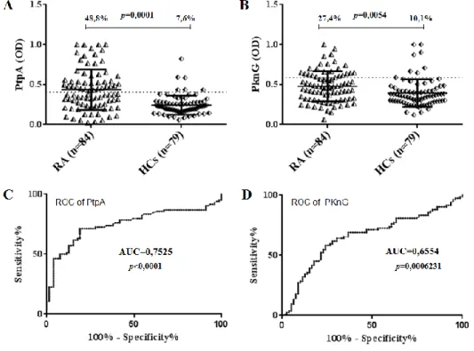
PtpA and PknG proteins secreted by Mycobacterium avium subsp. paratuberculosis are recognized by sera from patients with rheumatoid arthritis: a case-control study ... 59
Results ... 59
Discussion ... 64
Association between Lipoprotein Levels and Humoral Reactivity to Mycobacterium avium subsp. paratuberculosis in Multiple Sclerosis, Type 1 Diabetes Mellitus and Rheumatoid Arthritis... 68
Dr. Bo Marco – “Immune response against Mycobacterium avium subsp. paratuberculosis, Epstein-Barr virus, HERV-K and IRF5 in rheumatoid arthritis” – PhD thesis in Life Sciences and Biotechnologies – University of Sassari, Italy
16
Results ... 68
Discussion ... 77
Antibody response to epitopes of Epstein-Barr virus, Mycobacterium avium subsp. paratuberculosis and IRF5 in connective tissue diseases ... 81
Results ... 81
Discussion ... 86
Investigating the immune response against the homologous peptides (BOLF1305-320, MAP_402718-32, IRF5434-424) in AIA, CIA and CAIA sera of mice ... 90
Results ... 90
Discussion ... 91
CHAPTER 5: Conclusions... 93
CHAPTER 6: Materials and Methods ... 96
Patients and controls ... 96
Mice ... 97
Antigen-Induced Arthritis (AIA) ... 97
Clinical and histological methods ... 98
Fix/decalcification/OCT embedding of AIA knees for frozen sectioning ... 98
Peptides ... 99
Recombinant protein production and plate preparation ... 100
ELISA ... 100
Competitive inhibition assay ... 101
Quantification of lipoproteins in serum samples ... 101
Dr. Bo Marco – “Immune response against Mycobacterium avium subsp. paratuberculosis, Epstein-Barr virus, HERV-K and IRF5 in rheumatoid arthritis” – PhD thesis in Life Sciences and Biotechnologies – University of Sassari, Italy
17
REFERENCES ... 103
Dr. Bo Marco – “Immune response against Mycobacterium avium subsp. paratuberculosis, Epstein-Barr virus, HERV-K and IRF5 in rheumatoid arthritis” – PhD thesis in Life Sciences and Biotechnologies – University of Sassari, Italy
18
CHAPTER 1: Introduction Rheumatoid arthritis
Rheumatoid arthritis (RA) is a chronic inflammatory autoimmune disease that mainly affects diarthrodial joints. It can potentially involve every compartment in the organism such as skin, blood vessels, heart, lungs and muscles. At the level of the joint the inflammatory process has an erosive character and can lead to the destruction of the juxta-articular bony heads and to the ankylosis (Scott DL et al., 2010).
RA was first described by Augustin-Jacob Landré-Beauvais in the early 1800s. The term “rheumatoid arthritis” was coined by Alfred Bering Garrod in 1876 and was definitively adopted at the American Rheumatism Association (ARA) in 1941.
Epidemiology
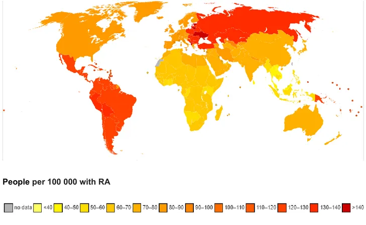
RA is the most common inflammatory arthritis, affecting from 0.5 to 1% of the general population worldwide. The prevalence ranges from 0.3 to 2% of the population, while yearly incidence is of 2-4 new cases per 10 000 inhabitants (Fig. 1). Females are predominantly affected with a male/female ratio of 1:4. The disease can occur at any age, but it generally occurs in subjects between 40 and 60 years old. In Italy, it was estimated that more than 5 million people suffer from RA with a higher prevalence among women, and that about one fifth have the most disabling and severe forms. If RA is not properly diagnosed and controlled in time, the joints may become permanently deformed resulting in progressive limitation and loss of self-sufficiency that leads in turn to a worsened quality of life, loss of working capacity and high social and economic costs.
Dr. Bo Marco – “Immune response against Mycobacterium avium subsp. paratuberculosis, Epstein-Barr virus, HERV-K and IRF5 in rheumatoid arthritis” – PhD thesis in Life Sciences and Biotechnologies – University of Sassari, Italy
19
People per 100 000 with RA
Fig. 1: Global prevalence of RA https://www.who.int/healthinfo/global_burden_disease/estimates_country/en/
Etiopathogenesis
The etiology and pathogenesis of RA are complex and multifaceted. Although RA etiology remains unknown, different studies supported the hypothesis that the interaction of environmental and genetic factors is responsible (Scott DL et al., 2010), (Klareskog L et al., 2009), (Feldmann M et al., 1996). The disease is characterized by an activation of the immune system which, through a complex series of events, involve both humoral and cellular immunity leading to the development of an acute inflammatory process and later to its maintenance and chronicity. As discussed later, this process implicates certain specific genes that can culminate in the breakage of immune tolerance and lead to autoreactivity.
Dr. Bo Marco – “Immune response against Mycobacterium avium subsp. paratuberculosis, Epstein-Barr virus, HERV-K and IRF5 in rheumatoid arthritis” – PhD thesis in Life Sciences and Biotechnologies – University of Sassari, Italy
20 From a genetic point of view, it has been pointed out that class II HLA-DR4 and DR1 antigens are the ones that confer greater susceptibility to the disease. The initiation of RA probably begins years before the onset of clinical symptoms. The loss of tolerance leads to autoreactivity and this process is due to specific genes, including class II major histocompatibility complex (MHC) genes, protein tyrosine phoshatase-22 (PTPN22) (Begovich AB et al., 2004), and peptidylarginine deiminases. The first description of a genetic link between HLA-DR and RA has been described in the 1970 century. HLA-DR4 was present in 70% of RA subjects and only 30% of healthy controls (HCs). Other HLA genes such as DRB*1301 that contain, in the third hypervariable region of DRβ-chains, the DERAA sequence, are associated with decreased susceptibility to RA (van der Woude D et al., 2010).
In addition, signal transduction gene polymorphisms, cytokine promoter polymorphisms and population-specific genes (e.g., PADI4 in Koreans and Japanese) are involved. PTPN22 and PADI4 increase the risk in some racial and ethnic groups, but not in all. It is likely that the earliest phases are characterized by repeated activation of innate immunity.
A number of environmental factors clearly contribute to RA susceptibility, although no specific exposure has been identified as the pivotal agent. Smoking is the best defined environmental risk factor for RA and the interaction between HLA-DR and tobacco exposure is perhaps the best example of how genes and environment conspire to enhance the risk.
Other triggering factors are represented by bacterial agents (E. coli, Streptococcus, mycoplasma and mycobacteria), viral agents (EBV, varicella zoster virus (VZV), rubella virus, Herpes Simplex, Parvovirus B19) and superantigens (Arleevskaya MI et al., 2016).
Dr. Bo Marco – “Immune response against Mycobacterium avium subsp. paratuberculosis, Epstein-Barr virus, HERV-K and IRF5 in rheumatoid arthritis” – PhD thesis in Life Sciences and Biotechnologies – University of Sassari, Italy
21 These agents can trigger joint inflammation with a direct and/or indirect mechanism. This process occurs often in normal people but is self-limited, while in subjects with a predetermined propensity for an immune hyper-reactivity or autoreactivity it could lead to a different outcome.
The indirect mechanism is the most popular mechanism in which we may observe the triggering of a cross immune response between microbial antigens and articular auto-antigens. In addition, environmental stresses can lead to post-transcriptional modifications of proteins in mucosal surfaces or the synovium, especially carbamylation or citrullination of arginine residues. People with a propensity for RA can develop antibodies (Abs) against these modified proteins with production of rheumatoid factors (RFs) and anti-citrullinated protein Abs (ACPAs).
Pathological anatomy
The most important alterations observed in RA patients are synovitis, nodules and rheumatoid vasculitis. Synovitis is initially characterized by exudation, cellular infiltration (lymphocytes and plasma cells) and proliferation (vessels, synoviocytes, fibroblasts). Proliferation is becoming increasingly important and leads to the formation of the synovial cloth that invades the subchondral bone and destroys the cartilage leading to ankylosis. The nodules are found in 15-30% of the cases, they can be superficial (skin, subcutaneous, tendinous sheaths) or deep (internal organs), single or multiple and their diameter varies from a few millimeters to 2 or more centimeters. Histologically they are characterized by a central zone of fibrinoid necrosis, an intermediate zone formed by one or more layers of cells arranged in a palisade and an external zone of granulation tissue infiltrated by
Dr. Bo Marco – “Immune response against Mycobacterium avium subsp. paratuberculosis, Epstein-Barr virus, HERV-K and IRF5 in rheumatoid arthritis” – PhD thesis in Life Sciences and Biotechnologies – University of Sassari, Italy
22 lymphocytes and plasma cells. Vasculitis is not specific, affects small vessels and can involve any organ.
Diagnostic classification
Different RA diagnostic classifications have been used during the years after the first description (Arnett FC et al., 1988), (Kasturi S et al., 2014). The most recent diagnostic classification is based on 2010 ACR/EULAR Classification Criteria for RA (criteria of the American College of Rheumatology) (Aletaha D et al., 2010).
Therapy
The therapy is multidisciplinary and consists of combining the pharmacological approach [NSAIDs (Nonsteroidal anti-inflammatory drugs), steroids, DMARDs (disease-modifying antirheumatic drugs), biological drugs] with non-pharmacological one (rehabilitation, surgical correction, psychological approach) (Pinals RS et al., 1981). The drugs used in RA are aimed at reducing inflammation and consequent structural damage. They are called disease-modifying antirheumatic drugs and can be classified into synthetic DMARDs and biological DMARDs. Synthetic DMARDs are also classified into conventional synthetic DMARDs and target synthetic DMARDs (Smolen JS et al., 2014).
Biologic drugs, recently introduced for the treatment of RA, have been very successful; these are molecules (monoclonal antibodies), obtained using recombined DNA technology, which can selectively block a cytokine or a particular cellular interaction identified as key points in the pathogenesis of the disease. Biological drugs allowed clinical remission in a high percentage of patients and proved to slow down (in some cases and conditions blocked) the radiological progression of arthritis. Those currently on the market for RA have as molecular targets the tumor necrosis factor (TNF), the interleukin receptor 1, the IL-6 receptor, the
CD-Dr. Bo Marco – “Immune response against Mycobacterium avium subsp. paratuberculosis, Epstein-Barr virus, HERV-K and IRF5 in rheumatoid arthritis” – PhD thesis in Life Sciences and Biotechnologies – University of Sassari, Italy
23 20, the CD-80 and the CD-86 (also known as B7-1 and B7-2, both of which are fundamental to the co-stimulation of B lymphocytes - T lymphocytes) (Nam JL et al., 2014). In Italy, Europe and USA, five anti-TNFα have been approved for the treatment of RA: Infliximab (originator, biosimilar), Etanercept (originator, biosimilar), Adalimumab, Golimumab and Certolizumab. All these drugs have been shown to be more effective on a clinical profile when administered together with methotrexate, especially in the inhibition of radiological damage (Maini RN et al., 1998). The biologicals with different mechanisms of action to date prescribed for the treatment of RA are: Abatacept, Tocilizumab, Anakinra and Rituximab (Maini RN et al., 1998).
Dr. Bo Marco – “Immune response against Mycobacterium avium subsp. paratuberculosis, Epstein-Barr virus, HERV-K and IRF5 in rheumatoid arthritis” – PhD thesis in Life Sciences and Biotechnologies – University of Sassari, Italy
24
CHAPTER 2: Infectious agents and RA Human endogenous retrovirus-K
For a long time, endogenous retroviruses (ERVs) and other repetitive elements have been considered as junk DNA having no impact on the host. However, over the years, it has been possible to discover the evolutionary interplay between ERVs and host biology, leading to interesting results. ERVs are viral elements that are part of the animal and human genome and that have probably originated from retroviruses. Today we know that ERVs constitute a quantitatively significant component of the human genome; it is estimated that there are roughly 98 000 sequences of complete ERVs and their fragments and that they represent approximately 8% of the whole human genome. If we add other retro elements such as retrotransposons (fragments of DNA that can be independently transcribed in an intermediate to RNA and consequently replicate in different positions of the genome), it is believed that more than 40% of the genome of an individual is of external origin. These sequences of endogenous elements inserted into the seminal line cells by retro-transcription mechanisms are inherited as stable mendelian genes (Temin HM., 1985).
It seems that Human endogenous retrovirus-K (HERV-K) could has a role in the autoimmune process of RA in which the immune system fails to discriminate self from non-self. Several factors can contribute to the phenomenon of molecular mimicry, such as genetic predisposition (HLA genes), environmental factors, age and sex (Freimanis G et al., 2010). In RA, it was pointed out that HERV-K-10 has amino acid sequences homologous to IgG1Fc, a target of rheumatoid factor. In patients with RA, it has been observed that there is a differential expression of the HERV-K (HML-2) genes and the transcription of Rec proteins.
Dr. Bo Marco – “Immune response against Mycobacterium avium subsp. paratuberculosis, Epstein-Barr virus, HERV-K and IRF5 in rheumatoid arthritis” – PhD thesis in Life Sciences and Biotechnologies – University of Sassari, Italy
25 Furthermore, it was observed that in the mononuclear cells of synovial fluid of RA patients there is a high expression of the HERV-K type 1 virus encoding a super antigen capable of stimulating a high number of reactive T lymphocytes (Tugnet N et al., 2013). The immune response against HERV-K is guided by an antigen and could be an initial event in the autoimmune mechanism that leads to the loss of tolerance to the organism's own antigens. A connection is therefore possible between the activation of HERV-K and the inflammatory process. Moreover, it was observed that in patients with RA there is a statistically significant association between the viral load of HERV-K1 in the plasma and the activity of the disease. This association could reflect the increased cellular activation found in peripheral blood during the period of active disease which, in turn, can facilitate the activation and perhaps the replication of HERV-K type 1 followed by a high viral load (Tugnet N et al., 2013). This association has been highlighted only with HERV-K type 1, which encodes the Np9 oncoprotein. In fact, RA is characterized by a chronic inflammation and an incorrect proliferation of invasive synoviocytes, such as the growth of cancer cells. This could suggest that the activation of HERV-K 1 could contribute to the chronicity of the process, where the synoviocytes show defects in apoptosis.
Finally, the differential activation and expression of HERV-K could be induced by environmental signals or by the activation of the immune system following exposure to triggering factors that may include exogenous viruses such as EBV, also involved in RA (Tugnet N et al., 2013).
Dr. Bo Marco – “Immune response against Mycobacterium avium subsp. paratuberculosis, Epstein-Barr virus, HERV-K and IRF5 in rheumatoid arthritis” – PhD thesis in Life Sciences and Biotechnologies – University of Sassari, Italy
26 Epstein-Barr virus
Epstein-Barr virus (EBV), known as human gammaherpesvirus 4, is one of the most common viruses in humans identified by Anthony Epstein and Yvonne Barr in 1964 using electron microscopy in biopsies of Burkitt’s Lymphoma (BL). The virus belongs to the
Herpesviridae family, gammaherpesvirinae subfamily and genus Lymphocryptovirus. EBV
is an ubiquitous virus and 90% of the worldwide population is infected. The virus is orally transmitted and infects epithelial cells of the oropharynx (Young LS et al., 2004).
EBV is about 122-180 nm in diameter and is composed of a linear, double helix of DNA which contains ~172.000 base pairs and 85 genes that codifies for more than 85 proteins. There are six nuclear antigens (EBNAs 1, 2, 3A, 3B, 3C and EBNA-LP); three latent membrane proteins (LMPs 1, 2A, 2B); small non-polyadenylated RNAs, EBERs 1 and 2; microRNAs (miR-BHRF1 and miR-BART); and several early lytic genes (Young LS et al., 2004).
Deoxyribonucleic acid is enclosed within a protein nucleocapsid, which is surrounded by a tegument made of protein, in turn surrounded by an envelope containing both lipids and surface projections of glycoproteins, which are essential to infect the host cell. EBV is usually found in the episomal form inside cells, although, in some cases it has been described as a viral DNA randomly integrating into the host’s genome (Young LS et al., 2004). Also, EBV has the ability to express different viral proteins according to different cells and differentiation stages of infected cells.
EBV is involved in several diseases, e.g. infectious mononucleosis (IM), BL, Hodgkin’s lymphoma, gastric cancer, nasopharyngeal carcinoma and other neoplasms. Recent reports show that EBV has been associated with autoimmune disease including multiple
Dr. Bo Marco – “Immune response against Mycobacterium avium subsp. paratuberculosis, Epstein-Barr virus, HERV-K and IRF5 in rheumatoid arthritis” – PhD thesis in Life Sciences and Biotechnologies – University of Sassari, Italy
27 sclerosis (MS), systemic lupus erythematosus (SLE), Sjogren’s syndrome (SS) and RA (Olsson T et al., 2017), (Ascherio A et al., 2015), (Trier NH et al., 2019).
Mycobacterium avium subspecies paratuberculosis
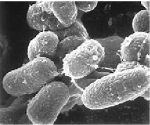
Mycobacterium avium subspecies paratuberculosis (MAP) belongs to the order of the Actinomycetales and is part of the group Mycobacterium avium complex (Mac) (Harris NB et al., 2001). MAP is an optional anaerobic Gram-positive intracellular bacterium, with a small rod shape, ranging from 0.15μm to 1.15μm (Fig. 2). It is strongly acid-alcohol resistant, asporiginal and capsule-free bacterium (Harris NB et al., 2001), (Lévy-Frébault VV et al., 1992).
Fig. 2. Mycobacterium avium subspecies paratuberculosis https://johnes.org/
It is a mandatory pathogen for ruminants that requires the presence of a favorable environment to multiply. MAP has a considerable tropism towards ruminant animal tissue and is the etiologic agent of severe gastroenteritis in ruminants known as Johne’s disease or paratuberculosis (Chiodini RJ et al., 1984), but other animal can be also affected.Recent research on a survey of 48 countries found paratuberculosis to be very common in livestock representing also a serious problem for public health (Whittington R et al., 2019), (Garvey M., 2018).
Dr. Bo Marco – “Immune response against Mycobacterium avium subsp. paratuberculosis, Epstein-Barr virus, HERV-K and IRF5 in rheumatoid arthritis” – PhD thesis in Life Sciences and Biotechnologies – University of Sassari, Italy
28 It is a bacterium that grows very slowly in culture media taking up to 6-8 weeks at 37°C for a visible growth, with duplication times of about 48-72 hours compared to 12-24 hours of Mycobacterium tuberculosis. This feature is mostly due to the bacterial wall rich in complex lipids that slows metabolic exchange with the external environment.
MAP may occur in two forms: bacillary or spheroplasty. The presence of a large number of bacteria in a bacillary form is necessary to give rise to overt disease (pluribacillary form) and can be detected with the coloring of Ziehl-Neelsen. Few bacteria in spheroplastic form, on the other hand, are able to cause the disease (paucibacillary form, characterized by the formation of granulomas) (Chiodini RJ et al., 1984).
MAP is transmitted via the orofecal route and is localized in the intestine of the host, it is excreted with the stool of the infected animals, it is able to withstand exposure to the environment for a long time, enhancing the risk for the cycles of re-infection and making it difficult to eradicate the disease in endemic areas. The presence of MAP in the environment is influenced by the type of soil, pH and soil moisture, however this microorganism tolerates a wide range of temperature excursions. Through rainwater, MAP can be transferred to waterways, such as lakes and rivers, and some controls have documented its presence in these types of water (Chiodini RJ et al., 1984), (Richardson H et al., 2019). The water intended for domestic use is subjected to various treatments that make it possible to eliminate the solids suspended in it and reduce the probable presence of MAP; however, its complete elimination is difficult due to the high resistance to chlorine and other biocides, which constitutes a danger of contamination for domestic supplies (Whan LB et al., 2001).
In all hosts, the most common route of infection is represented by the ingestion of feces or contaminated foods such as milk, colostrum or water. Once penetrated into the host, MAP
Dr. Bo Marco – “Immune response against Mycobacterium avium subsp. paratuberculosis, Epstein-Barr virus, HERV-K and IRF5 in rheumatoid arthritis” – PhD thesis in Life Sciences and Biotechnologies – University of Sassari, Italy
29 reaches the intestine, where, through transcytosis, it passes through the membrane of the M cells in the Peyer plaques (besides being able to infect the epithelial cells of the mucosa) and into the deep layer of the intestinal mucosa, where it is captured by macrophages. MAP has a marked capacity to resist the enzymatic degradation mechanisms implemented by macrophages during infection; this ability allows MAP not only to survive, but also to grow and multiply, albeit very slowly, within these cells. The mechanism by which the mycobacterium survives and multiplies inside the phagosome is still not clear. Phagosomes constitute an extremely hostile environment to many bacteria due to the rather acid pH, the presence of proteolytic enzymes and different antibacterial molecules such as defensins and toxic intermediates of oxygen and nitrogen. It is probable that MAP inhibits the action of oxygen and nitroxide by the synthesis of oxidase and alkyl hydroperoxidase C and D. It is also known that, in the case of mycobacterial infections, normal phagosome-lysosome fusion does not occur with subsequent acidification and the release of hydrolytic enzymes. Recent studies have shown that the components of the host cell, steroid cholesterol and a surface phagosomal protein called TACO (tryptophane aspartate-containing), promote the establishment of intracellular infection by mycobacteria (Pieters J et al., 2002), (Meena LS et al., 2010). During growth within the macrophage phagosome, mycobacteria develop a capsule similar to a lamellar lining. The presence of MAP also induces a significant increase in the expression of pro-inflammatory cytokines in the macrophage, such as IL-1α (interleukin-1α), IL-1, IL-6, IL-12 and TNF-α. The latter, together with IL-12, stimulates the natural killer (NK) lymphocytes to produce INF-γ, in turn responsible for the activation of other macrophages which accentuate the inflammatory process (Chiodini RJ et al., 1984).
This bacterium has been also associated to Crohn’s disease (McNees AL et al., 2015), (Kuenstner JT et al., 2017), (Feller M et al., 2007), (Sechi LA et al., 2005). The possible
Dr. Bo Marco – “Immune response against Mycobacterium avium subsp. paratuberculosis, Epstein-Barr virus, HERV-K and IRF5 in rheumatoid arthritis” – PhD thesis in Life Sciences and Biotechnologies – University of Sassari, Italy
30 involvement of MAP has been suggested for the first time in 1984 by Rodrick Chiodini by the close similarity from a clinical, pathological and epidemiological point of view, between Crohn's disease and Johne's disease of ruminants (Chiodini RJ 1984). Another proof was the first isolation of MAP from a Crohn's patient with a further successful cultivation (Chiodini RJ et al., 1986).
Following colonisation of the host, the parasitic organism alters the host immune system through molecular mimicry, displaying peptide sequences similar to that of the host cells and causing a disruption of self vs. non-self-recognition. Theoretically, the failure to recognize the invading organism from host cells may result in numerous autoimmune conditions.
In addition to Crohn’s disease, several studies show that MAP could be another triggering factor in other human autoimmune diseases such as MS (Frau J et al., 2013), (Cocco E et al., 2011) (Bo M et all., 2018b), Type 1 Diabetes (T1D) (Songini M et al., 2010) and Hashimoto's thyroiditis (Cossu D et al., 2015).
Dr. Bo Marco – “Immune response against Mycobacterium avium subsp. paratuberculosis, Epstein-Barr virus, HERV-K and IRF5 in rheumatoid arthritis” – PhD thesis in Life Sciences and Biotechnologies – University of Sassari, Italy
31
Aim of the study
The goals of my research can be summarized in the following points:
Deepening knowledge on the role of endogenous retroviruses in RA disease. Analysis of the synergistic effect between EBV, HERV-K and MAP in RA disease. Investigation of the molecular mimicry mechanism between EBV, MAP and host
peptides.
Starting from previous discoveries of anti-Interleukin-2 (IL-2) Abs present in subjects affected by autoimmune diseases and their possible role in alterations involving regulatory T cell responses, I investigated the immune response against two epitopes of IL-2 in RA Sardinian patients.
Analysis of reactivity to PtpA and PknG proteins secreted by MAP in the sera of RA patients and correlation analysis with immune response against BOLF1, MAP_4027 and IRF5 peptides.
Investigation of the correlation between humoral reactivity against MAP and serum lipoprotein levels in subjects at T1D risk (rT1D) grouped by geographical background and in patients affected by MS and RA.
Considering that anti-CCP antibodies are a biomarker for precocious diagnosis of RA, I’ve investigated the reactivity against wild type and citrullinated peptides of MAP, EBV and IRF5 in RA, SLE, systemic sclerosis (SSc) and SS patients in order to assess similitudes and/or differences.
Investigation of the reactivity against three homologous peptides IRF5, BOLF1 and MAP previously analyzed in human, in the serum of three arthritis models (AIA, Collagen-Induced Arthritis (CIA) and Collagen Antibody-Induced Arthritis (CAIA)) and comparison with human results.
Dr. Bo Marco – “Immune response against Mycobacterium avium subsp. paratuberculosis, Epstein-Barr virus, HERV-K and IRF5 in rheumatoid arthritis” – PhD thesis in Life Sciences and Biotechnologies – University of Sassari, Italy
32
CHAPTER 3: Mouse model of rheumatoid arthritis
RA is an autoimmune/inflammatory disease that causes joint damage, disability, decreased quality of life, and cardiovascular and other comorbidities. The etiology is unknown and pathogenesis is not clear yet. For this reason, in order to better understand the complexity of the pathogenesis of RA, animal models have been extensively used to understand the molecular mechanisms involved in human diseases with the objective to develop new therapeutic strategies. Obviously, a disease identical to RA cannot be developed in experimental animals because they are different species with different genetics and life environments, as compared with humans.
Many rodent models of arthritis have been developed through the decades of research in the field and, among all animal models, mouse models of RA have many features of human disease. This has contributed to numerous advances to better understand pathogenic pathways in RA and major advances in development of new drugs (Bevaart L et al., 2010), (McNamee K et al., 2015), (Kollias G et al., 2011). This way, by translating results from mouse to human, it has been possible to refine the understanding of molecular pathways involved in disease pathogenesis. These outcomes have been achieved by combining knowledge on RA-associated genes, environmental factors and the presence of serological elements.
Arthritis models can be induced in different ways that can be divided into 3 groups:
1) those elicited by active immunization, 2) those elicited by passive immunization,
3) those elicited by the administration of irritant chemicals resulting in chronic inflammation. CIA is an example of an active immunization strategy, whereas
Dr. Bo Marco – “Immune response against Mycobacterium avium subsp. paratuberculosis, Epstein-Barr virus, HERV-K and IRF5 in rheumatoid arthritis” – PhD thesis in Life Sciences and Biotechnologies – University of Sassari, Italy
33 antibody- induced arthritis models, such as collagen antibody–induced arthritis and K/BxN antibody transfer arthritis, represent examples of passive immunization strategies (Caplazi P et al., 2015), (Asquith DL et al., 2009).
Induced arthritis models
RA is characterized by immune perturbation in both the innate and adaptive compartments and subsequent chronic inflammation. The etiology is complex and, for this reason, it was indispensable to generate animal models that better represent RA disease for a simplified study of RA immunopathogenesis.
Collagen-induced arthritis
CIA is one of the models used. Arthritis is normally induced in mice by immunization with autologous or heterologous type II collagen in adjuvant. Susceptibility to collagen-induced arthritis is strongly associated with major histocompatibility complex class II genes, and the immunopathogenesis of CIA involved both a T-cell and B-cell specific response to type II collagen. The chief pathological features of CIA include a proliferative synovitis with infiltration of polymorphonuclear and mononuclear cells, pannus formation, cartilage degradation, erosion of bone, and fibrosis (Brand DD et al., 2007). As in RA, pro-inflammatory cytokines, such as tumour necrosis factor α (TNFα) and interleukin (IL)-1β, are abundantly expressed in the arthritic joints of mice with CIA, and blockade of these molecules results in a reduction of disease severity.
Antigen-induced arthritis
The above indicated model of arthritis can be developed when mice are primed with an antigen like methylated bovine serum albumin (mBSA) in complete Freund’s adjuvant (CFA), and subsequently challenged by intra-articular injection of the same antigen (Asquith DL et
Dr. Bo Marco – “Immune response against Mycobacterium avium subsp. paratuberculosis, Epstein-Barr virus, HERV-K and IRF5 in rheumatoid arthritis” – PhD thesis in Life Sciences and Biotechnologies – University of Sassari, Italy
34 al., 2009). Such models are useful in that mice of several strains can be investigated to establish a hierarchical role for given factors in adaptive immune-mediated articular damage. Subsequent pathology comprises immune complex-mediated inflammation with infiltration of polymorphonuclear cells, pannus formation, erosion of cartilage and bone, followed by articular T-cell-mediated responses and in particular is Th17 dependent (Asquith DL et al., 2009), (Egan PJ et al., 2008). Also, regarding the molecular pathway of immune response, a recent study has shown that synovial macrophages from the AIA joint have high levels of IRF5 that is an important transcription factor implicated in the adjustment of immune response, both innate and adaptive (Weiss M et al., 2013). Through the comparison of naïve joints and arthritis inflammed joints, researchers have shown that IRF5 deficient mice (IRF5-/-) had a significantly less swelling at day 2 in comparison with wild-type controls (WT). Reduced swelling corresponded to a decreased number of cells recovered from excised knee joints at that point. Also, IRF5 knockout mice showed a significantly lower synovial membrane thickening score in the mBSA-challenged knees than WT mice and reduced synovial membrane thickening has also been observed at day 7 (Weiss M et al., 2013).
Macrophages and neutrophils are the two major types of myeloid cells involved in RA inflammatory process. Considered the heterogeneity of macrophages (Murray PJ et al., 2014), (Sieweke MH et al., 2013) and the interesting role of neutrophils able to form distinct subsets and extracellular traps (NETs) (Khandpur R et al., 2013), a variety of destructive enzymes as well as autoantigens can be generated (Becher B et al., 2014), (Ericson JA et al., 2014). Further investigations must be carried out to understand the exact nature of the myeloid cells in various inflammatory diseases; this not only will provide other clues about pathogenesis, but can also contribute to the discovery of more effective therapeutics improving the quality of life of RA subjects.
Dr. Bo Marco – “Immune response against Mycobacterium avium subsp. paratuberculosis, Epstein-Barr virus, HERV-K and IRF5 in rheumatoid arthritis” – PhD thesis in Life Sciences and Biotechnologies – University of Sassari, Italy
35 In this research AIA mouse model has been used to analyse the immune response against viral and bacterial epitopes in order to better understand the possible role of some infections in RA pathogenesis and compare these results with those obtained in human.
Dr. Bo Marco – “Immune response against Mycobacterium avium subsp. paratuberculosis, Epstein-Barr virus, HERV-K and IRF5 in rheumatoid arthritis” – PhD thesis in Life Sciences and Biotechnologies – University of Sassari, Italy
36
CHAPTER 4: Investigating the role of viral and bacterial infections in RA and other rheumatic diseases
***
Identification of a HERV-K env surface peptide highly recognized in rheumatoid arthritis (RA) patients: a cross-sectional case-control study
Among environmental factors, the role of viruses in autoimmunity has been long postulated and endogenous retrovirus (HERV) is one potential candidate.
The hypothesis of a potential pathogenic link between HERV‐K and RA is based on several observations descending from both polymerase chain reaction (PCR)‐based and serological
studies (Trela M et al., 2016). The prevalence of insertionally polymorphic HERV‐K was reported increased significantly in the RA population (Krzysztalowska-Wawrzyniak M et al.,
2011). Moreover, a significantly increased expression of HERV‐K genes and transcription of viral proteins has been demonstrated in peripheral blood, synovial fluid and synoviocytes of RA patients compared to HCs (Hohn O et al., 2013), (Reynier F et al., 2009). In addition, common amino acid sequences between HERV‐K and host antigens have been reported in RA, suggesting that a mechanism of molecular mimicry may play a role in disease pathogenesis (Freimanis G et al., 2010), (Nelson PN et al., 2014).
Considering this background, we performed an in silico analysis to predict key antigenic regions of HERV‐K on the basis of their putative ability in igniting an RA‐specific humoral response. Candidate epitopes were then synthesized as short peptides, and an enzyme‐ linked immunosorbent assay (ELISA) was used to test the reactivity of RA and control serum to HERV‐K peptides. In silico analysis performed using IEDB Analysis Resource software, in particular the Antibody Epitope Prediction, Bepipred linear epitope prediction software
Dr. Bo Marco – “Immune response against Mycobacterium avium subsp. paratuberculosis, Epstein-Barr virus, HERV-K and IRF5 in rheumatoid arthritis” – PhD thesis in Life Sciences and Biotechnologies – University of Sassari, Italy
37 (http://www.iedb.org/home_v3.php), allowed us to identify different antigenic peptides derived from HERV‐K env surface protein (UniProtKB Accession number: Q69384).
Four peptides were selected for further analysis:
▪ HERV‐K env19–37 (VWVPGPTDDRCPAKPEEEG) ▪ HERV‐K env109–126(RPKGKTCPKEIPKGSKNT)
▪ ERV‐K env164–186 (SGQTQSCPSAQVSPAVDSDLTES) ▪ HERV‐K env205–226 (EKGISTPRPKIISPVSGPEHPE)
Results
Antibodies against HERV‐K env peptides were tested in serum of 70 RA patients and 71 HCs. HERV‐K env‐su19–37 antibodies were above positivity in the sera of 19% RA patients and in only 3% of HCs (AUC=0.84, RA versus HCs p<0.0025) (Fig. 3a).
Regarding HERV‐K env‐su109–126 peptide, 9% of RA patients and 3% of HCs were positive for antibodies in serum (p=0.165, AUC=0.71) (Fig. 3b).
No statistical differences were found regarding positivity against HERV‐K env‐su164–186 in RA patients (3%) versus HCs (3%) (AUC=0.56, Fig. 3c). HERV‐K env‐su205–226 peptide was recognized in the sera of 6% of RA patients and 3% of HCs (p=0.44, AUC=0.75) (Fig. 3d). No significant difference was found in the proportion of HERV‐K env‐su19–37 antibody positivity according to disease activity (moderate–severe RA versus low activity and remission RA) and type of immunosuppressive treatment.
Dr. Bo Marco – “Immune response against Mycobacterium avium subsp. paratuberculosis, Epstein-Barr virus, HERV-K and IRF5 in rheumatoid arthritis” – PhD thesis in Life Sciences and Biotechnologies – University of Sassari, Italy
38
Fig. 3. Prevalence of Abs against peptides derived from HERV‐K envelope (env) protein. Scatterplots present median values with interquartile range. Area under receiver‐operating characteristic (ROC) curve (AUC) as well as significant P‐values are displayed.
Mameli G et al. Clinical & Experimental Immunology, 2017
No correlation was found between HERV‐K env‐su19–37, antibody levels and RA descriptors [age, DAS‐28, HAQ, C‐reactive protein (CRP) and erythrocyte sedimentation rate (ESR)]. The same results were obtained with the other HERV‐K env‐su peptides tested (data not shown).
Discussion
HERV‐K mRNA transcripts and IgG anti‐HERV‐K Abs have been associated with autoimmunity and cancers, as well as neurological conditions (e.g. MS) and rheumatic diseases (Manghera M et al., 2016), (Balada E et al., 2010), (Tugnet N et al., 2013).
Dr. Bo Marco – “Immune response against Mycobacterium avium subsp. paratuberculosis, Epstein-Barr virus, HERV-K and IRF5 in rheumatoid arthritis” – PhD thesis in Life Sciences and Biotechnologies – University of Sassari, Italy
39
For the first time, a humoral response against selected surface epitopes of HERV‐K env‐su (HERV‐K env‐su19–37, HERV‐K env‐su109–126 and HERV‐K env‐su209–226) was searched in
patients with RA and controls. Among them, the most interesting peptide was HERV‐K env‐ su19–37, which was recognized in the sera of 19% of RA patients and in only 3% of controls.
HERV‐K env‐su19–37 has a high homology with different human antigens, including myosin
(with 61% of positive amino acids with sequence EAW76415.1) and netrin (73% of positive amino acids with sequence NP_006172.1). Evidence of a molecular mimicry as a pathogenic mechanism linking HERV‐K to RA was provided by several groups. Freimanis and co‐authors (Freimanis G et al., 2010) demonstrated a significant humoral response in RA patients against one HERV‐K epitope which shares residues with collagen type II, a common target of pathogenic RA autoantibodies. Similarly, IgGs against antigenic peptide on the matrix segment of HERV‐K10 sharing common sequence with IgG1Fc, a target of rheumatoid factor, were demonstrated in RA patients (Nelson PN et al., 2014). An intriguing explanation of this result would be that the surface epitope HERV‐K env‐su19–37 may function
as an ‘autoantigen’ driving a secondary immunological response.
Unfortunately, it was not possible to find any statistically significant correlation between antibody positivity and disease activity, systemic inflammation and type of immunosuppressive treatment. It may be possible that therapy could alter the humoral response. Notably, also in the series of RA patients described by Nelson and co‐authors, levels of antibodies to HERV‐K antigens showed no correlation with RA disease activity (Nelson PN et al., 2014).
However, it should be remembered that a number of conditions, both genetic and environmental, may modulate HERV‐K env protein transcription contributing to changes in
Dr. Bo Marco – “Immune response against Mycobacterium avium subsp. paratuberculosis, Epstein-Barr virus, HERV-K and IRF5 in rheumatoid arthritis” – PhD thesis in Life Sciences and Biotechnologies – University of Sassari, Italy
40
the amount of immunological response. HERV‐K transcription exhibits a significant increase in the presence of proinflammatory cytokines [tumour necrosis factor (TNF)‐α and interleukin (IL)‐6] in RA with regard to the general population (Freimanis G et al., 2010). Exogenous viruses such as EBV, reported to be linked to RA (Erre GL et al., 2015), may transactivate and stimulate HERV‐K expression (Nelson PN et al., 2003). Moreover, differential HERV‐K transcription has been shown to be regulated epigenetically by the degree of DNA methylation in RA (Neidhart M et al., 2000).
In conclusion, a specific humoral response against a superficial envelope epitope of HERV‐ K was demonstrated in RA patients. Results of this study clearly add to the evidence for a possible role of HERV‐K in RA pathogenesis.
Dr. Bo Marco – “Immune response against Mycobacterium avium subsp. paratuberculosis, Epstein-Barr virus, HERV-K and IRF5 in rheumatoid arthritis” – PhD thesis in Life Sciences and Biotechnologies – University of Sassari, Italy
41
Interferon regulatory factor 5 is a potential target of autoimmune response triggered by Epstein-Barr virus and Mycobacterium avium subsp. paratuberculosis in
rheumatoid arthritis: investigating a mechanism of molecular mimicry
As discussed above, 50% of the risk for RA is attributable to environmental contributors such as microbial infections (Duarte JH., 2015). EBV and mycobacteria were among pathogens reported to be involved in RA pathogenesis (Song YW et al., 2010), (Adtani P et al., 2015) (Ball RJ et al., 2015), (Erre GL et al., 2015), (Westergaard MW et al., 2015), (Rao DA et al., 2017).
In recent years, the potential role of IRF5 in the pathogenesis of RA has been frequently reported (Adtani P et al., 2015), (Eames HL et al., 2016), (Negi VS et al., 2014). IRF5 is a key transcription factor of the immune system, playing an important role in modulating inflammatory immune responses in many cell types including dendritic cells and macrophages by driving them toward a proinflammatory phenotype in concert with cytokines and chemokines expression, and by regulating B cell maturity and antibody production (Popko K et al., 2015), (Lino AC et al., 2016), (Dekkers J et al., 2016), (Olalekan SA et al., 2015), (Fillatreau et al., 2015), (Mahabadi M et al., 2016).
Recent in vivo studies identified the importance of IRF5 as a new link between the pathogenic activation of RNA-sensing Toll-like receptors and proinflammatory cytokine production in inflamed joints of arthritic mice (Duffau P et al., 2015).
Intriguingly, the presence of a specific seroreactivity against EBV and MAP epitopes cross-reacting with homologous human antigens such as IRF5 have been recently demonstrated in various autoimmune diseases (Cossu D et al., 2015), (Mameli G et al., 2015). Here it was supposed that significantly increased antibody titers following infection with MAP and EBV
Dr. Bo Marco – “Immune response against Mycobacterium avium subsp. paratuberculosis, Epstein-Barr virus, HERV-K and IRF5 in rheumatoid arthritis” – PhD thesis in Life Sciences and Biotechnologies – University of Sassari, Italy
42 may cross-react with homologous peptides of the transcription factor IRF5 leading to modulation of its expression.
Based on the above-mentioned preliminary findings, our study aimed at evaluating the presence of a cross-reactivity between three homologous peptides derived from EBV tegument protein BOLF1 (BOLF1305-320), the MAP_4027 antigen (MAP_402718-32) (Eames HL et al., 2016), (Negi VS et al., 2014) and the human interferon regulatory factor 5 (IRF5424-434) in a group of RA patients and HCs.
Results
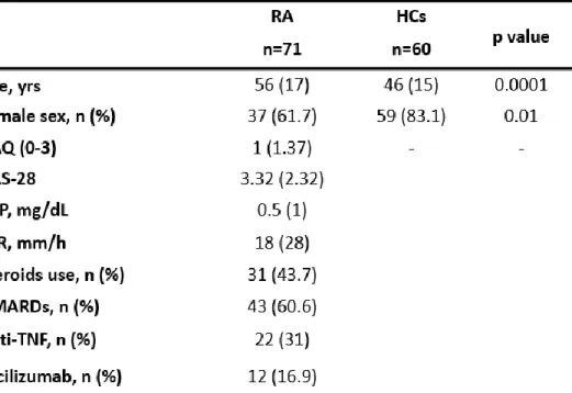
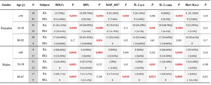
71 RA patients (11 males, 60 females; median age 56.18) and 60 HCs (23 males, 37 females; median age 46.1) were enrolled in the study; demographic and clinical features of RA patients and HCs are summarized in the Table 1.
Table 1. Demographic, clinical and laboratory features of RA patients and HCs.
Non normally distributed data are expressed as median (IQR). Health Assessment Questionnaire (HAQ); Disease Activity Score-28 (DAS-28); C-reactive protein (CRP); Erythrocyte sedimentation rate (ESR); Disease modifying anti-rheumatic drugs (DMARDs); Anti-Tumor Necrosis Factor alpha (Anti-TNF).
Dr. Bo Marco – “Immune response against Mycobacterium avium subsp. paratuberculosis, Epstein-Barr virus, HERV-K and IRF5 in rheumatoid arthritis” – PhD thesis in Life Sciences and Biotechnologies – University of Sassari, Italy
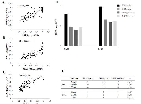
43 IRF5424-434 was found to have the highest Abs seroreactivity among the selected antigens being positive in 49 out of 71 RA patients (69%) and only in 5 out of 60 HCs (8%; AUC=0.9; p<0.0001; Fig. 4A and Fig. 4D).
Abs against BOLF1305-320 were observed in 41 out of 71 RA patients (58%) and in 5 out of 60 HCs (8%; AUC=0.9; p<0.0001; Fig. 4B and Fig. 4D). Humoral responses against MAP_402718-32 was detectable in 12 out of 71 RA patients (17%), whereas HCs showed Abs prevalence accounting for only 5% giving a p value close to the threshold of statistical significance (AUC= 0.6; p=0.051; Fig. 4C and Fig. 4D).
Globally, RA subjects displayed much more increased positivity to at least one of the assessed epitopes compared to HCs with a high degree of statistical significance (71% vs. 15%, respectively; p<0.0001; Fig. 4D).
Dr. Bo Marco – “Immune response against Mycobacterium avium subsp. paratuberculosis, Epstein-Barr virus, HERV-K and IRF5 in rheumatoid arthritis” – PhD thesis in Life Sciences and Biotechnologies – University of Sassari, Italy
44
Fig. 4: A-C) ELISA-based analysis of Abs reactivity against the homologous epitopes derived from human, EBV and MAP in RA patients and HCs. The sera were tested against plate-coated
IRF5424-434 (A), BOLF1305-320 (B) and MAP_402718-32 (C) peptides. Bars represent the median ± interquartile range. Cutoff values for Abs positivity are indicated by dashed lines. P-values, respective to each epitope showed a statistical difference between RA patients and HCs and are reported above the distributions. D) Prevalence of Abs directed against BOLF1305-320,
MAP_402718-32 and IRF5424-434 epitopes in RA patients and HCs. Total percentage of Abs positivity
to at least one peptide is represented by the first bar in each group. Other bars correspond to a single-peptide positivity relative to each epitope. E) Peptides alignment obtained using Clustal
W2. The results show the region of identity with a star (*), strong similar amino acid with a dot (.) and
a missing region in dashes (-).