C A S E R E P O R T
Early pathological gambling in co-occurrence with
semantic variant primary progressive aphasia: a
case report
This article was published in the following Dove Press journal: Clinical Interventions in Aging
Petronilla Battista1,2 Rosa Capozzo2 Giovanni Rizzo3,4 Chiara Zecca2 Antonio Anastasia5 Roberto De Blasi6 Giancarlo Logroscino2,7
1Istituti Clinici Scientifici Maugeri-SPA SB.
I.R.C.C.S. Institute of Cassano Murge,
Bari, Italy;2Neurodegenerative Disease
Unit, Department of Clinical Research in Neurology, University of Bari Aldo Moro, “Pia Fondazione Cardinale G. Panico”,
Tricase, Italy;3UOC Clinica Neurologica,
IRCCS Istituto Delle Scienze
Neurologiche di Bologna, Bologna, Italy;
4Department of Biomedical and
Neuromotor Sciences, University of
Bologna, Bologna, Italy;5Department of
Nuclear Medicine, Pia Fondazione di
Culto e Religione“Card.G.Panico”,
Tricase, Italy;6Department of Diagnostic
Imaging, Pia Fondazione di Culto e
Religione“Card.G.Panico”, Tricase, Italy;
7Department of Basic Medical Sciences,
Neuroscience and Sense Organs, University of Bari Aldo Moro, Bari, Italy
Abstract: We have comprehensively documented a case of semantic variant of primary progressive aphasia (sv-PPA) presenting with early-onset pathological gambling (PG). While a growing number of studies have shown the presence of behavioral alterations in patients with sv-PPA, PG has been observed only in the behavioral variant of frontotemporal dementia (bv-FTD). To date, no case of PG with the co-occurrence of prominent semantic deficits at the onset of the disease has been reported in the literature. Impulse disorders at onset may wrongly lead to a misdiagnosis (ie, psychiatric disorders). Therefore, a wider characterization of cognitive/aphasia symptoms in patients presenting impulse disorders and predominant language dysfunctions is recommended.
Keywords: impulse disorders, language disorders, neurodegenerative diseases, semantic dementia
Introduction
Semantic variant of primary progressive aphasia (sv-PPA), also known as“semantic dementia”, is one of the main clinical variants of frontotemporal dementia (FTD). From a clinical-pathological point of view, FTD degeneration is usually caused by TAR-DNA-binding protein (TDP-43), a 43 kilodaltons that has been reported as the underlying cause of the disease in approximately 68%–80% of subjects with sv-PPA.1 According to the current diagnostic criteria,2in order to identify patients with sv-PPA, two core features are necessary: the impaired naming of objects more than actions and impaired single-word comprehension. Moreover, several studies have demonstrated that the clinical features of sv-PPA may depend on the atrophy prevalently involved in the left hemisphere rather than in the right one.3For example, severe anomia and impaired single-word difficulties are more likely to be present in patients with a prevalently left hemisphere involvement,4while impaired recognition of familiar and famous faces is a typical consequence of predominantly right hemisphere atrophy.5 There is growing awareness of the presence of behavioral alterations in patients with sv-PPA. The most frequent clinical features are disinhibition, impulsivity, bizarre food choices/hyperphagia and repetitive/compulsive behavior that belong to the sphere of positive symptoms.6,7To date, impulse control disorder or, more specifically, patholo-gical gambling (PG) has been widely observed in the behavioral variant of FTD (bvFTD),8,9 but no cases of PG with the co-occurrence of semantic deficits at the onset of the disease have been reported in the literature. In this report, we describe the
Correspondence: Giancarlo Logroscino Department of Clinical Research in Neurology, University of Bari Aldo Moro, “Pia Fondazione Cardinale G. Panico”, Via S. Pio X, n. 4., Tricase, Lecce 73039, Italy Tel +39 083 377 3904
Fax +390 833 183 0670
Email [email protected]
Clinical Interventions in Aging
Dovepress
open access to scientific and medical research
Open Access Full Text Article
Clinical Interventions in Aging downloaded from https://www.dovepress.com/ by 31.40.210.160 on 22-Jan-2020
case of a 64-year-old male with symptoms of PG and the onset of co-occurrence of semantic impairment typical of sv-PPA.
Case report
In October 2016, an Italian-speaking, right-handed 64-year-old male, with only eight years of formal education, was referred to our Neurodegenerative Diseases Centre by his family. He presented with a 2-year history of progressive language deficits and impulse control disorders with PG evident from the onset. Before developing PG, the patient was described by his family as being parsimonious and care-ful with money. Over the two years, he had started to spend his money on gaming machines and scratch cards, squander-ing his salary and his savsquander-ings and borrowsquander-ing money from friends and relatives to continue gambling. From the onset of the disease, the patient also presented semantic deficits and started to use objects inappropriately because he seemed not to recognize them. At initial neurological examination, the patient presented with mild signs of Parkinson’s (ie, bradyki-nesia, hypomimia) and there was a family history of dementia (his father had Alzheimer’s disease). In the following month, the patient underwent a neuropsychological assessment that showed an entirely normal cognitive performance, even for executive tasks, with the exception of a slight but not sig-nificant impairment in verbal episodic memory that could be related to his lexical-semantic deficits. Activities of daily living were preserved. At the speech-language examination, he showed isolated semantic deficits, ie, impairment in sin-gle-word comprehension, nonverbal semantic association, surface dyslexia and word-finding difficulties as assessed by the Screening for Aphasia in Neurodegeneration battery (SAND).10,11At the spontaneous speech task, he showed a low speech rate associated with several false starts and a lot of lexical-semantic errors, ie, word-finding pauses with a ten-dency to produce high-frequency nouns and poor content resulting in a reduction of information units. The patient’s linguistic profile did not report articulatory, phonetic and phonemic errors. PG was confirmed from the Barratt Impulsiveness Scale12 and South Oaks Gambling Screen13 (Tables 1 and 2).
MRI brain scans (3 T) showed bilateral (right > left) fronto-temporo-insular and cingulate atrophy (Figure 1, Panels A-B-C); SPECT brain scan showed no right temporal lobe perfusion and hypoperfusion of the bilateral frontal lobes and left cingulate (Figure 2) and DAT-SCAN showed damage to the nigrostriatal dopamine system, ie right putamen, caudate nucleus (right > left) (Figure 3).
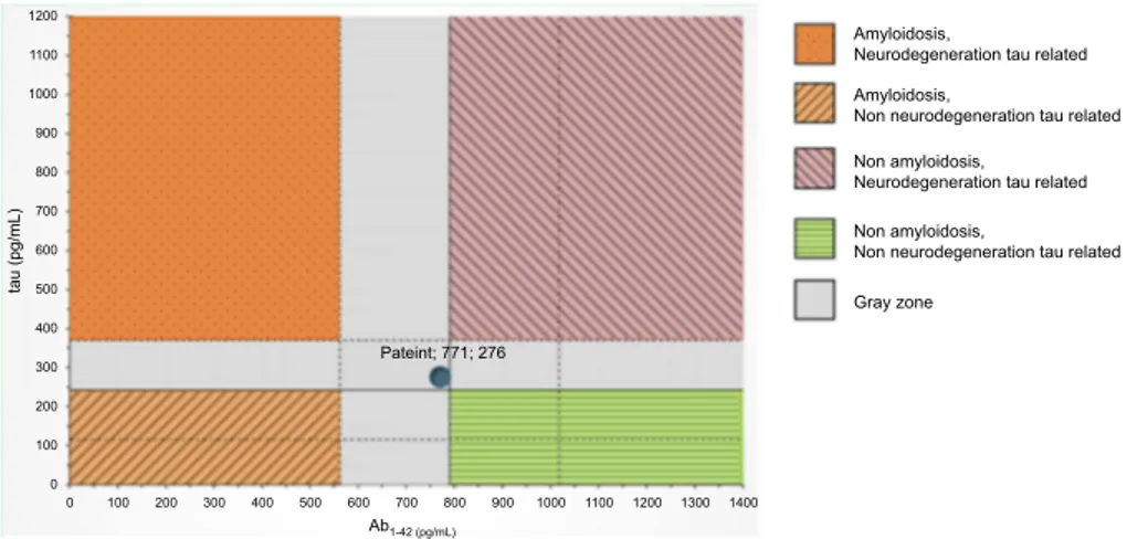
CSF biomarkers were within the normal range for age (Aβ1-42771 pg/mL; tTau 276 pg/mL; nd 42.79 pg/mL; pTau181: f). Imaging and CSF biomarkers indicated a neu-rodegenerative process that was not due to amyloidosis (Figure 4).
The patient was diagnosed as having sv-PPA according to the classification of Gorno-Tempini et al.2
In November 2017, the language deficit worsened to paragrammatic speech, a low speech rate and reduction of information content. Also, his behavioral changes increas-ingly worsened with apathy, disinhibition, compulsive/ stereotyped behaviors and hyperphagia, as did perfor-mance in executive/social cognition abilities, compatible with a frontal lobe dysfunction (Tables 1 and 2).
This clinical picture indicated the presence of a second phenotype, behavioral-frontotemporal dementia (bv-FTD), according to the criteria of Rascovsky et al14 with asso-ciated mild parkinsonism. A diagnosis of fronto-temporal lobar degeneration (FTLD) was established according to the Lund–Manchester criteria.15
PG was nonresponsive to pharmacological therapy (eg, levodopa) during the course of the disease and was con-sidered a significant clinical finding within the wider spec-trum of behavioral symptoms.
Discussion
From a clinical point of view, our patient’s behavioral and cognitive disorders showed similar trajectories. He had bilat-eral cerebral atrophy that resulted in the overlapping of beha-vioral and language symptoms. Behabeha-vioral symptoms are usually related to right hemispheric atrophy; therefore, as this was prevalent, we should have expected more evident beha-vioral symptoms compared to semantic deficits. However, behavioral symptoms became more pronounced during the course of the disease.
Other causes of neurodegenerative dementia were excluded, for example, an atypical frontal form of Alzheimer’s disease,16as the patient did not show low levels of beta-amyloid at CSF examination. At the second evalua-tion, the patient presented with dementia, spontaneous par-kinsonism and an altered DAT SCAN, but a diagnosis of Lewy body dementia17 was excluded because the clinical picture was unremarkable for two core features (hallucina-tions andfluctuating cognition) and cognitive functions were reasonably preserved compared to behavioral changes. Treatment with levodopa was started only several months after onset and therefore was not responsible for the PG.
Clinical Interventions in Aging downloaded from https://www.dovepress.com/ by 31.40.210.160 on 22-Jan-2020
The clinical information collected may also lead to an alternative, possibly even complementary, hypothesis related to the evolution of the patient’s symptoms.
As the disease progresses, the symptoms of the different phenotypes of FTLD can converge as an initially focal atrophy becomes more prominent and spreads to affect large areas in the frontal and temporal lobes. Over time, patients manifest global cognitive impairment and motor
deficits, including parkinsonism.18In our patient, parkinso-nian signs were very slight at thefirst clinical examination (without DAT-scan evidence), but had worsened by the second examination (with imaging evidence). This hypoth-esis sits well with previous studies in which early parkin-sonism is present in 18%–20% of patients with FTLD and is most often seen in patients with bv-FTD, while it is rarely seen in sv-PPA, only in about 3% (for a review see19). Table 1 Patient’s neuropsychological score and cutoff score
Test First assessment Follow up (1 year) Cutoff score
Global cognitive functioning
Mini-mental state examination 28 27 <23.8
Memory
Rey auditory verbal learning test
-Immediate recall 25 20 <28.53
-Delayed recall 6 5 <4.69
-Recognition 15 10
-Rey recall of copy of the -Rey-Osterriethfigure 21,5 16,5 <9.47
Digit span forward 6 5 <4.26
Attention & executive functions
Frontal assessment battery 14 12 <12.03
- Trial making test A 53 67 >94
- Trial making test B 158 175 >283
- Trial making test BA 105 108 >187
Digit span backward 4 4 <2.65
Stroop color and word test -time 28 24 >36.92
Stroop color and word test -errors 0 0 >4.24
Phonemic (F-A-S) 25 8 <17.35
Semantic verbalfluency 23 4 <25
Social cognition
Story-based empathy task 12 3 <8.29
Visuo-spatial abilities
Copy of the Rey-Osterrieth complexfigure 30 32 <28.88
Incomplete letters-VOSP battery 17 16 <16
Impulse dyscontrol
Barratt impulsiveness scale 65 68 <69,3
South Oaks gambling screen Positive Positive
-Behavioral scales
Apathy scale 2 12 >11
Geriatric depression scale 6 11 >10
Neuropsychiatric inventory 12 34
-Frontal behavioral inventory 10 37
-Functional autonomy
ADL 6/6 4/6
-IADL 7/8 3/8
-Note: Bold scores are pathological.
Abbreviations: VOSP, Visual Object and Space Perception Battery; ADL, Activities in Daily Living; IADL, Instrumental Activities in Daily Living.
Clinical Interventions in Aging downloaded from https://www.dovepress.com/ by 31.40.210.160 on 22-Jan-2020
sv-PPA with parkinsonism is not yet fully understood. One study reported that six sv-PPA patients showed parkinson-ism, but their clinical features were not defined in detail.20 Therefore, the case could be made for a patient with bv-FTD and prominent semantic involvement. The parkinson-ism seen in bv-FTD patients is usually characterized by akinetic rigid syndrome,21 the same kind that we found in our patient. Moreover, specific patterns of cortical and sub-cortical atrophy in the frontal lobe, insula and anterior cingulate cortex characterize bv-FTD.22,23 Our findings in this patient with parkinsonism show a neurodegenerative process involving not only anterior frontal areas, but also
posterior frontal regions, including M1 and the cortico-basal, ganglia-caudate, nucleus-thalamo-cortical motor loops that are also implied in PG behavior.24
At the onset, PG may wrongly lead to a misdiagnosis. It is extremely important to consider several differential diagnoses as these symptoms may overlap with other con-ditions, ie, late-onset psychiatric disorders. Moreover, pro-minent PG can lead to an underestimation of cognitive/ language deficits in the early phase of the disease. A wider characterization of impulse disorders in FTLD with pre-dominant language dysfunctions would be crucial for an early diagnosis.
Table 2 Patient’s speech and language score and cutoff score. Bold scores are pathological
Test First assessment Follow-up (1 year) Cutoff score
Speech-Language SAND
- Naming
- Sentence comprehension
- Single-word comprehension
- Repetition of word and nonwords
- Repetition of sentences - Reading - Writing - Semantic association - Connected speech Token test CAGI naming
CAGI single-word comprehension Pyramid and Palm Tree Test AAT repetition subtest
ENPA verbal sentence comprehension subtest
7/14 8/8 8/12 10/10 4/6 12/16 4/6 1/4 2/6 34/36 28/48 42/48 37/52 142/150 14/14 3/14 6/8 4/12 10/10 2/6 12/16 2/6 0/4 1/6 30/36 18/48 32/48 26/52 135/150 12/14 ≤9.969 ≤6.157 ≤10.258 ≤6.349 ≤2.455 ≤10.106 ≤2.132 ≤1.166 ≤3 <26.25 <41.48 <47.08 <40.15 -<11.6 Note: Bold scores are pathological.
Abbreviations: SAND, Screening for Aphasia in Neurodegeneration; AAT, Aachener Aphasie Test; ENPA, Esame Neuropsicologico per l’Afasia.
Figure 1 Neuroimaging examinations. The patient’s first MRI brain scan (3 tesla), performed about 2 years into his illness. Panel A: Sagittal FLAIR T2 image highlights the marked frontal and cingulate atrophy. Panel B: The axial 3D FFE T1-w image shows bilateral anterior temporal lobe atrophy (right > left). Panel C: Coronal FFE 3D T1-w image shows fronto-insular and anterior temporal cortical atrophy, mainly on the right side.
Abbreviation: MRI, magnetic resonance imaging.
Clinical Interventions in Aging downloaded from https://www.dovepress.com/ by 31.40.210.160 on 22-Jan-2020
However, we are aware that our report presents some limitations. In particular, the lack of an experimental task in the neuropsychological examinations. Prior reports have shown that, on a task like the Iowa Gambling Task, the pattern of gambling in PPA is different from bv-FTD.25
The experimental task could have been useful to distin-guish compulsion from gambling behavior.
To sum up, FTLD is a clinical-pathological syndrome that encompasses a heterogeneous group of neurodegen-erative disorders. The different phenotypes described may
Figure 2 SPECT brain scan shows hypoperfusion of the right temporal lobe and hypoperfusion of the bilateral frontal lobes and left cingulate. Abbreviation: SPECT, single-photon emission computed tomography.
Figure 3 Brain Dat-scan shows damage to the nigrostriatal dopamine system, in particular a loss of dopamine neurons in the right putamen and caudate nucleus (right > left).
Clinical Interventions in Aging downloaded from https://www.dovepress.com/ by 31.40.210.160 on 22-Jan-2020
have clinical and pathological characteristics that overlap, and it is possible that isolating a phenotype can be difficult because its borders may not be clearly evident as they may all belong to the same clinical-pathological spectrum.
Conclusion
In conclusion, this case is pertinent because of its atypical onset. Over the course of time, the evolution and progres-sion of symptoms and the distributions of pathological changes suggest an underpinning FTLD pathology. It is worth noting that, in order to increase the accuracy of the clinical diagnosis, appropriate and validated batteries26are essential since they are inexpensive and low-tech tools that can be used in a variety of clinical settings, such as primary, secondary or tertiary care and research studies.
Consent
Written informed consent was obtained from the patient for publication of this case report and accom-panying images. This case study was approved by the institutional ethics committee of AO Pia Fondazione Card. G. Panico.
Disclosure
The author reports no conflicts of interest in this work.
References
1. Rohrer J, Geser F, Zhou J, et al. TDP-43 subtypes are associated with distinct atrophy patterns in frontotemporal dementia. Neurology.
2010;75:2204–2211. doi:10.1212/WNL.0b013e3181ed735b
2. Gorno-Tempini ML, Hillis AE, Weintraub S, et al. Classification of
primary progressive aphasia and its variants. Neurology.
2011;76:1006–1014. doi:10.1212/WNL.0b013e31821103e6
3. Thompson SA, Patterson K, Hodges JR. Left/right asymmetry of atrophy
in semantic dementia behavioral–cognitive implications. Neurology.
2003;61(9):1196–1203. doi:10.1212/01.WNL.0000091868.28557.B8
4. Gefen T, Wieneke C, Martersteck A, et al. Naming vs knowing faces in primary progressive aphasia: a tale of 2 hemispheres. Neurology.
2013;81(7):658–664. doi:10.1212/WNL.0b013e3182a08f83
5. Snowden JS, Thompson JC, Neary D. Knowledge of famous faces
and names in semantic dementia. Brain. 2004;127(4):860–872.
doi:10.1093/brain/awh099
6. Rosen HJ, Allison SC, Ogar JM, et al. Behavioral features in seman-tic dementia vs other forms of progressive aphasias. Neurology.
2006;67(10):1752–1756. doi:10.1212/01.wnl.0000247630.29222.34
7. Harris JM, Saxon JA, Jones M, Snowden JS, Thompson JC. Neuropsychological differentiation of progressive aphasic disorders. J Neuropsychol. 2018. doi:10.1111/jnp.12149
8. Manes FF, Torralva T, Roca M, Gleichgerrcht E, Bekinschtein TA, Hodges JR. Frontotemporal dementia presenting as pathological
gam-bling. Nat Rev Neurol. 2010;6(6):347–352. doi:10.1038/nrneurol.2010.34
9. Tondo G, De Marchi F, Terazzi E, Sacchetti M, Cantello R. Frontotemporal dementia presenting as gambling disorder: when a psychiatric condition is the clue to a neurodegenerative disease. Cogn
Behav Neurol. 2017;30(2):62–67. doi:10.1097/WNN.0000000000
000122
10. Catricalà E, Gobbi E, Battista P, et al. SAND: a screening for aphasia in NeuroDegeneration. Development and normative data. Neurol Sci.
2017;38:1469–1483. doi:10.1007/s10072-017-3001-y
11. Battista P, Catricalà E, Piccininni M, et al. Screening for aphasia in neuro degeneration for the diagnosis of patients with primary progressive aphasia: clinical validity and psychometric properties. Dement Geriatr
Cogn Disord. 2018;46(3–4):243–252. doi:10.1159/000492632
12. Patton JH, Stanford MS, Barratt ES. Factor structure of the Barratt
impulsiveness scale. J Clin Psychol. 1995;51(6):768–774.
doi:10.1002/(ISSN)1097-4679
13. Lesieur HR, Blume SB. The South Oaks Gambling Screen (SOGS):
A new instrument for the identification of pathological gamblers. Am
J Psychiatry. 1987;144:9.
14. Rascovsky K, Hodges JR, Knopman D, et al. Sensitivity of revised diagnostic criteria for the behavioural variant of frontotemporal
dementia. Brain. 2011;134(9):2456–2477. doi:10.1093/brain/awr044
15. Brun A, Englund B, Gustafson L, et al. Clinical and neuropathologi-cal criteria for frontotemporal dementia. J Neurol Neurosurg Psychiatry. 1994;57(416):418.
16. Dubois B, Feldman HH, Jacova C, et al. Advancing research
diagnostic criteria for Alzheimer’s disease: the IWG-2 criteria.
Lancet Neurol. 2014;13(6):614–629. doi:10.1016/S1474-4422(14)
70090-0
Amyloidosis,
Neurodegeneration tau related 1100 1000 900 800 700 600 500 400 300 200 100 1200 0 100 200 300 400 500 600 700 Ab1-42 (pg/mL) tau (pg/mL) 800 900 1000 1100 Pateint; 771; 276 1200 1300 0 1400 Non amyloidosis, Neurodegeneration tau related Amyloidosis,
Non neurodegeneration tau related
Non amyloidosis,
Non neurodegeneration tau related Gray zone
Figure 4 CSF examination within the normal range for age (Aβ1-42771 pg/mL; tTau 276 pg/mL; nd 42.79 pg/mL; pTau181). Abbreviations: MRI, magnetic resonance imaging; SPECT, single-photon emission computed tomography; CSF, cerebrospinalfluid.
Clinical Interventions in Aging downloaded from https://www.dovepress.com/ by 31.40.210.160 on 22-Jan-2020
17. McKeith IG. Consensus guidelines for the clinical and pathologic diagnosis of dementia with Lewy bodies (DLB): report of the Consortium on DLB International Workshop. J Alzheimers Dis Rep.
2006;9(s3):417–423.
18. Lanata SC, Miller BL. The behavioural variant frontotemporal dementia (bvFTD) syndrome in psychiatry. J Neurol Neurosurg
Psychiatry. 2016;87(5):501–511. doi:10.1136/jnnp-2015-310697
19. Bang J, Spina S, Miller BL. Frontotemporal dementia. Lancet.
2015;386(10004):1672–1682. doi:10.1016/S0140-6736(15)00461-4
20. Seelaar H, Kamphorst W, Rosso SM, et al. Distinct genetic forms of
frontotemporal dementia. Neurology. 2008;71::1220–1226. doi:10.1212/
01.wnl.0000319702.37497.72
21. Park HK, Chung SJ. New perspectives on parkinsonism in fronto-temporal lobar degeneration. J Clin Mov Disord. 2013;6(1):1. doi:10.14802/jmd.13001
22. Pan PL, Song W, Yang J, et al. Gray matter atrophy in behavioral variant frontotemporal dementia: a meta-analysis of voxel-based
morphometry studies. Dement Geriatr Cogn Disord. 2012;33(2–
3):141–148. doi:10.1159/000338176
23. Seeley WW. Selective functional, regional, and neuronal vulnerabil-ity in frontotemporal dementia. Curr Opin Neurol. 2008;21(6):701. doi:10.1097/WCO.0b013e3283168e2d
24. Clark L, Boileau I, Zack M. Neuroimaging of reward mechanisms in Gambling disorder: an integrative review. Mol Psychiatry. 2018;1. In Press.
25. Gleichgerrcht E, Torralva T, Roca M, et al. Decision making
cogni-tion in primary progressive aphasia. Behav Neurol. 2012;25(1):45–
52. doi:10.1155/2012/606285
26. Battista P, Miozzo A, Piccininni M, et al. Primary progressive aphasia: a review of neuropsychological tests for the assessment of speech and
language disorders. Aphasiology. 2017;31:1359–1378. doi:10.1080/
02687038.2017.1378799
Clinical Interventions in Aging
Dovepress
Publish your work in this journal
Clinical Interventions in Aging is an international, peer-reviewed journal focusing on evidence-based reports on the value or lack thereof of treatments intended to prevent or delay the onset of maladaptive correlates of aging in human beings. This journal is indexed on PubMed Central, MedLine, CAS, Scopus and the Elsevier
Bibliographic databases. The manuscript management system is completely online and includes a very quick and fair peer-review system, which is all easy to use. Visit http://www.dovepress.com/ testimonials.php to read real quotes from published authors.
Submit your manuscript here: https://www.dovepress.com/clinical-interventions-in-aging-journal
Clinical Interventions in Aging downloaded from https://www.dovepress.com/ by 31.40.210.160 on 22-Jan-2020