A nationwide survey of Leishmania infantum
infection in cats and associated risk factors in
Italy
Roberta Iatta1, Tommaso Furlanello2, Vito ColellaID1,3, Viviana Domenica Tarallo1, Maria
Stefania Latrofa1, Emanuele Brianti4, Paolo TrerotoliID5, Nicola Decaro1,
Eleonora Lorusso1, Bettina Schunack6, Guadalupe MiròID7, Filipe Dantas-Torres1,8,
Domenico OtrantoID1*
1 Dipartimento di Medicina Veterinaria, Universitàdegli Studi di Bari, Bari, Italy, 2 Clinica Veterinaria San Marco, Veggiano, Padova, Italy, 3 Faculty of Veterinary and Agricultural Sciences, University of Melbourne, Parkville, Australia, 4 Dipartimento di Scienze Veterinarie, Universitàdegli Studi di Messina, Messina, Italy,
5 Dipartimento di Scienze Biomediche e Oncologia Umana, Universitàdegli Studi di Bari, Bari, Italy, 6 Bayer Animal Health GmbH, Leverkusen, Germany, 7 Departamento de Sanidad Animal, Facultad de Veterinaria, Universidad Complutense de Madrid, Spain, 8 Department of Immunology, Instituto Aggeu Magalhães, Fundac¸ão Oswaldo Cruz (Fiocruz), Recife, Brazil
*domenico.otranto@uniba.it
Abstract
Though scantly investigated, Leishmania infantum infection and clinical cases of leishmani-asis in cats have been recently reported in several countries of the Mediterranean basin, with large variability in prevalence data. A major limitation in the comparability of the data available is attributed to the differences in diagnostic techniques employed and cat popula-tions sampled. The aim of this study was to assess the prevalence of L. infantum infection in owned cats across Italy by serological and molecular tests and the identification of potential risk factors. Blood samples from 2,659 cats from northern (n = 1,543), central (n = 471) and southern (n = 645) Italy were tested for antibodies against L. infantum, by an immunofluores-cence antibody test and for the parasites’ DNA, by real-time PCR. Samples were addition-ally screened for feline leukemia virus (FeLV) and feline immunodeficiency virus (FIV) proviral DNAs. An overall cumulative L. infantum prevalence of 3.9% was recorded by serol-ogy (3.3%) and/or qPCR (0.8%), with a higher rate (10.5%) in southern Italy. The risk of L. infantum infection in cats was significantly associated to the geographical areas (South vs North and Centre; p<0.0001), age class (from 19 months to 6 years old vs�18 months old, p = 0.0003), neutering status (not neutered vs neutered, p = 0.0028) and FIV infection (p = 0.0051).Though the role of cats in the epidemiology of L. infantum is still debated, our find-ings indicate that cats are exposed to and/or infected by this protozoan, mainly in endemic regions of Italy. Hence, a standardization of procedures for a prompt diagnosis of L. infan-tum infection in cats and for screening cat population is crucial for a better understanding of the epidemiology of feline leishmaniasis, and of the potential role of cats in the transmission cycle of zoonotic visceral leishmaniasis.
a1111111111 a1111111111 a1111111111 a1111111111 a1111111111 OPEN ACCESS
Citation: Iatta R, Furlanello T, Colella V, Tarallo VD,
Latrofa MS, Brianti E, et al. (2019) A nationwide survey of Leishmania infantum infection in cats and associated risk factors in Italy. PLoS Negl Trop Dis 13(7): e0007594.https://doi.org/10.1371/ journal.pntd.0007594
Editor: David Harley, University of Queensland,
AUSTRALIA
Received: March 14, 2019 Accepted: July 2, 2019 Published: July 15, 2019
Copyright:© 2019 Iatta et al. This is an open access article distributed under the terms of the
Creative Commons Attribution License, which permits unrestricted use, distribution, and reproduction in any medium, provided the original author and source are credited.
Data Availability Statement: All data are fully
available without restriction.
Funding: This work was funded by Bayer Animal
Health GmbH (Germany, Europe). FDT is the recipient of a research fellowship from Conselho Nacional de Desenvolvimento Cientı´fico e Tecnolo´gico (CNPq; 313118/2018-3). The funders had no role in data collection, data analysis, decision to publish, or preparation of the manuscript.
Author summary
Zoonotic visceral leishmaniasis is a potentially fatal parasitic disease, which is caused by
Leishmania infantum. Its distribution is associated with the occurrence of the sand fly
vec-tors and reservoir hosts. SinceL. infantum infection can occur in cats with clinical or
sub-clinical outcomes, the role of cats in the epidemiology of zoonotic visceral leishmaniasis needs to be thoroughly assessed. This study aimed to evaluate the prevalence and associ-ated risk factors for infection withL. infantum in a large subset of cats across Italy, a
known endemic area with records of human cases of visceral leishmaniasis. Serum and blood samples from 2,659 cats from northern (n = 1,543), central (n = 471) and southern (n = 645) Italy were tested for antibodies againstL. infantum and parasites’ DNA,
respec-tively. A cumulativeL. infantum prevalence of 3.9% was recorded by serology (3.3%) and/
or real-time PCR (0.8%). The risk ofL. infantum infection in cats was associated to the
geographical areas, age class, neutering status and feline immunodeficiency virus infec-tion. These findings reveal that cats are exposed to and/or infected by this protozoan across the country, warranting further investigation to assess their role in the epidemiol-ogy of zoonotic visceral leishmaniasis to refine surveillance and prevention strategies against this veterinary and medically important ailment.
Introduction
Amongst vector-borne zoonoses, visceral leishmaniasis (VL) byLeishmania infantum is a
major global disease potentially fatal to humans. VL is one of the most important threats among the neglected tropical diseases causing an estimated 300,000 new cases and about 20,000 deaths in humans each year [1]. Its distribution is associated with the occurrence of the phlebotomine sand fly vectors of the genusPhlebotomus spp. and Lutzomyia spp., in the Old
and New World, respectively. Developing countries take the brunt of VL considering that mal-nutrition and low hygienic conditions represent risk factors for the spreading of the infection in human patients [2]. Dogs are the main reservoir hosts of the parasite [3] with usually more than 30% seropositive animals in endemic areas [4]. Other animal species, such as cats and some wild animals (e.g., foxes and hares) have been implicated in the epidemiology of the infection [5,6], with hares involved as reservoir hosts in the outbreak of VL in south-western Madrid, Spain [7]. Where canine leishmaniasis (CanL) is endemic, cats are often exposed toL. infantum, with seroprevalence ranging from 0.7% to 30% according to animal life style and
diagnostic technique used [8]. In a study conducted in southern Italy (i.e., the Aeolian Islands, Sicily), a cumulativeL. infantum serological and molecular prevalence of 25.8% in cats was
reported [9]. Differences in the feline immune function, such as an effective Th1 immunity which often allows spontaneous resolution of lesions, may play a role for the reduced clinical signs in infected cats [10] resulting in subclinical forms with only few reports of overt illness, mainly characterised by skin lesions and lymphadenomegaly [8]. Though cats are exposed to sand fly bites [11], the overall prevalence ofL. infantum infection is generally lower than in
dogs living in the same areas [8,9,12,13]. Consequently, feline leishmaniasis (FeL) has been for long time disregarded by veterinary practitioners and parasitologists and, as a result, the cur-rent distribution of this disease may be underestimated. Examples of neglected zoonotic VL are well embodied by the recent first report ofL. infantum in dogs and a cat from Bosnia and
Herzegovina, where human leishmaniasis is known to occur in people visiting the country and in the local population [14]. Further, the subclinical presentation of FeL makes the diagnosis of the infection a complex task [8]. Concomitant infections with viral agents such as feline
Competing interests: Dr. Bettina Schunack is
employed by a commercial company, Bayer Animal Health GmBbH (Germany, Europe). The remaining authors have declared that no competing interests exist.
leukemia virus (FeLV), feline immunodeficiency virus (FIV), feline coronavirus (FCoV) and the protozoonToxoplasma gondii have been diagnosed with FeL [13,15,16]. Since the first description of FeL in a domestic cat (Felis silvestris catus) [17], the number of reports of clinical cases and prevalence ofL. infantum infection in cats have steadily increased in endemic
areas, such as in the Mediterranean basin [12,13,18–20], the Middle East [21] and Brazil [22]. However, these data have been gained using different diagnostic methodologies since there is currently no consensus about the method of choice for diagnosing FeL. Serological [i.e., im-munofluorescence antibody test (IFAT) and enzyme-linked immunosorbent assay (ELISA)], and molecular (e.g., real time-PCR, qPCR) tests are primarily employed for the diagnosis of FeL either for clinical and research purposes [9,23] though few investigations have used multi-ple tests in combination [9,15,24]. CanL is endemic in Italy with up to 40% ofL. infantum
infected dogs in the highly endemic regions of south-central Italy [25] where the infection in cats has also been recorded [9,23,26]. However, the lack of information about the distribution ofL. infantum subclinical infections in cats is an hindrance to a clear understanding of the role
of cats in the epidemiology of zoonotic VL in endemic areas. In this study we assessed the prevalence ofL. infantum infection in a large number of cats across Italy by serological and
molecular tests and identified potential risk factors for FeL.
Materials and methods
Ethics statement
From June 2017 to August 2018, serum and blood samples of cats were sent from six veterinary analysis laboratories distributed throughout Italy, to the Parasitology Unit of the Department of Veterinary Medicine, University of Bari (Italy) for serological and molecular testing. Sam-ples were originally received for animal’s health check analyses. The protocol of this study was approved by the ethical committee of the Department of Veterinary Medicine of the University of Bari, Italy (Prot. Uniba 7/17).
Sample collection
Blood and serum samples were obtained from 2,659 cats living in the North (n = 1,543), the Centre (n = 471) and the South (n = 645) of Italy. Animal data (i.e., age, sex, breed, neutering status and the owner’s province) were recorded. Cats were grouped according to age in youn-ger than 18 months old (group 1, G1), between 19 months and 6 years old (group 2, G2) and elder than 6 years (group 3, G3).
Serological testing
Serum samples were tested for anti-L. infantum antibodies with a slightly modified IFAT
pro-tocol previously described in Otrantoet al., 2009 [27]. In particular, after the incubation of serum samples and fluoresceinated rabbit anti-cat immunoglobulin G (IgG) the slides were washed by immersion in phosphate-buffered saline three times for 10 min each by shaking. In addition, the conjugated anti-cat IgG was diluted 1:50 (Sigma-Aldrich, Germany). Serum sam-ples from a cat positive forL. infantum by cytological and molecular analyses, and from 10
healthy cats living in a non-endemic area (Westbrook, Maine, USA), were used as positive and negative controls, respectively. Samples were scored as positive when they produced a clear cytoplasmic and membrane fluorescence of promastigotes from a cut-off dilution of 1:80, as recommended by current LeishVet guidelines [8]. Positive sera were titrated by serial dilutions until negative results were obtained.
Molecular testing
Genomic DNA was extracted from blood using the GenUP DNA Kit (Biotechrabbit, Ger-many), following the producer’s recommendations. The detection of a fragment (120 bp) ofL. infantum kDNA minicircle was achieved by qPCR, using primers, probes and protocol
described elsewhere [28]. For all qPCR tests, DNA extracted from blood samples of a cat posi-tive toL. infantum by cytological examination (i.e., positive control) and of 10 healthy cats
liv-ing in a non-leishmania-endemic area (i.e., Westbrook, ME 04092 USA) (i.e., negative control) were included. Samples were scored as positive when a threshold cycle less than 37 was recorded. FeLV and FIV proviral DNAs were tested using primers and protocol previously described [29,30].
Mapping and statistical analysis
The minimum sample size was calculated based on the following assumptions: infinite popula-tion size; confidence level of 95%; expected prevalence values of 20%, 10% and 5% according to provenance from South, Central and North Italy, respectively [8,9]; maximum accepted error 5% (South Italy), 3% (Central Italy) and 2% (North Italy). The location ofL. infantum
positive cats was geo-referenced using a geographical information system (GIS, ArcGIS ver-sion 10.3 ESRI), according to the owner’s province. Categorical data were summarized as count and percentage. Comparisons between independent groups were performed by chi-square test. Concordance between IFAT and qPCR was evaluated by McNemar test for paired data and Cohen’s K was determined as a measure of concordance. The odds ratio and its 95% confidence interval ofL. infantum infection were calculated for each level of the categorical
variables through a univariate and multivariate logistic regression analyses to evaluate the asso-ciation between the risk ofL. infantum infection by IFAT or qPCR and independent variables
(i.e., sex, age class, neutering status and geographic area). In the multivariate analysis FIV and FELV results were included for evaluating the association betweenL. infantum and FIV or/
and FELV infections. The age is reported in months or years and is summarised as mean and standard deviation and comparisons between independent groups were evaluated with t-stu-dent test. Analyses were performed with the software SAS V9.4 for a personal computer. A p-value<0.05 was considered for statistical significance.
Results
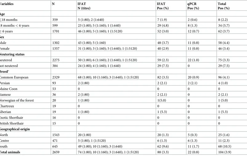
The number of sampled cats (i.e., 2,659) enrolled from northern (n = 1,543), central (n = 471) and southern (n = 645) Italy exceeded the minimum sample size (i.e., 457, 385 and 246 samples from northern, central and southern Italy, respectively). Owned cats enrolled in the study (i.e., n = 1,302 males and n = 1,357 females) aged from 1 month to 21.3 years (mean 8.3 years, median 9 years). Of these, 359 (13.5%) were less than 18 months old (G1), 599 (22.5%) between 18 months and 6 years old (G2) and 1,701 (64%) elder than 6 years (G3). Most of the animals were common European breed (n = 2,329, 87.6%) and neutered (n = 2,275, 85.5%) (Table 1). The prevalence ofL. infantum infection by serology and/or molecular test in association with
age, sex, breed, neutering status and cat origin is reported inTable 1. Overall, 104 (3.9%) cats were positive toL. infantum by IFAT (88/2,659, 3.3%) and/or by qPCR (22/2,659, 0.8%) with
statistically significant (McNemar test p<0.0001) difference and poor concordance between the techniques, K = 0.097 (95% CI: 0.0176–0.1766). Out of these 104 positive cats, 6 (5.8%) were positive to both qPCR and IFAT with IgG titres of 1:80 (n = 3), 1:640 (n = 2) and 1:5120 (n = 1). Out of the 22 PCR positive cats, 16 (72.7%) were seronegative. Of the seropositive cats, 84.1% (74/88) had an antibody titre of 1:80, whereas in the remaining the titres varied from 1:160 to 1:5120 (Table 1). The prevalence of positive cats detected by serological and/or
molecular tests in the different geographic areas was 10.5% (68/645) in the South, 2.3% (11/ 471) in the Centre and 1.6% (25/1543) in the North of Italy.
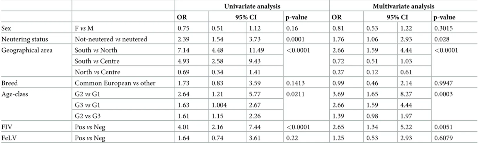
The results of the univariate and multivariate logistic regression analyses are shown in
Table 2. The risk ofL. infantum infection in cats, in the multivariate model (i.e., accounting
for sex and age class) was significantly associated with the geographical areas (p<0.0001). In particular, cats living in the South of Italy were related to higher risk ofL. infantum infection
than those in the North by the univariate (OR = 7.14; 95% CI: 4.48–11.49) and multivariate analysis (OR = 2.66, 95% CI: 1.59–4.44), and those in the Centre by the univariate analysis (OR = 4.93; 95% CI: 2.58–9.43). Animals positive toL. infantum (n = 104; 3.9%) aged from 6
months to 20.1 years old (mean 7.5 years, standard deviation 5 years), while negative cats aged from 1 month to 21.3 years (mean 8.4 years, standard deviation 5.4 years) old with no statisti-cally significant difference between the two groups (t = 1.795, p = 0.0728). For cats in G2 (i.e., 19 months to 6 years old) a higherL. infantum prevalence (5.7%, 34/599) (χ2= 6.806;p =
0.0333) was recorded. The multivariate regression analysis showed that these G2 cats were related to a higher risk ofL. infantum infection than those in G1 (i.e., �18 months) (OR =
3.69; 95% CI: 1.65–8.27). The neutering status was a statistically significant factor associated with positivity toL. infantum. In particular, the risk of not neutered compared to neutered cats
was OR = 1.76 (95% CI: 1.06–2.93) and statistically significant (p = 0.028). No statistical
Table 1. Association between variables: Age, sex, breed, reproductive status and cat origin and the serological and molecular positivity forLeishmania infantum.
Variables N IFAT N (titre) IFAT Pos (%) qPCR Pos (%) Total Pos (%) Age �18 months 359 5 (1:80); 2 (1:640) 7 (1.9) 2 (0.6) 8 (2.2) 18 months < 6 years 599 23 (1:80); 5 (1:160); 1 (1:640) 29 (4.8) 8 (1.3) 34 (5.7) � 6 years 1701 46 (1:80); 5 (1:160); 1 (1:5120) 52 (3.0) 12 (0.7) 62 (3.7) Sex Male 1302 43 (1:80); 5 (1:160) 48 (3.7) 11 (0.8) 58 (4.4) Female 1357 31 (1:80); 3 (1:160); 5 (1:640); 1 (1:5120) 40 (2.9) 11 (0.8) 46 (3.4) Neutering status neutered 2275 50 (1:80); 6 (1:160); 2 (1:640); 1 (1:5120) 59 (2.3) 22 (1.0) 75 (3.3) not neutered 384 24 (1:80); 4 (1:160); 1 (1:640) 29 (7.5) 0 29 (7.5) Breed� Common European 2329 68 (1:80); 10 (1:160); 3 (1:640); 1 (1:5120) 82 (3.5) 20 (0.9) 96 (4.1) Persian 93 2 (1:80) 2 (2.1) 2 (2.1) 4 (1.0) Maine Coon 53 0 0 0 0 Siamese 36 2 (1:80) 2 (2.1) 0 2 (2.1)
Norwegian of the forest 20 1 (1:80) 1(5.0) 0 1 (5.0)
Chartreux 19 0 0 0 0 Siberian 19 1 (1:80) 1 (5.3) 0 1 (5.3) Exotic Shorthair 16 0 0 0 0 British Shorthair 15 0 0 0 0 Geographical origin North 1543 20 (1:80) 20 (1.3) 5 (0.3) 25 (1.6) Centre 471 5 (1:80); 1 (1:5120) 6 (1.3) 6 (1.3) 11 (2.3) South 645 49 (1:80); 10 (1:160); 3 (1:640) 62 (9.6) 11 (1.7) 68 (10.5) Total animals 2659 74 (1:80); 10 (1:160); 3 (1:640); 1 (1:5120) 88 (3.3) 22 (0.8) 104 (3.9)
�only breeds with � 15 animals listed
association was found betweenLeishmania infected cats and sex in neither the univariate
(p = 0.016), nor the multivariate model (p = 0.3015). Equally no association was observed for the breed variable (univariate analysis: p = 0.1413; multivariate analysis: p = 0.9947). Overall, 115 and 101 cats were positive to FeLV (4.3%) or FIV (3.8%) respectively, of which 6.1% (7/ 115) for FeLV and 12.9% (13/101) for FIV were also positive toLeishmania. Out of the 13 cats
coinfected with FeLV and FIV, three (23.1%) were alsoLeishmania positive. Leishmania infan-tum infection was significantly associated only to FIV infection resulting in the multivariable
model with an OR = 2.65 (95% CI: 1.34–5.22, p = 0.0051).
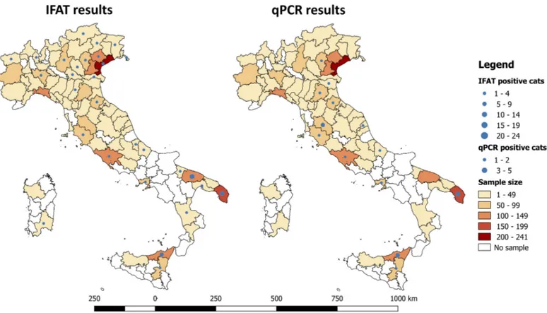
At the GIS analysis, the highest number of cats (i.e., >100 cats) was sampled in 8 out of 83 provinces examined in the study area (i.e., n = 4 in the North, n = 1 in the Centre and n = 3 in the South of Italy). The geographical analysis identified the presence of three areas with a significant higher proportion ofL. infantum seropositive cats, located in three provinces of southern Italy
(i.e., 13.5% in Bari, 11.4% in Messina and 6.4% in Lecce; Chi-square test for linear trend = 23.4224,
df = 1, p = <0.0001) (Fig 1). In two of these three provinces (i.e., Messina and Lecce) a higher pro-portion of qPCR positive samples (3.8% and 1.6%, respectively) was also recorded (Fig 1).
Discussion
The present study represents the largest epidemiological survey combining serological and molecular testing to assess the occurrence ofL. infantum in cats to date.
The prevalence of FeL recorded by serology (3.3%) and qPCR (0.8%) indicates that cats are exposed to, or infected by,L. infantum throughout Italy. The overall seroprevalence of 3.3% is
similar to that recorded in cats from central Spain (1.3–3.2%) and Portugal (2.8%) [12,31]. Higher seroprevalence data have been reported in cats from highly endemic regions of the Mediterranean basin for CanL, such as in southern Spain (28.3%) [19], central (16.3%) [26] and southern Italy (25.8%) [9] and Turkey (15.2%) [24]. It means that in areas where CanL is endemic, cats are likely to be more exposed toL. infantum as compared to areas where the
prevalence of CanL is low [9]. Nonetheless, in our study different proportions of the popula-tion of infected animals were recorded in northern (1.6%) and centre (2.3%) versus southern (10.5%) Italy. AlthoughL. infantum has spread throughout Italy [32], the southern and insular regions are still considered more endemic areas for human VL and CanL [25,33], as a result of the favourable geographical climate conditions that allow the presence and abundance of sand
Table 2. Odds ratio and 95% confidence interval from logistic regression analysis for cats positive toLeishmania infantum by IFATand/or qPCR.
Univariate analysis Multivariate analysis
OR 95% CI p-value OR 95% CI p-value
Sex Fvs M 0.75 0.51 1.12 0.16 0.81 0.53 1.22 0.3015
Neutering status Not-neuteredvs neutered 2.39 1.54 3.73 0.0001 1.76 1.06 2.93 0.028 Geographical area Southvs North 7.14 4.48 11.49 <0.0001 2.66 1.59 4.44 <0.0001
Southvs Centre 4.93 2.58 9.43 0.72 0.51 1.03
Northvs Centre 0.69 0.34 1.41 0.27 0.12 0.61
Breed Common European vs other 1.73 0.83 3.59 0.1413 0.99 0.46 2.14 0.9947
Age-class G2vs G1 2.64 1.21 5.77 0.0211 3.69 1.65 8.27 0.0003
G3vs G1 1.63 1.004 2.67 2.66 1.59 4.44
G2 vs G3 1.61 1.15 2.26 1.39 0.98 1.97
FIV Posvs Neg 4.01 2.16 7.44 <0.0001 2.65 1.34 5.22 0.0051
FeLV Posvs Neg 1.64 0.74 3.61 0.22 1.25 0.53 2.93 0.6079
CI: 95% confidence interval; OR: odds ratio
fly vectors in the whole country (e.g.,Phlebotomus perniciosus, Phlebotomus perfiliewi, Phlebo-tomus ariasi, and PhleboPhlebo-tomus neglectus) [25]. Accordingly, the occurrence ofL. infantum in
wild animals has been reported mainly in the centre and south of Italy [34–36].
In addition, current data available in the literature on the seroprevalence of FeL have been gained using different diagnostic methodologies and cut-off values, representing a limiting fac-tor for data comparison. For example, for the IFAT, the percentage of seropositive animals was higher at a cut-off of 1:40 (i.e., 25.8% and 28.3%) [9,19], than that at 1:64 (i.e., 0.7%) [37], 1:100 (3.2%) [31] and 1:80 (3.3%; present study). Though, an IFAT cut-off (1:80) has been recom-mended by LeishVet group [8], more comprehensive and standardized protocols and ideally a defined gold standard procedure would allow a more consistent diagnosis ofL. infantum
infec-tion in cats and contribute to a better understanding of the role of cats in the epidemiology of zoonotic VL.
The higher seroprevalence recorded in cats from southern (9.6%) versus central (1.3%) and northern (1.3%) regions of Italy suggests that cats are more exposed toL. infantum in the
southern regions, as reported by previous serologic investigations conducted in Liguria and Tuscany (i.e., 0.9%) [18] and in Sicily and Calabria (southern Italy) (i.e., 6.9%) [38]. This is fur-ther supported by the GIS analysis that indicates a significant higher proportion ofL. infantum
positive cats in three areas of southern Italy. Likewise, though the distribution pattern ofL. infantum infection is changing throughout the country as a result of many biological and
eco-logical factors [32], CanL mainly occurs in southern Italy [25].
Since no information on the cats’ life style (i.e., outdoor/indoor access) and on sand fly pop-ulation during the sample collection are available, the role of age and neutering status as risk factors for the occurrence of infections in cats, may be explained by differences in behaviour.
Fig 1. Geographical localization of areas, indicated by provinces, from where cats positive toLeishmania infantum by serological and molecular tests were
collected. The location ofL. infantum positive cats was geo-referenced using a geographical information system (GIS, ArcGIS version 10.3 ESRI).
Indeed, not neutered and older than 18 months cats are more prone to an outdoor life style than younger and neutered cats, due to more pronounced predatory instinct [39]. Therefore, these cats are more exposed to sand fly bites, which over time, results in a higher risk ofL. infantum infection. Undoubtedly, the prevalence of cats positive for FeL at the molecular tests
also depends on the cat life style and therefore exposure to sand fly bites with the higher num-ber of animals positive to DNA recorded in feral and stray cats from Spain (i.e., 8.7% and 26%) [5,15] and Portugal (30.4%) [40].
Leishmania infantum DNA has been detected in blood samples of only a few cats (0.8%) as
already reported in Portugal (0.3%) [41], northern (1.1%) [42] and southern Italy (2.1%) [9] and Cyprus (2.3%) [20]. This may suggest that blood is not the ideal tissue for molecular diag-nosis ofL. infantum infection in cats, as for dogs. Indeed, when comparing different matrixes
for the molecular diagnosis of FeL, conjunctival swab (16.7%) and lymph node aspiration (11.7%) showed to be more sensitive than blood (7.8%) [37], as also reported for the diagnosis of CanL [27,43]. Though the number of samples positive with molecular diagnosis (n = 22) was limited in this study, the molecular positivity forL. infantum parallels the occurrence of
higher seroprevalence in the examined animals, as reported previously [5,15]. The high per-centage of molecular positivity and thus occurrence ofL. infantum DNA in seronegative
ani-mals (72.7%, 16/22) might either indicate that these cats were at an early stage of the infection or endorse that they are less susceptible compared to dogs [10]. While there is a correlation between molecular and serological positivity in dogs infected for CanL [9,13], there is none between molecular and serological tests for FeL, supporting the different immune responsive-ness of dogs and cats toL. infantum.
Longitudinal studies in cats could contribute in determining the course ofL. infantum
infection after natural exposure over time. Further, transmission studies with competent sand fly vectors could provide more information about the role of cats as reservoir hosts ofL. infan-tum. So far, P. perniciosus has shown to feed on cats [44] and allow the developing of the parasite after the blood ingestion [11] thus potentially enabling further transmission ofL. infantum.
SinceP. perniciosus is the main vector of L. infantum in Italy [45] and the most abundant phle-botomine sand fly species in some areas of southern Italy [46], further observations are needed to elucidate the role of naturally infected cats in sustaining theL. infantum life cycle.
The significant association betweenL. infantum and FIV infections (p = 0.0051) was
previ-ously reported [16,47] and indicates that immunosuppressive agents, such as FIV, impair the cellular immune response, thus increasing the risk for FeL. This correlation is supported by lit-erature showing a high prevalence of FIV infection (i.e., 30%) inL. infantum infected cats [8]. Therefore, FIV andL. infantum coinfections might predispose animals to visceral forms, as an
effect of the viral immunosuppression, as recognized in HIV seropositive patients [48]. In conclusion, differences in immune responses between dogs and cats and scant data on the ability of vectors in the transmission ofL. infantum on natural infected cats, complicate the
appreciation of the role of cats inL. infantum epidemiology. As future perspectives, the
stan-dardization of procedures for a prompt diagnosis ofL. infantum infection and for screening
cat populations is a crucial task for a better understanding of the epidemiology of FeL, and the role of cats as reservoir hosts. In addition, prevention measures for providing protection against the infection and treatment strategies for cats infected byL. infantum need to be
fur-ther addressed.
Acknowledgments
The authors would like to thank Anna Attanasi (Centro Veterinario Salentino, San Cesario di Lecce, Lecce, Italy), Chiara Cavallaro (Laboratorio Analisi Veterinarie PET Diagnostic LAB,
Catania, Italy), Carmela Coccioli (Azienda Sanitaria Locale, Bari, Italy), Raffaele Maglione (Laboratorio Analisi Veterinarie LABFORVET, Napoli, Italy) and Romana Romano (Labora-torio Analisi Veterinarie Rtr Vet, Lecce, Italy) for their support with the sample collection.
Author Contributions
Conceptualization: Roberta Iatta, Domenico Otranto.
Data curation: Roberta Iatta, Tommaso Furlanello, Emanuele Brianti, Domenico Otranto. Formal analysis: Paolo Trerotoli.
Funding acquisition: Roberta Iatta, Domenico Otranto.
Investigation: Roberta Iatta, Maria Stefania Latrofa, Nicola Decaro.
Methodology: Roberta Iatta, Viviana Domenica Tarallo, Emanuele Brianti, Paolo Trerotoli,
Eleonora Lorusso.
Project administration: Roberta Iatta. Software: Paolo Trerotoli.
Supervision: Roberta Iatta, Domenico Otranto. Validation: Roberta Iatta.
Visualization: Roberta Iatta.
Writing – original draft: Roberta Iatta, Domenico Otranto.
Writing – review & editing: Roberta Iatta, Vito Colella, Emanuele Brianti, Bettina Schunack,
Guadalupe Mirò, Filipe Dantas-Torres, Domenico Otranto.
References
1. World Health Organization. Leishmaniasis 2018 (22nd December).http://www.who.int/mediacentre/ factsheets/fs375/en/.
2. Alvar J, Ve´lez ID, Bern C, Herrero M, Desjeux P, Cano J, et al. Leishmaniasis Worldwide and Global Estimates of Its Incidence. PLoS One 2012; 7(5):e35671.https://doi.org/10.1371/journal.pone. 0035671PMID:22693548
3. Quinnell RJ, Courtenay O. Transmission, reservoir hosts and control of zoonotic visceral leishmaniasis. Parasitology 2009; 136:1915–34.https://doi.org/10.1017/S0031182009991156PMID:19835643 4. Solano-Gallego L, Koutinas A, Miro´ G, Cardoso L, Pennisi MG, Ferrer L, et al. Directions for the
diagno-sis, clinical staging, treatment and prevention of canine leishmaniosis. Vet Parasitol. 2009; 165(1–2):1– 18.https://doi.org/10.1016/j.vetpar.2009.05.022PMID:19559536
5. Milla´n J, Ferroglio E, Solano-Gallego L. Role of wildlife in the epidemiology of Leishmania infantum infection in Europe. Parasitol. Res. 2014; 113:2005–14.https://doi.org/10.1007/s00436-014-3929-2
PMID:24804923
6. Otranto D, Cantacessi C, Pfeffer M, Dantas-Torres F, Brianti E, Deplazes P, et al. The role of wild canids and felids in spreading parasites to dogs and cats in Europe. Part I: Protozoa and tick-borne agents. Vet Parasitol. 2015; 213(1–2):12–23.https://doi.org/10.1016/j.vetpar.2015.04.022PMID:
26003669
7. Molina R, Jime´ nez MI, Cruz I, Iriso A, Martı´n-Martı´n I, Sevillano O, et al. The hare (Lepus granatensis) as potential sylvatic reservoir of Leishmania infantum in Spain. Vet Parasitol. 2012; 190(1–2):268–71.
https://doi.org/10.1016/j.vetpar.2012.05.006PMID:22677135
8. Pennisi MG, Cardoso L, Baneth G, Bourdeau P, Koutinas A, Miro´ G, et al. LeishVet update and recom-mendations on feline leishmaniosis. Parasit Vectors. 2015; 8:302. https://doi.org/10.1186/s13071-015-0909-zPMID:26041555
9. Otranto D, Napoli E, Latrofa MS, Annoscia G, Tarallo VD, Greco G, et al. Feline and canine leishmanio-sis and other vector-borne diseases in the Aeolian Islands: Pathogen and vector circulation in a
confined environment. Vet Parasitol. 2017; 236:144–51.https://doi.org/10.1016/j.vetpar.2017.01.019
PMID:28288759
10. Day MJ. Cats are not small dogs: Is there an immunological explanation for why cats are less affected by arthropod-borne disease than dogs? Parasit Vectors. 2016; 9(1):507.https://doi.org/10.1186/ s13071-016-1798-5PMID:27646278
11. Maroli M, Pennisi MG, Di Muccio T, Khoury C, Gradoni L, Gramiccia M. Infection of sandflies by a cat naturally infected with Leishmania infantum. Vet Parasitol. 2007; 145(3–4):357–60.https://doi.org/10. 1016/j.vetpar.2006.11.009PMID:17174035
12. Cardoso L, Lopes AP, Sherry K, Schallig H, Solano-Gallego L. Low seroprevalence of Leishmania
infantum infection in cats from northern Portugal based on DAT and ELISA. Vet Parasitol. 2010; 174
(1–2):37–42.https://doi.org/10.1016/j.vetpar.2010.08.022PMID:20851524
13. Maia C, Gomes J, Cristo´ vão J, Nunes M, Martins A, Rebêlo E, et al. Feline Leishmania infection in a canine leishmaniasis endemic region, Portugal. Vet Parasitol. 2010; 174(3–4):336–40.https://doi.org/ 10.1016/j.vetpar.2010.08.030PMID:20869810
14. Colella V, HodzˇićA, Iatta R, Baneth G, AlićA, Otranto D. Zoonotic Leishmaniasis, Bosnia and Herzego-vina. Emerg Infect Dis. 2019; 25(2).https://doi.org/10.3201/eid2502.181481PMID:30511917 15. Sherry K, Miro´ G, Trotta M, Miranda C, Montoya A, Espinosa C, et al. A serological and molecular study
of Leishmania infantum infection in cats from the Island of Ibiza (Spain). Vector Borne Zoonotic Dis. 2011; 11(3):239–45.https://doi.org/10.1089/vbz.2009.0251PMID:20804432
16. Sobrinho LS, Rossi CN, Vides JP, Braga ET, Gomes AA, de Lima VM, et al. Coinfection of Leishmania
chagasi with Toxoplasma gondii, Feline Immunodeficiency Virus (FIV) and Feline Leukemia Virus
(FeLV) in cats from an endemic area of zoonotic visceral leishmaniasis. Vet Parasitol. 2012; 187(1– 2):302–6.https://doi.org/10.1016/j.vetpar.2012.01.010PMID:22285010
17. Sergent E, Lonbard J, Quilichini M. La leishmaniose a Alger. Infection simultanee d’un enfant, d’un chien et d’un chat dans la meme habitation. Bull Soc Path Exot. 1912: 5;93–98.
18. Poli A, Abramo F, Barsotti P, Leva S, Gramiccia M, Ludovisi A, Mancianti F. Feline leishmaniosis due to
Leishmania infantum in Italy. Vet Parasitol. 2002; 106(3):181–91. PMID:12062507
19. Martı´n-Sa´nchez J, Acedo C, Muñoz-Pe´rez M, Pesson B, Marchal O, Morillas-Ma´rquez F. Infection by
Leishmania infantum in cats: epidemiological study in Spain. Vet Parasitol. 2007; 145(3–4):267–73. https://doi.org/10.1016/j.vetpar.2006.11.005PMID:17157440
20. Attipa C, Papasouliotis K, Solano-Gallego L, Baneth G, Nachum-Biala Y, Sarvani E et al. Prevalence study and risk factor analysis of selected bacterial, protozoal and viral, including vector-borne, patho-gens in cats from Cyprus. Parasit Vectors. 2017; 10(1):130. https://doi.org/10.1186/s13071-017-2063-2PMID:28285597
21. Akhtardanesh B, Sharifi I, Mohammadi A, Mostafavi M, Hakimmipour M, Pourafshar NG. Feline visceral leishmaniasis in Kerman, southeast of Iran: Serological and molecular study. J Vector Borne Dis. 2017; 54(1):96–102. PMID:28352052
22. Metzdorf IP, da Costa Lima MS Junior, de Fatima Cepa Matos M, de Souza Filho AF, de Souza Tsuji-saki RA, Franco KG, et al. Molecular characterization of Leishmania infantum in domestic cats in a region of Brazil endemic for human and canine visceral leishmaniasis. Acta Trop. 2017; 166:121–5.
https://doi.org/10.1016/j.actatropica.2016.11.013PMID:27851895
23. Persichetti MF, Pennisi MG, Vullo A, Masucci M, Migliazzo A, Solano-Gallego L. Clinical evaluation of outdoor cats exposed to ectoparasites and associated risk for vector-borne infections in southern Italy. Parasit Vectors. 2018; 11(1):136.https://doi.org/10.1186/s13071-018-2725-8PMID:29554931 24. Can H, Do¨şkaya M, O¨ zdemir HG,Şahar EA, Karakavuk M, PektaşB, et al., Seroprevalence of Leishmania
infection and molecular detection of Leishmania tropica and Leishmania infantum in stray cats ofİzmir, Tur-key. Exp Parasitol. 2016; 167:109–14.https://doi.org/10.1016/j.exppara.2016.05.011PMID:27260567 25. Maroli M, Rossi L, Baldelli R, Capelli G, Ferroglio E, Genchi C, et al. The northward spread of
leishmani-asis in Italy: evidence from retrospective and ongoing studies on the canine reservoir and phlebotomine vectors. Trop Med Int Health. 2008; 13(2):256–64.https://doi.org/10.1111/j.1365-3156.2007.01998.x
PMID:18304273
26. Vita S, Santori D, Aguzzi I, Petrotta E, Luciani A. Feline leishmaniasis and ehrlichiosis: serological investigation in Abruzzo region. Vet Res Commun. 2005; 29 Suppl 2:319–21.
27. Otranto D, Paradies P, de Caprariis D, Stanneck D, Testini G, Grimm F, et al. Toward diagnosing
Leish-mania infantum infection in asymptomatic dogs in an area where leishLeish-maniasis is endemic. Clin Vaccine
Immunol. 2009; 16(3):337–43.https://doi.org/10.1128/CVI.00268-08PMID:19129471
28. Francino O, Altet L, Sa´nchez-Robert E, Rodriguez A, Solano-Gallego L, Alberola J, et al. Advantages of real-time PCR assay for diagnosis and monitoring of canine leishmaniosis. Vet Parasitol. 2006; 137(3– 4):214–21.https://doi.org/10.1016/j.vetpar.2006.01.011PMID:16473467
29. Endo Y, Cho KW, Nishigaki K, Momoi Y, Nishimura Y, Mizuno T, et al. Molecular characteristics of malignant lymphomas in cats naturally infected with feline immunodeficiency virus. Vet Immunol Immu-nopathol. 1997; 57(3–4):153–67. PMID:9261955
30. Stiles J, Bienzle D, Render JA, Buyukmihci NC, Johnson EC. Use of nested polymerase chain reaction (PCR) for detection of retroviruses from formalin-fixed, paraffin-embedded uveal melanomas in cats. Vet Ophthalmol. 1999; 2(2):113–1 PMID:11397251
31. Miro´ G, Rupe´rez C, Checa R, Ga´lvez R, Herna´ndez L, Garcı´a M, et al. Current status of L. infantum infection in stray cats in the Madrid region (Spain): implications for the recent outbreak of human leish-maniosis? Parasit Vectors. 2014; 7:112.https://doi.org/10.1186/1756-3305-7-112PMID:24655361 32. Otranto D, Capelli G, Genchi C. Changing distribution patterns of canine vector borne diseases in Italy:
leishmaniosis vs. dirofilariosis. Parasit Vectors. 2009; 2 Suppl 1:S2. https://doi.org/10.1186/1756-3305-2-S1-S2PMID:19426441
33. Mannocci A, La Torre G, Chiaradia G, De Waure C, Mainelli MT, Cernigliaro A, et al. Epidemiology and direct medical costs of human leishmaniasis in Italy. J Prev Med Hyg. 2007; 48(1):27–36. PMID:17506235 34. Dipineto L, Manna L, Baiano A, Gala M, Fioretti A, Gravino AE, et al. Presence of Leishmania infantum
in red foxes (Vulpes vulpes) in southern Italy. J Wildl Dis. 2007; 43(3):518–20.https://doi.org/10.7589/ 0090-3558-43.3.518PMID:17699092
35. Verin R, Poli A, Ariti G, Nardoni S, Fanucchi Bertuccelli M, Mancianti F. Detection of Leishmania infan-tum DNA in tissues of free ranging red foxes (Vulpes vulpes) in Central Italy. Eur J Wildl Res. 2010; 56:689–692.
36. Piantedosi D, Veneziano V, Di Muccio T, Manzillo VF, Fiorentino E, Scalone A, et al. Epidemiological survey on Leishmania infection in red foxes (Vulpes vulpes) and hunting dogs sharing the same rural area in Southern Italy. Acta Parasitol. 2016; 61(4):769–775.https://doi.org/10.1515/ap-2016-0106
PMID:27787204
37. Silaghi C, Knaus M, Rapti D, Kusi I, Shukullari E, Hamel D, et al. Survey of Toxoplasma gondii and Neospora caninum, haemotropic mycoplasmas and other arthropod-borne pathogens in cats from Albania. Parasit Vectors. 2014; 7:62.https://doi.org/10.1186/1756-3305-7-62PMID:24517118 38. Pennisi MG, Lupo T, Malara D, Masucci M, Migliazzo A, Lombardo G. Serological and molecular
preva-lence of Leishmania infantum infection in cats from Southern Italy. J Feline Med Surg. 2012; 14:656–7.
39. Overall KL, Rodan I, Beaver BV, Carney H, Crowell-Davis S, Hird N, et al. Feline behavior guidelines from the American Association of Feline Practitioners. J Am Vet Med Assoc. 2005; 227(1):70–84. PMID:16013540
40. Maia C, Nunes M, Campino L. Importance of cats in zoonotic Leishmaniasis in Portugal. Vector Borne Zoonotic Dis. 2008; 8(4); 555–9.https://doi.org/10.1089/vbz.2007.0247PMID:18471058
41. Vilhena H, Martinez-Dı´az VL, Cardoso L, Vieira L, Altet L, Francino O, et al. Feline vector-borne patho-gens in the north and centre of Portugal. Parasit Vectors. 2013; 6:99. https://doi.org/10.1186/1756-3305-6-99PMID:23587366
42. Spada E, Canzi I, Baggiani L, Perego R, Vitale F, Migliazzo A, et al. Prevalence of Leishmania infantum and co-infections in stray cats in northern Italy. Comp Immunol Microbiol Infect Dis. 2016; 45:53–8.
https://doi.org/10.1016/j.cimid.2016.03.001PMID:27012922
43. Maia C, Ramada J, Cristo´vão JM, Gonc¸alves L, Campino L. Diagnosis of canine leishmaniasis: conven-tional and molecular techniques using different tissues. Vet J. 2009; 179(1):142–4.https://doi.org/10. 1016/j.tvjl.2007.08.009PMID:17936654
44. Gonza´ lez E, Jime´nez M, Herna´ndez S, Martı´n-Martı´n I, Molina R. Phlebotomine sand fly survey in the focus of leishmaniasis in Madrid, Spain (2012–2014): seasonal dynamics, Leishmania infantum infec-tion rates and blood meal preferences. Parasit Vectors. 2017; 10(1):368.https://doi.org/10.1186/ s13071-017-2309-zPMID:28764772
45. Maroli M, Feliciangeli MD, Bichaud L, Charrel RN, Gradoni L. Phlebotomine sandflies and the spreading of leishmaniases and other diseases of public health concern. Med Vet Entomol. 2013; 27(2):123–47.
https://doi.org/10.1111/j.1365-2915.2012.01034.xPMID:22924419
46. Tarallo VD, Dantas-Torres F, Lia RP, Otranto D. Phlebotomine sand fly population dynamics in a leish-maniasis endemic peri-urban area in southern Italy. Acta Trop. 2010; 116(3):227–34.https://doi.org/10. 1016/j.actatropica.2010.08.013PMID:20816927
47. Spada E, Proverbio D, Migliazzo A, Della Pepa A, Perego R, Bagnagatti De Giorgi G. Serological and molecular evaluation of Leishmania infantum infection in stray cats in a nonendemic area in Northern Italy. ISRN Parasitol. 2013; 2013:916376.https://doi.org/10.5402/2013/916376PMID:27335864 48. Alvar J, Aparicio P, Aseffa A, Den Boer M, Cañavate C, Dedet JP, et al. The relationship between
leish-maniasis and AIDS: the second 10 years. Clin Microbiol Rev. 2008; 21(2):334–59, table of contents.