Thoraco-abdominal coordination and
performance during uphill running at altitude
Eva Bernardi1*, Lorenza Pratali2, Gaia Mandolesi1, Maria Spiridonova1, Giulio Sergio Roi3, Annalisa Cogo11 Biomedical Sport Studies Centre, University of Ferrara, Ferrara, Italy, 2 Institute of Clinical Physiology, National Research Council, Pisa, Italy, 3 Isokinetic Medical Group, Education and Research Department, Bologna, Italy
Abstract
Introduction
Running races on mountain trails at moderate-high altitude with large elevation changes throughout has become increasingly popular. During exercise at altitude, ventilatory demands increase due to the combined effects of exercise and hypoxia.
Aim
To investigate the relationships between thoraco-abdominal coordination, ventilatory pat-tern, oxygen saturation (SpO2), and endurance performance in runners during high-intensity
uphill exercise.
Methods
Fifteen participants (13 males, mean age 42±9 yrs) ran a “Vertical Kilometer,” i.e., an uphill run involving a climb of approximately 1000 m with a slope greater than 30%. The athletes were equipped with a portable respiratory inductive plethysmography system, a finger pulse oximeter and a global positioning unit (GPS). The ventilatory pattern (ventilation (VE), tidal volume (VT), respiratory rate (RR), and VE/VT ratio), thoraco-abdominal coordination, which is represented by the phase angle (PhA), and SpO2were evaluated at rest and during
the run. Before and after the run, we assessed respiratory function, respiratory muscle strength and the occurrence of interstitial pulmonary edema by thoracic ultrasound.
Results
Two subjects were excluded from the respiratory inductive plethysmography analysis due to motion artifacts. A quadratic relationship between the slope and the PhA was observed (r = 0.995, p = 0.036). When the slope increased above 30%, the PhA increased, indicating a reduction in thoraco-abdominal coordination. The reduced thoraco-abdominal coordina-tion was significantly related to reduced breathing efficiency (i.e., an increased VE/VT ratio; r = 0.961, p = 0.038) and SpO2(r = -0.697, p<0.001). Lower SpO2values were associated
with lower speeds at 20% slope40% (r = 0.335, p<0.001 for horizontal and r = 0.36, a1111111111 a1111111111 a1111111111 a1111111111 a1111111111 OPEN ACCESS
Citation: Bernardi E, Pratali L, Mandolesi G, Spiridonova M, Roi GS, Cogo A (2017) Thoraco-abdominal coordination and performance during uphill running at altitude. PLoS ONE 12(3): e0174927.https://doi.org/10.1371/journal. pone.0174927
Editor: Laurent Mourot, University of France Comte´, FRANCE
Received: August 8, 2016 Accepted: March 18, 2017 Published: March 31, 2017
Copyright: © 2017 Bernardi et al. This is an open access article distributed under the terms of the Creative Commons Attribution License, which permits unrestricted use, distribution, and reproduction in any medium, provided the original author and source are credited.
Data Availability Statement: All relevant data are within the paper and its Supporting Information files.
Funding: The authors received no specific funding for this work.
Competing interests: The authors have declared that no competing interests exist.
p<0.001 for vertical). The reduced thoraco-abdominal coordination and consequent reduc-tion in SpO2were associated with interstitial pulmonary edema.
Conclusion
Reductions in thoraco-abdominal coordination are associated with a less efficient ventilatory pattern and lower SpO2during uphill running. This fact could have a negative effect on
performance.
Introduction
Running races on mountain trails at moderate-high altitude with large elevation changes throughout has become increasingly popular. High-intensity exercise under these environ-mental conditions requires extensive respiratory and cardiovascular system engagement. The respiratory and cardiac responses that occur during this type of exercise represent key constit-uents of endurance performance that can be affected by oxygen desaturation and increased respiratory work [1–3]. Indeed, during high-intensity exercise at altitude, ventilatory demands increase due to the combined effects of exercise and hypoxia, which affect the ventilatory sys-tem above 2000–2500 m [4–6]. Under such conditions, the respiratory muscles become strained and can experience progressive fatigue, and lung function can be impaired [3,7]. The postural tasks related to the presence of steep slopes and different types of terrain [8] may also affect the ventilatory system through modifications of thoraco-abdominal coordination, which involves the coordinated action of the diaphragm and the abdominal muscles to inflate and deflate the chest. A reduction in thoraco-abdominal coordination may in turn alter ventilatory patterns and reduce ventilatory efficiency [9]. Ventilatory efficiency is defined as the amount of ventilation (VE) required to achieve a given level of oxygen saturation (SpO2) during
spon-taneous breathing [10]. Specifically, ventilatory patterns characterized by a higher tidal volume (VT) and reduced respiratory rate (RR) have been found to be the most efficient due to the reduction in dead space ventilation [11–14]. This ventilatory pattern can play an important role in SpO2and therefore can influence respiratory and cardiac responses and exercise
perfor-mance at altitude [10,12].
Ventilatory pattern and thoraco-abdominal coordination are important for determining ventilatory efficiency and can represent a key tool for functional evaluations of athletes’ perfor-mance. To the best of our knowledge, despite the importance of these aspects, few studies have examined ventilatory pattern and thoraco-abdominal motion in healthy adults both at rest and during exercise [15–17]. Romei et al. [16] showed that chest wall kinematics and ventila-tory pattern are significantly influenced by body position during rest in healthy subjects. How-ever, no study has focused on changes in these parameters during dynamic changes in body position.
Moreover, the development of interstitial pulmonary edema due to both strenuous exercise and hypoxia [18,19] can contribute to changes in lung function. Studies have frequently shown a significant increase in pulmonary extravascular water following strenuous exercise, leading to mild interstitial pulmonary edema. Although small increases in interstitial water in the lungs do not cause overt clinical symptoms, they could contribute to lung-related limitations to exercise performance [20]. Interstitial pulmonary edema has been shown in endurance ath-letes after intense exercise at sea level using different imaging techniques such as chest X-ray, magnetic resonance imaging, computed tomography, and scintigraphy [21–23]. Currently,
chest ultrasounds are a common method to detect the presence of pulmonary extravascular water based on the presence of multiple diffuse bilateral B lines [24]. Ultrasound B lines (US-B lines) are considered a sonographic sign of lung interstitial syndrome. US-B lines are defined as discrete laser-like vertical hyperechoic reverberation artifacts that arise from the pleural line, extend to the bottom of the screen without fading, and move synchronously with the slid-ing of the lung [24].
In the present study, we hypothesized that during strenuous uphill exercise at moderate alti-tude, the preservation of a coordinated movement between the chest wall and abdomen (i.e., the two compartments move synchronously during the ventilatory cycle) would be associated with more efficient breathing, higher SpO2levels and reduced ventilatory demands. Moreover,
we hypothesized that all these factors would influence endurance performance and cardiore-spiratory responses. We predicted that steeper slopes might prompt subjects to change their postures, leading to reductions in thoraco-abdominal coordination and the above-reported effects.
To examine these hypotheses, we tested competitive athletes taking part in skyrunning dur-ing an uphill run above 2000 m. Skyrunndur-ing involves runndur-ing on mountain trails at an altitude of at least 2000 m and with ground slopes that can exceed 30%. Respiratory function and the effects of increased slopes on thoraco-abdominal coordination and breathing efficiency were examined via analyses of ventilatory parameters and SpO2, which was measured via finger
pulse oximetry (a method widely utilized and validated for field research) [25]. We also assessed the possible occurrence of interstitial pulmonary edema via thoracic ultrasound as a contributing factor limiting performance under these conditions.
Materials and methods
Fifteen endurance-trained, nationally competitive skyrunners (13 males) were recruited with the aid of the International Skyrunning Federation. The study was conducted in accordance with the Declaration of Helsinki and was approved by the Ethics and Research Committee of the Medical School of the University of Ferrara (protocol number 120387). All participants provided their informed written consent to participate in the study. The exclusion criteria were injuries and illnesses that impaired normal training in the three months prior to the study. No participant was excluded.
Experimental overview
The athletes performed a simulated individual Vertical Kilometer, i.e., a run with approxi-mately 1000 m of vertical climbing over variable terrain with a substantial incline, not exceed-ing five kilometers in length (International Skyrunnexceed-ing Federation rule 2.4.8) and with slopes that could exceed 30%. The simulated Vertical Kilometer was shortened to 794 m (from Cervi-nia, Italy, at 2030 m to a place slightly above Rifugio Oriondè, Italy, at 2824 m) due to the pres-ence of snow on the upper part of the trail. Along the route, the slopes varied from 2% up to 40% [26] with variable changes along the way. In fact, steep slopes were situated at the begin-ning as well as at the end of the trail (a detailed description is available in the website of Verti-cal Kilometer,www.cervinoxtrail.com). Immediately after the arrival at Rifugio Oriondè, the athletes ran back downhill to the starting point.
Anthropometric data, medical histories and altitude training information were collected using a questionnaire.
Respiratory function
Vital capacity (VC), forced expiratory volume in the first second (FEV1), FEV1/VC ratio and
inspiratory muscle strength (PImax) were measured at rest, before the uphill run (T0) and immediately after the descent (T1) using a portable spirometer (Spiropalm Cosmed; Roma, Italy) and a respiratory pressure meter (MicroRPM Carefusion, UK). All tests were performed according to the ATS/ERS guidelines [27,28]. The predicted reference values used were the val-ues reported in the Global Lung Function Initiative [29].
Ventilation monitoring
During the event, the athletes were outfitted with a portable respiratory inductive plethysmog-raphy system (Lifeshirt VivoMetrics, Inc, Ventura CA, USA), which consisted of a snugly fit-ting elastic garment that incorporated rib cage and abdominal inductance sensors containing shielded electrical conductors connected to signal-processing and recording unit for continu-ous data collection. A finger pulse oximeter was integrated in the system [30]. The sensors were placed by the same investigator for consistency. The recorded data included changes in ventilatory pattern (VE, RR, VT and VE/VT ratio) and thoraco-abdominal coordination, which was expressed as the phase angle index (PhA). The VE/VT ratio is useful for describing the ventilatory response to exercise and indicating the contribution of VT to VE during exer-cise [31,32]. These parameters were measured breath-by-breath throughout the run. The PhA was assessed by computing the contribution of the rib cage to VT and the phase shift between the rib cage and the abdominal excursion. A value of 0˚ indicates total synchrony, and a value of 180˚ indicates total asynchrony between the two compartments. The system was calibrated for each subject at rest before the event. Following the calibration procedure, the subjects were asked to breathe rapidly into a calibration bag with a fixed volume for seven breathing cycles that involved emptying and filling the bag with each breath. This procedure was repeated four times while alternating between sitting and upright positions to evaluate the contributions of the rib cage and abdomen to the VT at different body positions.
GPS
A global positioning system unit (GPS; Garmin Edge 305, USA) was positioned on the arm of the subjects to record the path traveled, altitude, slope and horizontal speed. The Garmin Edge 305 is a satellite-based navigation system provided with an antenna that acquires satellite infor-mation and a barometric altimeter to calculate the elevation. The system records the point-to-point distance; the horizontal speed is calculated as the ratio of distance over time and the slope as the change in elevation over the course of the run. The vertical speed can be subse-quently calculated as the ratio of difference in height over time. The reported accuracy of this GPS model is 5 m for both speed and distance. GPS errors can arise in obstructed conditions, such as indoors or in a narrow valley, but these scenarios were not the case for the run from Cervinia to Rifugio Oriondè. Other potential sources of GPS inaccuracy are atmospheric dis-turbances, but the present study was performed under good weather conditions and with no drop in the barometric pressure.
Ultrasound lung echo
To diagnose the presence of pulmonary extravascular water, ultrasound lung echo assessments were performed. This technique has been compared to gold standard methods [33–35], and many studies have addressed its use in the diagnosis of pulmonary edema, particularly in non-hospital settings [36–39]. Ultrasound chest examinations (Vivid I, General Electric Healthcare
Clinical System, Buckinghamshire, UK) were performed by an experienced cardiologist (L.P.) 30 min before the run and within 10 min after the descent to assess for the presence of intersti-tial pulmonary edema, which was identified based on US-B lines using a cardiac probe (2.5– 3.5 MHz). The cardiologist was unaware of the results of the pulmonary function test, SpO2or
the subjects’ performance. A total of 28 echographic windows were scanned in each anterior and lateral hemithorax from the second to the fourth (on the right side to the fifth) intercostal spaces and from the parasternal line to the axillary line. In each intercostal space, the number of US-B lines signs was recorded at the parasternal, midclavicular anterior axillary, and midax-illary sites. The sum of the US-B lines indicates the extent of extravascular fluid in the lungs. Zero is defined as a complete absence of US-B lines in the investigated area. A score of 5 US-B lines was defined as a normal echographic chest pattern [40].
Data and statistical analysis
The VE, PhA and SpO2data were analyzed with dedicated software (VivoLogic, VivoMetrics,
Inc, Ventura CA, USA).
Only data regarding the ascent were analyzed. Movement artifacts introduced during exer-cise were removed from the collected data by setting the minimal acceptable VT values for breath assessment to 25% of the basal value (i.e., for a VT of 400 mL a deviation less than 100 mL was not considered a breath). Similarly, we removed low SpO2values if they were not
sup-ported by a reasonable heart rate and were therefore likely due to poor signal or motion arti-fact. The ventilatory pattern (VE, VT and RR), PhA and SpO2data were averaged over
one-minute intervals and integrated with the GPS data. To simplify the graphic representation in the X-Y axis, we grouped all values into different classes. The slope values were grouped into four classes (i.e., 0–10%, 10–20%, 20–30%, and 30–40%). The same procedure was applied to the PhA values (i.e., 0–10, 10–13, 13–16, and >16) and the delta SpO2values, which were
expressed as the difference between the SpO2values at rest and those during exercise (i.e.,
-15, <-15 -10, <-10 -5 and <-5 0). All computations were performed using a statistical software package (SPSS version 20.0.0; IBM Corp., Armonk, NY, USA).
Normal distributions of the data were verified using the Kolmogorov-Smirnov test. Polyno-mial regression was used to test the nonlinear relationships of the slope with the PhA. For all ventilatory parameters, mean values and 95% confidence intervals (CI) were calculated. The differences between the means of each parameter measured at T0 and T1 were evaluated using paired Student’s t tests. The effect size was calculated as the standardized mean difference in respiratory function, ventilatory pattern and SpO2data between T0 and T1 and graded
accord-ing to Cohen’sd. Correlations were analyzed based on Pearson correlation coefficients for the dependent variables. Multivariable logistic regression analysis was performed considering speed as the dependent variable and the decrease in SpO2, the increase in VE/VT ratio, the
PhA and the slope as independent variables. The level of significance was set at p<0.05 for all statistical comparisons.
Results
Fifteen experienced and trained skyrunners (13 males, mean age 42±9 yrs, mean body mass index 21.7±2.2 kg/m2) participated in the study. One male and one female were excluded from
the respiratory inductive plethysmography analysis due to motion artifacts. The final study population included thirteen subjects (12 males) for the respiratory analysis while the spiro-metric and echo results refer to the whole group (15 subjects).
The athletes ran uphill for an average of 65.2±14.1 min at a mean horizontal speed of 5.7 ±1.1 km/h and a mean vertical speed of 792±158 m/h while simulating the Vertical Kilometer.
A significant correlation was found between horizontal and vertical speed (p = 0.01). With respect to the downhill run, subjects spent a mean of 30±2.5 min descending to the starting point.
All subjects exhibited normal spirometric values (FEV1, VC, FEV1/VC ratio) and PImax at
T0. VC (p = 0.041) and PImax (p = 0.023) were significantly reduced at T1 compared to T0, although the effect size was trivial (Table 1). The ventilatory pattern (VE, VT and RR) and SpO2data at rest and during the run, expressed as the mean± CI, are reported inTable 2. We
observed a significant and high reduction in SpO2during the run (p<0.001), in addition to the
obvious increase in VE and the ventilatory pattern components.
Only slopes between 0 and 40% were analyzed, as slopes >40% required climbing activity with the use of the hands, which involved unavoidable changes in posture.
When the slope increased to over 30%, we observed a significant increase in PhA, indicating a decreased thoraco-abdominal coordination (Fig 1). The best interpolation of these points was represented by the following quadratic curve: PhA = 12.897–0.773slope + 0.301slope2 (r = 0.995, r2= 0.990, F = 97.389, p = 0.036). Figs2and3present the mean decreases in SpO
2
(calculated as SpO2at rest—SpO2during exercise) and increases in the VE/VT ratio (delta
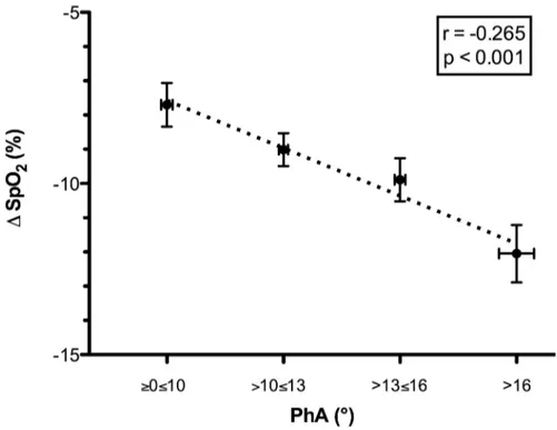
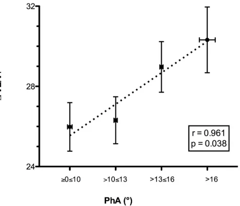
VE/VT), respectively, for each class of PhA. Increases in PhA were significantly related to decreases in SpO2(r = -0.265, p<0.001;Fig 2) and increases in the VE/VT ratio (r = 0.961,
p = 0.038;Fig 3). The increase in the VE/VT ratio significantly affected SpO2: greater decreases
in SpO2were associated with a higher VE/VT ratio (r = -0.697, p<0.001;Fig 4).
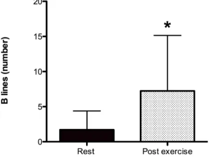
The number of US-B lines, assessed via chest ultrasound examination, increased in 86% of the athletes after the run (US-B lines at rest, 2.0±4.2; US-B lines immediately after exercise,
Table 1. Respiratory function of 15 subjects before (T0) and after the run (T1).
T0 T1 P Effect size (d) VC (L) 5.5 ± 0.4 5.3 ± 0.4 0.041 0.14 VC (% pred) 117 ± 7 114 ± 8 0.041 -FEV1(L) 4.1 ± 0.3 4.1 ± 0.2 0.921 0.0 FEV1(% pred) 109 ± 4 110 ± 5 0.968 -FEV1/VC 76.6 ± 9.0 75.3 ± 3.8 0.716 0.13 FEV1/VC (% pred) 93 ± 6 94 ± 5 0.744 -PImax (cmH2O) 115 ± 11 110 ± 11 0.023 0.22
Data are expressed as the mean ± 95%CI.
Abbreviations: T0, at rest before the run; T1, at rest after the descent. VC, vital capacity; VC (% pred), % of predicted value of vital capacity; FEV1, forced
expiratory volume in the first second; FEV1(% pred), % of predicted value of forced expiratory volume in the first second; PImax, maximal inspiratory
pressure.
https://doi.org/10.1371/journal.pone.0174927.t001
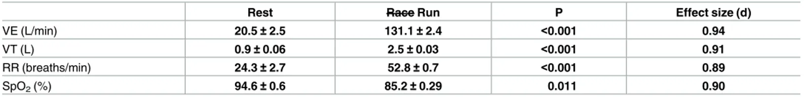
Table 2. Ventilatory pattern and oxygen saturation of 13 subjects at rest (T0) and during the run.
Rest Race Run P Effect size (d)
VE (L/min) 20.5 ± 2.5 131.1 ± 2.4 <0.001 0.94
VT (L) 0.9 ± 0.06 2.5 ± 0.03 <0.001 0.91
RR (breaths/min) 24.3 ± 2.7 52.8 ± 0.7 <0.001 0.89
SpO2(%) 94.6 ± 0.6 85.2 ± 0.29 0.011 0.90
Data are expressed as the mean ± 95%CI.
Abbreviations: VE, ventilation; VT, tidal volume; RR, respiratory rate; SpO2, oxygen saturation.
Fig 1. Relationship between ground slope and thoraco-abdominal coordination (PhA) during the run. The ground slope values were grouped into four classes, each associated with a mean PhA value. When the slope increased to over 30%, a significant increase in PhA was observed, representing a decrease in thoraco-abdominal coordination. The best interpolation of these points is represented by the quadratic curve
PhA = 12.897–0.773slope + 0.301slope2. Abbreviations: PhA, phase angle.
https://doi.org/10.1371/journal.pone.0174927.g001
Fig 2. Relationship between the mean decrease in oxygen saturation and thoraco-abdominal coordination (PhA) during the run. The PhA values were grouped into four classes, each associated with the mean decrease in SpO2(SpO2at rest—mean SpO2during the race). Reductions in thoraco-abdominal
coordination, indicated by an increase in PhA, were significantly related to decreases in SpO2. Abbreviations:
PhA, phase angle; Δ SpO2, mean decrease in oxygen saturation.
7.5±7.2; p = 0.001); (Fig 5). The differences in the number of US-B lines were linearly corre-lated with the decreases in the SpO2(r = 0.661, p = 0.032).
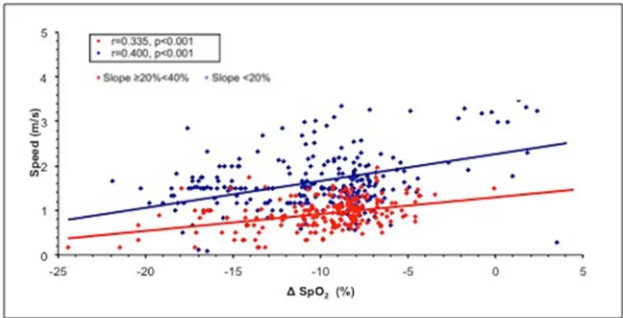
Lower SpO2values were associated with lower speeds at 20% slope40% (r = 0.335,
p<0.001 for horizontal and r = 0.36, p<0.001 for vertical) and 0%>slope<20% (r = 0.400, p<0.001 for horizontal and r = 0.15, p = 0.01 for vertical); (Fig 6).
In the multivariable analysis, the mean decrease in oxygen saturation, the PhA and the slope remained independent predictors of both horizontal (β = 0.657, p<0.001; full model R adjusted = 0.207) and vertical speed (β = 0.543, p<0.001; full model R adjusted = 0.291).
Because the range of ages in the population was rather large (34–60 years), we tested for possible effects of age on both oxygen desaturation and speed and did not find a significant correlation (p = 0.924 and 0.092, respectively).
Discussion
Studies on uphill running and mountain ultramarathons have considered biomechanical and neuromuscular adaptations [41,42] as well as respiratory modifications [3,7]. To the best of our knowledge, no study has investigated ventilation and ventilatory pattern during the run.
We hypothesized that during uphill exercise, the ground slope would influence the thoraco-abdominal coordination in breathing, which would modify the ventilatory pattern, possibly affecting gas exchange (i.e., SpO2) and athletic performance. Furthermore, we aimed to verify
Fig 3. Relationship between the delta VE/VT ratio and thoraco-abdominal coordination (PhA) during the run. The PhA values were grouped into four classes, each associated with the delta VE/VT (difference between the VE/VT ratio during the run and at rest). Reductions in thoraco-abdominal coordination, indicated by an increase in PhA, were significantly related to increases in the VE/VTratio, which represents a less
efficient ventilatory pattern. Abbreviations: PhA, phase angle; VE/VT, ratio between ventilation and tidal volume.
the development of asymptomatic interstitial lung edema after exercise that was strenuous but shorter than that described in the literature [18,19]. We studied a group of well-trained sky-runners during a run at moderate altitudes between 2000 m and 2800 m, where not only exer-tion but also mild hypoxia could affect the responses of the respiratory and cardiac systems.
Fig 4. Relationship between the delta VE/VT ratio and the mean decrease in oxygen saturation during the run. Reductions in SpO2(SpO2at rest—mean SpO2during the race) were grouped into four classes,
each associated with the delta VE/VT (difference between VE/VT ratio during the run and at rest). Increases in the VE/VT ratio were related to greater levels of oxygen desaturation. Abbreviations: Δ SpO2, mean
decrease in oxygen saturation; VE/VT, ratio between ventilation and tidal volume. https://doi.org/10.1371/journal.pone.0174927.g004
Fig 5. Thoracic ultrasound evaluation (US-B lines) at rest and after the run. Ultrasound echo evaluations performed before and immediately after the run showed increased interstitial lung edema, which was identified by the presence of US-B lines.*Student’s t-test, p<0.001.
Ventilation monitoring
The main finding of our investigation is that ground slope negatively influences thoraco-abdominal coordination during intense exercise at moderate altitude, as shown by the increase in the angle representing the excursions of the two compartments. This phenomenon was more evident at slopes >30%, supporting reduced synchrony between the rib cage and the abdomen with increasing slope that we found in a previous study in which participants exer-cised at sea level on a treadmill at slopes between 0% and 25% [43]. We believe that the reduc-tion in thoraco-abdominal coordinareduc-tion that resulted from increased slope was dependent on changes in posture, leading to a greater forward inclination of the trunk and a greater displace-ment of the center of mass to compensate for the increasing slope. Actually, a few studies on thoraco-abdominal kinematics [16,44] have reported an association between greater inclina-tions of the trunk and reducinclina-tions in rib cage displacement as a cause of reduced coordination between these two compartments. Moreover, the reduction in thoraco-abdominal coordina-tion in our subjects corresponded to changes in ventilatory patterns represented by an increased respiratory rate for the same amount of ventilation (i.e., a higher VE/VT ratio). This ventilatory pattern is less efficient in terms of gas exchange [10,13]. In fact, a ventilatory pat-tern characterized by a low respiratory rate and a high tidal volume is associated with a better maintenance of SpO2and is therefore considered more efficient due to the reduction in dead
space ventilation [10,11,13]. This phenomenon has been reported both in healthy subjects dur-ing exposure to high altitude and in patients with chronic respiratory and cardiac diseases [10–15,17].
In our study, changes in ventilatory patterns leading to decreased efficiency (i.e., higher VE/VT ratio) were associated with more severe decreases in SpO2, and this relationship was
stronger for slopes >20%. As the slope of the terrain varied throughout the run [26] and was not correlated with the altitude, the reduced SpO2observed during exercise at steeper slopes
was not related to increased environmental hypoxia. Greater decreases in SpO2appear to
nega-tively influence performance: athletes with higher levels of oxygen desaturation during the run had slower speeds (both horizontal and vertical). In fact, horizontal and vertical speeds are considered the best indicators for endurance performance in mountain environments. The relationship between decreases in SpO2and speed during this run at 2000–2800 m supports
the role of arterial oxygen desaturation in the decline in performance at altitude, as reported
Fig 6. Relationship between mean horizontal speed and mean decreases in oxygen saturation during the run. To simplify the analysis, the slopes were grouped into two ranges: <20% and 20%. Abbreviation: SpO2, oxygen saturation.
by Chapman et al. [45] in a study of highly trained distance runners under conditions of simu-lated hypoxia (2100 m). Many factors can influence run speed, and our results show that a more severe reduction in SpO2can contribute to affecting performance in highly trained
sub-jects during heavy exercise in a mildly hypoxic environment.
Respiratory function
Regarding lung function and respiratory muscle strength, our results are in agreement with data reported in the literature. Since the 1980s, it has been well known that athletes show a reduction in vital capacity and an increase in respiratory muscle fatigue after exercises of vari-ous intensities and durations [46–48]. In particular, during a 2.5-hour treadmill run, simulat-ing a marathon, a reduction in vital capacity of approximately 3% was reported after at least 90 min, while an average 8.6% reduction in post-race vital capacity was found after a marathon [46]. A similar reduction of approximately 10% was recently reported after an ultramarathon [3,7,49]. In our study we observed a small but significant 4% reduction in vital capacity after an uphill and downhill run with an average duration of 95 min. A possible cause of the decrease in vital capacity can be the reduced strength of the respiratory muscles, as reported by Vernillo et al [7]. A reduction in PImax is a sign of respiratory muscle fatigue, which can also play a significant role in performance limitations, contributing to reduced running speeds in prolonged running events [7,50–52]. Accordingly, we found a trivial but significant reduction in PImax after the entire run. We must emphasize that our results were obtained after the downhill part of the run, in contrast to previous studies of similar duration, where the same results were obtained after a run on level ground [46,47]. We wondered whether the descent could have affected the results. It has been reported that the energy cost of running is affected by slope in that cost linearly increases with increasing slope and linearly decreases with decreasing slope, with a minimum value at a -20% grade, after which it increases [41]. There-fore, even if the uphill run is more demanding in terms of cardiorespiratory work than the downhill run [41], the steepest parts of the descent can contribute to the cardiorespiratory work. In the trail covered in the present study, the steepest slopes were spread over the whole route and we think that the descent effort could have contributed to the results.
Ultrasound lung echo
The development of interstitial pulmonary edema, as shown by the higher number of US-B-lines in the athletes after the run, was found in 86% of the subjects and was significantly related to reductions in SpO2. Similar increases in extravascular lung water have previously been
described in outdoor environments in athletes immediately after both intense and prolonged exercise [18,20] and in trekkers during high-altitude exposure with and without exercise [19,53].
The pathophysiological mechanism of the development of asymptomatic interstitial pulmo-nary edema after intense exercise [18,54] is related to different factors, such as increases in pulmonary blood flow [55] and the cardiac changes that occur during exercise [18].
We speculate that the changes in thoraco-abdominal coordination and the consequent decrease in breathing efficiency, which affects SpO2, can contribute to the development of
asymptomatic interstitial pulmonary edema during intense exercise at moderate altitude. In the present study, the lung echo assessment was performed immediately after the descent, but we do not think that this timing affected the results. In fact, it has been reported that the radio-graphic evidence of mild interstitial edema persists for at least one and a half hours post-mara-thon [20]. Also the development of asymptomatic interstitial lung edema can be involved in the decrease in lung volumes, as previously reported in athletes after an ultramarathon [20].
To the best of our knowledge, this study is the first to analyze the combination of breathing coordination, ventilatory pattern, and SpO2during exercise in the field involving continuous
changes in ground slope. All these parameters were analyzed in relation to exercise perfor-mance and to the possible development of interstitial pulmonary edema.
We recognize that this study has certain limitations. One limitation is the absence of data regarding oxygen consumption and the work of respiratory muscles during the run to better identify their role in the performance. The lack of postural and kinematic investigation to support our suppositions constitute additional limitations. Finally, we did not control for the effect of increasing altitude alone (i.e., without exercise). We highlight that according to the literature and our own data [56,25], the reduction in SpO2between 2000 and 2800 m
in healthy subjects at rest is approximately 4%. Therefore, at 2800 m, the SpO2at rest is
expected to be approximately 90%, a value higher than the values found in our study during heavy exercise.
Conclusions
This study presents new data on ventilation during strenuous uphill exercise documenting the role that the respiratory system can play in performance under such conditions.
No study has yet monitored the ventilatory pattern and thoraco-abdominal coordination during an uphill run. The combined use of inductive plethysmography, which has been shown to accurately estimate ventilation during exercise [30], and GPS data provided us the unique opportunity to investigate ventilatory changes during this type of race.
In athletes performing intense exercise between 2000 m and 2800 m, steep slopes led to a reduction in thoraco-abdominal coordination, which was associated with a less efficient venti-latory pattern and decreased SpO2. These effects were more evident at slopes greater than 20–
30%. Therefore, we suggest that ventilatory patterns play a crucial role in breathing efficiency and SpO2and can be a contributing factor to declines in running speed and the development
of interstitial pulmonary edema. Furthermore, we confirm the previous findings regarding the reduced strength of respiratory muscles and decreased lung function as well the development of interstitial lung edema after strenuous exercise.
From a practical perspective, these results reinforce findings indicating that athletes and trainers should pay particular attention to the respiratory system. Future investigations of pos-ture during exercise at different slopes, at sea level, and under conditions of simulated hypoxia should provide additional information on the role of posture in determining ventilatory patterns.
Supporting information
S1 Dataset. Anthropometric, inductive plethysmography, SpO2and GPS data.
Abbrevia-tions: BMI, body mass index; VT, tidal volume; VE/VT, ratio between ventilation and tidal vol-ume; Ti, inspiratory time; Te, expiratory time; Tt total breath time; Ti/Tt fractional inspiratory time; Ti/Te, ratio between inspiratory and expiratory time; PhAng, phase angle; HR, heart rate; SpO2, oxygen saturation; delta SpO2, mean decrease in oxygen saturation.
(XLSX)
Acknowledgments
The study was conducted in collaboration with the International Skyrunning Federation and EV-K2-CNR.
Author Contributions
Conceptualization: GM AC LP GSR. Formal analysis: GM MS EB. Investigation: GM MS LP. Methodology: GM.
Project administration: AC LP GSR. Supervision: AC LP GSR.
Visualization: EB.
Writing – original draft: AC LP GM. Writing – review & editing: AC LP GSR.
References
1. Dempsey J, Johnson B, Saupe K. Adaptations and limitations in the pulmonary system during exercise. Chest. 1990; 97: 81–87.
2. Dempsey J, Romer L, Rodman J, Miller J, Smith C. Consequences of exercise-induced respiratory muscle work. Respir Physiol Neurobiol. 2006; 151: 242–250.https://doi.org/10.1016/j.resp.2005.12. 015PMID:16616716
3. Wu¨thrich T, Marty J, Kerherve H, Millet G, Verges S, Spengler C. Aspects of respiratory muscle fatigue in a mountain ultramarathon race. Med Sci Sports Exerc. 2015 Mar; 47(3): 519–527.https://doi.org/10. 1249/MSS.0000000000000449PMID:25033264
4. Cibella F, Cuttitta G, Romano S, Grassi B, Bonsignore G, Milic-Emili J. Respiratory energetics during exercise at high altitude. J Appl Physiol. 1999; 86: 1785–1792. PMID:10368338
5. West J, Hackett P, Maret K, Milledge JS, Peters RM Jr, Pizzo CJ, et al. Pulmonary gas exchange on the summit of Mount Everest. J Appl Physiol. 1983; 55: 678–687. PMID:6415007
6. Pugh L, Gill M, Lahiri S, Milledge J, Ward M, West J. Muscular exercise at great altitudes. J Appl Phy-siol. 1964; 19: 431–440. PMID:14173539
7. Vernillo G, Rinaldo N, Giorgi A, Esposito F, Trabucchi P, Millet GP, et al. Changes in lung function dur-ing an extreme mountain ultramarathon. Scand J Med Sci Sports. 2015; 25(4): e374–380.https://doi. org/10.1111/sms.12325PMID:25262823
8. Gandevia S, Butler J, Hodges P, Taylor J. Balancing acts: respiratory sensations, motor control and human posture. Clin Exp Pharmacol Physiol. 2002; 29(1–2): 118–121. PMID:11906469
9. Cavalcanti A, Lima C, de Sa´ R, Reinaux CM, Braz Ju´nior DS, Teixeira AL, et al. Influence of posture on the ventilatory pattern and the thoraco-abdominal kinematics of patients with chronic obstructive pulmo-nary disease (COPD). Physiother Theory Pract. 2014; 30(7): 490–494.https://doi.org/10.3109/ 09593985.2014.901458PMID:24678754
10. Bernardi L, Passino C, Spadacini G, Bonfichi M, Arcaini L, Malcovati L, et al. Reduced hypoxic ventila-tory response with preserved blood oxygenation in yoga trainees and Himalayan Buddhist monks at alti-tude: evidence of a different adaptive strategy? Eur J Appl Physiol. 2007; 99: 511–518.https://doi.org/ 10.1007/s00421-006-0373-8PMID:17206440
11. Bernardi L, Spadacini G, Bellwon J, Hajric R, Roskamm H, Frey AW. Effect of breathing rate on oxygen saturation and exercise performance in chronic heart failure. Lancet. 1998; 351: 1308–1311.https:// doi.org/10.1016/S0140-6736(97)10341-5PMID:9643792
12. Bilo G, Revera M, Bussotti M, Bonacina D, Styczkiewicz K, Caldara G, et al. Effects of slow deep breathing at high altitude on oxygen saturation, pulmonary and systemic hemodynamics. PLoS One. 2012; 7(11): e49074.https://doi.org/10.1371/journal.pone.0049074PMID:23152851
13. Casaburi R, Porszasz J, Burns M, Carithers ER, Chang RS, Cooper CB. Physiologic benefits of exer-cise training in rehabilitation of patients with severe chronic obstructive pulmonary disease. Am J Respir Crit Care Med. 1997; 155: 1541–1551.https://doi.org/10.1164/ajrccm.155.5.9154855PMID:9154855 14. Pomidori L, Campigotto F, Amatya T, Bernardi L, Cogo A. Efficacy and tolerability of yoga breathing in
patients with chronic obstructive pulmonary disease: a pilot study. J Cardiopulm Rehabil Prev. 2009; 29: 133–137.https://doi.org/10.1097/HCR.0b013e31819a0227PMID:19305239
15. Goldman M, Grimby G, Mead J. Mechanical work of breathing derived from rib cage and abdominal V-P partitioning. J Appl Physiol. 1976; 41: 752–763. PMID:993163
16. Romei M, Lo Mauro A, D’Angelo M, Lichtenstein DA, Mathis G, Kirkpatrick AW, et al. Effects of gender and posture on thoraco-abdominal kinematics during quiet breathing in healthy adults. Resp Physiol Neurobiol. 2010; 172: 184–191.
17. Wells J, Smyth R, Rebuck A. Thoracoabdominal motion in response to treadmill and cycle exercise. Am Rev Respir Dis. 1986; 134: 1125–1128.https://doi.org/10.1164/arrd.1986.134.5.1125PMID:3098144 18. Pingitore A, Garbella E, Piaggi P, Menicucci D, Frassi F, Lionetti V, et al. Early subclinical increase in
pulmonary water content in athletes performing sustained heavy exercise at sea level: ultrasound lung comet-tail evidence. Am J Physiol Heart Circ Physiol. 2011; 301(5): H2161–2167.https://doi.org/10. 1152/ajpheart.00388.2011PMID:21873499
19. Pratali L, Cavana M, Sicari R, Picano E. Frequent subclinical high altitude pulmonary edema detected by chest sonography as ultrasound lung comets in recreational climbers. Crit Care Med. 2010; 38(9): 1818–1823.https://doi.org/10.1097/CCM.0b013e3181e8ae0ePMID:20562696
20. Zavorsky GS, Milne EN, Lavorini F, Rienzi JP, Cutrufello PT, Kumar SS, et al. Small changes in lung function in runners with marathon-induced interstitial lung edema. Physiol Rep. 2014; 27; 2(6). 21. Hanel B, Law I, Mortensen J. Maximal rowing has an acute effect on the blood-gas barrier in elite
ath-letes. J Appl Physiol. 2003; 95: 1076–1082.https://doi.org/10.1152/japplphysiol.00082.2002PMID: 12716865
22. Prediletto R, Fornai E, Catapano G, Carli C, Garbella E, Passera M, et al. Time course of carbon mon-oxide transfer factor after breath-hold diving. Undersea Hyperb Med. 2009; 36: 1–9.
23. Zavorsky GS. Evidence of pulmonary oedema triggered by exercise in healthy humans and detected with various imaging techniques. Acta Physiol (Oxf). 2007; 189: 305–317.
24. Volpicelli G, Elbarbary M, Blaivas M, Lichtenstein DA, Mathis G, Kirkpatrick AW, et al. International Liai-son Committee on Lung Ultrasound (ILC-LUS) for International Consensus Conference on Lung Ultra-sound (ICC-LUS). International evidence-based recommendations for point-of-care lung ultraUltra-sound. Intensive Care Med. 2012; 38(4): 577–591https://doi.org/10.1007/s00134-012-2513-4PMID: 22392031
25. Mandolesi G, Avancini G, Bartesaghi M, Bernardi E, Pomidori L, Cogo A. Long-term monitoring of oxy-gen saturation at altitude can be useful in predicting the subsequent development of moderate-to-severe acute mountain sickness. Wilderness Environ Med. 2014; 25(4): 384–91.https://doi.org/10. 1016/j.wem.2014.04.015PMID:25027753
26. Route slopes.http://www.openrunner.com/?id=5381544
27. Miller M, Hankinson J, Brusasco V, Burgos F, Casaburi R, Coates A, et al. ATS/ERS Task Force. Stan-dardisation of Spirometry. Eur Respir J. 2005; 26: 319–338.https://doi.org/10.1183/09031936.05. 00034805PMID:16055882
28. American Thoracic Society/European Respiratory Society. ATS/ERS Statement on respiratory muscle testing. Am J Respir Crit Care Med. 2002; 166(4): 518–624.https://doi.org/10.1164/rccm.166.4.518 PMID:12186831
29. Quanjer PH, Stanojevic S, Cole TJ, Baur X, Hall GL, Culver BH, et al; ERS Global Lung Function Initia-tive. Multi-ethnic reference values for spirometry for the 3-95-yr age range: the global lung function 2012 equations. Eur Respir J. 2012; 40(6): 1324–43.https://doi.org/10.1183/09031936.00080312 PMID:22743675
30. Clarenbach C, Senn O, Brack T, Kohler M, Bloch K. Monitoring of ventilation during exercise by a porta-ble respiratory inductive plethysmograph. Chest. 2005; 128: 1282–1290.https://doi.org/10.1378/chest. 128.3.1282PMID:16162719
31. Kalsås K, Thorsen E. Breathing patterns during progressive incremental cycle and treadmill exercise are different. Clin Physiol Funct Imaging. 2009; 29(5): 335–8;https://doi.org/10.1111/j.1475-097X. 2009.00874.xPMID:19453565
32. Bernardi E, Melloni E, Mandolesi G, Uliari S, Grazzi G, Cogo A. Respiratory Muscle Endurance Training Improves Breathing Pattern in Triathletes. Ann Sports Med Res. 2014; 1(1): 1003.
33. Jambrik Z, Monti S, Coppola V, Agricola E, Mottola G, Picano E. Usefulness of ultrasound lung comets as a nonradiologic sign of extravascular lung water. Am J Cardiol. 2004; 93: 1265–70.https://doi.org/ 10.1016/j.amjcard.2004.02.012PMID:15135701
34. Cardinale L, Priola AM, Moretti F, Volpicelli G. Effectiveness of chest radiography, lung ultrasound and thoracic computed tomography in the diagnosis of congestive heart failure. World J Radiol. 2014; 28; 6(6): 230–7.https://doi.org/10.4329/wjr.v6.i6.230PMID:24976926
35. Agricola E, Bove T, Oppizzi M, Zangrillo A, Fragasso A, Picano E. Ultrasound comet-tail images: a marker of pulmonary edema. A comparative study with wedge pressure and extravascular lung water. Chest. 2005; 127: 1690–5.https://doi.org/10.1378/chest.127.5.1690PMID:15888847
36. Enghard P, Rademacher S, Nee J, Hasper D, Engert U, Jorres A, et al. Simplified lung ultrasound proto-col shows excellent prediction of extravascular lung water in ventilated intensive care patients. Crit Care. 2015; 19: 36.https://doi.org/10.1186/s13054-015-0756-5PMID:25656060
37. Liteplo AS, Marill KA, Villen T, Miller RM, Murray AF, Croft PE, et al. Emergency thoracic ultrasound in the differentiation of the etiology of shortness of breath (ETUDES): sonographic B-lines and N-terminal pro-brain-type natriuretic peptide in diagnosing congestive heart failure. Acad Emerg Med. 2009; 16: 201–210.https://doi.org/10.1111/j.1553-2712.2008.00347.xPMID:19183402
38. Lichtenstein D. Lung ultrasound in the critically ill. Review. Annals Intensive Care. 2014; 4: 1–12. 39. Emergency echocardiography: the European Association of Cardiovascular Imaging
recommenda-tions. Neskovic AN, Hagendorff A, Lancellotti P, Guarracino F, Varga A, Cosyns B, et al; European Association of Cardiovascular Imaging. Eur Heart J Cardiovasc Imaging. 2013; 14(1): 1–11.https://doi. org/10.1093/ehjci/jes193PMID:23239795
40. Lichtenstein D, Meziere G, Biderman P, Gepner A, Barre O. The comet-tail artifact. An ultrasound sign of alveolar-interstitial syndrome. Am J Respir Crit Care Med. 1997; 156: 1640–6.https://doi.org/10. 1164/ajrccm.156.5.96-07096PMID:9372688
41. Vernillo G, Giandolini M, Edwards WB, Morin JB, Samozino P, Horvais N, Millet GY. Biomechanics and Physiology of Uphill and Downhill Running. Sports Med. 2016 Aug 9. [Epub ahead of print].
42. Vernillo G, Savoldelli A, Zignoli A, Skafidas S, Fornasiero A, La Torre A, Bortolan L, Pellegrini B, Schena F. Energy cost and kinematics of level, uphill and downhill running: fatigue-induced changes after a mountain ultramarathon. J Sports Sci. 2015; 33(19): 1998–2005.https://doi.org/10.1080/02640414. 2015.1022870PMID:25751128
43. Pomidori L, Grazzi G, Uliari S, Grassi G, Bernardi L, Rinaldi A, et al. Breathing pattern at rest and during different exercises at sea level (SL) and at high altitude (HA) Eur Respir J. 2006; 28: Suppl 50; 174. 44. Baydur A, Behrakis P, Zin W, Jaeger M, Weiner J, Milic-Emili J. Effect of posture on ventilation and
breathing pattern during room air breathing at rest. Lung. 1987; 165: 341–351. PMID:3123805 45. Chapman R, Stager J, Tanner D, Stray-Gundersen J, Levine B. Impairment of 3000-m run time at
alti-tude is influenced by arterial oxyhemoglobin saturation. Med Sci Sports Exerc 2011; 43: 1649–1656. https://doi.org/10.1249/MSS.0b013e318211bf45PMID:21311361
46. Farrell PA, Maron MB, Hamilton LH, Maksud MG, Foster C. Time course of lung volume changes during prolonged treadmill exercise. Med Sci Sports Exerc. 1983; 15(4): 319–24. PMID:6621323
47. Maron MB, Hamilton LH, Maksud MG. Alterations in pulmonary function consequent to competitive marathon running. Med Sci Sports. 1979; 11(3):244–9. PMID:522634
48. O’Kroy JA, Loy RA, Coast JR. Pulmonary function changes following exercise. Med Sci Sports Exerc. 1992; 24(12):1359–64. PMID:1470019
49. Warren G, Cureton K, Sparling P. Does lung function limit performance in a 24-hour ultramarathon? Respir Physiol. 1989; 78(2): 253–263. PMID:2609032
50. Dempsey J, Sheel A, Haverkamp H, Babcock M, Harms C. Pulmonary system limitations to exercise in health. Can J Appl Physiol. 2003; 28 Suppl: S2–24.
51. Harms C, Wetter T, St Croix C, Pegelow D, Dempsey J. Effects of respiratory muscle work on exercise performance. J Appl Physiol. 2000; 89: 131–138. PMID:10904044
52. Sheel A, Derchak P, Morgan B, Pegelow D, Jacques A, Dempsey J. Fatiguing inspiratory muscle work causes reflex reduction in resting leg blood flow in humans. J Physiol. 2001; 537: 277–289.https://doi. org/10.1111/j.1469-7793.2001.0277k.xPMID:11711580
53. Strapazzon G, Vezzaro R, Hofer G, Dal Cappello T, Procter E, Balkenhol K, et al. Factors associated with B-lines after exposure to hypobaric hypoxia. Eur Heart J Cardiovasc Imaging. 2015; 16(11): 1241– 1246.https://doi.org/10.1093/ehjci/jev074PMID:25851323
54. Hopkins SR, Schoene RB, Henderson WR, Spragg RG, Martin TR, West JB. Intense exercise impairs the integrity of the pulmonary blood-gas barrier in elite athletes. Am J Respir Crit Care Med. 1997; 155 (3): 1090–4.https://doi.org/10.1164/ajrccm.155.3.9116992PMID:9116992
55. Younes M, Bshouty Z, Ali J. Longitudinal distribution of pulmonary vascular resistance with very high pulmonary blood flow. J Appl Physiol (1985). 1987; 62(1): 344–58.
56. Lorente-Aznar T, Perez-Aguilar G, Garcı´a-Espot A, Benabarre-Ciria S, Mendia-Gorostidi JL, Dols-Alonso D, et al. Estimation of arterial oxygen saturation in relation to altitude. Med Clin (Barc). 2016 Sep 27; pii:S0025-7753(16)30373-6. [Epub ahead of print].