Abstract. – OBJECTIVE: Cardiac surgery is often performed by cardiopulmonary by-pass (CPB), generally associated with organ dysfunc-tion. The aim of this work was to determine if and how this phenomenon is related to mito-chondrial damage. To this purpose, the effect of the addition of serum from CPB patients to hu-man fibroblasts cultures on mitochondrial respi-ratory chain and oxidative phosphorylation (OX-PHOS) activities was investigated.
PATIENTS AND METHODS: Serum samples of five patients were obtained before (pre-CPB) and after 6 h CPB weaning (CPB). Mitochondri-al OXPHOS activities were examined by polaro-graphic and spectrophotometric assays, and re-active oxygen species (ROS) production was measured by a spectrofluorimeter.
RESULTS: Addition of CPB serum to fibro-blasts determined a decrease of mitochondri-al oxygen consumption due to an inhibition of mitochondrial respiratory chain and some OX-PHOS enzymes activities. This inhibition seems to be mainly related to a reduced activity of com-plex I.
CONCLUSIONS: Our data represent the first translational research evidence showing that CPB determines mitochondrial dysfunction which leads to impairment of OXPHOX activities and to an increase in ROS production, compro-mising tissue bioenergetic efficiency.
Key Words:
Translational research, Cardiopulmonary by-pass (CPB), Mitochondrial respiratory chain, Oxidative phosphorylation (OXPHOS), NADH-coenzyme Q oxy-doreductase (complex I), Multiple organ dysfunction syndrome (MODS), Ischemia-reperfusion injury, Sys-temic inflammatory response syndrome (SIRS), Reac-tive oxygen species (ROS).
Introduction
Cardiac surgery is often performed with the help of cardiopulmonary by-pass (CPB), which maintains cardiopulmonary function by a pump and an oxygenator when the heart is stopped; CPB is not free of complications in the postoper-ative period.
Twenty percent of “low-risk” cardiac patients develop postoperative complications1,2 and the in-cidence of multiple organ dysfunction syndrome (MODS) following CPB is 11% with a mortality rate of 41%1,3. Ischemia-reperfusion injury, endotoxemia, and contact of blood with the extracorporeal circuit release pro-inflammatory cytokines such as IL-1, IL-6, IL-8, TNF-α that may promote a systemic inflammatory response syndrome (SIRS)1,4-8. CPB
A. GNONI
1, A. BALLINI
2,3, R. TRENTADUE
1, F. TAURINO
1, L. SANTACROCE
4,5,
P. FERRARA
6, F. MASSARO
6, N. BRIENZA
6,A.M. MASSARI
1, N. SARDARO
1,
G. DIPALMA
7, F. INCHINGOLO
7, S. SCACCO
11Department of Basic Medical Sciences, Neurosciences and Sense Organs, University of Bari “Aldo Moro” Policlinico, Bari, Italy
2Department of Biosciences, Biotechnologies and Biopharmaceutics, Campus Universitario “Ernesto Quagliariello”, University of Bari “Aldo Moro”, Bari, Italy
3Department of Precision Medicine, University of Campania “Luigi Vanvitelli”, Naples, Italy 4Ionian Department, University of Bari “Aldo Moro”, Bari, Italy
5School of Technical Medical Sciences, “A. Xhuvani” University, Elbasan, Albania
6Anaesthesia and Intensive Care Unit, Department of Emergency and Organ Transplantation, University of Bari “Aldo Moro” Policlinico, Bari, Italy
7Interdisciplinary Department of Medicine, University of Bari “Aldo Moro”, Bari, Italy
Antonio Gnoni and Andrea Ballini contributed equally to this work as co-first authors
Francesco Inchingolo and Salvatore Scacco contributed equally to this work as co-last authors
Induction of mitochondrial dysfunction in
patients under cardiopulmonary by-pass:
preliminary results
may also promote apoptosis due to increased activ-ity of Fas9,10. It has been demonstrated that serum of patients after weaning from CPB, when incubated with endothelial cells, showed apoptotic proper-ties11,12. Apoptosis is related to mitochondrial dys-function13-15. A dominant role of mitochondria is to produce energy of the cell Adenosine triphosphate (ATP) through respiration and oxidative phosphory-lation (OXPHOS) system. OXPHOS consists of four multienzyme complexes (I-IV), forming the mito-chondrial respiratory chain. The electron transfer along the respiratory chain creates a proton gradient across the inner mitochondrial membrane, which is used by Complex V (ATP synthase) to drive the ATP synthesis. Mitochondria are also the quanti-tative source of reactive oxygen species (ROS) in mammalian16. Abnormalities of the electron trans-port chain and OXPHOS are the most common causes of increased ROS production and mitochon-drial diseases17.
It has been shown that in several pathologi-cal conditions, serum levels of some mitochon-dria-related substances can be changed, includ-ed mesenchymal stem cells. Their levels have been associated to the pathology progression or have been considered as prognostic parameters. Plasma level of mitochondrial Coupling Factor 6 (CF6), an essential component of ATP synthase complex, increases in patients with coronary heart disease18, acute myocardial infarction19, stroke20 or with type 2 diabetes mellitus21. Serum levels of mitochondrial Inhibitory Factor 1 (IF1), an inhibitor of ATPase activity, are independently associated with long-term prognosis in coronary artery diseases22. Mitochondrial dysfunction and oxidative stress in human fibroblasts cultures ex-posed to serum from septic patients have been re-ported23. In experimental models of sepsis, Com-plex I, ComCom-plex III, and ComCom-plex IV activities of the respiratory chain are reduced24-27.
In this translational research work, we evaluate if CPB is able to affect mitochondrial function and determine “cytopathic hypoxia”. For this purpose, we have measured the OXPHOS capacity in fibro-blasts cell cultures incubated with serum samples obtained from patients before and after CPB.
Patients and Methods
Patients and MethodsAll patients gave permission after they signed a written informed consent in accordance with the Helsinki Declaration, for the re-use of human
bio-specimens in scientific research. The inves-tigation was conducted in accordance with the current medical protocol as described by the Ital-ian Government’s NIH legislation. The procedure followed a precise individual medical anamnesis together with the required clinical evaluations performed at the Medical School-University of Bari “Aldo Moro” (Bari, Italy), in collaboration with the Elbasan University (School of Technical Medical Sciences, “A. Xhuvani”), Albania (Eth-ical Committee approval N. 496/1). Procedures were conducted according to Good Clinical Prac-tice (GCP) and manufacture specifications.
In seven patients undergoing first elective heart surgery with CPB, blood samples were collected from the central venous catheter after induction of anaesthesia, and 6 h after weaning from CPB. After blood centrifugation at 500 g for 10 min at 25°C, the supernatant was preserved at –20°C until analysis. Anaesthesia management included premedication with morphine 10 mg s.c. 1 h be-fore access to the operating room.
Anaesthesia was induced with propofol, 1-2 mg/kg, midazolam, 0.1-0.2 mg/kg, and fentanest 3-4 μg/kg. Neuromuscular blockade was obtained with rocuronium, 0.1 mg/kg for the intubation. Anaesthesia was maintained with continuous in-fusion of propofol 10 mg/kg/hr, fentanest, bolus of 50 μg every 30 min, and rocuronium 7 μg/kg/ min. Extracorporeal circulation was conducted with aortic and atriumcaval cannulation. Cardio-plegia was obtained with a cold solution. Pump flow was set from 4 to 4.5 L/min. Activated clot-ting time of 480 s was obtained with heparin bo-lus intravenous (i.v).
After surgery, clinical data and blood tests were collected to determine systemic inflamma-tory response syndrome (SIRS) occurrence. SIRS was defined according to The American College of Chest Physicians and the Society of Critical Care Medicine (ACCP/SCCM)27.
Only patients with SIRS were enrolled in the study. Clinical conditions were determined by the APACHE II score (Acute Physiology and Chronic Health Evaluation II)28 and SOFA (Sequential Or-gan Failure Assessment) score29.
Cell Cultures and Mitoplasts Preparation Neonatal normal human dermal fibroblasts (NHDF-neo; Cambrex, CC-2509, Paullo, Milan, Italy) were grown in the exponential phase in high glucose Dulbecco’s Modified Eagle’s Medi-um (DMEM, Euroclone, Pero, MI, Italy) supple-mented with 10% fetal bovine serum (FBS;
Sig-ma-Aldrich, MI, Italy), plus 2 mM L-glutamine (Euroclone, Pero, MI, Italy), 100 μg/ml penicillin (Euroclone, Pero, MI, Italy) and 100 μg/ml strep-tomycin (Euroclone, Pero, MI, Italy).
The cultures were incubated in 5% CO2 hu-midified atmosphere at 37°C. FBS was replaced 12 h before the assay with 10% pre and post-CPB patient serum, while other components were not modified. For mitoplasts (i.e., mitochondria de-void of outer membrane) preparation, fibroblasts were harvested with 0.05% trypsin and 0.02% ethylenediaminetetraacetic acid (EDTA), and washed in phosphate-buffered saline (PBS) pH 7.4. Cells in PBS were exposed for 10 min on ice to 0.5 mg digitonin/mg cellular protein. Mito-plasts were prepared as in Papa et al30, were pel-leted at 10,000 g and resuspended in PBS31. Cellular Respiration
The cellular respiratory activity was measured polarographically31 with a Clark-type oxygen electrode in a water-jacketed chamber, magneti-cally stirred at 37°C8. The cells were collected by trypsinization and centrifugation to 500 g per 3 min at 37°C and resuspended at 1-3×10⁶ cells/ml in a buffer containing Sucrose 75 mM, Tris-HCl 30 mM, KCl 50 mM, EDTA 0.5 mM, Magnesium chloride (MgCl2) 0.5 mM, KH2PO4 2mM. Cell suspensions were transferred to the polarographic chamber, and an aliquot was used for cell count-ing and protein determination with the Bradford method.
The substrates and inhibitors of mitochondri-al OXPHOS were added at the following con-centrations: oligomycin (1 μg/μl), carbonyl cy-anide m-chlorophenyl hydrazone (CCCP) (0.25 μM), Rotenone (1 μg/μl), succinate (1 M), digi-tonin (10%), antimycin A (1 μg/μl), ascorbate (1 M)+ N,N,N′,N′-tetramethyl-p-phenylenediamine (TMPD; 0.1 M) and potassium cyanide (KCN; 1.0 mM).
Respiratory control ratio (RCR) represents State III uncoupled/State IV oxygen consumption ratio (state IIIu/state IV). Respiration rates are ex-pressed as femtomoles of molecular oxygen con-sumed per milligram of mitochondrial protein. Assay of Mitochondrial Respiratory Complex Activities
The activity of NADH-coenzyme Q oxidore-ductase (complex I, EC 1.6.5.3) was assayed es-sentially32. Mitoplasts were exposed to ultrasound energy for 15 s at 0°C, and Vmax and Km were determined using NADH as electrons donor to
360-374 nm with the Δε of 2.01 mM-1. In 800 μl of Mix (K-Phosphate buffer 50 mM pH 7.4, MgCl2 2 mM, K-EDTA 2 mM, KCN 3 mM, antimycin A 1 μg/ml) 30 μg of mitochondrial proteins were add-ed in presence of decylubiquinone 0.2 mM. The reaction was started with different concentrations of NADH (1.3-2.8 μM). The measurements were determined with and without rotenone 1μg/ml to discriminate the complex I activity. The Vmax and Kmof complex I were determined with Line-weaver-Burk equation following the oxidation of NADH. Cytochrome-C oxidase (complex IV, EC 2.3.3.1) activity was determined on mitoplasts in 700 μl of buffer (10 mM phosphate buffer, KCN, MgCl2 2 mM, pH 7.4) following the oxidation of ferrocytochrome c 10 μM.
Citrate synthase (CS) activity, used as enzy-matic marker of the mitochondrial matrix, was assayed according to Taurino et al33. Briefly, mito-chondrial proteins (30 μg/ml), 0.5 mM acetyl-co-enzyme A, and 0.5 mM 5,5′-dithiobis (2-nitro-benzoic acid) (DTNB) were added to a Tris-HCl buffer (100 mM), pH 8.0. The reaction was start-ed by the addition of 0.5 m oxalacetate, and the initial rate was measured following the reduction of DTNB at 419 nm (Δε=163 mM−1·cm−1).
The determination of ATPase (complex V) ac-tivity consisted of a coupled double enzymatic as-say, as reported in Cavallo et al34. Since the mito-plasts were exposed to ultrasound energy, in 700 μl of Mix (sucrose 250 mM, Potassium chloride (KCl) 50 mM, MgCl2 5 mM and Tris-HCl 20mM pH 7.5) 30 μg of proteins were added in presence of pyruvate kinase 2 U, lactate dehydrogenase 2.5 U, phosphoenolpyruvate 1 mM, and NADH 200 μM. When ATP 0.5 mM is added, phosphoe-nolpyruvate is converted to pyruvate and pyru-vate to lactate with the oxidation of NADH to 340 nm31.
Membrane Potential Measurement
The mitochondrial membrane potential was measured following the fluorescence quenching of safranin at 525 nm (excitation) and at 575 nm (emission) with a spectrofluorimter (JASCO FP 6200). 1x10⁶ cells were permeabilized with 20 μg/1x106 of digitonin and resuspended in 1 ml of buffer ICR (sucrose 75 mM, Tris-Cl 30 mM pH 7.4, KCl 50 mM, K-EDTA 0.5 mM, MgCl2 2 mM, Potassium phosphate monobasic (KH2PO4) 2 mM, pH 7.4) with safranin (5 μM), and ciclo-sporyn A (33nM). The potential is generated by glutamate/malate (10/10 mM) and succinate (10 mM) addition.
ROS Measurements
Fibroblasts were treated with 10 μM dichlo-rofluorescein-diacetate DCF-DA (503-570 λ) for the detection of H2O2 with fluorimetric analysis. ROS production was measured using the cell per-meant probe 2′-7′dichlorodihydrofluorescin dia-cetate (DCF-DA) which passively diffuses into cells where intracellular esterases cleave acetate groups to form the impermeable DCF-H2 which remains trapped within the cell. Cells were col-lected by trypsinization and resuspended in a small volume of PBS and were then incubated in 800 μl of PBS with 10 μM DCF-DA for 20 min in the dark at 37°C. The linear fluorescence in-crease (507 nm excitation and 530 nm emission wavelength), produced by the ROS-dependent ox-idation of DCF-H2 to the fluorescent compound dichlorofluorescein (DCF), was measured with a Jasco FP6200 spectrofluorimeter.
Statistical Analysis
Clinical data are presented as absolute num-bers, with standard deviation, or percentages. ROS generation, expressed as arbitrary fluo-rescence units, was calculated as percentage of those recorded before CPB cultured fibroblasts treatment. Results are analyzed with Student’s t-test. p<0.05 was considered statistically signif-icant.
Results
The pre-CPB and post-CPB serum were used to determine whether circulating factors can af-fect mitochondrial functions. Patients without clinical signs of SIRS in the post-operative time were excluded from the study. Clinical data are summarized in Table I.
A significant difference between Complex I activity of cells incubated with serum pre-CPB and post-CPB can be observed. Results in Figure 1 demonstrate that, when compared to serum pre-CPB, the addition of serum post-CPB to human fibroblasts determines a significant decrease (i.e., nearly to 50%, p= 0.017) of complex I activity. Interestingly, a similar but less pronounced inhi-bition of ATPase activity was observed (Figure 1).
No statistically significant differences were found for Cytochrome c oxidase (COX) and ci-trate synthase activities in post-CPB vs., pre-CPB serum-treated fibroblasts (p =0.681 and p =0.614, respectively) (data not shown). Figure 2 shows that the Vmax ratios of Complex I and Citrate Syn-thase (CI/CS) activities were significantly lower in post-CPB as compared to pre-CPB serum (CI/ CS, p =0.012). The Vmax ratios of Complex I and Cytochrome c oxidase appears to be lowered in the post-CPB vs. pre-CPB samples, but the data is not statistically significant (CI/COX, p =0.039). To prove the biological relevance of alteration in mitochondrial respiratory chain activity in-duced by serum addition to fibroblasts cultures, oxidative damage was then measured. ROS were significantly increased in cells incubated with post-CPB compared to pre-CPB serum samples (p<0.001) as reported in Figure 3. The membrane potential semi-quantitative analysis by fluores-cence quenching, showed that after
glutamate/ma-Table I. Clinical data.
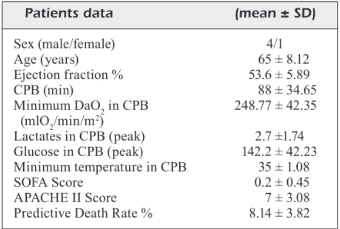
Patients data (mean ± SD)
Sex (male/female) 4/1 Age (years) 65 ± 8.12 Ejection fraction % 53.6 ± 5.89 CPB (min) 88 ± 34.65 Minimum DaO2 in CPB 248.77 ± 42.35 (mlO2/min/m2) Lactates in CPB (peak) 2.7 ±1.74 Glucose in CPB (peak) 142.2 ± 42.23 Minimum temperature in CPB 35 ± 1.08 SOFA Score 0.2 ± 0.45 APACHE II Score 7 ± 3.08
Predictive Death Rate % 8.14 ± 3.82
Figure 1. Effect of pre-CBP serum and post-CBP serum addition to human fibroblasts. The complex I activity in these cells showed a decrease of about 50% (p= 0.017) when post-CBP serum was added, with respect to the addition of pre-post-CBP serum. A similar but less pronounced inhibition was noted regarding the ATPase activity. Each bar is the mean ± standard error (SEM); the results are analyzed with Student’s t-test. p
< 0.05 was considered statistically significant. *Significantly
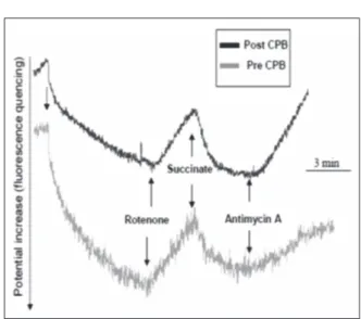
late addition a plateau value was rapidly achieved in the post-CPB samples measurements, whilst in the pre-CPB samples no saturation of membrane potential no potential was observed in the time-scale of the experiments (Figure 4).
Rotenone addition reverses the potential in both conditions and successive succinate pulse re-turns to the pre-inhibition levels the fluorescence
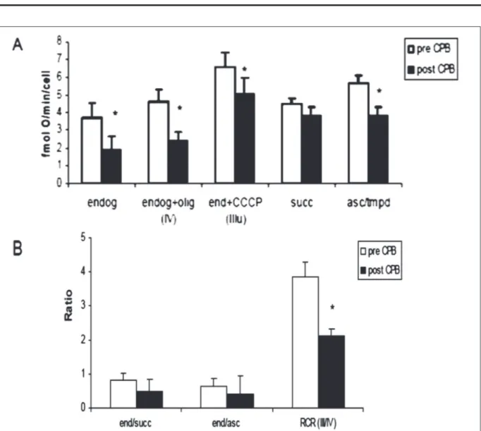
signals (Figure 4). Of note, successive inhibition by Antimycin A leads to membrane potential collapse with significantly different kinetics, as shown in Figure 4. The polarographic analysis underlined (Figure 5A) the reduction of endog-enous respiration in cells incubated with serum post-CPB samples (p = 0.008).
A similar decrease was observed after incuba-tion with oligomycin (state IV) and addiincuba-tion of the uncoupler CCCP (state IIIu; Figure 5A). The state IV and IIIu of respiratory chain decreased after post-CPB addition, (p=0.018 and p=0.001), while the succinate respiration was unaffected (Figure 5A). The ascorbate/TMPD respiration, a measure of isolated COX respiratory activity, was reduced (p=0.01; Figure 5A). Figure 5B shows that whilst the ratios between endogenous respiration, and succinate or ascorbate/TMPD respiration are not statistically different in the two different exper-imental conditions, the respiratory control ratio (RCR) shows a marked decrease in the post-CPB condition (p=0.007; Figure 5B).
Discussion
The data of this translational study show that ad-dition to human fibroblasts cultures of serum from patients submitted to cardiac surgery with CPB induces abnormalities of mitochondrial functions. In particular, a decrease in cellular endogen res-piration, associated with inhibition of some
activ-Figure 2. Ratios between Vmax of Complex I and Citrate Sintase (Cx I/CS) and between Vmax of Complex I and Cytochrome c oxidase (Cx I/COX) activities in pre-CBP and post-CBP serum-treated fibroblasts. Lower values were observed in post-CBP serum-treated cells (Cx I/CS,
p= 0.012; Cx I/COX, p= 0.039, respectively). Each bar is
the mean ± standard error (SEM); the results are analyzed with Student’s t-test. p< 0.05 was considered statistically significant. *Significantly different with respect to pre-CBP serum addition.
Figure 3. ROS measurements performed to prove the ox-idative damage and the relevant alteration of mitochondrial respiratory chain activity induced by post-CBP serum ad-dition to human fibroblasts. ROS levels in these cells were significantly higher after post-CBP serum incubation, com-pared to pre-CBP serum-treated fibroblasts. ROS generation was expressed as arbitrary fluorescence units and calculated as percentage of those recorded before CPB cultured fibro-blasts treatment. Each bar is the mean ± SEM; the results are analyzed with Student’s t-test. p < 0.05 was considered sta-tistically significant.**Significantly different with respect to pre-CBP serum addition, with p < 0.001.
Figure 4. Membrane potential measurements by
spectro-fluorimetric analysis. It was observed a decrease of values after glutamate/malate but not succinate addition in pre-CBP serum-treated fibroblasts.
ities of the OXPHOS system and of mitochondrial membrane potential, was observed. Conversely, an increase of the oxidative stress, determined by ele-vated ROS production, was detected.
When we assayed in a selective approach the activities of single components of the respirato-ry chain, we observed a reduced activity of mi-tochondrial complex I and complex V, but not of complex II activity.
It is well known that alterations of mitochon-drial efficiency and function are mainly related to alterations in mitochondrial mass, a amount of respiratory enzymes or changes in enzyme activ-ities28,29. The relative preservation of citrate syn-thase activity we found indicates that total mito-chondrial content remains largely unchanged and resistant to CPB injury, thus excluding changes in mitochondrial mass. Conversely, the complex
Figure 5. Polarographic analysis of cellular respiratory activity in cellular suspensions of pre-CBP and post-CBP
serum-treated human fibroblasts. A, Marked reduction in cellular endogenous respiration was observed in fibroblasts incubated with post-CBP serum (p = 0.008); a similar decrease was noted after addition of oligomycin (state IV) and of the uncoupler CCCP (state uIII); an impairment of the state IV and uIII of respiratory chain appeared after post-CBP serum addition (p = 0.018 and p=0.001). Ascorbate/TMPD respiration was reduced (p=0.01) too. *Significantly different with respect to pre-CBP serum addition. B, Rates between endogenous activity and Complex II oxymetry (ratio end/succ) and between endogenous activity and Complex IV oxymetry (ratio end/asc) were reduced after CPB with p = 0.015 and p = 0.047, respectively. RCR index (State uIII/State IV ratio) decreased significantly after CPB treatment (p = 0.007). Each bar is the mean ± SEM; the results are analyzed with Student’s t-test. p< 0.05 was considered statistically significant. * Significantly different with respect to pre-CBP
serum addition. End= endogenous respiration; oligo = oligomycin; uIII = uncoupled state III; succ = succinate; asc = ascorbate; RCR = respiratory control ratio (Ratio between uIII state and IV state of respiration).
I/complex IV and complex I/citrate synthase ra-tios show a reduction after CPB indicating a mi-tochondrial enzymatic complex I defect. Note that the deficiency of complex I activity has been indicated as a major cause of reduced mitochon-drial oxygen consumption and of increased ROS production.
The decrease of Ascorbate/TMPD respiration can be associated to an impairment of complex IV activity. The alteration of RCR index indi-cates a mitochondrial uncoupling, namely a mi-tochondrial membrane injury with cytochrome c loss, which is an expression of the apoptotic process. Taken together, these results showed that CPB induces damage of some mitochondri-al complexes, particularly the Complex I, and even to a lesser extent Complex IV and V. The obtained results add further support to those of Aebert et al11 and Schmid et al12 who reported higher proportion of apoptotic endothelial cells in culture plates incubated with serum samples obtained at 6 h after weaning from CPB, when compared to plates incubated with pre-operative samples.
When mitochondrial complex activities have been evaluated in septic patients, similarly to our data, a reduction in Complex I activity was ob-served, indicating a strong association between septic shock and mitochondrial damage35-38. Fur-thermore, using spectrophotometric assay, cyto-chrome c function has been evaluated in septic baboons26,27. In agreement with our findings, it has been reported that after infusion of Esche-richia coli, Cytochrome c oxidase activity was found to be reduced, and, similarly to our data, 6 h after application of the noxious stimulus, cy-tochrome c function was lost37,38. Taken togeth-er, these findings should be also seen in the light of the whole membrane moiety, including lipids such as cardiolipin and quinones, which plays a key role in the function of the mitochondrial re-spiratory complexes39,40.
Proinflammatory cytokines have been linked to depression of myocardial contractility both in patients with acute septic shock and in in vitro models employing isolated myocytes exposed to serum from such patients41,42. In sepsis such as in post-CPB SIRS is evident an association between proinflammatory cytokines and mito-chondrial damage41.
The loss of efficiency by OXPHOS, as a con-sequence of the reduced ATPase activity that we observed in post-CPB-treated fibroblasts (Figure 2), might contribute to explain some
major post-CPB complications such as acute kidney injury, or pulmonary and neurological dysfunctions.
Traditionally, postoperative complications have been related to the critical role of low oxygen de-livery during CPB. Low hematocrit and pump flow have been identified as factors promoting renal dysfunction43, and the finding of high lactate lev-els have been viewed as index of low oxygen de-livery8,44,45. Our data may offer new insights. The target of maintaining an adequate oxygen delivery during CPB may be insufficient to guarantee aero-bic metabolism, if CBP determines mitochondrial chain impairment and consequent cytopathic hy-poxia. Evidence has been reported that the latter, rather than inadequate oxygen delivery, may play a more important role in the development of multiple organ dysfunction syndrome (MODS)46-48. In this connection, as mitochondria are the primary con-sumers of cellular oxygen, mitochondrial function and dysfunction are crucial in the development of cytopathic hypoxia49-50. Certainly, future studies are warranted to deeply understand the relation-ship between postoperative organ damage and mi-tochondrial dysfunction.
Conclusions
Mitochondria play a central role in the intra-cellular events associated with CPB. Altered me-tabolism can lead to rapid ROS overproduction and damage of the mitochondrial protein with a generalized reduction in the capacity to generate ATP. We have shown that post-CPB SIRS is as-sociated with a condition similar to “cytopathic hypoxia”. Although this phenomenon is consid-ered to be self-limiting, an exaggerated and pro-longed inflammatory status with mitochondrial dysfunction may be critical in the pathogenesis of post-operative complications. More translational research studies are needed to clarify the role of mitochondrial dysfunction in the various phases of CPB, before testing therapeutic strategies tar-geting mitochondria.
Conflict of Interest
The Authors declare that they have no conflict of interests.
Authors’ Contribution
A.G. conceived and designed the research. R.T., F.T., and A.M.M. performed the laboratory research; S.S. supervised
the manuscript and gave the final approval of the version to be published. G.D., N.S., and L.S. participated in bib-liographic research. A.B. contributed to statistical analy-sis and manuscript revision. F.I. contributed to bibliograph-ic research, reagents, materials, and analysis tools. All au-thors read and approved the final version of the manuscript. P.F., F.M., and N.B. collected the biological material. All au-thors read and approved the final version of the manuscript.
References
1) Laffey JG, BoyLan J, ChenG DC. The systemic
in-flammatory response to cardiac surgery. Anes-thesiology 2002; 97: 215-252.
2) Grover fL. The Society of Thoracic Surgeons
Na-tional Database: current status and future direc-tions. Ann Thorac Surg 1999; 68: 367-373. 3) KoLLef Mh, WraGGe T, Pasque C. Determinants of
mortality and multiorgan dysfunction in cardiac surgery patients requiring prolonged mechanical ventilation. Chest 1995; 107: 1395-1401.
4) PaPareLLa D, yau TM, younG e. Review:
cardio-pulmonary bypass induced inflammation: patho-physiology and treatment. An Update. Eur J Car-diothorac Surg 2002; 21: 232-244.
5) haLL rI, sMITh Ms, roCKer G. The systemic
in-flammatory response to cardiopulmonary bypass: pathophysiological, therapeutic, and pharmaco-logical considerations. Anesth Analg 1997; 85: 766-782.
6) MarIaPPan n, sooraPPan rn, haque M, srIraMuLa s,
franCIs J. TNF-α induced mitochondrial oxidative
stress and cardiac dysfunction: restoration by su-peroxide dismutase mimetic Tempol. Am J Physi-ol Heart Circ PhysiPhysi-ol 2007; 293: H2726-H2737. 7) DavIes MG, haGen Po. Review: systemic
inflam-matory response syndrome. Br J Surg 1997; 84: 920-935.
8) DeMers P, eLKourI s, MarTIneau, r, CouTurIer, a,
CarTIer r. Outcome with high blood lactate levels
during cardiopulmonary by-pass in adult cardiac operation. Ann Thorac Surg 2000; 70: 2882-2086. 9) Bone rC, BaLK ra, Cerra fB, DeLLInGer rP, feIn aM,
Knaus Wa, sCheIn rM sIBBaLD WJ. Definition for
sep-sis and organ failure and guidelines for the use of innovative therapies in sepsis. The ACCP/SC-CMConsensus Conference Committee. Ameri-can College of Chest Physiacians /Society of Crit-ical Care Medicine. Chest 1992; 101: 1644-1655. 10) KaWahITo K, MIsaWa y, fuse K. Transient rise in
se-rum soluble Fas in patients undergoing cardiac surgery. Artif Org 2000; 24: 628-631.
11) aeBerT h., KIrChner s, KeIser a, BIrnBauM De, hoL -Ler e, anDreesen r, eIssner G. Endothelial
apopto-sis is induced by serum of patient after cardiopul-monary bypass. Eur J Cardiothor Surg 2000; 18: 589-593.
12) sChIMD f, vuDaTTu n, fLoerChInGer B, hILKer M, eIss -ner G, hoenICKa M, hoLLer e, BIrnBauM De.
Endo-thelial Apoptosis and circulating endoEndo-thelial cells after bypass grafting with and without cardiopul-monary bypass. Eur J Cardiothorac Surg 2006; 29: 496-500.
13) MuravChICK s, Levy r. Clinical implications of
mi-thocondrial dysfunction. Anesthesiology 2006; 105: 819-837.
14) huBBarD W, BLanD KI, ChauDry Ih. The role of the
mitochondrion in trauma and shock. Shock 2004; 22: 395-402.
15) Brenner C, Marzo I, KroeMer G. Revolution in
apop-tosis: from a nucleocentric to a mitochondriocen-tric perspective. Exp Gerontol 1998; 33: 543-553. 16) GonzáLez-fLeCha B, CuTrIn JC, BoverIs a. Time
course and mechanism of oxidative stress and tissue damage in rat liver subjected to in vivo ischemia-reperfusion. J Clin Invest 1993; 91: 456-464.
17) PaPa s, rasMo DD, TeChnIKova-DoBrova z, PaneL -LI D, sIGnorILe a, sCaCCo s, PeTruzzeLL a v, PaPa f,
PaLMIsano G, GnonI a, MICeLLI L, sarDaneLLI aM.
Respiratory chain complex I, a main regulatory target of the cAMP/PKA pathway is defective in different human diseases. FEBS Lett 2012; 586: 568-577.
18) ChaI sB, huI yM, LI XM, TanG Cs. Plasma level of
mitochondrial coupling factor 6 increases in pa-tients with coronary heart disease. Circ J 2007, 71, 693-697.
19) DInG Wh, Chu sy, JIanG hf, CaI Dy, PanG yz, TanG
Cs, qI yf. Plasma mitochondrial coupling factor 6
in patients with acute myocardial infarction. Hy-pertens Res 2004; 27: 717-722.
20) osanaI T, fuJIWara n, sasaKI s, MeToKI n, saIToh G,
ToMITa h, nIshIMura T, shIBuTanI s, yoKoyaMa h, Kon -Ta y, MaGoTa K, oKuMura K. Novel pro-atherogenic
molecule coupling factor 6 is elevated in patients with stroke: a possible linkage to homocysteine. Ann Med 2010; 42: 79-86.
21) LI XL, XInG qC, DonG B, Gao yy, XInG ss, PanG yz,
JIanG hf, TanG Cs. Plasma level of mitochondrial
coupling factor 6 increases in patients with type 2 diabetes mellitus. Int J Cardiol 2007, 117, 411-412.
22) TaurIno f, GnonI a. Systematic review of
plas-ma-membrane ecto-ATP synthase: a new player in health and disease. Exp Mol Pathol 2018; 104: 59-70.
23) TrenTaDue r, fIore f, Massaro f, PaPa f, Iuso a, sCa -CCo s, sanTaCroCe L, BrIenza n. Induction of
mito-chondrial dysfunction and oxidative stress in hu-man fibroblast cultures exposed to serum from septic patients. Life Sci 2012; 91: 237-243. 24) TruMBeCKaITe s, oPaLKa Jr, neuhof C, zIerz s, GeLL
-erICh fn. Different sensitivity of rabbit heart and
skeletal muscle to endotoxin-induced impairment of mitochondrial function. Eur J Biochem 2001; 268: 1422-1429.
25) Levy rJ. Mitochondrial dysfunction,
bioenerget-ic impairment, and metabolbioenerget-ic down-regulation in sepsis. Shock 2007; 28, 24-28.
26) GeLLerICh fn, TruMBeCKaITe s, oPaLKa Jr, GeLLerICh
Jf, Chen y, neuhof C, reDL h, WerDan K, zIerz s.
Mi-tochondrial dysfunction in sepsis: evidence from bacteraemic baboons and endotoxaemic rabbits. Biosci Rep 2002; 22: 99-113.
27) GeLLerICh fn TruMBeCKaITe s, herTeL K, zIerz s,
MuLLer-WerDan u, WerDan K, reDL h, sChLaG G.
Im-paired energy metabolism in hearts of septic ba-boons: diminished activities of complex I and complex II of the mitochondrial respiratory chain. Shock 1999;11: 336-341.
28) Knaus Wa, DraPer ea, WaGner DP, zIMMerMan Je.
Apache II: a severity of disease classification sys-tem. Crit Care Med 1985; 13: 818-829.
29) vInCenT JL, Moreno r, TaKaLa J, WILLaTTs s, De Men -Donça a, BruInInG h, reInharT CK, suTer PM, ThIJs
LG. The SOFA (Sepsis-related Organ Failure As-sessment) score to describe organ dysfunction/ failure. On behalf of the Working Group on Sep-sis-Related Problems of the European Society of Intensive Care Medicine. Intensive Care Med 1996; 22: 707-710.
30) PaPa f, LIPPoLIs r, sarDaro n, GnonI a, sCaCCo s. All
trans retinoic acid depresses the content and ac-tivity of the mitochondrial ATP synthase in human keratinocytes. Biochem Biophys Res Commun 2017; 482: 301-304.
31) TaurIno f, GIannoCCaro C, sarDaneLLI aM, CavaLLo
a, De LuCa e, sanTaCroCe s, PaPa s, zanoTTI f, GnonI
a. Function and expression study uncovered he-patocyte plasma membrane ecto-ATP synthase as a novel player in liver regeneration. Biochem J 2016; 473: 2519-2530.
32) CavaLLo a, PrIore P, GnonI Gv, PaPa s, zanoTTI f,
GnonI a. 3,5-Diiodo-L-thyronine administration to
hypothyroid rats rapidly enhances fatty acid oxi-dation rate and bioenergetic parameters in liver cells. PLoS One. 2013; 8: e52328.
33) TaurIno f, sTanCa e, sICuLeLLa L, TrenTaDue r, PaPa s,
zanoTTI f, GnonI a. Mitochondrial proteome
anal-ysis reveals depression of the Ndufs3 subunit and activity of complex I in diabetic rat brain. J Pro-teomics 2012; 75: 2331-2341.
34) CavaLLo a, TaurIno f, DaMIano f, sICuLeLLa L, sar -DaneLLI aM, GnonI a. Acute administration of
3,5-diiodo-L-thyronine to hypothyroid rats stimu-lates bioenergetic parameters in liver mitochon-dria. J Bioenerg Biomembr 2016; 48: 521-529. 35) KroeMer G, DaLLaPorTa B, resChe-rIGon M. The
mi-tochondrial death/life regulator in apoptosis and necrosis. Annu Rev Physiol 1998; 60: 619-642. 36) PaGLIarInI DJ, CaLvo se, ChanG B, sheTh sa, vafaI sB,
onG se, WaLforD Ga, suGIana. C, Boneh a, Chen
WK, hILL De, vIDaL M, evans JG, ThorBurn Dr, Carr
sa, MooTha vK. A mitochondrial protein
compen-dium elucidates complex I disease biology. Cell 2008; 134: 112-123.
37) sIMonson sG, WeLTy-WoLf K, huanG yT, GrIeBeL Ja,
CaPLan Ms, fraCICa PJ, PIanTaDosI CA. Altered
mito-chondrial redox responses in Gram negative sep-tic shock in primates. Circ Shock 1994; 43: 34-43. 38) Iuso a, sCaCCo s, PICCoLI C, BeLLoMo f, PeTruzzeL -La v, TrenTaDue r, MInuTo M, rIPoLI M, CaPITanIo n,
zevIanI M, PaPa s. Dysfunctions of cellular
oxida-tive metabolism in patients with mutations in the NDUFS1 and NDUFS4 genes of complex I. J Biol Chem 2006; 281: 10374-10380.
39) LoBasso s, PaLese LL, anGeLInI r, CorCeLLI a.
Rela-tionship between cardiolipin metabolism and ox-ygen availability in Bacillus subtilis. FEBS Open Bio 2013; 3: 151-155.
40) BossIs f, De GrassI a, PaLese LL, PIerrI CL. Prediction
of high- and low-affinity quinol-analogue-binding sites in the aa3 and bo3 terminal oxidases from Bacillus subtilis and Escherichia coli1. Biochem J 2014; 461: 305-314.
41) BreaLey D, BranD M, harGreaves I, heaLes s, LanD J,
sMoLensKI r, DavIes na, CooPer Ce, sInGer M.
As-sociation between mitochondrial dysfunction and severity and outcome of septic shock. Lancet 2002; 360: 219-223.
42) KuMar a, MIChaeL P, BraBanT D, ParIssenTI a, raMa -na C, Xu X, ParrILLo J. Human serum from patients
with septic shock activates transcription factors STAT1, IRF1, and NF-kB and induces apoptosis in human cardiac myocytes. J Biol Chem 2005; 280: 42619-42626.
43) ranuCCI M, roMITTI f, IsGrò G, CoTza M, BrozzI s,
BonCILLI a, DITTa a. Oxygen delivery during
car-diopulmonary by-pass and acute renal failure af-ter coronary operations. Ann Thorac Surg 2005; 80: 2213-2220.
44) ChIoLéro rL, reveLLy JP, Leverve X, GersBaCh, P, Cay -euX MC, BerGer MM, TaPPy L. Effect of
cardiogen-ic shock on lactate and glucose metabolism after heart surgery. Crit Car Med 2000; 28: 3784-3791. 45) BuTLer J, roCKer GM, WesTaBy s. Inflammatory
re-sponse to cardiopulmonary bypass. Ann Thorac Surg 1993; 55: 552-559.
46) fInK MP. Cytopathic hypoxia. Mitochondrial
function as mechanism contributing to organ dys-function in sepsis. Crit Care Clin 2001; 17: 219-237.
47) fInK MP. Bench-to-bedside review: cytopathic
hy-poxia. Crit Care 2002; 6: 491-499.
48) fInK MP. Cytopathic hypoxia in sepsis. Acta
An-aesthesiol Scand 1997; 110: 87-95.
49) LIu Gh, Wen y, yanG P, LIu Gf. Regulation by
Pink1 on the mitochondrial dysfunction in endo-thelial cells post the hypoxia mimetic agent Co-Cl2 treatment. Eur Rev Med Pharmacol Sci 2018; 22: 5704-5711.
50) shen W, JIanG XX, LI yW, he q.
Mitochondria-me-diated disturbance of fatty acid metabolism in proximal tubule epithelial cells leads to renal in-terstitial fibrosis. Eur Rev Med Pharmacol Sci 2018; 22: 810-819.