UNIVERSITA’ DEGLI STUDI DEL PIEMONTE ORIENTALE
“AMEDEO AVOGADRO”
Sede di Alessandria
DOTTORATO DI RICERCA IN SCIENZE AMBIENTALI
(ACQUE INTERNE E AGROECOSISTEMI) (XXVIII ciclo)
TESI DI DOTTORATO DI RICERCA
TRANSLATIONAL CONTROL IN MALIGNANT PLEURAL
MESOTHELIOMA
Candidato:
Stefania Oliveto
Coordinatore:
Giorgio Malacarne
Tutor:
Stefano Biffo
"Se saprai ricordarmi, sarò sempre con te"
A te che, in silenzio,
mi hai dato la forza di andare avanti.
ACRONYMS AND ABBREVIATIONS
4E-BPs: eIF4E binding proteins AGO: argonaute
ALL: acute lymphoblastic leukemia BAP1: BRCA1-associated protein-1 BCL2: B-cell lymphoma 2
CDKN2A/ARF: cyclin-dependent kinase inhibitor 2A/alternative reading frame CDS: coding sequence
CLL: chronic lymphocytic leukemia
CPEB: cytoplasmic polyadenylation element binding protein CPI-17: C-kinase potentiated Protein phosphatase-1
CT: computed tomography DAG: dyacilglycerol
DGCR8: DiGeorge syndrome critical region gene 8 DLBCL: diffuse large B cell lymphoma
EFL1: elongation factor-like 1 eIF6: eukaryotic initiation factor 6 ECM: extracellular matrix
EMT: epithelial-mesenchymal transition EPP: extrapleural pneumonectomy FACS: fluorescence-activated cell sorting FGF: fibroblast growth factor
GCN2: general control nonderepressible-2 GEF: guanine nt exchange factor
GEO: Gene Expression Omnibus
GRB10: growth factor receptor–bound protein 10 GSK: glycogen synthase kinase-3
HCC: hepatocellular carcinoma HGF: hepatocyte growth factor HOXD10: homeobox D10 HRI: heme-regulated inhibitor IRES: internal ribosome-entry site
ii MCL1: myeloid cell leukemia sequence 1
MPM: Malignant Pleural Mesothelioma mTOR: mammalian target of Rapamycin
MTT: (3-(4, 5-dimethylthiazolyl-2)-2, 5-diphenyltetrazolium bromide MVB: multivesicular bodies
MYPT1-PP1 δ: myosin phosphatase targeting subunit 1-protein phosphatase 1δ NF2: neurofibromatosis type 2
NOD-SCID: non-obese diabetic-severe combined immunodeficiency PABP: polyA binding protein
PDGF: plateled-derived growth factor PET: positron emission tomography
PERK: protein kinase RNA-like endoplasmic reticulum kinase PKC: protein kinase C
PKR: protein kinase RNA activated PI3K: phosphoinositide 3-kinase PMA: phorbol 12-myristate 13-acetate PRAS40: proline-rich Akt substrate 40 kDa PS: phosphatidylserine
RACK1: receptor for activated C Kinase 1 RHOC: Ras homolog gene family, member C RISC: RNA-induced silencing complex SBDS: Swachman Bodian Diamond Syndrome SNP: single nucleotide polymorphism
SRL: sarcin-ricin loop
TGFβ: transforming growth factor β Tif6: translation initiation factor 6 TOP: terminal oligopyrimidine TRBP: TAR RNA binding protein
ULK1: Unc-51 like autophagy activating kinase 1 uORF: upstream open reading frame
UPR: unfolded protein response UTR: untranslated region XPO5: exportin 5
iii VEGF: vascular endothelial growth factor
iv
Contents
1. ABSTRACT ... 1
2. INTRODUCTION ... 3
2.1 MALIGNANT MESOTHELIOMA ... 3
2.1.1 Pleura: structure, functions and pathological conditions ... 3
2.1.2 Malignant Pleural Mesothelioma ... 5
2.1.3 Pathogenesis, diagnosis and therapies of Malignant Pleural Mesothelioma ... 7
2.2 TRANSLATION ... 11
2.2.1 Cap-dependent translation ... 12
2.2.2 The translational regulation ... 15
2.2.3 Novel concepts in translational control: regulation by microRNA ... 22
2.3 EUKARYOTIC INITIATION FACTOR 6(EIF6) ... 23
2.3.1 eIF6 on ribosome biogenesis and antiassociation activity ... 26
2.3.2 eIF6 and translation ... 27
2.3.3 eIF6 and cancer ... 29
2.4 MICRORNAS ... 31
2.4.1 microRNA and translation ... 34
2.4.2 microRNAs and cancer ... 37
2.5 PROTEIN KINASE C Β AND ENZASTAURIN ... 42
2.6 AIM AND MAIN CONCLUSIONS OF THE WORK ... 46
3. MATERIALS AND METHODS ...48
3.1 MICE ... 48
3.2 CELL LINES AND LENTIVIRAL VECTORS ... 48
3.3 ANTIBODIES AND REAGENTS ... 49
3.4 RNA EXTRACTION AND REAL TIME RT-PCR ... 49
3.5 CELL PROLIFERATION, CELL CYCLE AND CELL DEATH ANALYSIS ... 50
3.6 POLYSOMAL PROFILE ... 50
3.7 DATAMINING ... 51
3.8 IMMUNOHISTOCHEMISTRY ... 52
3.9 TWO-DIMENSIONAL (2D) GEL ELECTROPHORESIS ... 52
3.10 MEASUREMENTS OF LACTATE SECRETION AND ATP CONTENT ... 53
3.11 MICRORNAS PROFILING ... 53
3.12 STATISTICAL ANALYSIS ... 54
4. RESULTS ...55
4.1 EIF6 AND MALIGNANT PLEURAL MESOTHELIOMA ... 55
4.1.1 eIF6 is a marker of aggressive Malignant Pleural Mesothelioma (MPM) ... 55
4.1.2 eIF6 hyperphosphorylation in REN, a MPM cell line ... 58
4.1.3 eIF6 antiassociation activity is important for recycling inactive 80S ... 61
4.1.4 eIF6 reduction and dephosphorylation slow cell growth in cultured cells ... 64
4.1.5 eIF6 depletion and Enzastaurin administration have an antitumoral effect, in vivo ... 67
4.1.6 eIF6 depletion and Enzastaurin cause metabolic changes of cancer cells ... 70
4.2 MICRORNAS SUBCELLULAR DISTRIBUTION IN MPM ... 71
4.2.1 microRNAs association with polysomes define the subcellular distribution of miRNAs ... 71
4.2.2 microRNAs exhibit different ratios of association with polysomes ... 74
4.2.3 Cell cycle pathways are related to miRNAs which are associated to polysomes... 76
5 DISCUSSION ...78
5.1 EIF6 IN MALIGNANT PLEURAL MESOTHELIOMA ... 78
5.2 MICRORNAS IN MPM ... 81
1
1. ABSTRACT
Protein synthesis is a cellular process finely regulated during growth and development and its deregulation can lead to cell apoptosis or disease. Translational control is rate-limiting in cancer growth and translation initiation step is emerging as an attractive therapeutic target. eIF6 is an antiassociation factor that regulates the availability of active 80S. Its activation is driven by the RACK1/PKCβ axis, in a mTORc1 independent manner. We previously described that eIF6 haploinsufficiency causes a striking survival in the Eµ-Myc mouse lymphoma model, with lifespans extendend up to 18 months. microRNAs have been shown to regulate a wide range of biological processes destabilizing messenger RNAs and by repressing the translation of these mRNAs. Involvement of microRNAs in repression of translation suggests that they might be associated with polysomes. Here we screen for 1) eIF6 expression in human cancers and 2) association of microRNAs with polysomes in Malignant Pleural Mesothelioma (MPM). We show that MPM tumors and a MPM cell line (REN cells) contain high levels of hyperphosphorylated eIF6. Enzastaurin is a PKC beta inhibitor used in clinical trials. We prove that Enzastaurin treatment decreases eIF6 phosphorylation rate, but not eIF6 protein stability. The growth of REN, in vivo, and metastasis are reduced by either Enzastaurin treatment or eIF6 shRNA. Molecular analysis reveals that eIF6 manipulation affects the metabolic status of malignant mesothelioma cells. Less glycolysis and less ATP content are evident in REN cells depleted for eIF6 or treated with Enzastaurin (Anti-Warburg effect). We propose that eIF6 is necessary for Malignant Mesothelioma growth, in vivo, and can be targeted by kinase inhibitors. Finally we found that the MPM miRNA signature was characterized also by differential miRNAs subcellular distribution. In particular, only some miRNAs were expressed in the polysomal pool with variability in miRNAs occupancy, indicating that some miRNAs can repress translation, while others cannot. Particularly, we
2 evidenced that polysome-bound miRNAs present a correlation with the cell cycle pathway in REN cell, a MPM epithelioid cell line, suggesting that their polysomal localization could explain how these miRNAs may regulate cell cycle components translation.
3
2. INTRODUCTION
2.1 Malignant Mesothelioma
Malignant mesothelioma is a rare but highly aggressive tumour and its mortality is one of the highest associated with cancers, up to 1% (Carbone, Albelda et al. 2007). There are two major localizations of malignant mesothelioma: the pleura and peritoneum, sporadically it may also arise in the pericardium or tunica vaginalis testis (Chekol and Sun 2012).
2.1.1 Pleura: structure, functions and pathological conditions
The chest cavity surrounds the heart and lungs and comprises the ribs, associated muscles and connective tissue. This cavity is covered by the parietal pleura, which is attached to the chest wall and by a continuous parietal mesothelial cell layer. The lungs themselves are enclosed by the visceral pleura which is integral to the lung surface and which has a surface visceral mesothelial layer. The close fitting of the lungs to the inside of the chest wall means that there is a thin space between the two mesothelial layers that contains the pleural fluid and a population of pleural macrophages (Donaldson, Murphy et al. 2010). The pleural mesothelial cells derive from the mesoderm and cover the surface in an epithelial-like manner, hence the word mesothelium (Michailova and Usunoff 2006). The normal mesothelial cell layer appears glistening, smooth, and semi-transparent. Mesothelial cells may vary from a row of flattened and elongated ovoid nuclei widely separated by cytoplasm to columnar or cuboidal cells with round basal nuclei and a cuboidal luminal surface (Batra and Antony 2015). These cells have microvilli and multiple intercellular adherens junctions and focal adhesions that anchor the mesothelial cell onto the extracellular membrane via integrins (Batra and Antony 2015). The connective tissue is intersected with blood vessels, lymphatic vessels, immune cells and fibroblast-like cells. It has been shown that fibroblast-like cells may differentiate and replace
4 the damaged mesothelium following injury (Mutsaers, Whitaker et al. 2002; Michailova and Usunoff 2006). In the pleural cavity between the two layers there is a small amount of fluid for lubrication that reduces friction between the visceral and parietal layers during breathing. This fluid is continuously circulating and is produced by the mesothelial cells and then drained into the lymphatic circulation. The pleural mesothelium also controls several tissue functions such as regulation of fibrinolysis, trans-membrane material flux, maintenance of serosal integrity by producing growth factors (GF) and extracellular matrix (ECM) components (Agostoni and Zocchi 2007). The most common growth factors found in the pleural space are vascular endothelial growth factor (VEGF), transforming growth factor β (TGFβ), fibroblast growth factor (FGF), platelet-derived growth factor (PDGF), hepatocyte growth factor (HGF) and heparin-binding epidermal growth factor-like growth factor.
Common cytokines and chemokines include interleukins IL1, IL6, IL8 and IL15, SDF-1and
prostaglandins (Michailova and Usunoff 2006).
Many pathogens and carcinogens can hit pleura and cause cellular injuries; the consequent infections and inflammation led to big challenges in diagnosis and treatment (Murthy, Raja et
al. 2012). Long-term inhalation of several natural and industrial fibers irritates the pleura
causing chronic inflammation and scarring. Inflammation and injury result in cellular responses both from the immune system and the mesothelium itself (Cagle and Allen 2011). Cancers in the pleural cavity are often metastatic adenocarcinomas from other organs (Cagle and Allen 2011), as , stomach, breast, lung and ovary (Batra and Antony 2015), but there are also tumors arising from the pleural tissue (Cagle and Allen 2011). Solitary fibrous tumors of the pleura are mostly benign mesenchymal tumors without the standard mesothelial immunophenotype, but when malignant transformation occurs is highly difficult to distinguish it from the most common primary malignancy of pleural origin, the malignant pleural mesothelioma (Usami, Iwano et al. 2007)
5 2.1.2 Malignant Pleural Mesothelioma
The pleural form of malignant mesothelioma is the most common type accounting for more than 70% of all mesothelioma cases (Chen and Pace 2012). Malignant pleural mesothelioma represents a common malignant disease (Ismail-Khan, Robinson et al. 2006) arising from mesothelial cells of the pleura and showing a close relationship with previous exposure to asbestos fibers. Asbestos is the most common causative agent for MPM (Yang, Testa et al. 2008), with 80% of mesothelioma patients reporting asbestos exposure (Pass et al., 2008). However only a fraction of subjects exposed to high levels of asbestos develop MPM, suggesting that additional factors, such as genetic predisposition, may render some individuals more susceptible to asbestos carcinogenicity (Testa, Cheung et al. 2011). Recently, it has been reported that DAS, an artificial clay used as a toy and teaching material, contains a large amount of asbestos. This striking discovery changes the scenario of number of subjects exposed to asbestos fibers, showing that the presence of 30% of asbestos in DAS composition may cause exposure to a different variety of users, including teachers, artists and children. Since DAS has been used not only in Italy, it is essential that mesothelioma patients should be asked about their use of DAS, in particular if they do not report a past asbestos exposure (Silvestri, Di Benedetto et al. 2016). Wagner and colleagues were the first to describe the relationship between asbestos and MPM in 1960 when he published a series of MPM cases in asbestos mine workers from United States, Western Europe, South Africa, Japan, India, China, Australia, Indonesia and Vietnam (Porpodis, Zarogoulidis et al. 2013). This relationship is one of the clearest between a carcinogen and its associated cancer. Asbestos is a group of hydrated fibrous silicate minerals that occur in nature and is distinguished in two major groups: the white asbestos and the blue asbestos. The white asbestos include serpentines and chrysotile, instead the blue asbestos include the amphiboles, crocidolite and amosite (Yang, Testa et al. 2008). While white asbestos comprises 90% of the
6 world’s entire asbestos consumption, the blue form is the most carcinogenic (Yang, Testa et al. 2008). Due to its remarkable heat-resistant capacities, asbestos has been called a miracle-fiber and has been extensively used in industry (Ismail-Khan, Robinson et al. 2006). The people most exposed to this carcinogen are therefore asbestos miners, plumbers, electricians, shipyard workers, construction workers and people in similar professions (Craighead 2011; Chen and Pace 2012). Since most asbestos exposure is work-related, mesothelioma is considered an occupational disease and, considering that asbestos exposure is more common in occupations with a predominantly male workforce, the incidence of MPM is higher among men than among women (5:1 ratio) (Nasreen, Khodayari et al. 2012). The risk fraction attributable to occupational asbestos exposure is lower than 40% in women and higher than 80% in men. Environmental mesotheliomas are linked either with a natural exposure in areas in the world where asbestos exists as a geological components of the soil or with neighborhood exposure in people living close to asbestos factories or mines (van Meerbeeck, Scherpereel et al. 2011). The commercial use of asbestos peaked between 1930 and 1960 (Kaufman and Pass 2008), but asbestos has been strongly restricted or banned, in several countries since then (Kao, Reid et al. 2010). Inhaled asbestos fibers accumulate in the mesothelium leading to a status of chronic inflammation and signaling activation, favoring the carcinogenic process (Yang, Rivera et al. 2010). Asbestos fibers of a certain length and width are inhaled all the way out to the alveoli. Over time, these fibers migrate out to both layers of the pleura. These fibers are like thin spears that can penetrate plasma membranes without killing the cells. Upon ingestion by macrophages and other cells the asbestos fibers become covered by iron-rich proteins and iron deposit. These ferruginous bodies may lead to increased formation of reactive oxygen species (ROS). ROS lead to cellular damage, especially DNA mutations, and have been linked to tumor progression (Wu 2006). Macrophages that try to phagocytize the asbestos fibers fail, but in the process produce more
7 cytokines and ROS. This is called frustrated phagocytosis and is part of a chronic inflammation in the lung and pleura (Wu 2006). Properties of the asbestos fibers and the increased ROS production during the inflammation process are thought to be some of the main biological causes of Malignant Pleural Mesothelioma. Incidence of MPM reach 100 cases/million/year in occupationally exposed populations opposed to 1 case/million/year in the general populations (Porret, Madelaine et al. 2007), although there are prominent differences in incidence of MPM reported from different countries worldwide varying from 7 per million (Japan) to 40 per million (Australia) inhabitants per year. In Europe the incidence is around 20 per million with large intercountry variation. It is logical that these differences are due to differences in historical asbestos import and consumption (Pass and Carbone 2009). All individuals who have been exposed to asbestos are considered as a population at risk. The mean latency of MPM after exposure to asbestos is around 30-40 years. The median age at diagnosis in Western countries is 69 years with an increasing fraction of patients with co-morbidities (van Meerbeeck, Scherpereel et al. 2011). Although 50 years have passed since the discovery of the first incidence of MPM, an optimal strategy has not been yet established, as the diagnosis, staging and treatment remains highly complex.
2.1.3 Pathogenesis, diagnosis and therapies of Malignant Pleural Mesothelioma The most common symptoms of MPM are shortness of breath and pain (90%), tiredness (36%), cough (22%), sweating (22%), worry (29%) and constipation (22%) (Muers, Stephens et al. 2008). There are three major histopathological subtypes of MPM: epithelioid (60%), sarcomatous (10%) and biphasic (30%). The phenotypes are closely linked to patient survival, the median survival time has been reported to fall from 12 months for epithelioid mesotheliomas, associated with the best prognosis, to only 4 months for sarcomatoid
8 mesotheliomas (Pinto, Novello et al. 2013). Given that the disease is infrequent and only a few pathologists have extensive experience with mesothelioma, the diagnosis is sometimes delayed (Porpodis, Zarogoulidis et al. 2013). The clinical procedure is first imaging with chest x-ray that can show the effusion and the tumor or pleural thickening. Computed tomography (CT) can show a pleural mass and invasion. More advanced imaging techniques, such as magnetic resonance imaging and positron emission tomography (PET) can be helpful in evaluating tumour likelihood, invasiveness and staging. Staging is useful in planning surgical management however is of little importance for medical management of malignant mesothelioma (Robinson, Musk et al. 2005). Video-assisted thoracoscopy is the best biopsy technique (accuracy of 98%) and cytology, a reliable diagnostic tool for experienced
cytopathologists, can offer additional tissue confirmation. Thus, several
immunohistochemical panels are proposed to distinguish between sub-types of mesothelioma, secondary carcinoma and other malignant tumors metastatic to serosal membranes (Henderson, Reid et al. 2013). Calretinin is the most commonly used antibody, positive for mesothelioma with a reported specificity of 87% and sensitivity of 95%. Other useful antigens include thrombomodulin, mesothelin and cytokeratin 5 (Yaziji, Battifora et al. 2006). Molecular genetic analysis has revealed three key genetic alterations in MPM: cyclin-dependent kinase inhibitor 2A/alternative reading frame (CDKN2A/ARF), neurofibromatosis type 2 (NF2) and BRCA1-associated protein-1 (BAP1) genes. CDKN2A)/ARF gene is the most frequently inactivated tumor suppressor gene in human MPM (Musti, Kettunen et al.
2006). CDKN2A encodes p16INK4a whereas ARF encodes p14ARF. p16INK4a controls the cell
cycle via the CDK4/cyclin D retinoblastoma protein pathway, whereas p14ARF regulates p53
protein. The homozygous deletion of CDKN2A/ARF causes the inactivation of two major tumor suppressing pathways of retinoblastoma and p53 in the cell. It has been reported that MPM cases of epithelioid type showed ~70% of homozygous deletion of CDKN2A whereas
9 sarcomatoid type showed ~100% of homozygous deletion. Moreover, although p53 is the most frequently inactivated tumor suppressor genes in human malignancies, only a limited number of MPM cases show a p53 mutation. (Sekido 2013). It has been demonstrated that miR-31 is co-deleted with CDKN2A, and reintroduction of miR-31 in MPM cells shows a suppressive effect on mesothelioma cells (Ivanov, Goparaju et al. 2010). Mouse studies
showed that mice deficient for Arf, but not p16INK4a, were susceptible to accelerated
asbestos-induced MPM (Altomare, Menges et al. 2009). Instead the inactivation of both Arf and p16INK4a may cooperate to accelerate asbestos-induced tumorigenesis in vivo (Altomare, Menges et al. 2011). NF2 gene encodes merlin, a tumor suppressor protein that can be inactivated not only genetically but also with other mechanisms, such as phosphorylation of CPI-17, an oncogene product that inhibits the merlin phosphatase MYPT1-PP1 δ (Thurneysen, Opitz et al. 2009). Merlin is able to regulate multiple cell signaling cascades including mTOR and the Hippo pathways, which regulate cell proliferation and growth. Furthermore, a study suggested that upregulation miR-885-3p might target NF2 (Guled, Lahti et al. 2009), however it still remains unclear how much these inactivation mechanisms are actually involved in MPM cases. To clarify the mechanism of NF2 mutation in MPM it has been developed a NF2 knockout mouse model. Asbestos exposed Nf2 (+/−) knockout mice exhibited accelerated MPM tumor formation compared with asbestos-treated wild-type (Altomare, Vaslet et al. 2005). Finally, BAP1 encodes a nuclear ubiquitin C-terminal hydrolase, a class of deubiquitinating enzymes. It has been implicated in various biologic processes including DNA damage, response and regulation of cell cycle and growth (Eletr and Wilkinson 2011). BAP1 is also involved in histone modification and its inactivation induces the impairment of global gene expression profiling. Germline mutations of BAP1 gene were detected in two families with a high incidence of MPM and some BAP1 mutations occur in the families developed other types of tumors including uveal melanoma (Testa, Cheung et al.
10 2011). BAP1 was also shown to be frequently mutated in uveal melanomas of the eye (Harbour, Onken et al. 2010) and germline mutation of BAP1 was identified in families carrying melanocytic tumors (Wiesner, Obenauf et al. 2011). It is an important tumor suppressor in multiple tissues and its germline mutation may have a causative role in uveal and cutaneous melanoma, mesothelioma, melanocytic BAP1-mutated atypical intradermal tumors and other cancers (Sekido 2013).
Treatment of MPM can be classified into radical procedures such as surgery and into palliative measures which consist in the removal of pleural effusions and the preventing of their recurrence in order to relieve the symptoms such as dyspnea and chest pain (Porpodis, Zarogoulidis et al. 2013). Today, once the diagnosis is made, there are no accepted or published guidelines to establish a standard surgical approach, as extrapleural pneumonectomy and pleurectomy. It is a fact that surgery is not an option for the majority of the patients due to the diffuse spreading growth of this tumor (Porpodis, Zarogoulidis et al. 2013). Moreover several factors should be taken into account concerning the choice of surgery treatment such as patient’s cardiopulmonary reserve, disease stage, surgeon’s experience and the extent of planned adjuvant therapy (Kaufman and Flores 2011). However, since the role of surgery as single-modality therapy in MPM remains controversial, the management of MPM consists of combinations between platinum-based chemotherapy, surgery and radiation. Similarly to surgical treatment, there is no evidence of survival benefit concerning radical radiotherapy of the hemithorax when compared to best supportive care (Porpodis, Zarogoulidis et al. 2013). Actually, multimodality strategies include EPP or pleurectomy combined with adjunctive therapies such as immunotherapy, radiotherapy and chemotherapy. However, frequently, the only choice available is palliative treatment (West and Lee 2006). In locally advanced or metastatic disease, chemotherapy improves the quality of life and induces symptomatic relief. However the tumor is generally characterized by
11 chemoresistance, and it has been observed that most single agents exhibit low intrinsic activity (Montanaro, Rosato et al. 2009). The current standard first line therapy for systemic treatment of advanced MPM is represented by combined chemotherapy with cisplatin and antifolate. The median response rate to chemotherapy is only 30% and its impact on overall survival (OS) is negligible. According to a recent study run by Zalcman and colleagues, a
phase 3 clinical trial showed that addition of bevacizumab to pemetrexed and cisplatin
improved overall survival of malignant pleural mesothelioma, with tractable toxic effects, suggesting that it should be a new acceptable treatment for MPM (Zalcman, Mazieres et al. 2015). Other approaches have been tested in MPM clinical trials, such as instillation of cytokines, antibodies, vaccines, immunogene therapy and adoptive transfer of T cell (Pinton, Manente et al. 2012).
By now, several investigations are necessary to understand, on one hand how improving the quality patients life, on the other hand trying to define new therapies.
2.2
Translation
Protein synthesis, or translation, is essential for cell growth. It is regulated by ribosomes synthesis in the nucleolus and by ribosome usage in the cytoplasm. Translation is deregulated in cancer cells (Silvera, Formenti et al. 2010; Loreni, Mancino et al. 2014). Recent works have shown that the translational machinery plays an active role in transformation and tumor malignancy, suggesting that it can be a therapeutic target (Sonenberg 2008; Ruggero 2013). Translation is the cellular process in which mRNA, previously transcribed from DNA and processed, is decoded by ribosomes to make proteins. Ribosomes are constituted by ribosomal RNA (rRNA) and structural proteins. In Eukaryotes they are formed by a small subunit (40S) and by a large subunit (60S). The joining between 40S and 60S subunits generates a
12 translational competent ribosome (80S). Translation can be divided in four major steps: initiation, elongation, termination and ribosomal recycling. Each of these steps is assisted by protein factors - called eukaryotic initiation factors (eIFs), eukaryotic elongation factors (eEFs) and eukaryotic termination factors (eRFs), which transiently associate with the ribosome and/or the mRNA. Translation initiation consists of the events that led up to the positioning of an elongation-competent 80S ribosome at the start codon of the mRNA. Polypeptide synthesis takes place during the elongation phases. The completed polypeptide is released after the ribosome encounters a stop codon during translation termination (Lackner and Bahler 2008). The importance and complexity of translation initiation compared to elongation and termination is further underscored by the fact that only few dedicated factors are needed for the elongation and termination processes, whereas more than 25 proteins are needed to guarantee a proper translation initiation (Holcik and Pestova 2007).
2.2.1 Cap-dependent translation
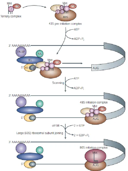
Translation initiation can be subdivided into four steps: 1) binding of the specific initiator Met-tRNA, to the small ribosomal subunit (40S), 2) binding of the formed complex to the cap structure at the 5’ end of mRNA, 3) scanning of the 5’untranslated region (5’UTR) of the mRNA and start codon recognition, 4) joining of the large ribosomal subunit (60S) to generate a translation competent ribosome (80S). As physiological conditions favor the association of 40S and 60S ribosomal subunit to form complete 80S ribosomes, but only free ribosomal subunits can initiate translation, it is important that post termination ribosomes dissociate (Preiss and Hentze 2003). The eukaryotic initiation factors eIF3, eIF1, eIF1A and eIF6 are thought to promote this dissociation in eukaryotes (Holcik and Pestova 2007). As reported in Figure 1, the first step in 43S preinitiation complex formation is the assembly of a ternary complex, consisting of eIF2, methionyl tRNA (met-tRNA) and GTP. Its assembly is
13 stimulated by the guanine nt exchange factor (GEF) eIF2B. GTP is hydrolyzed after recognition of the AUG start codon producing eIF2 bound to GTP (Sonenberg and Hinnebusch 2007). eIF2B promotes GDP-GTP exchange to regenerate active eIF2. Binding of the ternary complex to the 40S ribosomal subunit is supported by eIF1, eIF1A and eIF3 in mammalian cells (Preiss and Hentze 2003; Holcik and Pestova 2007). The 43S preinitiation complex is ready to bind to the 5’ end of the mRNA.
Figure 1. Cap-mediated translation initiation (Gebauer and Hentze 2004)
The eIF4F complex bounds to the 5’m7GpppN cap structure and promotes the recruitment of
14 eIF4F is composed of the cap-binding protein eIF4E, the scaffold protein eIF4G and the ATP-dependent helicase eIF4A that, assisted by eIF4B, unwinds secondary structures in the 5’UTR of the mRNA. The binding of the preinitiation complex to the mRNA involves the cooperative activities of eIF4F, eIF3, eIF4B and PABP. PABP was identified as a protein that associated with polyA tail at the 3’UTR of the mRNA. The PABP-eIF4G interaction is thought to promote a circularization of the mRNA molecule forming a closed loop. This circularization provides a possible framework by which 3’UTR-binding proteins can regulate translation initiation (Gebauer and Hentze 2004). Once assembled near the 5’ end of the mRNA, the 48S complex scan along the mRNA to find the AUG starts codon. In eukaryotes, recognition of an AUG as a start codon critically depends on its surrounding sequence. The scanning process requires ATP and a study using a reconstitute mammalian translation initiation system suggests that this requirement reflects the necessity of unwinding secondary structures in the 5’ UTR by the eIF4A and eIF4B RNA helicases (Pestova and Kolupaeva 2002). Furthermore eIF1 and eIF1A have been shown to play an important role in the scanning process as well as in the recognition of the corresponding initiation codon. Several events take place in order for the 60S subunit to join the 48S complex and form the 80S ribosome. Joining of the 60S ribosomal subunit to the 48S complex requires hydrolysis of two GTP molecules. First, eIF5 triggers GTP hydrolysis by eIF2, which leaves the complex thereafter in the GDP bound state together with eIF5 (Unbehaun, Borukhov et al. 2004). eIF1 and eIF3 remain associated with the complex until eIF5B, a second GTPase, binds to the 43S preinitiation complex and allows the 60S subunit to join. Finally, GTP hydrolysis in eIF5B, triggered by 60S subunit joining, results in the dissociation of eIF5B in the GDP bound form and the formation of an elongation competent 80S ribosome (Pestova, Lomakin et al. 2000).
15 2.2.2 The translational regulation
Protein synthesis, in comparison to the other biosynthetic processes, is the most energetically expensive process going on within the cells; therefore translation has to be highly regulated. Translational regulation is involved in the response to cellular stress (Holcik and Sonenberg 2005), in the misregulation of gene expression during cancer, in apoptosis and in development (Hinton, Coldwell et al. 2007). The need for translational control is also important for systems where transcriptional control is not possible, such as RNA viruses and reticulocytes, where the nucleus is absent. These systems provided us much of our understanding of translational regulation, e.g. reticulocytes are the most efficient cell-free protein synthesis in vitro. Translational regulation can be divided into global regulation of translation and mRNA specific regulation (Gebauer and Hentze 2004): global regulation affects the translation efficiency of most mRNAs through a general tuning of translation, while mRNA specific regulation affects the translation of specific mRNAs. Global regulation of translation is generally mediated through modifications of translation initiation factors that transform the information from external compartments to the cell. Initiation phase of translation is the limiting step for a given mRNA, and initiation factors act as regulators, downstream of signaling events (Sonenberg and Hinnebusch 2009). Certain mRNA can be specifically regulated, usually by proteins that bind to cis-regulatory sequences present in 5’ and/or 3’ UTRs of a given mRNA. The ribosome itself can be targeted to exhert translational regulation, and several of its protein constituents can stand posttranslational modifications (Lackner and Bahler 2008).
Regulation of ternary complex formation. One of the best studied examples of the
translational downregulation is the control of the active ternary complex formation. Binding
of Met-tRNAiMet to the 40S subunit through the ternary complex is an essential step in
16 phosphorylated and inhibits the exchange GDP-GTP by eIF2B, and the formation of active ternary complexes is highly reduced, downregulating global translation (Holcik and Sonenberg 2005; Oyadomari, Harding et al. 2008) (Figure 2).
Figure 2. Integration of stress responses by the phosphorylation of eIF2α (Holcik and Sonenberg 2005).
Induction of p-eIF2α serves as an important regulator, under which general protein synthesis and cell proliferation are blocked, thus allowing cells to recuperate from stress or be eliminated if the damage is beyond repair (Koromilas 2015). Phosphorylation of eIF2α is mediated by PKR, an interferon (IFN)-inducible protein with pro-inflammatory and antiviral properties, which is activated by binding to double-stranded (ds) RNA; a family of kinases consisting of the heme-regulated inhibitor (HRI), which is activated by heme deficiency; the
17 endoplasmic reticulum (ER)-resident protein kinase PERK/PEK, which is activated by the accumulation of misfolded proteins in the ER; and finally the general control non-derepressible-2 (GCN2), which is activated by accumulation of uncharged tRNAs caused by amino-acid or nutrient deprivation (Chen 2007; Koromilas 2015). Phosphorylation of the α-subunit of eIF2 inhibits the GDT-GTP exchange reaction mediated by eIF2B due to a reduced dissociation of eIF2 from eIF2B. As a result, less eIF2B is available to promote GDP-GTP exchange and global translation is inhibited (Holcik and Sonenberg 2005).
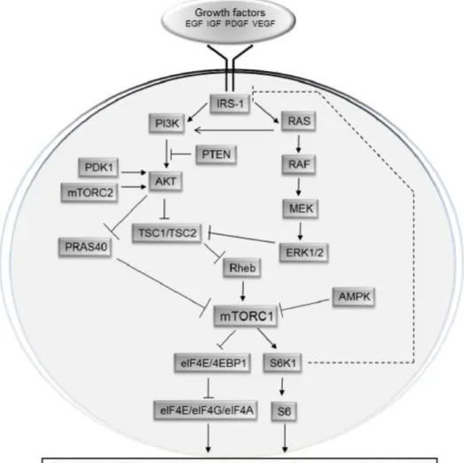
Regulation of cap-dependent translation and mTOR pathway. Most of the cap-dependent
translation is regulated by the pathway of the mammalian Target of Rapamycin (mTOR). mTOR is an evolutionarily conserved Ser/Thr kinase, which regulates proliferation and growth in response to cellular energy status, growth factors, hormones, and nutrient availability (Zoncu, Efeyan et al. 2011). mTOR exists in two functionally and structurally distinct protein complexes: mTOR complex 1 and 2 (mTORC1 and mTORC2). These two complexes regulate disparate cellular functions by phosphorylating distinct sets of substrates. Several substrates of mTORC1 have been identified including the eIF4E-binding proteins (4E-BPs), S6 kinases 1 and 2 (S6Ks), PRAS40, Ser/Thr kinase Ulk1 (also known as hATG1), and growth factor receptor-bound protein 10 (Grb10) (reviewed by (Caron, Ghosh et al.
2010). The function, the upstream regulators and the associated substrates of mTORC2 are
less understood (Oh and Jacinto 2011). mTORC2 phosphorylates AGC kinase family members and controls cytoskeletal organization and cell survival (Guertin and Sabatini 2007; Garcia-Martinez and Alessi 2008). mTORC2 also associates with the ribosome (Zinzalla, Stracka et al. 2011) where it phosphorylates residues in nascent polypeptide chains that are important for optimal protein folding (Oh, Wu et al. 2010). mTORC1 plays a central role in the regulation of proliferation and cell growth (Ma and Blenis 2009), cellular processes that are directly proportional to translational activity. Hormones, growth factors and glucose
18 stimulate mTORC1, up-regulate translation and stimulate cellular growth and proliferation. Conversely, under conditions in which energy production, oxygen supply and nutrients are inadequate, mTORC1 signaling is down-regulated, resulting in inhibition of translation, reduction in cellular growth proliferation, and induction of autophagy. Rapamycin is a naturally occurring allosteric inhibitor of mTORC1 (Guertin and Sabatini 2007). mTOR inactivation, by treatment with rapamycin, mimics deprivation of nutrients, both in mammals and in yeast. Main downstream targets of mTOR kinase are eIF4E-binding proteins (4E-BPs), rpS6 kinases (S6K) and eEF2 kinase (Hay and Sonenberg 2004). 4E-BPs and S6Ks are the most extensively studied and best-understood downstream effectors of mTORC1, which have been implicated in the regulation of translation (Figure 3).
19 The first step of cap-dependent translation initiation is the assembly of the eIF4F complex on the mRNA cap structure (Topisirovic and Sonenberg 2011). eIF4E binds to the 5’ cap structure of eukaryotic mRNAs and provides the first contact between the translational machinery and the mRNA in de novo translation initiation. eIF4E interacts with several types of protein binding partners. It binds the scaffold protein eIF4G, which in turn, interacts with the RNA helicase eIF4A, the multisubunit eIF3 which provides the association to the 40S subunit, and the poly (A)-binding protein (PABP). The eIF4E/4G/4A complex is referred as the eIF4F complex which is thought to be of key importance in mediating normal, cap-dependent translation initiation. A second group of eIF4E binding proteins comprises low molecular mass proteins that block its interaction with eIF4G. In mammals three eIF4E binding proteins are known, 4E-BP1/2/3. 4E-BPs interferes with the assembly of the eIF4F complex by competing with eIF4G for binding to eIF4E. On activation, mTORC1 phosphorylates residues corresponding to Thr37 and Thr46 on human 4E-BP1, which act as priming sites for the phosphorylation of Ser65 and Thr70. Phosphorylation of 4E-BPs on these four residues, leads to their dissociation from eIF4E, allowing the assembly of the eIF4F complex. In addition to 4E-BPs, TOR regulates translation by activating the S6Ks (Ma and Blenis 2009). Although Drosophila expresses a single S6K protein (dS6K), mammals express two variants of S6K (S6K1 and S6K2). rpS6 was the first identified S6K substrate. Five phosphorylation sites (Ser235, Ser236, Ser240, Ser244, and Ser247 in humans and rodents) are clustered in the carboxyl terminus of rpS6 (Meyuhas 2008). It has been demonstrated that, using S6K1/S6K2 double knockout mice, both S6K1 and S6K2 isoforms contribute to the regulation of basal and inducible rpS6 phosphorylation at S235/236 and S240/244 sites (Pende, Um et al. 2004; Chauvin, Koka et al. 2014). Notably, S6K2 knockout mice display a reduction of rpS6 phosphorylation only at S235/236 while S6K1-deficient mice show no alterations (Bhattacharya, Kaphzan et al. 2012). Several studies have demonstrated that S6K1
20 regulates translation initiation through the phosphorylation of the cap binding complex component eIF4B at S422 (Raught, Peiretti et al. 2004). Finally, for many years it has been believed that the phosphorylation of rpS6 had an effect on the translation of a specific subset of mRNAs bearing a 5′ terminal oligopyrimidine tract (TOP). Actually, this model has been changed by studies showing that both double mutant S6K1/2 MEFs and rpS6 knockin mouse exhibit normal TOP translation (Ruvinsky, Sharon et al. 2005).
Regulation of cap independent translation (IRES)
An important mode of translational regulation during stress is the selective recruitment of mRNA through internal ribosome-entry site (IRES). The IRES directly recruits ribosomes, bypassing the requirements for the mRNA 5’ cap structure and eIF4E (Johannes and Sarnow 1998; Hellen and Sarnow 2001). Expression of genes bearing IRES elements in their mRNAs is controlled by multiple molecular mechanisms, with IRES-mediated translation favored when cap-dependent translation is compromised (Komar and Hatzoglou 2011). The translation initiation of several IRES-containing mRNAs occurs predominantly during stress and apoptosis (Holcik and Sonenberg 2005). By this alternative mechanism, even if cap-dependent translation is reduced, some cellular mRNAs can be efficiently translated (Figure 4). Kozak points out that evidence cited in support of the internal initiation hypothesis is often flawed, in fact, when putative IRESs are examined more carefully, they often turn out to harbor cryptic promoters or splice sites (Kozak 2005). Nevertheless it is clear that IRES-mediated translation is used relatively frequently under both normal physiological and pathological conditions.
21
Figure 4. Cap dependent (a) versus IRES dependent (b) translation initiation (Komar and Hatzoglou 2011)
Other mechanisms of mRNA translational regulation
Of great importance in the translational mechanism are some regulatory sequences represented by upstream open reading frame (uORF), which interferes with the expression of the CDS. uORFs, particularly common in transcripts for oncogenes and growth factors, are present in 10% of mRNAs (Sachs and Geballe 2006). uORFs interpose a barrier to prevent ribosomal access to initiation codon, preventing so the translation of the downstream ORF and affecting gene expression and mRNA stability. Even if the ribosome recognizes an initiation codon and translates the uORF, it might reinitiate at a downstream AUG codon thereby overcoming the barrier, typically in conditions of reduced translation driven by impaired eIF2-GTP-Met-tRNA ternary complex formation during specific cellular stress, as amino acid deprivation and unfolded protein response (UPR) (Baird and Wek 2012). For instance, when misfolded proteins accumulate in ER, eIF2α may be phosphorylated by PERK causing a reduction in global translation and favoring reinitiation at downstream ORF (Sonenberg and Hinnebusch 2009).
Several lines of evidence indicate that mRNAs exist in an actively translated and associated with polysomes form and in a translationally repressed and associated with P-bodies state. The idea that the recruitment of mRNAs to P-bodies interferes with translation initiation is supported by the finding that inhibition of translation elongation causes P-bodies to disappear, while inhibition of translation initiation increases the size and number of P-bodies. In
22 mammalian cells, several proteins with established roles in translational repression localize to P-bodies: eIF4E inhibitory protein eIF4E-T, RCK/p54 and CPEB (reviewed by (Decker and Parker 2012). The exact mechanism of how mRNAs shuttle into P-bodies and become translationally repressed is yet unknown.
2.2.3 Novel concepts in translational control: regulation by microRNA
miRNA biology is associated, in the last years, to translation mechanism. microRNAs are short non coding RNA of 21-26 nt emerged as key posttranscriptional regulators of gene expression in metazoan animals, plants, and protozoa. Current studies estimate that human genome encodes hundreds of different miRNAs and that they could regulate almost the 60% of all genes (Friedman, Farh et al. 2009). In animals, miRNAs form imperfect hybrids with sequences in the mRNA 3’-untranslated region (3’ UTR), with the miRNA 5’-proximal “seed” region (positions 2–8) providing most of the pairing specificity (reviewed in (Bushati and Cohen 2007; Filipowicz, Bhattacharyya et al. 2008). Until very recently, it appeared that plant miRNAs generally base-pair to mRNAs with perfect complementarity and trigger endonucleolytic mRNA cleavage by the RNA interference (RNAi) mechanism. Generally, the binding partially complementary to target mRNAs, leads to mRNA degradation and
translation inhibition (Iorio and Croce 2012) recruiting the decapping and deadenylating
machinery. Recently, however, some reports identify miRNAs as activator of mRNA translation during cell quiescence (Vasudevan, Tong et al. 2008; Niepmann 2009), as reported for miRNA 369-3: the direct base pairing between miRNA 369-3 and its target is required for translational upregulation after serum starvation (Vasudevan, Tong et al. 2008). Most studies affirm that miRNA mechanism acts at initiation of translation and can work as tumor suppressor or accelerating factor. The example of tumor suppressor is represented by miR-21
23
whose targets are PI3K and the apoptotic pathways(Loreni, Mancino et al. 2014). In addition
to classical tumor suppressor and oncogene functions, miRNAs can be also implicated in cell migration and metastasis, as the highly expressed miR10-b in metastatic breast cancer that
positively regulates cell migration and invasion(Ma, Teruya-Feldstein et al. 2007). miRNAs
associate with Ago proteins to form RNA-induced silencing complexes (RISCs), through which they can modulate gene expression components. Components of miRISC and repressed mRNAs are enriched in processing bodies, which are cytoplasmic structures involved in the storage or degradation of translationally repressed mRNAs. Some P-bodies components are important for effective repression of protein synthesis by miRNAs. Recently, multivesicular bodies (MVBs) and endosomes were also identified as cellular organelles contributing to miRNA function or miRISC turnover (reviewed in (Fabian, Sonenberg et al. 2010). Regulation of gene expression via small RNAs and sequestration to P bodies, and interplay between miRNA translation inhibition and mRNA decay add further complexity to cellular posttranscriptional control. As 60% of genes are potential miRNA targets (Lewis, Burge et al. 2005), miRNAs could exhert their function in a dual way: a mRNA could be regulated by several miRNAs and a miRNA could target several mRNAs. Moreover, both their expression and action is cell and tissue specific, as microRNA can target different mRNAs in different cell and tissues. This implicates that the action of microRNA is not conserved, but depends from its environment. Elucidation of the molecular events behind these mechanisms is needed.
2.3
Eukaryotic Initiation Factor 6 (eIF6)
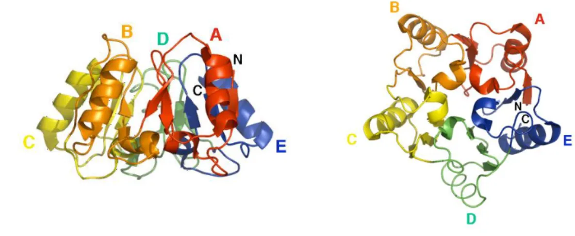
Eukaryotic Initiation Factor 6 is an evolutionary conserved protein. The primary sequence shows two main features: 1) the protein is 245 aa long and it is 77% identical between humans and yeast (Biffo, Sanvito et al. 1997), 2) eIF6 primary sequence is evolutionarily
24 unique, with no conserved motifs. eIF6 structure has been solved, according to X-ray data: it is a rigid protein organized with a cyclic fold, called pentein or star-like structure, formed by 5 stretches of α/β subdomains arrayed about a five-fold axis of psuedosymmetry (Groft, Beckmann et al. 2000). The structure encloses a cavity that contains sixteen well-ordered water molecules, with limited degree of motility (Figure 5).
Figure 5. Structure of eIF6. The protein has a unique star-like structure known as pentein, which is formed by five quasi identical subdomains (A–E) (Groft, Beckmann et al. 2000).
Structural data have identified that eIF6 binds to intersubunit space of the large ribosomal subunit (Klinge, Voigts-Hoffmann et al. 2011). It is able to interact with the hydrophobic C-terminal chain of the ribosomal protein L23 (rpL23). The sarcin-ricine loop and rpL24 also contribute to the interaction of eIF6 with the 60S subunit. Since the steric hindrance, it prevents binding between 60S and 40S.
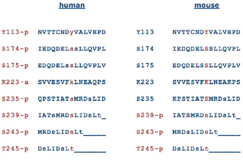
The semiconserved C-terminal tail seems a candidate region for eIF6 regulation and transport due to its flexibility. The C-terminus of eIF6 is characterized by the presence of many phosphorylation sites that are highly conserved in mammalian cells (Figure 6).
25
Figure 6. eIF6 phosphorylation sites (http://www.phosphosite.org)
The phosphorylation sites Ser174 and Ser175 are located at the accessible surface of eIF6, not involved in the interaction with the 60S subunit. Also the flexible C-terminal sequence that contains Ser235 phosphorylation site is located at the outer surface of eIF6 (Gartmann, Blau et al. 2010). Mutation of the yeast homologues of eIF6, called Tif6p, at Serine-174 to Alanine reduced phosphorylation drastically and caused loss of cell viability and growth. When both Ser-174 and Ser-175 were mutated to alanine, phosphorylation of Tif6p was abolished. Furthermore, while wild-type Tif6p was distributed both in nuclei and the cytoplasm of yeast cells, the mutant Tif6p, containing Ser174Ala and Ser175Ala, became a constitutively nuclear protein (Basu, Si et al. 2003). Several studies have shown that eIF6 has a dual function: it is necessary for the maturation of 60S ribosomal subunit in the nucleus and possesses a ribosomal antiassociation activity (Miluzio, Beugnet et al. 2009), and it is involved in translation in the cytoplasm (reviewed by Biffo et al., A. Parsyan ed., 2014).
Data collected through genomic sequencing projects reveal that evidences for eIF6 gene duplication do not exist, suggesting a strong evolutionary pressure for control of the protein
26 concentration. Human eIF6 gene is constitutively expressed in vitro, but modulated in vivo, since protein level in vivo are variable among different organs. Studies on levels of eIF6 in several metazoan tissues show that the protein is expressed at low level in muscle and high in brain. Furthermore, eIF6 is particularly expressed in stem cells or in cycling cells, but undetectable in some postmitotic cells (Donadini, Giodini et al. 2001).
2.3.1 eIF6 on ribosome biogenesis and antiassociation activity
Ribosome biogenesis is a very important process that occurs in the nucleolus and leads to the production of large and small mature ribosomal subunits and to their export to the cytoplasm. The small and large subunit are separately processed and exported, although they derived from the same rRNA precursor (47S in mammals). The large subunit matures through intermediate steps known as 90S-66S-pre60S-60S. Several lines of direct and indirect evidences support the necessity of eIF6 in ribosome biogenesis. Deletion of the yeast homolog Tif6 leads to a loss of the 60S ribosomal subunit that can be rescued by the ectopic expression of human eIF6 (Sanvito, Piatti et al. 1999; Si and Maitra 1999; Brina, Grosso et al. 2011). Moreover biochemical evidences converge to the role of eIF6 in ribosome biogenesis. The protein is identified in molecular complexes from 66S to mature 60S. In agreement with this finding, a pool of eIF6 is localized in the nucleolus of both yeast and mammalian cells (Sanvito, Vivoli et al. 2000). The molecular mechanism by which eIF6 regulates 60S biogenesis is not completely clear. rRNA pulse-chain has shown that yeast cells depleted of eIF6 have defective pre-RNA processing. This causes the reduced formation of mature 25S and 5.8S rRNA relative to 18S rRNA, which may account for the selective deficit of 60S ribosomal subunit. Thus, eIF6 acts in biogenesis of 60S subunit, rather than in its stabilization
27 (Basu, Si et al. 2001; Woolford and Baserga 2013). However, our knowledge of the eIF6 function in the biogenesis of 60S subunit is not clear and requires further studies.
Moreover, eIF6 has a relevant biochemical activity, preventing binding of 40S and 60S in the absence of mRNA and thus avoiding an accumulation of inactive 80S subunit. In this way eIF6 is able to keep the small and large subunit available for initiation of translation (reviewed by Biffo et al., A. Parsyan ed. 2014). eIF6 was initially identified on the basis of its antiassociation activity in calf liver (Valenzuela, Chaudhuri et al. 1982) and wheat germ (Russell and Spremulli 1979), but it cannot dissociate preformed 80S complexes. It has been published the crystal structure of the 60S ribosomal subunit in complex with eIF6 in
Tetraymena termophyla (Klinge, Voigts-Hoffmann et al. 2011). The structure reveals
interactions between eukaryotic specific ribosomal proteins in the stabilization of the active site. The site of the eIF6 binding to 60S was mapped to the 40S-60S interface, close to sarcin-ricin loop (SRL) and ribosomal protein rpL23 e rpL24, where it would prevent binding of the 40S subunit. It is rationale to speculate that the antiassociation activity of eIF6, as observed in
vitro, is relevant for translational control in vivo (Biffo et al., A. Parsyan ed. 2014).
Furthermore, although eIF6 is dispensable for translation in vitro, low concentrations of eIF6 have a slight stimulatory effect on translation, whereas higher concentrations inhibit it (Russell and Spremulli 1979).
2.3.2 eIF6 and translation
Ceci et al showed that eIF6 is able to repress translation after binding to 60S ribosomal subunit and similar observations were made with eIF6 bound to 50S (Benelli, Marzi et al. 2009). Furthermore, mammalian and yeast eIF6 have common properties, such as the mainly cytoplasmic localization, which correlates with a role of eIF6 in the control of translation.
28 Since the binding of eIF6 to the large ribosomal subunits is able to affect translational initiation, it is possible to assume that there is a mechanism that favors its release leading to dissociation of eIF6 from the 60S ribosomal subunit. Two models for eIF6 release have been proposed: 1) 60S, bound to eIF6, is translocated from the nucleus to the cytoplasm. Here, the interaction between the Swachman-Bodian-Diamond Syndrome protein (SBDS) and the GTPase Efl1p with the 60S subunit leads to an allosteric change of 60S mediating the release of eIF6 (Wong, Traynor et al. 2011). This mechanism is relevant during the maturation step of the 60S subunit (Bussiere, Hashem et al. 2012). 2) Release of eIF6 is mediated by RACK/PKC complex. RACK1 acts as a scaffold receptor protein for active PKC and binds to the small ribosomal subunit (Ceci, Gaviraghi et al. 2003; Volta, Beugnet et al. 2013). Activated PKC translocates from endomembrane to the small subunit, comes in vicinity with eIF6 bound to 60S subunit and catalyzes the phosphorylation of eIF6 on Ser235 and its subsequent release (Brina, Grosso et al. 2011) (Figure 7).
Figure 7. The two models of eIF6 release that regulate the interaction of the two ribosomal subunits.
29 Mouse model of eIF6 haploinsufficiency evidence that eIF6 is critical for translation initiation (Gandin, Miluzio et al. 2008). eIF6-null mice are embryonic lethal in mammals, but heterozygous mice, presenting a 50% of the eIF6 protein level, are viable. This reduction of the protein affects the cytoplasmic pool, and not the nuclear levels, leading to a proper biogenesis of the 60S ribosomal subunit. This confirm the notion that the function of the protein is cytoplasmic, translation related, and not nuclear, ribosomal biogenesis-related. The analysis of polysomal profiles of eIF6 heterozygous mice shows an increase in the 80S peak and a decrease in polysomes, confirming the role of eIF6 in initiation of translation. All mouse tissues of heterozygous mice have levels of the eIF6 protein reduced of 50% compared to the wild-type controls. The liver of eIF6 heterozygous mice shows an accumulation of inactive 80S complexes, and hepatocytes have normal level of translation but are not able to regulate its response to insulin. Thus full levels of eIF6 are necessary to perform the translation program induced by insulin of the cell, in vivo. The expression of eIF6 is rate limiting for tissue growth, as mice haploinsufficient for eIF6 have smaller livers than wild-type and reduced white fat mass. The deficit in insulin-stimulated translation occurring in eIF6+/– cells correlates with a high insulin sensitivity in tissues. Hepatocytes, fibroblasts and adipocytes from heterozygous eIF6 cells show a delayed G1-to-S phase progression but are normal in size, and have normal apoptosis and senescence (Gandin, Miluzio et al. 2008).
2.3.3 eIF6 and cancer
The research into the role in of eIF6 in cancer is still in the twilight. In tumors oncogenic pathways that promote tumor development and cellular transformation are hyperactivated and deregulation in translational control are endpoint of these these pathways (Silvera, Formenti et al. 2010). eIF6 is overexpressed in several cancer types, such as head and neck cancer (Rosso
30 et al., 2004), lung metastasis (Martin, Sanz et al. 2008), acute promyelocitic leukemia (Harris, Ozpolat et al. 2004) and malignant mesothelioma (Biffo, Sanvito et al. 1997). The mechanism that explains the eIF6 overexpression in cancer is unclear. eIF6 overexpression may reflect an increased demand for the protein by proliferating cancer cells, and not its role in etiology and cancer development. Cells with halved level of eIF6 protein show a reduction in MYC or HRAS-mediated oncogenic transformation (Gandin, Miluzio et al. 2008). MYC-induced lymphomagenesis is reduced in murine lymphoma with reduced eIF6 levels resulting in prolonged tumor free survival in the absence of negative side effects (Miluzio, Beugnet et al. 2011). Mutation of eIF6 in the PKC consensus site Ser235 reduces the rate of transformation, suggesting its role in tumorigenesis (Miluzio, Beugnet et al. 2011). The most relevant information related to the regulation of the eIF6 activity by signaling are: 1) eif6 is hyperphosphorylated in cancer cells, in the C-terminus at Ser235, Ser239 and Thr243 (Ceci, Gaviraghi et al. 2003; Dephoure, Zhou et al. 2008); 2) mutation of Ser235 to Ala reduces translation and tumorigenesis (Gandin, Miluzio et al. 2008; Miluzio, Beugnet et al. 2011); 3) eIF6 activity is independent from mTORC1 activation but essential for growth factor and insulin activation (Gandin, Miluzio et al. 2008); 4) eIF6 interacts with RACK1 (Ceci, Gaviraghi et al. 2003; Guo, Wang et al. 2011), which is able to affect translation (Volta, Beugnet et al. 2013). The PKC isoform that binds RACK1 is PKCβ, and only the PKCβII isoform show a higher affinity for RACK1 receptor (Stebbins and Mochly-Rosen 2001). Moreover PKCβ inhibition reduces translation not affecting mTORC1 targets (Grosso, Volta et al. 2008), suggesting a role for the PKC axis in the regulation of translation. However it is possible that eIF6 activity is affected by mTORC2, upstream of several PKCs (Hagiwara, Cornu et al. 2012). These data suggest a role of eIf6 as a modulator of tumorigenesis and tumor growth.
31
2.4
MicroRNAs
microRNAs are endogenous, small non-coding single-stranded RNAs of ~22 nucleotides in length, found in both plants and animals. They act as negative regulators of gene expression in several cellular processes and, in mammals, they are able to control the activity of more than 60% of all protein-coding genes (Friedman, Farh et al. 2009). miRNAs regulate protein synthesis by base-pairing to target mRNAs. In animals, miRNAs form imperfect hybrids with sequences in the 3’UTR of mRNA, with the miRNA 5’-proximal “seed” region (positions 2– 8) providing most of the pairing specificity. In contrast plant miRNAs base-pair to mRNAs with perfect complementarity and trigger mRNA cleavage by the RNA interference (RNAi) mechanism (Filipowicz, Bhattacharyya et al. 2008; Bartel 2009). This is the typical strategy used by miRNAs to reduce the translation and stability of mRNAs, including those of genes that mediate processes in tumorigenesis, such as cell cycle regulation, inflammation, stress response, differentiation, apoptosis and invasion (Iorio and Croce 2012).
miRNAs were originally shown to be important in timing of larval development in C.
Elegans , leading to the identification of the best known miRNAs lin-4 and let-7. Initial
understanding of miRNA-mRNA target recognition came from observations of sequence complementarity of the lin-4 RNA to multiple conserved sites within the lin-14 3’UTR; molecular genetic analysis showed that this complementarity was required for the repression of lin-14 by lin-4 (Lee, Feinbaum et al. 1993; Wightman, Ha et al. 1993; Reinhart, Slack et al. 2000).
microRNA biogenesis is divided into two main processing steps that take place in the nucleus and in the cytoplasm: primary microRNAs are first processed into the nucleus by RNAse III Drosha, associated to the double stranded RNA-binding protein DGCR8 (DiGeorge syndrome critical region gene 8; Pasha in flies) known as the microprocessor complex, that generates a ~70 nucleotides precursor miRNA products, which fold into stable secondary stem-loop
32 structures. The latter are recognized by the Ran-GTP-dependent transporter Exportin 5, which mediates the translocation to the cytoplasm. Here Dicer, a RNAse III enzyme, associated to TRBP (TAR RNA-binding protein) and Argonaute proteins (AGO1-4), cleave the miRNA precursor hairpin and generate a transitory miRNA/miRNA* duplex (also named respectively miR-3p/miR-5p), which includes the mature miRNA guide, selected by thermodynamic properties, and the complementary passenger strand, usually subjected to degradation. This duplex is then loaded into the miRNA-associated RNA induced silencing complex (RISC or miRISC), including the mature single-stranded miRNA molecule and AGO proteins, where the mature miRNA could regulate gene expression, binding through partial complementarity to target messenger RNAs (mRNAs) and leading to translation inhibition or mRNA degradation, depending on the sequence complementarity between the miRNA and the target mRNA (reviewed by (Iorio and Croce 2012). Recent reports have tried to clarify the complex mechanisms regulating miRNA function on target mRNAs: microRNAs mainly recognize complementary sequences in the 3´ UTR of their target mRNAs, but recent studies have reported that they may also bind to the 5´ UTR or the open reading frame (Lytle, Yario et al. 2007; Orom, Nielsen et al. 2008; Moretti, Thermann et al. 2010). Sites located in coding regions appear to be less robust than those in the 3’UTR (Gu, Jin et al. 2009) and, surprisingly, miRNAs can upregulate translation upon growth arrest conditions (Henke, Goergen et al. 2008; Orom, Nielsen et al. 2008; Vasudevan, Tong et al. 2008) (Figure 8). Moreover it has been evidenced that the mature form of microRNAs may also be localized in the nucleus (Hwang, Wentzel et al. 2007).
33
Figura 8. microRNAs biogenesis and function (Iorio and Croce 2012)
miRNAs interact with their mRNA targets via base-pairing. The most important requirement is a contiguous and perfect Watson-Crick base-pairing of the seed region of the miRNA, the 5’ nucleotides 2–8, guide for the base-pairing. However, functional miRNA sites that contain bulged nucleotides or mismatches in the seed region have also been identified as shown for
34 the Lin-41 mRNA targeted by let-7 miRNA in Caenorhabditis elegans (Vella et al., 2004). Moreover miRNA-mRNA duplexes containing mismatches and bulges in the central region (miRNA positions 10–12) could prevent endonucleolytic cleavage of mRNA. AU-rich sequence context and structural accessibility of the sites could improve their efficacy (Bartel 2009). Multiple sites, for the same or different miRNAs, are required for effective repression, and when the sites are close to each other, they tend to act cooperatively (Grimson, Farh et al. 2007).
2.4.1 microRNAs and translation
Several studies tried to clarify the mechanisms of protein synthesis suppression by microRNAs. These studies showed that miRNAs could inhibit translation of target mRNAs or facilitate their deadenylation and subsequent degradation. All miRNA-mRNA interactions seem to downregulate gene expression at post-trancriptional level, but the scale of regulation vary and depends on the specific miRNA-mRNA target combination. Whether this event is due by accessibility of the mRNA to miRNAs or by other factors is unknown (Maroney et al., 2006). How do miRNAs regulate gene expression? Early analysis indicated that regulation was at the level of translation: the abundance of the regulated mRNA does not change, but the abundance of proteins encoded by these mRNAs was reduced (reviewed by (Maroney, Yu et al. 2006). The first question in clarifying the mechanism of translational repression by miRNAs is to determine whether miRNAs suppress the initiation of translation or act at the postinitiation stage. Lin-4 was discovered in C. elegans, and causes inhibition of translation of lin-14 without a reduction in mRNA levels or a shift in polysomes, leading to the conclusion that miRNAs could inhibit mRNA translation at the elongation step of the translation process (Olsen and Ambros 1999). In other experimental models several results denoted defects in the control of translation initiation and mRNA stability. miRNA-mediated repression of